CYTOPLASMIC MESSENGER RIBONUCLEOPROTEINS AND TRANSLATIONAL CONTROL DURING GROWTH AND DIFFERENTIATION OF EMBRYONIC MUSCLE CELLS
SATYAPRIYA SARKAR JNANANKUR BAG
Department of Muscle Research Boston Biomedical Research Institute
Department of Neurology Harvard Medical School
Boston, Massachusetts
I. INTRODUCTION
Although a large amount of information about the physico- chemical properties of the myofibrillar proteins and their role in the contraction-relaxation cycle of muscle tissue is currently available (for a review see Taylor, 1972), very little is known about myofibrillogenesis and its regulation during the growth and differentiation of muscle cells. Recent studies from a number of laboratories have suggested that translational control may exert a subtle regulation of protein synthesis during myogenesis of the skeletal and cardiac muscle fibers. In cultures of skeletal muscle cells, which are used by many investigators as a model sys-
389
tem for studying various aspects of terminal differentiation, the mononucleated rapidly proliferating muscle cells undergo several cycles of cell division, then withdraw from the mitotic cycle and fuse to form a multinucleated embryonic myotube (for a review see Holtzer et al., 1972). It is at this stage, namely the time of cell fusion and the formation of the myotube, that the intensive synthesis of myosin and a number of muscle-specific enzymes (re- viewed by Yaffe and Dym, 1972) is first observed. In cultures of rat myoblast cells treatment with actinomycin D at, or just prior to, fusion does not block the immediate appearance of myosin and other muscle-specific proteins (Yaffe and Dym, 1972). Buckingham et al. (1974) have recently reported that during the transition from non-differentiated myoblasts to the differentiated myotube stage, rapidly labeled poly (A)-containing RNAs of different sizes are stabilized with a significant increase in their half-lives.
They have shown that a 26 S RNA species, considered as the puta- tive mRNA coding for the 200,000 dalton large subunit of myosin (also referred to as the myosin heavy chain), accumulated as a free messenger ribonucleoprotein (mRNP) particle sedimenting as 110-120 S in sucrose gradients of cell lysates. After cell fusion this RNA species was located by pulse-chase experiments in a group of very large polysomes (40-60 ribosomes) which are known to syn- thesize myosin heavy chain in embryonic muscles (Sarkar and Cooke, 1970; Heywood and Rich, 1968). Chacko and Xavier (1974) have shown that when precardiac mesodermal cells, which are the pro- genitors of cardiac tissue, were treated with 5-bromodeoxyuridine
(BrdU), a thymidine analog or actinomycin D at a non-differenti- ated stage of growth in culture, they were able to continue growth and express their differentiated phenotype. These studies suggest that critical regulatory controls of gene expression may operate at a post-transcriptional level during the terminal differentia- tion of skeletal and cardiac muscle cells. However, the precise biochemical nature of these regulatory mechanisms remains to be understood.
MESSENGER RIBONUCLEOPROTEINS 391 Actin and myosin or myosin-like proteins are also present in a wide variety of non-muscle eukaryotic cells (for a review see Pol- lard and Weihing, 1974). It has been suggested that non-muscle
(also referred to as cytoplasmic) actin and myosin may play im- portant roles in processes such as cellular motility, adhesion, contraction and cytokinesis in eukaryotic cells. Rubinstein et al. (1974) have recently suggested that the cytoplasmic contrac- tile system found in almost all eukaryotic cells, including myo- blasts and BrdU-suppressed myogenic cells (Holtzer et al., 1972), may be controlled by a genetic program which is insensitive to BrdU. In contrast, the myofibrillar contractile system, which is specific for differentiated muscle cells, may arise from the read- out of a separate genetic program whose expression is sensitive to BrdU (Rubinstein et al., 1974). If indeed this is the case, the mechanisms by which the expression of these different genetic pro- grams is regulated in a myogenic cell remain to be understood.
The polymorphism of various muscle proteins adds another com- plexity in the problem of differentiation of muscle tissue. Re- cent evidence suggests that at least in the case of myosin, a major heteropolymeric myofibrillar protein, different sets of structural genes coding for myosin subunits (also referred to as the light and heavy chains) are expressed in various types of differentiated muscle fibers (e.g. slow, fast, cardiac, smooth) resulting in different isozymic forms of myosin characteristic of each muscle type (Sarkar et al., 1971; Sarkar, 1972; Lowey and Risby, 1971; Weeds and Frank, 1972; for a review see Taylor, 1972). The expression of such a "family of genes" coding for the polymorphic forms of muscle proteins must be precisely regulated during the life cycle of the muscle cell.
In order to elucidate the regulatory mechanisms involved in myogenesis we have selected the myosin heavy chain and actin as two suitable muscle-specific markers and probed whether the bio- synthesis of these proteins is regulated at a cytoplasmic level.
We report here the isolation and characterization of cytoplasmic
nonpolysomal mRNP particles containing actin and myosin heavy chain mRNAs from embryonic muscle cells. The significance of these results in relation to some of the cellular processes in muscle cells, outlined above, is also discussed.
II. RESULTS
A. Subribosomal mRNP Particles Containing Actin mRNA
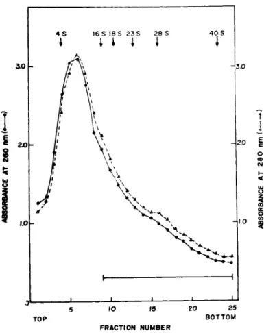
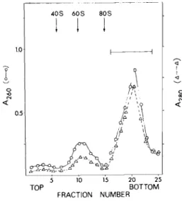
Since a large fraction of cellular mRNAs coding for 15,000- 35,000 dalton polypeptides sediment in sucrose gradients between 8-20 S (for a review see Brawerman, 1974), the expected size of the cytoplasmic mRNP particles, which may contain such mRNAs, should be within the range 20-40 S (Spirin, 1972). A crude sub- ribosomal pellet of embryonic muscle cells was, therefore, used to search for nonpolysomal mRNP particles. The subribosomal pellet was obtained by centrifugation of the postribosomal supernatant of homogenates of 14-day old chick embryonic leg and breast muscles at 255,000 g for 4 hr and details of the preparative procedures are described in our published reports (Bag and Sarkar, 1975; Bag et al., 1975). When subribosomal particles were centrifuged through linear 5-20% sucrose gradients, about 60% of the uv-ab- sorbing material sedimented as a broad peak of about 8-10 S (Fig.
1). The remaining material showed a heterogeneous distribution sedimenting between 16-40 S (tubes, 9-25, Fig. 1). Based on two considerations, size and heterogeneity of the sedimentation pro- file, this 16-40 S fraction of the gradient runs was pooled to look for cytoplasmic mRNP particles.
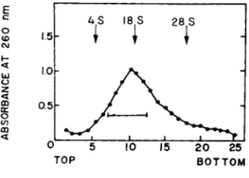
When the RNA isolated from the 16-40 S particles was analyzed by centrifugation through 5-20% linear sucrose gradients, the pro- file shown in Fig. 2 was obtained. The major part of the RNA sedimented as a broad peak of about 18-20 S. However, a signifi- cant amount of the uv-absorbing material showed a very hetero- geneous profile with S values equal to and larger than that of 28
MESSENGER RIBONUCLEOPROTEINS 393
FIGURE 1 Sedimentation profile of subribosomal particles in 5-20% linear sucrose gradients in 0.01 M Tris-HCl, pH 7.6 contain- ing 0.5 M KCl, 0.002 M EDTA and 500 \ig/ml of heparin. Centrifuga- tion was done for 17 hr at 24,000 rpm at 2° in a Spinco SW 25.1 rotor. For details see Bag and Sarkar (1975). Fractions indi- cated by the bar (16-40 S) were pooled for the isolation of RNA fractions with messenger activity. Reproduced from Bag and Sarkar
(1975) .
S rRNA run as a marker. Morris et al. (1972) have previously re- ported that a 10-17 S RNA fraction isolated from muscle polysomes can direct the synthesis of actin in a heterologous cell-free sys- tem. We, therefore, pooled an 8-20 S fraction (indicated by the bar in Fig. 2) from the gradient runs in order to test whether
FIGURE 2 Sedimentation profile of RNA fractions isolated from the pooled 16-40 S subribosomal particles (Fig. 1). Centri- fugation was done at 2° for 17 hr at 24,000 rpm in a Beckman SW 25.1 rotor. For details see Bag and Sarkar (1975). The bar in- dicates the pooled fractions (8-20 S), used for in vitro transla- tion. Reproduced from Bag and Sarkar (1975).
this nonpolysomal RNA fraction indeed programs the synthesis of actin or any other muscle protein of similar (40,000 - 50,000 dalton) size.
The preincubated S-30 system prepared from wheat germ embryos according to the method of Roberts and Paterson (1973) was used to test the mRNA activity of the RNA fractions isolated from the 16-40 S particles. Both the heterogeneous RNA isolated from these particles (tubes 9-25 in Fig. 1) and the 8-20 S RNA frac- tion (Fig. 2) strongly stimulated amino acid incorporation, about 12-15 fold, in this system (Table I). This stimulatory activity was comparable to that obtained with globin mRNA isolated and partially purified from rabbit reticulocyte polysomes. These re- sults indicate that the RNA fraction associated with the 16-40 S subribosomal particles exhibits a strong messenger activity in an in vitro cell-free system.
MESSENGER RIBONUCLEOPROTEINS 395 TABLE I Stimulation of Amino Acid Incorporation
by RNA Fractions Isolated from 16-40 S Subribosomal Particles in Wheat Germ Embryo Cell-Freea System
[3 5S] met
pg of RNA incorporated Degree of Source of RNA used (cpm/assay) stimulation
Minus RNA 2,400 Control Rabbit globin mRNA 2.5 30,000 12.5-fold Chicken 4S RNA 80 2,600
Chicken 28S rRNA 10 2,300
Total RNA from pooled 12 29,000 12-fold tubes 9-25 (Fig. 1)
8-20S RNA fraction 8 36,000 15-fold (Fig. 2)
aAssays were performed as described by Bag and Sarkar (1975).
B. In Vitro Translation of actin mRNA isolated from subribosomal Particles
Incubation mixtures using wheat germ embryo S-30 system pro- grammed with the 8-20 S RNA fraction, as described above, were used for the identification of actin as one of the possible prod- ucts of cell-free translation. After addition of highly purified chicken actin as carrier and dialysis to remove excess radioactive amino acids, the in vitro products were copurified by three cycles of reversible salt-dependent transformation of globular (G) to fibrillar (F) actin. Polymerization of the products to F-actin was done by dialyzing the sample against 5 mM N-2-hydroxyethyl- piperazine-N1-2-ethane sulfonic acid (Hepes), pH 7.0, 0.1 M KCl and 0.003 M MgCl2- The F-actin and ribosomes were pelleted by centrifugation at 160,000 g for 2 hr. The sample was then dial- yzed against a depolymerizing medium, 5 mM Hepes, pH 7.0, 0.2 mM CaCl2 and 0.2 mM ATP and the ribosomes were pelleted at 160,000 g for 2 hr leaving the soluble G-actin in the supernatant. Details of these steps are described elsewhere (Bag and Sarkar, 1975; Bag
300Γ
DISTANCE ( c m )
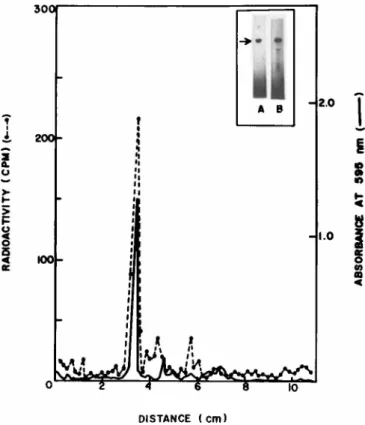
FIGURE 3 Sodium dodecyl sulfate Polyacrylamide gel electro- phoresis of labeled products of in vitro translation of 8-20 S RNA copurified with chicken actin. F-actin samples containing 1400 cpm (specific activity 25,000-30,000 cpm/mg) were analyzed using 10.5 cm of 10% gels at 4 mA/gel for 4 hr. The gels were stained with Coomassie brilliant blue, scanned, sliced and pro- cessed for radioactivity (Bag and Sarkar, 1975). Insert: Elec- trophoretograms of gel runs of purified actin (gel A) and the purified labeled product (gel B). Reproduced from Bag and Sar- kar (1975).
et al., 1975). About 60% of the actin, initially added as car- rier, was recovered in the final pellet of F-actin after three cycles of copurification. The final pellet contained about 10%
of the initial nondialyzable radioactivity incorporated in the cell-free system.
When portions of the F-actin pellet, purified as described above, were analyzed by acrylamide gel electrophoresis in the
MESSENGER RIBONUCLEOPROTEINS 397
i
T"
II II I I I I î ' t
J400
ο
1
H200
oc
ο <
ο Û
>
DISTANCE ( c m )
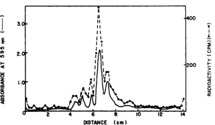
FIGURE 4 Sodium dodecyl sulfate Polyacrylamide gel electro- phoresis of CNBr peptides of purified products of in vitro trans- lation of 8-20 S RNA. For details see Bag and Sarkar (1975).
Samples containing 3000-3500 cpm were analyzed using 14 cm 12.5%
Polyacrylamide high cross-linked gels (bisacrylamide ratio of 1:10) in the presence of 8 M urea and 1% dodecyl sulfate. Gels were run at 2 mA/gel for 20 hr, and then stained, scanned at 595 nm, sliced and counted. Reproduced from Bag and Sarkar (1975).
presence of sodium dodecyl sulfate, about 70% of the radioactivi- ty applied to the gel was recovered in a densitometric peak of the actin band (Fig. 3). A number of additional minor bands, also observed in the radioactivity profile, may be due to other poly- peptides synthesized in the in vitro system, such as tropomyosin and troponin components of the thin filaments. These proteins are copurified with F-actin due to strong mutual interactions and mi- grate in the same region of the gel where the minor radioactive peaks (Fig. 3) are observed (Potter and Gergely, 1974).
When portions of the in vitro products, copurified with F- actin as described above, were electrophoresced in the presence of 8 M urea and absence of sodium dodecyl sulfate, the only major radioactive peak obtained in the gel run comigrated with the ac- tin band (data not presented here; Bag and Sarkar, 1975). About 80% of the radioactivity applied to the gel was recovered in the peak corresponding to actin band. These results indicate that as
judged by mobilities in two kinds of Polyacrylamide electrophor- etic systems, one based on charge and the other based on size, the in vitro products showed properties similar to that of muscle actin.
In order to prove that the 8-20 S RNA fraction indeed programs the de novo synthesis of the 42,000 dalton polypeptide chain of actin, the in vitro products were labeled with a mixture of R e - labeled amino acids and then copurified with chicken actin, as described above. The sample was aminoethylated and cleaved with CNBr and the resulting peptides were analyzed by sodium dodecyl sulfate Polyacrylamide gel electrophoresis using high cross- linked gels, as described by Swank and Munkres (1971). The re- sults are shown in Fig. 4. The densitometric scan shows that one major and five minor peaks were resolved in the gel run. Some of these peaks presumably represent mixtures of peptides which are not completely resolved (Elzinga, 1970). The correspondence of radioactive peaks from the in vitro translated products with the densitometric peaks and the absence of any significant amount of radioactivity in other regions of gel indicate that total syn- thesis of the polypeptide chain was achieved. Control experi- ments in which the 8-20 S RNA was omitted did not give any de- tectable amount of radioactivity above the background level in the actin band. These results indicate that the 8-20 S RNA frac- tion indeed directs the de novo synthesis of muscle actin in a heterologous translation system.
C. Myosin Heavy Chain mRNP in Embryonic Muscle Cells
The mRNA coding for the 200,000 dalton chick embryonic myosin heavy chain has been recently purified and characterized in our laboratory. The purified mRNA has a molecular weight of about 2.23 x 1 06 consisting of about 6400 nucleotides with a poly(A) segment of about 170 adenylic acid residues at the 31 end (Sar- kar et al., 1973; Mondai et al., 1974; Sarkar, 1974, 1975). The myosin heavy chain mRNA sediments in sucrose gradients as 25-27 S
MESSENGER RIBONUCLEOPROTEINS 3 9 9 (Heywood and Nwagwu,1969) but migrates in Polyacrylamide gels as 30-32 S (Sarkar et al., 1973; Mondai et al., 1974). Due to the very large size of myosin heavy chain mRNA, the expected size of an mRNP particle containing this mRNA should be much larger than the 40 S ribosomal subunit (Spirin, 1972). Therefore, the post- poly somal supernatant of embryonic muscle cells containing 80 S monosomes, 60 S and 40 S ribosomal subunits was selected in order to look for myosin heavy chain mRNP. This choice was justified by our initial experiments which showed that an RNA fraction, 24- 32 S, isolated from the post-polysomal supernatant of embryonic muscle cells, showed a strong mRNA activity in the wheat germ em- bryo S-30 system.
The procedure used for the isolation of myosin heavy chain mRNP includes the following steps: centrifugation of the post- polysomal supernatant (Bag and Sarkar, 1975) at 255,000 g for 2 hr to obtain a pellet of oligosomes, monosomes, ribosomal sub- units and myosin heavy chain mRNP; treatment of the post-polyso- mal particles with EDTA and separation of the ribosomal subunits
from myosin heavy chain mRNP by centrifugation in sucrose gradi- ents containing EDTA. Since ribosomal subunits are unfolded in the presence of EDTA whereas mRNP particles are resistant to EDTA treatment, the myosin heavy chain mRNP sedimented in the bottom one-fifth of the sucrose gradients where particles larger than 80 S were located. The myosin heavy chain mRNP was detected by its characteristic ^ 2 5 9 / ^ 2 8 0 r a t^-° °f about 1.2-1.3, which is lower than the corresponding value (1.6-1.8) of ribosomal subunits.
Details of these preparative methods are described elsewhere (Bag et al., 1975; Bag and Sarkar, 1976). When samples of myosin heavy chain mRNP, pooled from the gradient fractions, were recentrifuged through a second sucrose gradient in the presence of 0.5 M KCl, the mRNP was further purified, free from the contaminating ribosomal subunits. The results are shown in Fig. 5. About 60-65% of the uv-absorbing material applied to the sucrose gradient was recovered in a distinct peak which
4 0 S 6 0 S 8 0 S
FIGURE 5 Purification of myosin heavy chain mRNP by sucrose gradient centrifugation. Crude samples of myosin heavy chain mRNP obtained by sucrose gradient centrifugation of EDTA-treated postpolysomal particles were pooled from gradient fractions and centrifuged through a 15-40% linear sucrose gradient in 20 mM Tris-HCl, pH 7.6; 0.5 M KCl; 0.005 M magnesium acetate at 47f000 rpm for 90 min in a Spinco SW 50.1 rotor. For details see Bag et al. (1975). Fractions (16-25), indicated by the bar, were pooled and processed further for the isolation of myosin heavy chain mRNA.
showed an Á26è/Á280 r a ti ° °f about 1.2. The approximate S value of this peak calculated by the method of Martin and Ames (1961) was about 120. When the RNA isolated from the 120 S particles was assayed for mRNA activity in the wheat germ embryo S-30 sys- tem, a strong stimulatory activity, about 10-12 fold, was ob- served (Bag et al., 1975).
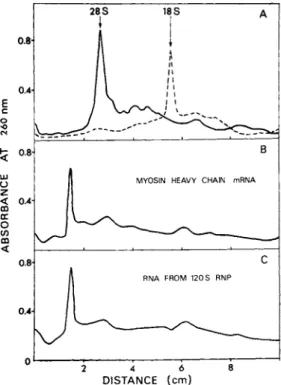
The RNA isolated from the 120 S particles (Fig. 5) was ana- lyzed by agarose-polyacrylamide gel electrophoresis using chick embryonic muscle ribosomal RNAs (28 S and 18 S) and myosin heavy chain mRNA purified from the myosin-synthesizing polysomes (Sar- kar et al., 1973; Mondai et al., 1974; Sarkar, 1974, 1975) as markers. The results are shown in Fig. 6. Densitometric scans
MESSENGER RIBONUCLEOPROTEINS 401
0.8-
0.4- E c ο •ο es
h 0.8
<
LU
<J Æ < 0.4- 00 oc Ο (Ë
m <
0.8-
0.4
2 4 6 8 D I S T A N C E ( c m )
FIGURE 6 Densitometrie scans of Polyacrylamide gel runs of RNA isolated from 120 S mRNP particles. For details see Bag et al. (1975). Panel A: 30 y g of 28 S chick embryonic muscle rRNA; 25 \ig of 18 S rRNA. Panel B: 28 ]ig of myosin heavy chain mRNA purified from myosin synthesizing polysomes
(Sarkar et al., 1973; Mondai et al., 1974). Panel C: 32 \ig of RNA isolated from 120 S mRNP particles (Fig. 5). Electro- phoresis was carried out using 2.5% gels containing 0.5% agarose cross-linked with 0.175% bisacrylamide. For details see Sarkar et al. (1973). The gels were scanned at 260 nm in a Gilford re- cording spectrophotometer fitted with a linear transport device for scanning gels.
of the gel runs indicate that purified myosin heavy chain mRNA isolated from muscle polysomes migrated slower than the 28 S rRNA
(panels A and B). This is in agreement with the published re- ports on the electrophoretic mobility and size of myosin heavy chain mRNA (Mondai et al., 1974; Sarkar, 1974, 1975; Morris et al., 1972). The RNA isolated from the 120 S particles gave a
densitometric peak which corresponded to that of purified myosin heavy chain mRNA (panels  and C). These results indicate that a unique species of RNA, which is larger than 28 S ribosomal RNA and is identical to myosin heavy chain mRNA in electrophoretic mobili- ty, is present in the 120 S particles.
D. In Vitro Translation of mRNA Isolated from 120 S Myosin Heavy Chain mRNP Particle
Based on the results presented in the preceding section, it was of interest to test whether the RNA isolated from the 120 S particles can indeed direct the synthesis of myosin heavy chain in a heterologous cell-free system. The rabbit reticulocyte lysate system, previously used by us for in vitro translation of highly purified myosin heavy chain mRNA (Mondai et al., 1974; Sarkar, 1974, 1975) was used for these studies. The products, labeled with a mixture of [1 4C] amino acids, were subjected to the follow- ing purification steps: coprecipitation at low ionic strength with highly purified chick embryonic myosin added as carrier after removal of excess radioactive amino acids by dialysis; chroma- tography of the myosin on a column of DEAE-cellulose; and Poly- acrylamide gel electrophoresis in the presence of sodium dodecyl sulfate of the column-purified myosin. Details of these methods have been previously reported by us (Sarkar and Cooke, 1970; Sar- kar et al., 1973; Mondai et al., 1974).
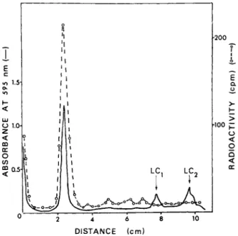
The radioactivity profile and the densitometric scan of the sodium dodecyl sulfate-gel runs of the in vitro products, puri- fied by DEAE-cellulose chromatography, are shown in Fig. 7. A- bout 70% of the counts applied to the gel was recovered in a peak which corresponded to the densitometric peak of the 200,000 dal- ton myosin heavy chain. No detectable amount of radioactivity above the background level was found in the myosin light chain regions of the gels (indicated as LC-^ and L C2 in Fig. 7). This indicates that the mRNAs coding for the light chains (average molecular weight of 20,000) was absent in the 120 S particles.
MESSENGER RIBONUCLEOPROTEINS 403
D I S T A N C E ( c m )
FIGURE 7 Sodium dodecyl sulfate Polyacrylamide gel electro- phoresis of labeled products of in vitro translation of RNA iso- lated from 120 S mRNP particles. The products were copurified with chick embryonic leg muscle myosin added as a carrier by low ionic strength precipitation and DEAE-cellulose chromatography.
For details see Bag et al. (1975). The purified products contain- ing 1200-1400 cpm (specific activity 25,000 cpm/mg) were analyzed using 10.5 cm of 5% gels at 4 mA/gel for 2.5 hr. After staining with Coomassie brilliant blue the gels were scanned at 595 nm, sliced and counted as described by Bag and Sarkar (1975). The migration of the light chains present in the carrier myosin are indicated with arrows and marked as LCj and LC2»
Control experiments using the reticulocyte lysate alone, incu- bated in the absence of exogenously added mRNA, indicated that globin was predominantly synthesized. As judged by the absence of any detectable level of counts above the background in the my- osin heavy band (data not shown), it is concluded that the re- ticulocyte lysate alone does not synthesize any myosin heavy chain. These results indicate that the translated products of the RNA from 120 S particles behaved like chick embryonic myosin heavy chain as judged by a number of properties.
E. Proteins Associated with Actin and Myosin Heavy Chain mRNP Particles
In order to prove that the cytoplasmic mRNP particles de- scribed in the previous section are true cellular entities, the nature of the protein moieties of these particles was investi- gated using two approaches. The relative protein and RNA con- tents were estimated by determining the buoyant densities after fixing the mRNP particles with formaldehyde (Spirin, 1972). The protein components of these particles were also analyzed by sodium dodecyl sulfate Polyacrylamide gel electrophoresis.
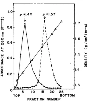
When samples of formaldehyde-fixed 120 S particles and 16-40 S subribosomal particles were centrifuged to equilibrium in pre- formed CsCl gradients, the absorbance profile gave a single peak at a density of 1.40 g/cm3 (Fig. 8 ) . The protein content of these particles, calculated from this value by the method of Spirin
(1972), amounted to 75%. In contrast, the 80S monosomes prepared from embryonic chick muscles gave a peak at a density of 1.57 g/
cm3 (Fig. 8). The buoyant densities of embryonic muscle ribosomal subunits usually ranged between 1.58-1.585 for 60 S and 1.52-1.53 g/cm3 for 40 S subunits, respectively (data not shown here;
Bag and Sarkar, 1976]. These values are in good agreement with those reported in the literature for eukaryotic ribosomes and ribosomal subunits Cfor a review see Spirin, 1972). As judged by the absorbance profile and the absence of any detect- able peak in the density range of 1.5-1.6 g/cm3, the mRNP parti- cles were free of contaminating ribosomal subunits. Further- more, the characteristic buoyant density of 1.4 g/cm3 appears to be a common property of muscle mRNP particles irrespective of their sizes, namely 120 S versus 16-40 S and the nature of the mRNA present, e.g. actin mRNA versus myosin heavy chain mRNA.
The buoyant density of the cytoplasmic muscle mRNP particles re- ported here is in good agreement with the published values in the literature which were obtained mainly by pulse-labeling of RNA in order to detect free mRNPs in a variety of eukaryotic systems
MESSENGER RIBONUCLEOPROTEINS 405
FIGURE 8 Buoyant densities of cytoplasmic mRNP particles and 80 S chick embryonic muscle monosomes. For details see Bag and Sarkar (1975). The pooled subribosomal fractions (16-40 S) as indicated in Fig. 1 and the 120 S particles (Fig. 5) were used as mRNP particles. Centrifugation was done at 35,000 rpm for 40 hr in a Beckman SW 50.1 rotor at 20° using preformed CsCl gradients.
·, absorbance of mRNP particles. The same profile was obtained with both 16-40 S and 120 S particles, o, absorbance of mono- somes. Δ, density of gradient fractions.
(for a review see Spirin, 1972). Since RNA is pelleted in CsCl gradients, the absence of any detectable pellet in the CsCl gradi- ent runs of mRNP particles makes it highly unlikely that the mRNA activity reported in the previous sections is due to any protein- free mRNA species present in the preparations.
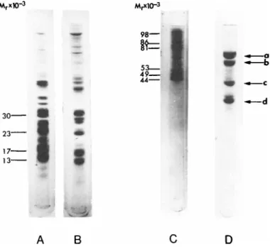
We have also analyzed the carboxymethylated protein samples of ribosomal subunits and mRNP particles by sodium dodecyl sul- fate Polyacrylamide gel electrophoresis. The electrophoretograms are shown in Fig. 9. The majority of typical ribosomal proteins in the molecular weight range of 15,000-30,000, which migrated in the middle region of the gel runs of 60 S and 40 S subunits (gels A and  ) , could not be detected in the mRNP particles (gel C). A
FIGURE 9 Sodium dodecyl sulfate Polyacrylamide gel electro- phoresis of mRNP particles and chick embryonic muscle ribosomal subunits. For details see Bag and Sarkar (1975) and Bag et al.
(1975). Electrophoresis of carboxymethylated samples of mRNP particles and ribosomal subunits was carried out using 10 cm long 10% gels at 4 mA/gel for 4 hr. The gels were stained with Coomas- sie brilliant blue. The amounts of samples applied to the gels are: (A) 0.3 A2QQ unit of 60 S ribosomal subunit; (Â) 0.4 A280 unit of 40 S ribosomal subunit; (C) 0.2 A28O u n it o f 1 2 0 s mRNP
(Fig. 5); (D) a mixed sample of the following markers: 12 \xg of conalbumin (molecular weight 78,000); 8 \ig of bovine serum albu- min (molecular weight 68,000); 6 ]ig of chicken actin (molecular weight 42,000) and 8 ygr of chicken tropomyosin (molecular weight 34,000). The bands corresponding to these 4 marker proteins are indicated with arrows (a-d) in decreasing order of molecular weight.
number of distinct polypeptides, seven to eight, ranging in mo- lecular weight from 44,000-100,000, which do not appear to repre- sent the typical ribosomal proteins, were present in the upper one-third segment of the electrophoretograms of the mRNP parti- cles (gel C ) . The major band of these proteins is a 44,000 com- ponent which was also absent in the ribosomal subunits. As
MESSENGER RIBONUCLEOPROTEINS 407 judged by these characteristic gel patterns and the buoyant densi- ties, it is concluded that the mRNP particles are cytoplasmic en- tities unrelated to and distinct from monoribosome, ribosomal sub- units and mRNA-containing "initiation complexes" of embryonic muscle cells.
F. Are the Protein Moieties Identical in All mRNP Particles In order to test whether cytoplasmic mRNP particles contain- ing mRNAs coding for different myofibrillar proteins contain the same or different polypeptide components, the proteins of two mRNP particles of unequal size, one containing myosin heavy chain mRNA and the other containing actin mRNA, were compared by dodecyl
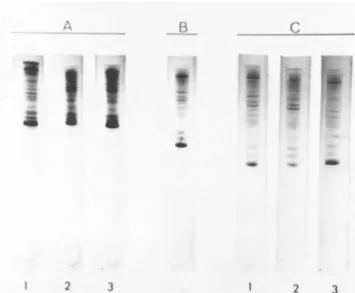
sulfate-polyacrylamide gel electrophoresis. The results are shown in Fig. 10. The electrophoretograms of actin mRNP (gel 2) and a mixed sample of actin and myosin heavy chain mRNPs (gel 3) show that the three faster components in the 44,000-53,000 dalton range (see also Fig. 9 for assignment of molecular weights) are present in both mRNP particles. Three distinct and a number of minor protein bands in the molecular weight range of 44,000-
53,000 (see also Fig. 9) were consistently found in gel runs of myosin heavy chain mRNP. Although protein bands of similar mobi- lities were also found in electrophoretograms of actin mRNP par- ticles, the relative amounts of these high molecular weight com- ponents were always lower than those present in the myosin heavy chain mRNP (panel A, gels 1 and 2). When mixed samples of the two mRNPs were electrophoresed, the three faster components co- migrated together (panel A, gel 3). This pattern did not change even when electrophoresis was run for a longer time (panel Â), or a combination of decreased sample load and increased time of electrophoresis was used to facilitate resolution of the bands
(panel C ) . The results indicate that the protein components of different mRNP particles are very similar. However, the subtle variation in the intensity and number of protein bands in the 81,000-86,000 dalton range of actin and myosin heavy chain mRNP
FIGURE 10 Electrophoresis of carboxymethylated proteins of myosin heavy chain mRNP and actin mRNP particles. For details see Bag and Sarkar (1975) and legends to Fig. 9. The following amounts of samples were used; Panel A : 0.08 A280 unit of actin mRNP (gel 1); 0.1 ¢28Ο unit of myosin heavy chain mRNP (gel 2);
and a mixture of 0.04 ¢230 unit of actin mRNP and 0.05 ¢28Ο unit of myosin heavy chain mRNP9 Panel B: a mixture of 0.04 ¢28Ο unit of actin mRNP and 0.05 A28O unit of myosin heavy chain mRNP.
Panel C: 0.03 A28O unit of actin mRNP (gel 1); 0.04 A2so unit of myosin heavy chain mRNP (gel 2); and a mixture of 0.03 A28O unit of actin mRNP and 0.04 A28O unit of myosin heavy chain mRNP (gel 3). Conditions for electrophoresis was increased to 5 hr for the mixed sample of mRNPs shown in panel  and 6.5 hr for the gels shown in panel C.
particles suggests that there may be subpopulations of mRNP parti- cles containing additional or different proteins. In order to un- derstand the molecular basis of this phenomenon we are continuing these studies using mRNP particles prepared and purified by dif- ferent methods.
G. In Vitro Translation of mRNP Particles
Since mRNP particles have been implicated in the process of stabilization of mRNAs during the transition of myoblasts to myo- tube stages (Buckingham et al., 1974), it is of interest to study whether or not the mRNP particles represent "non-translatable"
MESSENGER RIBONUCLEOPROTEINS 409 TABLE II Translation of Myosin Heavy Chain mRNP Particle
and Protein-free mRNA in Rabbit Reticulocyte Lysate Systema
mRNP added % of total cpm (equivalent incorporated in mRNA added y g y g RNA) myosin heavy chain band*3
Not detectable
6 2.65
10 5.10 15 5.70 20 5.84 30 5.71 5 - 2.95 10 - 5.84 15 - 7.43 20 - 8.31 30 - 8,37
translation of 120 S myosin heavy chain mRNP (Fig. 5) and protein-free mRNA isolated from the mRNP were done as described by Bag et al. (1975).
bThe in vitro products copurified with carrier myosin (for de- tails see Mondai et al., 1974; Bag et al., 1975) were electro- phoresed and the radioactivities in the myosin heavy chain bands were determined.
forms of mRNAs. If the mRNP particles are found to be inactive in an in vitro translation system, this would suggest that they may regulate the biosynthesis of myofibrillar proteins through a negative control, i.e., the mRNP particles in the myoblast stage are blocked in translation. Removal of this negative control, e.
g., in the myotube stage, would lead to an accelerated rate of cell-specific protein synthesis. In order to test this possibili- ty we have compared the relative efficiency of in vitro transla- tion of myosin heavy chain mRNP with that of protein-free myosin heavy chain mRNA using the rabbit reticulocyte lysate system.
The products from the in vitro incubations were copurified with carrier myosin and analyzed by sodium dodecyl sulfate gel electrophoresis as described in the previous section. The radio-
activity recovered in the myosin heavy chain band is calculated as per cent of the total radioactivity incorporated in the cell-free
system. The results presented in Table II indicate that the myo- sin heavy chain mRNP particle was able to program the de novo synthesis of the myosin heavy chain in a heterologous system.
Further, at low concentrations of mRNA or equivalent amount of mRNP the relative efficiencies of translation did not show any significant difference. However, at saturating concentration about 30% higher incorporation was achieved with protein-free mRNA. These results clearly indicate that the mRNP particle, at
least in the rabbit reticulocyte lysate system, does not exhibit any stringent negative control in translation. Since it is quite likely that the in vivo regulation of translation of mRNAs may be different from the in vitro conditions, other possible interpre- tations regarding the role of mRNP particles in myogenesis during the terminal differentiation of muscle cells should also be care- fully considered. Further discussion along these lines is pre- sented in the next section.
III. DISCUSSION
The studies presented here show that mRNAs coding for two im- portant myofibrillar proteins, actin and myosin heavy chain, are present as nonpolysomal cytoplasmic mRNP particles in embryonic muscle cells. These particles are true cytoplasmic entities un- related to ribosomes, as shown by the absence of typical ribosom- al proteins, by their characteristic buoyant density and the presence of a unique class of nonribosomal protein moieties firm- ly bound to them. The mRNAs isolated from these particles direct the synthesis of polypeptides in cell-free systems which are in- distinguishable from authentic skeletal muscle actin and myosin heavy chain. These results, considered together with the fact that the myosin heavy chain mRNP itself can be translated in het-
MESSENGER RIBONUCLEOPROTEINS 411
erologous system, indicate that the mRNAs are present in these particles in a form which can be translated without further proc- essing.
It is currently believed that mRNAs exist as ribonucleopro- tein particles in the nucleus and cytoplasm of a large number of eukaryotic cells (for a review see Spirin, 1972). However, the nature and possible role of these proteins bound to these parti- cles is not thoroughly understood. Cytoplasmic polyribosomal mRNPs isolated by dissociation of polysomes from a number of
species and tissues contain, as judged by sodium dodecyl sulfate gel electrophoresis, two major protein bands of approximate mo- lecular weights 52,000 and 78,000 and a large number of minor bands ranging from 6-13 (for a review see Blobel, 1973). The 78,000 dalton component, which is present in both nuclear and polysomal mRNPs, is specifically bound to the poly (A) segment at the 3*-end of eukaryotic mRNAs (Blobel, 1973; Quinlan et al., 1974; Firtel and Pederson, 1975). The results presented here in- dicate that the cytoplasmic muscle mRNP particles contain seven to eight distinct polypeptides including a prominent 44,000 dalton component. Among these major protein components are bands cor- responding to the 52,000 and 78,000 dalton components previously reported in many eukaryotic mRNPs. The electrophoretic pattern of the cytoplasmic mRNA-associated proteins described in these
studies supports the recent view that proteins associated with eukaryotic mRNPs are much more complex with respect to size and number than previously thought (for a review see Barrieux et al., 1975).
Previous reports have shown that specific mRNP particles, viz.
globin á-chain mRNP, globin á and 3 chain mRNPs and histone mRNP, are present in rabbit reticulocytes (Jacobs-Lorena and Baglioni, 1972), chick erythrocytes (Spohr et al., 1972) and unfertilized sea urchin eggs (Gross et al., 1973) respectively. While the sea urchin embryo during the early stages of development may be con- sidered as an undifferentiated cell, the reticulocyte and eryth-
rocyte represent a specific type of cell engaged primarily in the synthesis of only one protein, viz. globin, which accounts for about 80% of the cellular proteins. Embryonic muscle cells, on the other hand, are a terminally differentiated eukaryotic system which exhibit many unique characteristics. These include a post- mitotic and non-replicating chromosome, a highly ordered contrac- tile apparatus formed by the post-translational assembly of a large number of myofibrillar proteins and translational control of gene expression during differentiation. The work presented here describes the first successful isolation and partial charac- terization of actin and myosin heavy chain mRNP particles. As- suming that highly purified myosin heavy chain mRNP contains 1 mole of mRNA per mole, we have calculated the molecular weight of the mRNP from the known molecular weight of the purified mRNA and the RNA/protein ratio of the mRNP. This estimate gives a value of 8.11 x 1 06 which is in the expected range for an mRNP of this size (Spirin, 1972). Previous reports in the literature on myosin heavy chain mRNP were based on detection using either pulsed RNA
(Buckingham et al., 1974; Buckingham and Gross, 1975) or nonpoly- somal sedimentation of the mRNA in gradient runs (Heywood et al., 1975).
The ability of the myosin heavy chain mRNP to program the in vitro synthesis of myosin heavy chain, as reported here, indi- cates that the binding of specific proteins to the mRNA per se does not exert a stringent negative control on the translation process. In view of these results the stabilization of muscle- specific mRNAs as cytoplasmic mRNPs during the myoblast to myo- tube transition of cultured muscle cells (Buckingham et al., 1974) may be interpreted as a mechanism by which the concentra- tion of "translatable" mRNAs of the cell is increased. According to this view the mRNAs are stored as biologically active rather than inactive species in a cytoplasmic pool. The entry of the mRNAs from this compartment to the polysomes may be a key regula- tory event in translation during the terminal differentiation of
MESSENGER RIBONUCLEOPROTEINS 413 muscle cells. This model of translational control involving
mRNPs also explains the accelerated rate of cell-specific protein synthesis observed in the post-fusion stages of cultured muscle cells. The details of the transit of mRNAs from mRNP particles to polysomes is now being investigated by us. The 44,000 dalton protein, which we have observed in all classes of mRNPs and which
seem to be absent in polysomal mRNPs derived from embryonic mus- cle cells (manuscript in preparation) may be involved in some as- pect of this translational control.
Bester et al. (1975) have recently reported the isolation of a dialyzable, low molecular weight RNA species from the postpoly- somal supernatant of embryonic muscles. They have postulated that this RNA species exerts a negative control of translation on cy- toplasmic mRNP particles. In view of the results presented here, the regulation of translation by biologically active cytoplasmic mRNP particles during the growth and differentiation of muscle cells through a positive control, as discussed above, is favored by us.
ACKNOWLEDGMENTS
The authors thank Drs, John Gergely, Henry Paulus and Helga Boedtker for many helpful discussions; Dr. Paul C. Leavis for his advice on the isolation and characterization of actin; and Dr.
Raman K. Roy for his help in the preparation of globin mRNA and wheat germ embryo S-30 cell-free system. It is a pleasure to acknowledge the skillful technical assistance of the Misses Ann Sutton and Ven-jim Chen. This work was supported by grants from the National Institutes of Health (AM 13238), the American Heart Association (No. 71-915), and the Muscular Dystrophy Associations of America, Inc. This work was initiated during the tenure of a research fellowship to J. Bag from the American Heart Associa- tion, Massachusetts Affiliate, Inc.; more recent work has been
carried out during the tenure of a fellowship of the Medical Foun- dation, Inc., Boston, Massachusetts.
REFERENCES
Bag, J. and Sarkar, S. (1975)t Biochemistry 14, 3800-3807.
Bag, J. and Sarkar, S. (1976). J. Biol. Chem. 251, 7600-7609 Bag, J., Roy, R. Ê., Sutton, A. and Sarkar, S. (1975). In "Regula-
tion of Growth and Differentiated Function in Eukaryote Cells"
(G. P. Talwar, ed.), pp. 111-127. Raven Press, New York.
Barrieux, Á., Ingraham, Ç. Á., David, D. N. and Rosenfeld, M. G.
(1975). Proc. Nat. Acad. Sei. U.S.A. 72, 1523-1527.
Blobel, G. (1973). Proc. Nat. Acad. Sei. U.S.A. 70, 924-928.
Brawerman, G. (1974). Ann. Rev. Biochem. 43, 621-642.
Buckingham, M. E., Caput, D., Cohen, Á., Whalen, R. G. and Gross, F. (1974). Proc. Nat. Acad. Sei. U.S.A. 71, 1466-1470.
Buckingham, M. E. and Gross, F. (1975). FEBS Lett. 53, 355-359.
Chacko, S. and Xavier, J. (1974). Dev. Biol. 40, 340-354.
Elzinga, M. (1970). Biochemistry 9, 1365-1374.
Firtel, R. Á., and Pederson, T. (1975). Proc. Nat. Acad. Sei.
U.S.A. 72, 301-305.
Gross, K. W., Jacobs-Lorena, M., Baglioni, C. and Gross, P. R.
(1973). Proc. Nat. Acad. Sei. U.S.A. 70, 2614-2618.
Heywood, S. M. and Rich, A. (1968). Proc. Nat. Acad. Sei. U.S.A.
59, 590.
Heywood, S. M. and Nwagwu, M. (1969). Biochemistry 8, 3839-3845.
Heywood, S. Ì., Kennedy, D. S. and Bester, A. J. (1975). FEBS Lett. 53, 69-72.
Holtzer, Ç., Sanger, J. W., Ishikawa, H. and Strahs, K. (1972).
Cold Spring Harbor Symp. Quant. Biol. 37, 549-566.
Jacobs-Lorena, M. and Baglioni, C. (1972). Proc. Nat. Acad. Sei.
U.S.A. 69, 1425-1428.
Lowey, S. and Risby, D. (1971). Nature 234, 81-85.
Martin, R. G. and Ames, Â. N. (1961). J. Biol. Chem. 236, 1372-
MESSENGER RIBONUCLEOPROTEINS 415
1379.
Mondai, H., Sutton, Á., Chen, V. and Sarkar, S. (1974). Biochem.
Biophys. Res. Commun. 56, 988-995.
Morris, G. Å., Buzash, Å. Á., Rourke, A. W., Tepperman, Ê., Thomp- son, W. C. and Heywood, S. M. (1972). Cold Spring Harbor Symp.
Quant. Biol. 37, 535-541.
Pollard, T. D. and Weihing, R. R. (1974). Crit. Rev. Biochem. 2, 1-65.
Potter, J. D. and Gergely, J. (1974). Biochemistry 13, 2697-2703.
Quinlan, T. J., Billings, P. B. and Martin, T. E. (1974). Proc.
Nat. Acad. Sei. U.S.A. 71, 2632-2636.
Roberts, Â. E. and Paterson, Â. M. (1973). Proc. Nat. Acad. Sei.
U.S.A. 70, 2330-2334.
Rubinstein, Ν. Á., Chi, J. C. H. and Holtzer, H. (1974). Biochem.
Biophys. Res. Commun. 57, 438-445.
Sarkar, S. and Cooke, P. H. (1970). Biochem. Biophys. Res. Commun.
41, 918-925.
Sarkar, S., Sreter, F. A. and Gergely, J. (1971). Proc. Nat. Acad.
Sei. U.S.A. 68, 946-950.
Sarkar, S. (1972). Cold Spring Harbor Symp. Quant. Biol. 37, 14- 17.
Sarkar, S., Mukherjee, S. P., Sutton, Á., Mondai, H. and Chen, V.
(1973). Prep. Biochem. 3, 583-604.
Sarkar, S. (1974). In "Exploratory Concepts in Muscular Dystrophy II" (A. T. Milhorat, ed.), pp. 172-184. Excerpta Medica, Am- sterdam.
Sarkar, S. (1975). In "Eukaryotes at the Subcellular Level: De- velopment and Differentiation" (J. Last, ed.), pp. 315-368.
Marcel Dekker, New York.
Spirin, A. S. (1972). In "The Mechanism of Protein Synthesis and Its Regulation" (L. Bosch, ed.), pp. 515-537. North-Holland Publishing, Amsterdam.
Spohr, G., Kayibanda, B. and Scherrer, K. (1972). Eur. J. Bio- chem. 31, 194-208.
Swank, R. T, and Munkres, K. D. (1971). Anal. Biochem. 39, 462- 477.
Taylor, E. W. (1972). Ann. Rev. Biochem. 41, 577-616.
Weeds, A. G. and Frank, G. (1972). Cold Spring Harbor Symp. Quant.
Biol. 37, 9-14.
Yaffe, D. and Dym, H. (1972). Cold Spring Harbor Symp. Quant.
Biol. 37, 543-547.