nanomaterials
Review
From Single-Core Nanoparticles in Ferrofluids to Multi-Core Magnetic Nanocomposites:
Assembly Strategies, Structure, and Magnetic Behavior
Theodora Krasia-Christoforou1, Vlad Socoliuc2 , Kenneth D. Knudsen3 , Etelka Tombácz4 , Rodica Turcu5,* and Ladislau Vékás2,*
1 Department of Mechanical and Manufacturing Engineering, University of Cyprus, 75 Kallipoleos Avenue, P.O. Box 20537, Nicosia 1678, Cyprus; krasia@ucy.ac.cy
2 Laboratory of Magnetic Fluids, Center for Fundamental and Advanced Technical Research, Romanian Academy–Timisoara Branch, Mihai Viteazul Ave. 24, 300223 Timisoara, Romania;
vsocoliuc@gmail.com
3 Department for Neutron Materials Characterization, Institute for Energy Technology (IFE), 2027 Kjeller, Norway; Kenneth.knudsen@ife.no
4 Soós Ern˝o Water Technology Research and Development Center, University of Pannonia, Zrínyi M. Str. 18., H-8800 Nagykanizsa, Hungary; tombacz@chem.u-szeged.hu
5 Department of Physics of Nanostructured Systems, National Institute for Research and Development of Isotopic and Molecular Technologies, Donat Str. 67-103, 400293 Cluj-Napoca, Romania
* Correspondence: rodica.turcu@itim-cj.ro or rodica.turcu14@gmail.com (R.T.);
vekas.ladislau@gmail.com or vekas@acad-tim.tm.edu.ro (L.V.)
Received: 19 September 2020; Accepted: 20 October 2020; Published: 31 October 2020
Abstract:Iron oxide nanoparticles are the basic components of the most promising magnetoresponsive nanoparticle systems for medical (diagnosis and therapy) and bio-related applications. Multi-core iron oxide nanoparticles with a high magnetic moment and well-defined size, shape, and functional coating are designed to fulfill the specific requirements of various biomedical applications, such as contrast agents, heating mediators, drug targeting, or magnetic bioseparation. This review article summarizes recent results in manufacturing multi-core magnetic nanoparticle (MNP) systems emphasizing the synthesis procedures, starting from ferrofluids (with single-core MNPs) as primary materials in various assembly methods to obtain multi-core magnetic particles. The synthesis and functionalization will be followed by the results of advanced physicochemical, structural, and magnetic characterization of multi-core particles, as well as single- and multi-core particle size distribution, morphology, internal structure, agglomerate formation processes, and constant and variable field magnetic properties. The review provides a comprehensive insight into the controlled synthesis and advanced structural and magnetic characterization of multi-core magnetic composites envisaged for nanomedicine and biotechnology.
Keywords: magnetic nanoparticle systems; ferrofluids; magnetic fluids; single core; multi-core;
clusters; synthesis; functional coating; physical–chemical properties; structural characterization;
magnetic characterization; small-angle scattering techniques; nanomedicine; biotechnology
1. Introduction
Hybrid structures of colloidal nanoparticles designed for nanobiotechnology and nanomedicine [1–8], among them multifunctional magnetic nanoparticle–biomolecule–polymer hybrid systems with complex composition and topology [9–12], are receiving continuously increasing interest for medical
Nanomaterials2020,10, 2178; doi:10.3390/nano10112178 www.mdpi.com/journal/nanomaterials
Nanomaterials2020,10, 2178 2 of 67
diagnosis and treatment due to the newly acquired performances [13–24]. The required stability of superparamagnetic iron oxide nanoparticle (NP) systems in biological media target specific functionalities and selective drug delivery toward targeted locations [25–29] are ensured by molecular design of the dispersant/functional shell around the magnetic core [30–32]. Various interactions—van der Waals, electrostatic, molecular, entropic, hydrophobic, and magnetic—are contributing to the nanoscale self-assembly of nanoparticles [33–43] and provide colloidal nanoparticle clusters [44,45], in particular magnetoresponsive nanocomposite particles by merging magnetic and polymer materials with new collective properties, such as enhanced long-term stability and magnetic field-driven functionalities [43,46–49]. Among these, multi-core composites built up by magnetic nanoparticles embedded in non-magnetic matrices offer a composition, size, and structure dependent, sometimes nonlinear response to a constant or time-varying magnetic field [50–56]. In this way, a large variety of carefully engineered magnetoresponsive particles manufactured over time proved to be highly promising for nanomedicine, magnetic cell sorting, magnetic separations in biotechnology and environment purification, actuation, or catalysis [47,57–74].
It is essential to evidence that in contrast with agglomerates of single-core particles encountered in ferrofluids having weak colloidal stability, multi-core particles are the result of assembling a number of cores within a matrix. The number of cores in a multi-core particle is not changing with time, even in a magnetic field or intense shearing. The packing density, i.e., the distance between cores and also the size of the cores determines the intensity of magnetic interactions within the system and, finally, the cooperative magnetic behavior of multi-core particles [75–77]. Compared with single magnetic nanoparticles, the multi-core magnetic nanoparticles with a higher magnetic moment afford a considerable enhancement of the magnetic response [44,78,79], providing a significant driving force needed by most of the applications mentioned above. In order to assess the magnetic targeting/fixing applicability of magnetic particles, the magnetic moment of the particles is more relevant than mass magnetization [80,81].
In the above context, it is worthwhile mentioning that “bio-ferrofluids” with single and multi-core [82,83] or mostly multi-core magnetic particles (also commercial products) [84,85]
extended significantly the category of “true” ferrofluids, containing practically only single-core magnetic nanoparticles dispersed in the carrier liquid. Among others, the addition of multi-core magnetic particles into a single-core ferrofluid reduces the long-term colloidal stability and changes completely the flow behavior in the magnetic field of the initially Newtonian ferrofluid [86]. To ensure kinetic stability and to avoid spontaneous aggregation in biorelevant media (for certain pH, salt, and protein concentration values) optimized and application determined surface coating (e.g., with polyelectrolytes) of iron oxide NPs is required for advanced bio-ferrofluid products manufactured for in vivo usage [87,88]. IONP’s surface chemistry and different coating/functionalization strategies to enhance the colloidal stability of bio-ferrofluids, involving natural polymers (dextran, chitosan, alginate), synthetic polymers (PEG, poly(ethylene glycol);
PVP, poly(vinylpyrrolidone); PVA, poly(vinyl alcohol); PAA, poly(acrylic acid), etc.), dendrimers, dendrons, silanes, non-porous and porous silica, were thoroughly evaluated by Felder-Flesch and collaborators, including the relationship between coating and magnetic properties [89].
Colloidally stable ferrofluids with organic or aqueous carriers, in particular the single-core magnetic nanoparticles in their composition, proved to be a highly versatile and well-defined primary nanomaterial for manufacturing controlled magnetoresponsive superstructures of various morphologies [55,90–94]. In addition, nanostructures are assembled using the magnetostatic interaction between effectively diamagnetic and paramagnetic particles within a magnetized ferrofluid [35,95,96], the “magnetic holes” mechanism [97], allowing for a reversible assembling–disassembling process of practically non-magnetic particles [98] and also for the label-free manipulation and separation of cells and microorganisms using a ferromicrofluidic platform [99]. The magnetic hole mechanism was extended to the magnetic assembly of diamagnetic and magnetic particles immersed in a ferrofluid considered as a tunable magnetic continuum that controls the interactions between particles of
Nanomaterials2020,10, 2178 3 of 67
different magnetization and sizes [100]. Specially designed composite spheres with embedded monodispersed micromagnets in a suspension—described as magnetic suspensions with shifted dipoles [101]—can self-organize in well-defined reconfigurable multi-core structures by simple magnetostatic interactions [102,103].
Among the remotely controlled endogenous (pH variation, enzymes etc.) or exogenous (e.g., light, temperature, electric field, magnetic field) stimuli-responsive nanoassemblies, designed to ensure dosage, spatial, and temporal controllability, the magnetic field driven bio-nanocomposites attracted tremendous scientific and technological interest [6,43,104]. In this context of magnetism-based nanomedicine and biotechnology, we will focus mainly on theferrofluid-based generation of multi-core magnetic nanocomposites, which are motivated by the relevance and maturity of magnetic fluids technology in providing large quantities of high-performance ferrofluids for various biomedical and engineering applications [105–111]. In addition to numerous assembly procedures for different shapes of multi-core magnetic particles with an application-specific design of composition and functionalization, the paper presents the results of advanced characterization methods (transmission electron microscopy, TEM; scanning electron microscopy, SEM; high-resolution electron microscopy, HRTEM; dynamic light scattering, DLS) and zeta potential, X-ray, and neutron scattering techniques (small-angle x-ray scattering, SAXS; small-angle neutron scattering, SANS; polarization analyzed SANS, PASANS; very small-angle neutron scattering, VSANS; neutron reflectometry, NR; and magnetometry) and discusses magnetic properties in a constant and variable magnetic field, as well as particle structure (size, polydispersity, stabilizing shell thickness, composition of particle core and shell), magnetic structure (magnetic size and composition), particle interaction (interparticle potential, magnetic moment correlation, phase separation), cluster and supraparticle formation (developed aggregation and chain/bundle formation).
2. Multi-Core Superparamagnetic Nanospheres and Microspheres
2.1. Emulsion Procedures
Magnetic emulsions [112,113] composed of ferrofluid droplets dispersed in a non-miscible liquid can be successfully turned into superparamagnetic nanocomposite particles, usually of spherical shape. The controlled clusterization of magnetic nanoparticles using the miniemulsion technique [90,114–116], followed by encapsulation of the densely packed magnetic clusters in a polymer shell [62,117,118], is a successful joining of ferrofluid technology and emulsion procedures to provide highly magnetoresponsive multi-core particles [45,119–125]. This two-step colloidal assembly process has several important advantages [126]: (a) high-quality MNP building blocks can be produced by wet-chemical synthetic procedures of ferrofluids; (b) the MNPs are kinetically stable in the ferrofluid, and their clustering is initiated by some external trigger depending on the nature and surface chemistry of MNPs; and (c) size-controlled MNP clusters can be prepared if the MNP–MNP interaction potentials are engineered in an appropriate fashion. It is important to emphasize that MNP cluster formation is closely related to ferrofluid colloidal stability, i.e., the absence of aggregates in the ferrofluid used as primary nanomaterial is an essential feature [127].
The formation of ferrofluid droplets in emulsion followed by solvent evaporation to trigger nanoparticle clustering is a facile process to obtain closely packed and size-controlled clusters of magnetic NPs [128]. The basic procedure is represented schematically in Figure1I [122]. To exemplify, a small volume of hexane-based ferrofluid (200µL, concentration varies from 1 to 100 mg/mL) was added to 4 mL of 0.1 M CTAB (cetyl trimethylammonium bromide) aqueous solution. Then, all of the liquid was gently mixed by handshaking, followed by sonication for 2 min to form a stable micelle suspension. Afterwards, the mixture was heated in an 80◦C water bath and stirred at 500 rpm for 5 min so that the majority of the hexane was evaporated. Alternatively, hexane evaporation can be done by stirring under ambient conditions for several hours. Then, the solution was removed from the heat and stirred under a vacuum for 30 min to completely remove the hexane. By finishing solvent
Nanomaterials2020,10, 2178 4 of 67
evaporation, the interparticle van der Waals attractions increase and the particles stick to one another, forming densely packed spherical nanoparticle clusters (NPC) [55,119,129].
Nanomaterials 2020, 10, 2178 4 of 67
(I)
(II) Figure 1.Cont.
Nanomaterials2020,10, 2178 5 of 67
Nanomaterials 2020, 10, 2178 5 of 67
(III)
(IV)
Figure 1.Cont.
Nanomaterials2020,10, 2178 6 of 67
Nanomaterials 2020, 10, x FOR PEER REVIEW 6 of 69
(V)
Figure 1. Magnetic nanoclusters prepared by emulsion procedures. (I) Preparation of clusters of magnetic nanoparticles: schematic of the oil‐in‐water miniemulsion procedure using hexane (or toluene)‐based ferrofluid (Reprinted with permission from [122]. Copyright 2010 American Chemical Society); (II) Magnetic nanoparticle clusters stabilized with SDS: (a) TEM image (the scale bar is 200 nm); (b) the diameter distribution. (c) HRTEM image of magnetic clusters coated with polyacrylic
acid; (d) HRTEM image of magnetic clusters coated with pNIPA (poly(N‐isopropylacrylamide)–pAA
(polyacrylic acid) (republished with permission of Royal Society of Chemistry, from [127]; permission conveyed through Copyright Clearance Center, Inc.); (III) (A) Dynamic light scattering and transmission electron microscopy image (insert) of PpIX‐coated SPION nanoclusters; (B) Magnetic resonance (MR) relaxometry measurements of nanoclusters. An MR phantom image (inset) of nanoclusters at various concentrations in a microplate was also collected (republished with the permission of John Wiley and Sons, from [130]; permission conveyed through Copyright Clearance Center, Inc.). (IV) TEM and SEM micrographs of amphiphilic block copolymer (PDMAEMA) stabilized magnetic latex particles synthesized by a seeded semi‐batch emulsion polymerization of styrene in the presence of increasing amounts of DVB: 12 wt %, 23 wt %, and 38 wt % (based on overall monomer mass), introduced either in the initial load or in both the initial load and the feed. Scale bar:
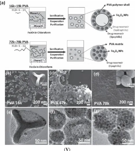
100 nm (republished with permission of Royal Society of Chemistry, from [131]. (V) Single emulsion particles (SEP) and double emulsion capsules (DEC). Primary material: chloroform‐based Fe3O4 ferrofluid (a) Schematic of one‐step emulsion synthesis incorporating iron oxide (IO) nanoparticles and single‐component polymer poly(vinyl alcohol) (PVA). PVA with a MW of 16,000 and 19,000 g/mol gives double emulsion capsules, PVA with a MW of 72,000 and 78,000 g/mol gives single emulsion particles (SEP), PVA with a MW of 23,000 to 67,000 g/mol gives mixtures of both types. SEM and corresponding TEM images of dried DEC‐IO and SEP‐IO are shown in (b,e) for PVA‐16k, (c,f) for PVA‐47k, and (d,g) for PVA‐78k. (Republished with permission of John Wiley and Sons, from [132];
permission conveyed through Copyright Clearance Center, Inc.).
Figure 1. Magnetic nanoclusters prepared by emulsion procedures. (I) Preparation of clusters of magnetic nanoparticles: schematic of the oil-in-water miniemulsion procedure using hexane (or toluene)-based ferrofluid (Reprinted with permission from [122]. Copyright 2010 American Chemical Society); (II) Magnetic nanoparticle clusters stabilized with SDS: (a) TEM image (the scale bar is 200 nm);
(b) the diameter distribution. (c) HRTEM image of magnetic clusters coated with polyacrylic acid;
(d) HRTEM image of magnetic clusters coated with pNIPA (poly(N-isopropylacrylamide)–pAA (polyacrylic acid) (republished with permission of Royal Society of Chemistry, from [127];
permission conveyed through Copyright Clearance Center, Inc.); (III) (A) Dynamic light scattering and transmission electron microscopy image (insert) of PpIX-coated SPION nanoclusters; (B) Magnetic resonance (MR) relaxometry measurements of nanoclusters. An MR phantom image (inset) of nanoclusters at various concentrations in a microplate was also collected (republished with the permission of John Wiley and Sons, from [130]; permission conveyed through Copyright Clearance Center, Inc.). (IV) TEM and SEM micrographs of amphiphilic block copolymer (PDMAEMA) stabilized magnetic latex particles synthesized by a seeded semi-batch emulsion polymerization of styrene in the presence of increasing amounts of DVB: 12 wt %, 23 wt %, and 38 wt % (based on overall monomer mass), introduced either in the initial load or in both the initial load and the feed. Scale bar:
100 nm (republished with permission of Royal Society of Chemistry, from [131]. (V) Single emulsion particles (SEP) and double emulsion capsules (DEC). Primary material: chloroform-based Fe3O4
ferrofluid (a) Schematic of one-step emulsion synthesis incorporating iron oxide (IO) nanoparticles and single-component polymer poly(vinyl alcohol) (PVA). PVA with a MW of 16,000 and 19,000 g/mol gives double emulsion capsules, PVA with a MW of 72,000 and 78,000 g/mol gives single emulsion particles (SEP), PVA with a MW of 23,000 to 67,000 g/mol gives mixtures of both types. SEM and corresponding TEM images of dried DEC-IO and SEP-IO are shown in (b,e) for PVA-16k, (c,f) for PVA-47k, and (d,g) for PVA-78k. (Republished with permission of John Wiley and Sons, from [132]; permission conveyed through Copyright Clearance Center, Inc.).
Nanomaterials2020,10, 2178 7 of 67
Once the oil phase (hexane or toluene) is left to evaporate (water is added to keep the volume constant), the primary droplets transform into clusters, while the solvent swollen micelles form a mixture of empty micelles and free surface-active agent (CTAB). Throughout the ripening process, the primary droplets maintain a constant number of MNPs, since the nanoparticles do not have high enough water solubility to diffuse from the primary droplet [123]. The controlled clustering of magnetic nanoparticles from ferrofluids allows tailoring the size and magnetic moment of the particles. Using a toluene-based ferrofluid containing oleic acid-coated Fe3O4nanoparticles, high magnetization magnetic clusters were prepared by the oil-in-water miniemulsion method [127,133]. The ferrofluid was added to the aqueous phase containing the surfactant (SDS or CTAB), and the mixture was ultrasonicated for 2 min to obtain small stable droplets of magnetic fluid in water. Then, the as-prepared miniemulsion was heated at 100◦C to remove the toluene and then carefully washed several times with a methanol–water mixture, magnetically separated, and redispersed in water. In a second step, these magnetic clusters coated with the surfactants SDS or CTAB were encapsulated into polymers.
The TEM image in Figure1IIa evidences the closed packed spherical clusters of approximately 100 nm mean size (Figure1IIb) prepared from a toluene-based ferrofluid. The magnetization curves of magnetic clusters at room temperature show no measurable hysteresis or coercivity, which is consistent with the superparamagnetic behavior. The saturation magnetization of magnetic clusters has relatively high values: MS=63.9 A·m2/kg for magnetic clusters stabilized with SDS and MS=76.7 A·m2/kg for magnetic clusters stabilized with CTAB. Figure1IIc,d provides the HRTEM images of closely packed magnetite NP clusters with pAAc and pNIPA–pAAc functional polymer coatings, respectively.
Anion exchange and cation exchange magnetic microgels manufactured using the SDS and CTAB stabilized clusters provided remarkable performances in High Gradient Magnetic Separation (HGMS) processes [127,134]. Protoporphyrin IX (PpIX)-coated SPION nanoclusters were formed in a highly reproducible microemulsion procedure [130] by dissolving PpIX and small hydrophobic SPIONs (physical diameter=7.3±1.0 nm) in toluene, adding this mixture to water, followed by sonication.
The PpIX-coated SPION nanoclusters dispersed in water have an average hydrodynamic diameter of≈37 nm with a polydispersity index (PDI) of 0.22 (Figure1IIIA), which is in good agreement with the sizes of tightly packed spherical cores of SPIONs evidenced by transmission electron microscopy (TEM). The superparamagnetic nanoclusters showed enhanced T2-contrast (i.e., hypointensity) compared to the control samples (Figure1IIIB inset). The same emulsification/solvent evaporation technique was applied using a commercial ferrofluid (fatty acid-coated magnetic nanoparticle powder (Ferrotec Co.) dispersed in toluene) to obtain iron oxide NP clusters used as seeds in a semi-continuous procedure of surfactant-free emulsion polymerization of styrene (with or without DVB (divinylbenzene), cross-linking agent) [131]. The resulting poly(2-dimethylaminoethyl methacrylate)-b-polystyrene (PDMAEMA-b-PS) amphiphilic block copolymer encapsulated magnetite nanoclusters have a well-defined core shell structure for a higher amount of DVB, as evidenced in SEM and TEM micrographs (Figure1IV). The preparation of core–shell type, double emulsion capsules (DEC) usually requires a two-step emulsifying process. According to [132] (Figure1Va), these capsules are manufactured by a surfactant-free one-step emulsifying process, in which the oleic acid coated iron oxide nanoparticles of a chloroform-based ferrofluid were acting as DEC stabilizers, while poly(vinyl alcohol) (PVA) acted as both the shell constituent and the surfactant. SEM and corresponding TEM images of dried double emulsion and single emulsion iron oxide composite particles (DEC-IO and SEP-IO) are shown in Figure1Vb–e.
In addition, in an emulsion procedure involving a second generation and clinically used photosensitizer (chlorin e6) and toluene-based ferrofluid with OA-coated Fe3O4 nanoparticles, nanoclusters with an average hydrodynamic diameter of 96.38± 4.6 nm were manufactured for dual mode imaging and photodynamic therapy [135]. A chloroform-basedγ-Fe2O3/Fe3O4ferrofluid was used as the polymer solvent/oil phase in the emulsion solvent evaporation process (ESE) for manufacturing SPION/polymer hybrid particles, starting from an oil-in-water emulsion procedure [136].
The initial core sizes in the ferrofluid ranged from 4 to 15 nm, whereas the assembling process using
Nanomaterials2020,10, 2178 8 of 67
polystyrene dissolved also in chloroform resulted in flower-shaped surfactant-stabilized polystyrene beads with sizes between 50 and 250 nm [137]. The magnetic ordering of single-core NPs accomplished by their close assembly within the nanoflower multi-core domains resulted in a significant enhancement of the specific absorption rate (SAR), in comparison to single domain iron oxide nanoparticles subjected to the same alternating magnetic field conditions.
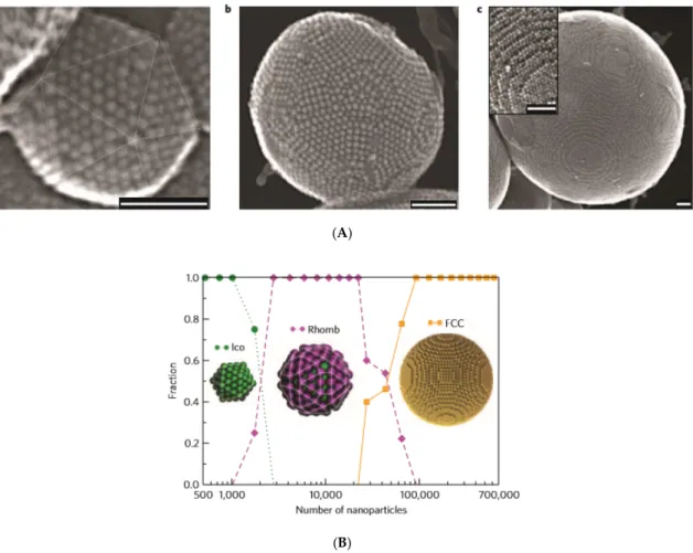
An oil-in-water miniemulsion procedure was applied to synthesize large magnetoresponsive supraparticles by entropy-driven clustering in a spherical confinement of oleic acid-coated cobalt–ferrite NPs of an apolar organic solvent (cyclohexane)-based ferrofluid [138], Figure2A,B.
Nanomaterials 2020, 10, x FOR PEER REVIEW 8 of 69
polystyrene beads with sizes between 50 and 250 nm [137]. The magnetic ordering of single‐core NPs accomplished by their close assembly within the nanoflower multi‐core domains resulted in a significant enhancement of the specific absorption rate (SAR), in comparison to single domain iron oxide nanoparticles subjected to the same alternating magnetic field conditions.
An oil‐in‐water miniemulsion procedure was applied to synthesize large magnetoresponsive supraparticles by entropy‐driven clustering in a spherical confinement of oleic acid‐coated cobalt–
ferrite NPs of an apolar organic solvent (cyclohexane)‐based ferrofluid [138], Figure 2A,B.
(A)
(B)
Figure 2. Entropy‐driven supraparticle assembly. (A) Secondary electron scanning transmission electron microscopy (SE‐STEM) images of typical supraparticles containing oleic acid coated cobalt–
iron oxide nanoparticles initially dispersed in cyclohexane (core diameter 6 nm; hydrodynamic diameter 9 nm). (a) Supraparticle with a diameter of 105 nm with Mackay icosahedral symmetry, as indicated by the thin lines. (b) 216 nm supraparticle with anti‐Mackay rhombicosidodecahedral structure. (c) 734 nm supraparticle consisting of a single face‐centered cubic (FCC) crystal domain.
Inset: a magnified view of the step edges of the FCC supraparticle. All scale bars are 50 nm. (B) Size dependence of the cluster structure–event‐driven molecular dynamics (EDMD) numerical simulation.
Structural transition from a Mackay icosahedron (Ico) to an anti‐Mackay rhombicosidodecahedron (Rhomb) to a face‐centered cubic (FCC) cluster, as observed for supraparticles consisting of nanoparticles. The fraction of structures, based on 121 supraparticles is plotted as a function of the number of nanoparticles per supraparticle. Fourteen icosahedra, 63 rhombicosidodecahedra, and 44 FCC clusters were observed. (Reprinted by permission from Copyright Clearance Center: Nature, Nature Materials, [138], Copyright 2015).
The cluster‐size dependence in event‐driven molecular dynamics (EDMD) simulations (Figure 2B) shows that the transitions from Mackay to an anti‐Mackay to face‐centered cubic (FCC) ordering approximately matched those shown in Figure 2A, which was observed experimentally. Entropy and
Figure 2. Entropy-driven supraparticle assembly. (A) Secondary electron scanning transmission electron microscopy (SE-STEM) images of typical supraparticles containing oleic acid coated cobalt–iron oxide nanoparticles initially dispersed in cyclohexane (core diameter 6 nm; hydrodynamic diameter 9 nm). (a) Supraparticle with a diameter of 105 nm with Mackay icosahedral symmetry, as indicated by the thin lines. (b) 216 nm supraparticle with anti-Mackay rhombicosidodecahedral structure. (c) 734 nm supraparticle consisting of a single face-centered cubic (FCC) crystal domain. Inset: a magnified view of the step edges of the FCC supraparticle. All scale bars are 50 nm. (B) Size dependence of the cluster structure–event-driven molecular dynamics (EDMD) numerical simulation. Structural transition from a Mackay icosahedron (Ico) to an anti-Mackay rhombicosidodecahedron (Rhomb) to a face-centered cubic (FCC) cluster, as observed for supraparticles consisting of nanoparticles. The fraction of structures, based on 121 supraparticles is plotted as a function of the number of nanoparticles per supraparticle.
Fourteen icosahedra, 63 rhombicosidodecahedra, and 44 FCC clusters were observed. (Reprinted by permission from Copyright Clearance Center: Nature, Nature Materials, [138], Copyright 2015).
The cluster-size dependence in event-driven molecular dynamics (EDMD) simulations (Figure2B) shows that the transitions from Mackay to an anti-Mackay to face-centered cubic (FCC) ordering approximately matched those shown in Figure2A, which was observed experimentally. Entropy and
Nanomaterials2020,10, 2178 9 of 67
spherical confinement proved to be sufficient for the formation of stable icosahedral MNP clusters without the contribution of interparticle attractive interactions. The same slow evaporation technique of emulsion droplets was applied to evidence that sharp cubic and rounded cubic NPs self-assemble also into spherical supraparticles [139].
In addition, large, micrometer-size superparamagnetic microparticles for affinity separation were synthesized in a four-step procedure using a hexane-based ferrofluid [120,140]: (1) creation of an oil-in-water emulsion in which OA-coated hydrophobic iron oxide nanoparticles of 5.3 nm mean physical size and a UV-activated initiator were distributed in hexane; (2) formation of uniform microparticles through emulsion homogenization and the evaporation of hexane; (3) functionalization of the microparticle with a PEG-functionalized surfactant and acrylic acid; and (4) polymerization of the microparticles.
The miniemulsion procedure proved to be useful to obtain magnetic nanocomposite particles with spatially separated functionalities [69,141] or to encapsulate magnetic nanoparticles together with fluorescent components [142,143].
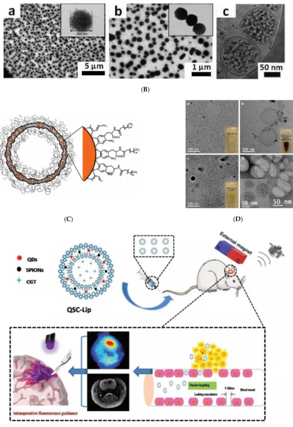
An efficient procedure for assembling CdSe–CdS QDs with Fe3O4MNPs into colloidal supraparticles (SPs) with a core–shell superstructure is given in [144]. In a typical synthesis process, 1 mL of chloroform solution containing 4 mg 9.0-nm size QDs and 6 mg 5.9-nm size MNPs was injected into 1 mL DTAB (dodecyltrimethylammonium bromide as a surfactant) aqueous solution (20 mg/mL in Nanopure water), followed by thorough mixing. After removing the chloroform, the co-assembling process (Figure3Aa) resulted in multifunctional multi-core particles of approximately 120 nm size consisting of a close-packed magnetoresponsive core and a fluorescent quantum dots shell (Figure3Ab–d). An additional thermal annealing process results in supercrystalline core–shell-structured supraparticles. After functionalizing with polyethylene glycol (PEG), the supraparticles can be magnetically manipulated inside living cells while being optically tracked.
Nanomaterials 2020, 10, x FOR PEER REVIEW 9 of 69
spherical confinement proved to be sufficient for the formation of stable icosahedral MNP clusters without the contribution of interparticle attractive interactions. The same slow evaporation technique of emulsion droplets was applied to evidence that sharp cubic and rounded cubic NPs self‐assemble also into spherical supraparticles [139].
In addition, large, micrometer‐size superparamagnetic microparticles for affinity separation were synthesized in a four‐step procedure using a hexane‐based ferrofluid [120,140]: (1) creation of an oil‐in‐water emulsion in which OA‐coated hydrophobic iron oxide nanoparticles of 5.3 nm mean physical size and a UV‐activated initiator were distributed in hexane; (2) formation of uniform microparticles through emulsion homogenization and the evaporation of hexane; (3) functionalization of the microparticle with a PEG‐functionalized surfactant and acrylic acid; and (4) polymerization of the microparticles.
The miniemulsion procedure proved to be useful to obtain magnetic nanocomposite particles with spatially separated functionalities [69,141] or to encapsulate magnetic nanoparticles together with fluorescent components [142,143].
An efficient procedure for assembling CdSe–CdS QDs with Fe3O4 MNPs into colloidal supraparticles (SPs) with a core–shell superstructure is given in [144]. In a typical synthesis process, 1 mL of chloroform solution containing 4 mg 9.0‐nm size QDs and 6 mg 5.9‐nm size MNPs was injected into 1 mL DTAB (dodecyltrimethylammonium bromide as a surfactant) aqueous solution (20 mg/ ml in Nanopure water), followed by thorough mixing. After removing the chloroform, the co‐assembling process (Figure 3a) resulted in multifunctional multi‐core particles of approximately 120 nm size consisting of a close‐packed magnetoresponsive core and a fluorescent quantum dots shell (Figure 3b–d). An additional thermal annealing process results in supercrystalline core–shell‐
structured supraparticles. After functionalizing with polyethylene glycol (PEG), the supraparticles can be magnetically manipulated inside living cells while being optically tracked.
a
(A)
Figure 3.Cont.
Nanomaterials2020,10, 2178 10 of 67
Nanomaterials 2020, 10, x FOR PEER REVIEW 10 of 69
(B)
Figure 3. Co‐assembling quantum dots and magnetic nanoparticles (NPs) of a ferrofluid in a magnetoresponsive core–shell nanostructure. (A) Synthesis and characterizations of core–shell‐
structured supraparticles (CS‐SPs). (a) Schematic of the formation of the CS‐SPs. A set of TEM images of CS‐SPs at different magnifications. Scale bars, 500 nm, 100 nm, and 10 nm (b–d). Ferrofluid:
magnetite NPs in chloroform carrier. Characteristic sizes: 9.0 nm for QDs (CdSe‐CdS) and 5.9 nm for magnetite NPs (reprinted by permission from Copyright Clearance Center: Springer Nature, Nature Communications,[144], Copyright 2014); (B) (a) Compositional analysis (EDX) of a PLGA nanostructure denoting the presence of elements of both magnetic IONPs and QDs (PbS). (inset) Schematic diagram of the magnetic NPs and QDs in the PLGA nanostructure. (b) TEM image of a typical PLGA nanostructure. (c) A detailed TEM image of a PLGA nanostructure revealing the presence of two types of NPs inside the structure. (d) Size distribution of both types of particles obtained from TEM images. The size distribution corresponds to the sizes of the magnetic NPs (15 nm) and QDs (4 nm). Ferrofluid: oleic acid‐coated magnetite NPs in hexane carrier. (reprinted with permission from [145]. Copyright 2016 American Chemical Society).
Polylactic‐co‐glycolic‐acid (PLGA) hybrid nanostructures were synthesized by a double‐
emulsion (water‐in‐oil‐in‐water) technique, involving a hexane‐based ferrofluid and PbS quantum dots (QDs) dispersed in toluene [145]. The ferrofluid mediated the encapsulation of magnetic and infrared emitting nanoparticles (PbS) in a polymeric matrix (Figure 3B a–d) to provide magnetic–
fluorescent imaging abilities to the resulting multi‐core particles.
Oleic acid‐coated PbS/CdS QDs and Fe
3O
4NPs dispersed in chloroform were made water‐
dispersible by micellar encapsulation, which was due to hydrophobic van der Waals interactions between the hydrocarbon chains of oleic acid and DTAB used as surfactant [146]. The resulting self‐
assembled Fe
3O
4and PbS/CdS supernanoparticles are aimed at synergistic dual‐mode heating treatment for cancer therapy.
Figure 3. Co-assembling quantum dots and magnetic nanoparticles (NPs) of a ferrofluid in a magnetoresponsive core–shell nanostructure. (A) Synthesis and characterizations of core–shell-structured supraparticles (CS-SPs). (a) Schematic of the formation of the CS-SPs. A set of TEM images of CS-SPs at different magnifications. Scale bars, 500 nm, 100 nm, and 10 nm (b–d). Ferrofluid: magnetite NPs in chloroform carrier. Characteristic sizes: 9.0 nm for QDs (CdSe-CdS) and 5.9 nm for magnetite NPs (reprinted by permission from Copyright Clearance Center: Springer Nature, Nature Communications [144], Copyright 2014); (B) (a) Compositional analysis (EDX) of a PLGA nanostructure denoting the presence of elements of both magnetic IONPs and QDs (PbS). (inset) Schematic diagram of the magnetic NPs and QDs in the PLGA nanostructure. (b) TEM image of a typical PLGA nanostructure. (c) A detailed TEM image of a PLGA nanostructure revealing the presence of two types of NPs inside the structure. (d) Size distribution of both types of particles obtained from TEM images. The size distribution corresponds to the sizes of the magnetic NPs (15 nm) and QDs (4 nm). Ferrofluid: oleic acid-coated magnetite NPs in hexane carrier. (reprinted with permission from [145]. Copyright 2016 American Chemical Society).
Polylactic-co-glycolic-acid (PLGA) hybrid nanostructures were synthesized by a double-emulsion (water-in-oil-in-water) technique, involving a hexane-based ferrofluid and PbS quantum dots (QDs) dispersed in toluene [145]. The ferrofluid mediated the encapsulation of magnetic and infrared emitting nanoparticles (PbS) in a polymeric matrix (Figure3Ba–d) to provide magnetic–fluorescent imaging abilities to the resulting multi-core particles.
Oleic acid-coated PbS/CdS QDs and Fe3O4 NPs dispersed in chloroform were made water-dispersible by micellar encapsulation, which was due to hydrophobic van der Waals interactions between the hydrocarbon chains of oleic acid and DTAB used as surfactant [146]. The resulting
Nanomaterials2020,10, 2178 11 of 67
self-assembled Fe3O4and PbS/CdS supernanoparticles are aimed at synergistic dual-mode heating treatment for cancer therapy.
2.2. Induced Destabilization of a Ferrofluid
Highly ordered soft magnetic nanoclusters [147] were obtained by strongly polar solvent (acetonitrile) or amphiphilic polymer (poly(styrene-co-maleic anhydride) (PScMA)) induced the destabilization of a volatile (toluene, chloroform, or tetrahydrofuran-based) ferrofluid [148,149], Figure4i–iv.
Nanomaterials 2020, 10, x FOR PEER REVIEW 11 of 69
2.1. Induced Destabilization of a Ferrofluid
Highly ordered soft magnetic nanoclusters [147] were obtained by strongly polar solvent (acetonitrile) or amphiphilic polymer (poly(styrene‐co‐maleic anhydride) (PScMA)) induced the destabilization of a volatile (toluene, chloroform, or tetrahydrofuran‐based) ferrofluid [148,149], Figure 4i–iv.
(i)
(ii)
Figure 4.Cont.
NanomaterialsNanomaterials 2020, 10, x FOR PEER REVIEW 2020,10, 2178 12 of 69 12 of 67
(iii)
(iv)
Figure 4.Controlled colloidal assembly of iron oxide nanoparticles from highly volatile ferrofluids.
(i) (a) Starting from the same hydrophobic iron oxide nanocubes (IONCs) (23±3 nm), IONCs are transferred in water by mixing the hydrophobic IONCs in toluene with the gallol-bearing PEG ligand
Nanomaterials2020,10, 2178 13 of 67
(GA-PEG-OH) in the presence of a base (1a). The solution is shaken for a few seconds, and after acetone addition, the PEG-IONCs are extracted in water (2a). Finally, after organic solvent evaporation at reduced pressure, the PEG-IONCs solution is dialyzed to remove the excess of GA-PEG-OH (3a).
This protocol provides the single-coated nanocubes in water. The MNBs instead are obtained by mixing the hydrophobic IONCs with a poly(maleic anhydride-alt-1-octadecene) polymer (PC18) in CHCl3(1b). The solution is shaken for few seconds, and then, 1 mL of acetonitrile is added at a flow rate of 2 mL min−1 (2b). The MNBs are collected by magnetic sorting and redissolved in water (3b) (Reprinted with permission from [148]. Copyright 2015 American Chemical Society). (ii) Polymer encapsulated colloidal-ordered assemblies (COA). TEM images of polymer–COA at higher (A) and lower (B) resolution. The dark pattern (A) results from the ordering of the closed packed assemblies within the nanobeads, while the brighter gray ring is caused by the polymer shell (lower electron density) of around 20 nm thickness.) (Reprinted with permission from [65]. Copyright 2015 American Chemical Society). (iii) Scheme of the clustering protocol using 20 nm core–shell iron oxide nanocubes.
Representative TEM micrographs of IONCs@PScMA in water and just after they have been prepared at a ratio of (A) 16.5, (B) 33, (C) 50 and (D) 66 polymer chains/nm2of particle surface. (E–H) A collection of TEM images at higher magnification of dimers and trimers formed at the ratio of 33. Schematic representation of the formation of soft colloidal nanoclusters. (iv) Tuning the mean hydrodynamic diameter of clusters by different polymer amounts. Volume distribution of hydrodynamic size of soft colloidal clusters measured in water starting from 20 nm IONCs. The hydrodynamic diameter was adjusted between 38 and 99 nm. No aggregation of clusters was detected, as polydispersity index (PDI) values were between 0.07 and 0.14 (see inset) (reprinted with permission from [149]. Copyright 2017 American Chemical Society).
2.3. Magnetoliposomes
Liposomes and, especially, magnetoresponsive liposomes are among the most promising vesicular drug carrier vehicles [150,151]. Allowing for encapsulation, retention, the membrane sealing off the interior of a hydrophilic volume from the environment, and the magnetic field-triggered release of the drug are the main attractive features of membranes-protected hollow nanocarriers for nanomedicine [152]. The loading of liposomes with iron oxide nanoparticles [153–155], mostly clusters preformed from aqueous [64,156,157] and organic [158,159] ferrofluids gives rise to magnetoliposomes with high magnetophoretic mobility and MRI contrast [160]. LipoMag composites [158] are assembled as oleic acid-coated magnetite nanocrystal cores with cationic lipid shells from a chloroform-based magnetic fluid through hydrophobic interactions [161,162] (Figure5A).
Nanomaterials 2020, 10, x FOR PEER REVIEW 13 of 69
Figure 4. Controlled colloidal assembly of iron oxide nanoparticles from highly volatile ferrofluids.
(i) (a) Starting from the same hydrophobic iron oxide nanocubes (IONCs) (23 ± 3 nm), IONCs are transferred in water by mixing the hydrophobic IONCs in toluene with the gallol‐bearing PEG ligand (GA‐PEG‐OH) in the presence of a base (1a). The solution is shaken for a few seconds, and after acetone addition, the PEG‐IONCs are extracted in water (2a). Finally, after organic solvent evaporation at reduced pressure, the PEG‐IONCs solution is dialyzed to remove the excess of GA‐
PEG‐OH (3a). This protocol provides the single‐coated nanocubes in water. The MNBs instead are obtained by mixing the hydrophobic IONCs with a poly(maleic anhydride‐alt‐1‐octadecene) polymer (PC18) in CHCl3 (1b). The solution is shaken for few seconds, and then, 1 mL of acetonitrile is added at a flow rate of 2 mL min−1 (2b). The MNBs are collected by magnetic sorting and redissolved in water (3b) (Reprinted with permission from [148]. Copyright 2015 American Chemical Society). (ii) Polymer encapsulated colloidal‐ordered assemblies (COA). TEM images of polymer–COA at higher (A) and lower (B) resolution. The dark pattern (A) results from the ordering of the closed packed assemblies within the nanobeads, while the brighter gray ring is caused by the polymer shell (lower electron density) of around 20 nm thickness.) (Reprinted with permission from [65]. Copyright 2015 American Chemical Society). (iii) Scheme of the clustering protocol using 20 nm core–shell iron oxide nanocubes. Representative TEM micrographs of IONCs@PScMA in water and just after they have been prepared at a ratio of (A) 16.5, (B) 33, (C) 50 and (D) 66 polymer chains/nm2 of particle surface.
(E–H) A collection of TEM images at higher magnification of dimers and trimers formed at the ratio of 33. Schematic representation of the formation of soft colloidal nanoclusters. (iv) Tuning the mean hydrodynamic diameter of clusters by different polymer amounts. Volume distribution of hydrodynamic size of soft colloidal clusters measured in water starting from 20 nm IONCs. The hydrodynamic diameter was adjusted between 38 and 99 nm. No aggregation of clusters was detected, as polydispersity index (PDI) values were between 0.07 and 0.14 (see inset) (reprinted with permission from [149]. Copyright 2017 American Chemical Society).
2.2. Magnetoliposomes
Liposomes and, especially, magnetoresponsive liposomes are among the most promising vesicular drug carrier vehicles [150,151]. Allowing for encapsulation, retention, the membrane sealing off the interior of a hydrophilic volume from the environment, and the magnetic field‐
triggered release of the drug are the main attractive features of membranes‐protected hollow nanocarriers for nanomedicine [152]. The loading of liposomes with iron oxide nanoparticles [153–
155], mostly clusters preformed from aqueous [64,156,157] and organic [158,159] ferrofluids gives rise to magnetoliposomes with high magnetophoretic mobility and MRI contrast [160]. LipoMag composites [158] are assembled as oleic acid‐coated magnetite nanocrystal cores with cationic lipid shells from a chloroform‐based magnetic fluid through hydrophobic interactions [161,162] (Figure 5A).
(A)
Figure 5.Cont.
Nanomaterials2020,10, 2178 14 of 67
Nanomaterials 2020, 10, x FOR PEER REVIEW 14 of 69
(B)
(C) (D)
(E)
Figure 5. (A) Schematic showing the preparation (upper) and assembly (lower) of LipoMag. Oleic acid‐coated magnetic nanocrystal cores and the lipid shells form through hydrophobic interactions (reprinted by permission from Copyright Clearance Center: Springer Nature, Nature Nanotechnology, [158], Copyright 2009). (B) (a,b) TEM and (c) cryo‐TEM micrographs of UMLs prepared by an REV process. At low magnification, a large number of dense vesicles are observed with diameters 200 nm in average. MNPs are trapped inside unilamellar vesicles (c) and dipole−dipole interaction can occur as exemplified by magnification (b). (Reprinted with permission from [64]. Copyright 2012 American Chemical Society). (C) Schematic of liposomes containing iron Figure 5. (A) Schematic showing the preparation (upper) and assembly (lower) of LipoMag.
Oleic acid-coated magnetic nanocrystal cores and the lipid shells form through hydrophobic interactions (reprinted by permission from Copyright Clearance Center: Springer Nature, Nature Nanotechnology, [158], Copyright 2009). (B) (a,b) TEM and (c) cryo-TEM micrographs of UMLs prepared by an REV process. At low magnification, a large number of dense vesicles are observed with diameters 200 nm in average. MNPs are trapped inside unilamellar vesicles (c) and dipole−dipole interaction can occur as exemplified by magnification (b). (Reprinted with permission
Nanomaterials2020,10, 2178 15 of 67
from [64]. Copyright 2012 American Chemical Society). (C) Schematic of liposomes containing iron oxide NPs in their bilayer. NitroDOPA–palmityl-stabilized iron oxide NPs are embedded in liposome membranes consisting of PEGylated and unmodified lipids; (D) Liposomes functionalized with iron oxide NPs. Cryo-TEM images of DSPC liposomes containing 5 mol % PEG(2)–PE that (a) were unmodified and incorporated (b) oleic acid-coated and (c) palmityl–nitroDOPA stabilized small iron oxide NPs. Insets show photographs of the respective PbS-based liposome dispersions where the lipid concentration was kept constant at 5 mg/mL. A comparison between (a) and (c) reveals no significant change of the spherical shape of liposomes upon loading their membranes with small, individually stabilized, iron oxide NPs. However, agglomerated, oleic acid-stabilized NPs seem to significantly distort the liposome shape. (d) TEM image of trehalose-fixed DSPC liposomes containing palmityl–nitroDOPA stabilized small NPs in their membranes. Liposomes were fixed with trehalose and air-dried on a carbon-supported Cu TEM grid where the carbon film had 3.5µm diameter holes. While the large image was taken in a hole that was spanned by trehalose, the inset was imaged on the carbon support.
Individually stabilized NPs with core diameters<5.5 nm are associated with liposomes. No NPs with core diameters>5.5 nm are seen. The inset indicates a high NP density of liposomes that were collapsed on the carbon support upon drying in air. (Reprinted with permission from Amstad et al.
2011a. Copyright 2011 American Chemical Society). (E) Liposome-integrated multiple-imaging agents and therapeutic drug for glioma-targeted delivery under exogenous magnetic field to accurately localize glioma. CGT, cilengitaide; QDs, quantum dots and SPIONs, superparamagnetic iron oxide nanoparticles were initially dispersed in chloroform. (Republished with permission of John Wiley and Sons, from [163]; permission conveyed through Copyright Clearance Center, Inc.).
Maghemite nanoparticles (9 or 7 nm) coated with citrate ligands and dispersed in water (Massart ferrofluid) or in a buffer were used for the preparation of Ultra Magnetic Liposomes (UMLs) encapsulating iron oxide nanoparticles in a volume fraction of up to 30% [64], Figure 5B. UMLs were prepared by a modified version of the reverse phase evaporation (REV) method [164]. This remarkable magnetic charge provides UMLs with high magnetic mobilities, MRI relaxivities, and heating capacities for magnetic hyperthermia [157]. Palmityl–nitroDOPA-stabilized iron oxide NPs were found to be spontaneously incorporated into liposome bilayers, whereas oleic acid-stabilized NPs agglomerated to form micelles [165], as seen in Figure5C,D. The observed significant difference between palmityl–nitroDOPA and oleic acid-stabilized NPs can be related to the irreversible binding of nitroDOPA versus the reversible adsorbing of oleic acid to the iron oxide surfaces, which favors the agglomeration of OA-coated NPs. Hydrophobic SPIONs and QDs initially dispersed in chloroform were co-encapsulated inside a lipid membrane to provide “all in one” nanocarriers–theranostics liposomes for glioma targeting [163], as shown in Figure5E.
The encapsulation of hydrophobically coated IONPs from a tetrahydrofuran-based ferrofluid together with camptothecin anticancer drug into a PPO block of Pluronic vesicles (Pluronic L121 (PEO–PPO–PEO, as purchased or carboxylated by succinic anhydride)) provided a scalable continuous manufacturing procedure to obtain multi-core theranostic drug delivery vehicles [166], as schematically represented in Figure6.
2.4. Co-Assembling in Aqueous Solution
Magnetic nanoparticles attached to a silica core with variable size provide a composite particle with a tunable-induced magnetic moment. Moreover, by applying an external silica coating, the thickness of the shell allows tuning also the dipolar interactions between particles at contact. The preparation procedure of these composites developed in [52] uses aqueous ferrofluid with maghemite or cobalt ferrite nanoparticles and an aqueous dispersion of silane-coupling agent-coated silica particles, ensuring the chemical attachment of magnetic nanoparticles to the silica core. Depending on the core size and shell thickness, the overall size of these superparamagnetic composite particles is between 150 and 200 nm (Figure7A).
Nanomaterials2020,10, 2178 16 of 67
Nanomaterials 2020, 10, x FOR PEER REVIEW 16 of 69
Figure 6. Continuous preparation of loaded magnetic polymersomes using a tetrahydrofuran‐based ferrofluid: the starting polymer solution (PEO–PPO–PEO in tetrahydrofuran) is diluted with water, the selective solvent for the PEO block, and induces polymersome self‐assembly. The microstructured mixing device is a stainless steel caterpillar micromixer with twelve mixing steps and a mixing channel with an inner volume of 10 mL. Hydrophobic agents were loaded in situ by simply adding the cargo (magnetic nanoparticles or drug molecules) to the starting polymer solution prior to mixing.
Due to the hydrophobicity of those compounds, incorporation in the hydrophobic part of the vesicle membrane occurs. Prior carboxylation of the end‐groups of the polymer enables further surface functionalization and conjugation to specific targeting moieties. (Republished with permission of John Wiley and Sons, from [166]; permission conveyed through Copyright Clearance Center, Inc.).
2.3. Co‐Assembling in Aqueous Solution
Magnetic nanoparticles attached to a silica core with variable size provide a composite particle with a tunable‐induced magnetic moment. Moreover, by applying an external silica coating, the thickness of the shell allows tuning also the dipolar interactions between particles at contact. The preparation procedure of these composites developed in [52] uses aqueous ferrofluid with maghemite or cobalt ferrite nanoparticles and an aqueous dispersion of silane‐coupling agent‐coated silica particles, ensuring the chemical attachment of magnetic nanoparticles to the silica core.
Depending on the core size and shell thickness, the overall size of these superparamagnetic composite particles is between 150 and 200 nm (Figure 7A).
Figure 6.Continuous preparation of loaded magnetic polymersomes using a tetrahydrofuran-based ferrofluid: the starting polymer solution (PEO–PPO–PEO in tetrahydrofuran) is diluted with water, the selective solvent for the PEO block, and induces polymersome self-assembly. The microstructured mixing device is a stainless steel caterpillar micromixer with twelve mixing steps and a mixing channel with an inner volume of 10 mL. Hydrophobic agents were loaded in situ by simply adding the cargo (magnetic nanoparticles or drug molecules) to the starting polymer solution prior to mixing. Due to the hydrophobicity of those compounds, incorporation in the hydrophobic part of the vesicle membrane occurs. Prior carboxylation of the end-groups of the polymer enables further surface functionalization and conjugation to specific targeting moieties. (Republished with permission of John Wiley and Sons, from [166]; permission conveyed through Copyright Clearance Center, Inc.).
Magnetoresponsive chondroitin sulfate-functionalized CaCO3microparticles (Figure7B) were obtained by crystallization from supersaturated aqueous solutions in the presence of oleic acid-stabilized magnetite nanoparticles as a water-based magnetic fluid and a natural strong–weak polyanion, chondroitin sulfate A (CSA) [167]. Accordingly, the growth mechanism of superparamagnetic microparticles involves the chains of CSA and the surfactant-coated magnetite nanoparticles, which could electrostatically accumulate a large amount of Ca2+ and carbonate ions; Ca2+ ions form an ionically cross-linked network with the carboxylate groups on the CSA and oleic acid. The microparticle characteristics investigated by physicochemical methods (SEM, TEM, X-ray diffraction, Raman spectroscopy, flow particle image analysis, particle charge density, and electrokinetic measurements) depend on the initial MF amount and polymer concentration.
Biocompatibility and also enhanced pH stability make their use in bio-related applications attractive.
Nanomaterials2020,10, 2178 17 of 67
Nanomaterials 2020, 10, x FOR PEER REVIEW 17 of 69
(a) (b)
(A)
(B)
Figure 7. (A) (a) TEM (top) and corresponding SEM picture (bottom) of silica–cobalt ferrite nanocomposite particles (b) Silica growth onto composite particles monitored by TEM, which shows the gradually increasing silica layer thickness from no silica present in picture 1 to a fully grown silica layer in picture 4. (Reprinted with permission from [52] Copyright 2005 American Chemical Society).
(B) Magnetoresponsive chondroitin sulfate‐functionalized CaCO3 microparticles. The influence of CSA and magnetic fluid (MF) content on the CaCO3−MF−CSA composite size, shape, and morphology, as evidenced by SEM (reprinted with permission from [167]. Copyright 2013 American Chemical Society).
Magnetoresponsive chondroitin sulfate‐functionalized CaCO3 microparticles (Figure 7B) were obtained by crystallization from supersaturated aqueous solutions in the presence of oleic acid‐
stabilized magnetite nanoparticles as a water‐based magnetic fluid and a natural strong–weak polyanion, chondroitin sulfate A (CSA) [167]. Accordingly, the growth mechanism of
Figure 7. (A) (a) TEM (top) and corresponding SEM picture (bottom) of silica–cobalt ferrite nanocomposite particles (b) Silica growth onto composite particles monitored by TEM, which shows the gradually increasing silica layer thickness from no silica present in picture 1 to a fully grown silica layer in picture 4. (Reprinted with permission from [52] Copyright 2005 American Chemical Society). (B) Magnetoresponsive chondroitin sulfate-functionalized CaCO3microparticles.
The influence of CSA and magnetic fluid (MF) content on the CaCO3−MF−CSA composite size, shape, and morphology, as evidenced by SEM (reprinted with permission from [167]. Copyright 2013 American Chemical Society).
Nanomaterials2020,10, 2178 18 of 67
3. Non-Spherical Multi-Core Superparamagnetic Assemblies
Superparamagnetic, multi-core nanocomposites built up by nanometer-sized magnetic nanoparticles exhibiting anisotropic morphologies [168] are highly interesting in the biomedical field as drug delivery systems, magnetic hyperthermia mediators, biosensors, MRI contrast agents, and bioseparators, due to their anisotropic magnetic response, high surface area, high magnetic moment, and high magnetic mobility [169].
Different synthetic approaches have been developed so far for the generation of anisotropically shaped superparamagnetic supraparticles starting from ferrofluids, including evaporation-guided, emulsion-templated and magnetic field-assisted self-assembly, supramolecular polymerization, electrospinning, ink-jet printing, and lithography-based approaches. In the following sub-sections, selected literature examples related to the aforementioned fabrication routes are provided and briefly discussed.
3.1. Evaporation-Guided Self-Assembly
The evaporation-guided self-assembly of colloidal ferrofluids has been employed by several research groups for obtaining superparamagnetic anisotropic supraparticles of various shapes and with sizes ranging from severalµm up to mm.
In this synthetic route, a liquid-repellent surface is used, on which ferrofluid droplets are left to dry in a controlled manner, resulting in the formation of superparamagnetic multi-core supraparticles [170,171].
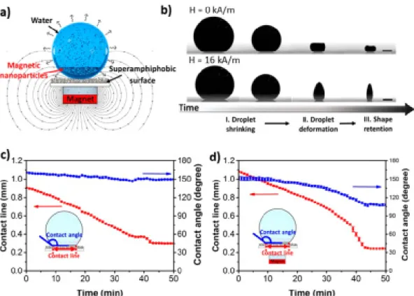
Hu and co-workers described the preparation of superparamagnetic supraparticles having distinct anisotropic shapes, starting from a magnetic colloidal aqueous suspension consisting of hybrid Fe3O4/polystyrene nanoparticles, stabilized with sodium dodecyl sulfate (SDS) [172]. The ferrofluid was left to dry on a superamphiphobic surface [173] in the presence of an externally applied magnetic field, resulting in the entrapment of the transient suspension droplet shapes upon evaporation. The authors demonstrated that different 3D anisotropic morphologies can be obtained including cones, barrels, and two towers (Figure8) upon altering the magnetic orientation, strength, and initial nanoparticle concentration. Moreover, since the presented approach is based on the evaporation-induced self-assembly starting from ferrofluids, it allows for the assembly of co-suspensions consisting of different NP types (such as TiO2), thus providing anisotropic binary supraparticles.
In another example, evaporation-induced self-assembly applied on Fe3O4/SiO2 core–shell nanoparticle aqueous dispersions deposited on curved, superhydrophobic surfaces was conducted to the formation of ellipsoidal anisometric magnetic Janus supraparticles [174]. The latter was accomplished in the presence of a magnetic field, guiding the accumulation of the magnetic nanoparticles at specific locations within the supraparticles.
Magnetic halloysite nanotubes (HNT), promising nanocomposites for MRI or magnetic hyperthermia, were prepared by loading preformed oleic acid-coated superparamagnetic magnetite nanoparticles (physical size ≈6 nm; hydrodynamic diameter of ≈10 nm) into HNT [175]. The OA-stabilized MNPs of a hexane-based ferrofluid were selectively loaded on the tetradecylphosphonic acid (TDP)-modified inner lumen of halloysite nanotubes in a slow evaporation process exploiting vacuum–N2cycles.
O’Mahony and co-workers prepared superparamagnetic microparticles of various morphologies, including dimpled and crumpled microparticles, by means of emulsion templated self-assembly, using oil-in-water emulsions [125]. Ferrofluids consisting of hydrophobic oleic acid-coated Fe3O4
NPs were employed for this purpose. The authors demonstrated among others that the ferrofluid concentration and the density of the oleic acid chains covering the nanoparticle surfaces significantly affect the morphology of the resulting microparticles. Such systems, exhibiting high magnetic mobilities, high surface areas, and consequently high binding affinities, are very promising in magnetic bioseparation processes.
Nanomaterials2020,10, 2178 19 of 67
Nanomaterials 2020, 10, x FOR PEER REVIEW 19 of 69
Figure 8. Formation of large 3D superparamagnetic structures by making use of the magnetic properties of ferrofluids. (a) Experimental system used for the production of supraparticles by evaporation‐guided assembly of a magnetic NPs dispersion on a superamphiphobic surface. (b) Evolution of a 3 wt % droplet during drying without (upper panel) and with (bottom panel) magnetic field. Scale bars are 0.5 mm. Drying curve of the droplet (c) without and (d) with magnetic field. Insets represent the dimensions measured during drying (reprinted with permission from [172]. Copyright 2019 American Chemical Society).
In another example, evaporation‐induced self‐assembly applied on Fe
3O
4/SiO
2core–shell nanoparticle aqueous dispersions deposited on curved, superhydrophobic surfaces was conducted to the formation of ellipsoidal anisometric magnetic Janus supraparticles [174]. The latter was accomplished in the presence of a magnetic field, guiding the accumulation of the magnetic nanoparticles at specific locations within the supraparticles.
Magnetic halloysite nanotubes (HNT), promising nanocomposites for MRI or magnetic hyperthermia, were prepared by loading preformed oleic acid‐coated superparamagnetic magnetite nanoparticles (physical size ≈6 nm; hydrodynamic diameter of ≈10 nm) into HNT [175]. The OA‐
stabilized MNPs of a hexane‐based ferrofluid were selectively loaded on the tetradecylphosphonic acid (TDP)‐modified inner lumen of halloysite nanotubes in a slow evaporation process exploiting vacuum–N
2cycles.
O’Mahony and co‐workers prepared superparamagnetic microparticles of various morphologies, including dimpled and crumpled microparticles, by means of emulsion templated self‐assembly, using oil‐in‐water emulsions [125]. Ferrofluids consisting of hydrophobic oleic acid‐
coated Fe
3O
4NPs were employed for this purpose. The authors demonstrated among others that the ferrofluid concentration and the density of the oleic acid chains covering the nanoparticle surfaces significantly affect the morphology of the resulting microparticles. Such systems, exhibiting high magnetic mobilities, high surface areas, and consequently high binding affinities, are very promising in magnetic bioseparation processes.
In a final example, 2D and 3D mesocrystalline films were generated from colloidal dispersions of oleic acid‐stabilized magnetite nanocubes in toluene via self‐assembly under slow evaporation conditions [176]. The self‐assembly process is driven by the NP dipolar magnetic attractive forces and the presence of an external magnetic field. In the case of the 2D mesocrystalline films, the generation of two distinct Fe
3O
4nanocube superstructures was observed, having the same orientational order and p 4 mm and c 2 mm layer symmetries, while slightly distorted fcc superlattices were found in the case of the 3D mesocrystalline films.
Figure 8.Formation of large 3D superparamagnetic structures by making use of the magnetic properties of ferrofluids. (a) Experimental system used for the production of supraparticles by evaporation-guided assembly of a magnetic NPs dispersion on a superamphiphobic surface. (b) Evolution of a 3 wt % droplet during drying without (upper panel) and with (bottom panel) magnetic field. Scale bars are 0.5 mm. Drying curve of the droplet (c) without and (d) with magnetic field. Insets represent the dimensions measured during drying (reprinted with permission from [172]. Copyright 2019 American Chemical Society).
In a final example, 2D and 3D mesocrystalline films were generated from colloidal dispersions of oleic acid-stabilized magnetite nanocubes in toluene via self-assembly under slow evaporation conditions [176]. The self-assembly process is driven by the NP dipolar magnetic attractive forces and the presence of an external magnetic field. In the case of the 2D mesocrystalline films, the generation of two distinct Fe3O4nanocube superstructures was observed, having the same orientational order and p4 mm andc2 mm layer symmetries, while slightly distorted fcc superlattices were found in the case of the 3D mesocrystalline films.
3.2. Magnetic Field-Assisted Self-Assembly
Highly stable aqueous ferrofluids consisting of superparamagnetic maghemite (γ-Fe2O3) nanoparticle clusters encapsulated within silica shells of controllable thicknesses were synthesized and further used in the generation of 1D, highly anisotropic nanostructures. These included nanochains and nanobundles (Figure9) formed in the presence of a magnetic field, which was due to the development of magnetic dipole–dipole interactions [177]. The incorporation of polyvinylpyrrolidone (PVP) in the aqueous superparamagnetic NP cluster suspensions provided stability to the assembled 1D structure that was irreversibly “locked” into fixed nanochain/nanobundle morphologies by incorporating an additional silica layer via hydrolysis/condensation reactions.