DEVELOPMENTAL BIOLOGY SUPPLEMENT 2 , 1 1 8 - 1 5 0 ( 1 9 6 8 )
The Emergence of Pattern in the Cell Walls of Higher Plants
J. HESLOP-HARRISON
Institute of Plant Development, University of Wisconsin, Madison, Wisconsin 53706
INTRODUCTION
Cell differentiation in the higher plant is usually accompanied by, or based upon, changes in cell wall organization. Most plant cells change shape as they grow, and insofar as shape modifications are determined intracellularly, they are related to the deposition of cellulose microfibrils in particular orientations in the primary wall.
In all cells destined for specialized roles in conduction, protection and support, the wall plays a major functional part, and this commonly depends upon the apposition of secondary layers and encrustation with compounds different from those of the primary wall. The de
velopment of walls patterned in various ways is therefore one of the principal manifestations of cell differentiation in plants.
Because of this special importance of the cell wall, it has been the target of considerable research. Earlier application of physical and optical methods provided a remarkably full picture of wall structure ( Frey-Wyssling, 1948; Preston, 1952), and subsequently electron microscopy has led to a burgeoning of knowledge concerning the ways walls grow. Developments in electron microscopic preparation technique are now improving our appreciation of the nature of cytoplasmic participation, particularly in respect to the emergence of pattern. It is the purpose of this contribution to provide a brief survey of some of this work; but at the outset it has to be acknowl
edged that there are numerous lacunae in our comprehension of causal sequences. Currently, we are finding ourselves able to describe in more and more detail the events accompanying the establishment of wall pattern, without yet seeing how all the various steps in the patterning process are interrelated. As with other manifestations of biological morphogenesis, the basic gap is in the understanding of how genetic information carried in the nucleus comes to be translated
118
PATTERN IN PLANT CELL WALLS 119 into three-dimensional form. For the moment we can do little more than probe this problem, but some aspects of it of particular signif
icance in relation to the control of wall pattern are discussed in later paragraphs.
TYPES OF PATTERNING IN PLANT CELL WALLS
We may begin with a brief review of the ways plant cell walls do reveal patterning. It is useful here to preserve the distinction between "primary" and "secondary" walls, the primary wall being that formed during the early growth of the cell, and the secondary the component laid down on the inner face of the primary wall as the cell matures and becomes specialized for particular functional roles.
Patterning in the primary wall is manifest in all three dimensions—
in stratification, and in the disposition of structural elements in the plane of the wall. In higher plants it is expressed principally in the orientation of cellulose microfibrils. In the young wall, cellulose represents only some 20-30% of the material present, the remainder being made up mainly of various pectic substances and hemicelluloses (Setterfield and Bayley, 1961; Roelofson, 1965); but cellulose is the only microfibrillar component, and constitutes the only part of the wall for which it can be argued from existing evidence that the spatial arrangement of molecules is determined by "purposive" forces acting from within the cell, if the impropriety of language may be excused. This is not to say that the matrix materials play no part in growth; indeed it is probably through the bonding between these encasing and encrusting molecules that the cell exerts control over wall extensibility. But anisotropy in the wall is established by the factors ordering the cellulose microfibrils, and in this manner the mechanical properties are imposed which determine that extension should occur more readily in certain directions than in others.
In the walls of young cells about to undergo growth in length, the microfibrils tend to be oriented transversely on the side walls, and more or less randomly in those at right angles to the prospective direction of growth. During elongation, newly deposited microfibrils preserve a similar pattern on side walls. However, extension brings about a reorientation of the microfibrils in older parts of the wall away from the transverse and toward the axial direction, so that the wall becomes stratified, the mean orientation of the microfibrils changing
120 J . HESLOP-HARRISON
progressively from the plasmalemma outward. This interpretation of wall growth in elongating cells is the "multinet" scheme due to Roelofson and Houwink (1953), and various aspects of it, as well as certain exceptions and weaknesses, are discussed at length in the reviews of Wilson (1964) and Roelofson (1965). For the present discussion the significant point is that patterned distribution of the microfibrils in the walls is here seen as a determinant of the directions of cell growth, and so of cell shape.
Pattern may also be seen in primary walls in the distribution of plasmodesmata and pit fields. The disposition of plasmodesmata in walls separating daughter cells of a division often appears more or less random, but plasmodesmata on the side walls of cells undergoing extension growth are usually aggregated in groups of 10-50 in pit fields. These fields are commonly ellipsoidal, with the long axis trans
verse to the direction of growth. The microfibrils in the area of the pit field itself form a sparse network, but the margin is built up from thicker aggregates of microfibrils, which curve round the field, some
times forming a distinct lip (Fig. 1). Pit fields do not increase in number during extension growth, but instead become spaced out on the lateral walls. According to Wilson (1957, 1958), new pit fields arise during or shortly after cell division, when preexisting fields are divided by the deposition of new material across their length.
Some parenchymatous cells continue to show wall thickening after the cessation of enlargement, but secondary wall formation is most conspicuously revealed in the differentiation of tracheids, xylem elements, fibers, and the like. The distinctive phenomenon is the deposition of further lamellae on the inner surface of the primary wall, often so as to produce patterns of bars, loops, helices, and networks of various kinds. In all studied cases, the pattern in secondary thickening is initiated through the localized deposition of closely packed, highly oriented, cellulose microfibrils, and it is of course to this that the strong birefringence of the secondary wall is due ( Fig. 2 ).
Insofar as encrusting substances such as lignin also show patterned distribution in secondary walls, it may be supposed that this is re
lated, initially at least, to the disposition of the cellulose component (Preston, 1952). There are instances where pattern in or upon a wall may be expressed first in the distribution of a noncellulosic component, as for example in the strangely formed waxy coatings of the epidermal cells of leaves and fruits, strikingly illustrated by Juniper (1959), and
PATTERN IN PLANT CELL WALLS 121
FIG. 1. Pit field in the wall between two cells of bean root tip. The plane of the section is slightly oblique, so that in the lower part of the field plasmadesmata are cut across, while in the upper part the surface layer of the protoplast of one of the cells may be seen. The wall in the area of the pit field is made up of a thin layer of microfibrils. Around the margin of the field primary wall thickening has continued, and the way microfibril orientation is concerned in defining the shape of the field is clearly visible. Electron micrograph by Dr. E. H. Newcomb.
X ca. 50,000.
in the elaborately sculptured exines of pollen grains. Cellulose does, however, play a part in setting up the dies and matrices for these patterns, and the special case of the pollen exine is considered in some detail in later paragraphs.
A general point to be made about the nature of pattern in plant cell walls is that it reveals that familiar characteristic of biological form, the quality one might term "order with diversity." Consider the pollen grains illustrated in Fig. 14; all differ, yet all show a pattern of the same general type. Insofar as the pattern is an expression of
122 J . HESLOP-HARRISON
FIG. 2. Sectioned endothecium of lily anther in late stage of development, by polarized light. The vertical bars of thickening show strong birefringence; the primary wall in between them is by this time essentially isotropic. X ca. 375.
the genome, it is evident that its working out in each cell is modulated and modified by the proximity of other cells, and no doubt also by fortuitous factors within each cell.
In many tissues, such as the endothecium to be discussed later (Fig. 3), intercellular coordination in wall thickening is not a func
tional necessity, but in other situations it clearly is. Thus pit fields are shared by two cells; in their formation and relocation during growth, function can be preserved only if the wall-building processes in the neighboring cells are correlated. Similarly, in the formation of a conducting vessel in the xylem, a successful outcome will result only from coordination of a file of cells, both in growth, and in the selection and execution of the appropriate form of secondary wall thickening (Sinnott and Bloch, 1945). Again it is necessary to con
clude that cellular interactions are playing a dominant role, not only in deciding which genes are activated (if it is indeed at the level of gene activation that we must seek for the control of wall pattern in each individual cell, a point discussed further below), but also in determining those geometrical attributes through which patterning is coordinated in neighboring cells.
PATTERN IN PLANT CELL WALLS 123
THE CYTOPLASM-WALL INTERFACE
To the extent that the organization of a cell wall is an expression of the genome, the emergence of pattern in it must depend upon the function of the cytoplasm as an intermediary in the conveyance of pattern information. The cytoplasm must therefore not only possess the biosynthetic machinery determining the kind and amount of the molecules—usually long-chain polymers—that contribute to the wall substance, but also contain mechanisms acting to govern their dis
position and orientation. Since wall synthesis occurs at or near the cell surface, it is natural to look here for manifestations of pattern- determining processes. The nature of the cytoplasm-wall interface in young cells during the deposition of the primary wall, and in older cells during secondary wall formation, is therefore a matter of some interest.
It is of course possible to query whether it is useful to think of an
"interface" between wall and cytoplasm at all. Preston (1955) ex
pressed the view that in the young cell "the cytoplasm interpene
trates the wall. . . . the wall is not a dead envelope but marks in
stead the outer limits of the living cytoplasm." For many years there have been suggestions that a protein component is present in the primary wall, and Lamport (1965) has expressed the view that this component contains not only enzymes, but also unique structural proteins possibly concerned with growth regulation. The view that there is a special wall protein has, however, been contested (Steward et al., 1967). Whatever may be the status of special wall proteins, there are indications from the work of Setterfield and Bayley (1957, 1959) that cellulose synthesis may occur at sites remote from the cytoplasm, which implies the presence of extracellular enzymes. We shall revert to the point in a later paragraph.
Against the view that the primary wall is no more than the outer layer of the cytoplasm must be placed fine-structural evidence in
dicating that, even during an active growth period, a plasmalemma, consisting of a single unit membrane, usually intervenes between the cytoplasm and the wall proper. The vesicles that aggregate to form the cell plate are invested by a unit membrane (e.g., Hepler and Newcomb, 1967), and these membranes form the precursors of the plasmalemma later seen to be apposed to the primary wall. The processes involved in the further accretion of wall material are now
124 J . HESLOP-HARRISON
reasonably well understood. Vesicles containing precursors of wall compounds are budded from dictyosomes within the cytoplasm;
these progress to the wall and discharge their contents through the plasmalemma (Mollenhauer et al., 1961; Wooding and Northcote, 1964; Northcote and Pickett-Heaps, 1966; Pickett-Heaps and North
cote, 1966; Wooding, 1968). The discharge is evidently achieved by a fusion of the vesicle with the plasmalemma, the vesicle then opening toward the cell wall and contributing its own membrane to the plasmalemma ( Frey-Wyssling et al., 1964). At the most, the process does not involve more than a very local dissolution of the plas
malemma, and there is no indication that the integrity of the mem
brane is lost at any time during growth in consequence of the secretion of materials through it. Accordingly, if the plasmalemma is defined as the outer limit of the cytoplasm, the wall lies outside of it, and is not to be regarded as part of the protoplast (Whaley et al., 1964).
An implication of this conclusion is that wall proteins, enzymatic and structural, are extracellular secretion products.
CYTOPLASMIC STRUCTURES ASSOCIATED WITH WALL PATTERNING:
THE MICROTUBULE
In any catalog of cytoplasmic structures likely to be concerned with wall-pattern determination, the microtubule must now take pride of place. Although microtubules were observed in plant cells sporadically before 1963, the paper by Ledbetter and Porter of that year served to show that they were regularly present in plant cytoplasm, and to establish the important fact that they were concerned in some intimate way with the deposition of cell wall materials. In the root cells of Phleum and Juniperus examined by Ledbetter and Porter, micro
tubules were found to be concentrated in the cortical region of the cytoplasm, just beneath the wall. Near end walls their disposition was random, but in the vicinity of the lateral walls, they were arranged circumferentially. Their orientation was therefore correlated closely with that of the cellulose microfibrils of the wall, from which the conclusion was drawn by Ledbetter and Porter that the microtubules might play a role in governing the orientation and deposition of the microfibrils.
All subsequent observations on the primary wall have supported the proposition that cellulose microfibril orientation is in some way related to that of microtubules in the adjacent cytoplasm, and the
PATTERN IN PLANT CELL WALLS 125 view has been powerfully suported by observations on the growth of patterned secondary walls. Hepler and Newcomb (1964) demon
strated that in the formation of bands of thickening in the tracheary elements produced during xylem regeneration in wounded Coleus stems, the microtubules were localized to cytoplasm in the vicinity of the new wall growths, and that the same kind of correlation existed between the orientation of the cellulose microfibrils of the developing thickenings and the microtubules as had been pointed out by Ledbetter and Porter for primary cell walls in the root. Similar observations were made by Wooding and Northcote (1964) for the differentiating xylem elements in Acer pseudoplatanus, and by Cron- shaw and Bouck (1965) for the same type of cells in the oat coleoptile.
As a model system revealing some of the ways in which micro
tubules are associated with patterned growth in a secondary wall, we may take the endothecium. The endothecium is the subepidermal layer of the anther wall that functions to open the loculi at the time of pollen dispersal. This is achieved by the development of differential tensions as the mature cells dry out. Each cell is roughly cylindrical and carries wall thickenings of the general form seen in Fig. 3. As cells with walls of this type lose water, it is obvious that shrinkage will be greater at the outer end than at the inner. Additional details of the architecture of the thickenings can be grasped from the scanning electron micrographs of Figs. 4 and 5. The thickenings are initially cellulosic, and the cellulose microfibrils are oriented con
sistently along the long axis of the bars, which are accordingly highly biréfringent (Fig. 2).
Growth of the primary wall of the endothecial cells of the lily anther is completed shortly after release of the spores from the meiotic tetrads; the cells are then cylindrical, thin-walled, and highly vacuolated. All cells of the endothecial layer begin secondary thick
ening together, and several cell layers in the connective, the tissue between the pollen sacs, embark simultaneously on a similar process of wall thickening to produce patterns of the same general character.
As may be seen from Figs. 3 and 4, the thickenings are not closely coordinated in different cells, and in this respect the endothecium differs from tissues like the xylem, where there is often a relationship between the disposition of thickening bars in neighboring cells (Sinnott and Bloch, 1945).
Electron microscopy shows that the siting of the thickenings is
126 J. HËSLOP-HARBISON
M^im
l$f&ry m.&
FIG. 3. Endothecium of lily anther, viewed from the pollen sac side. Each cell shows the characteristic thickening, consisting of a basal plate of cellulose, from which arise branching bars on the lateral walls. These bars terminate in points curving over the outer face of each cell. X ca. 500.
FIG. 4. Cells of the endothecium cut across; Stereoscan electron micrograph.
PATTERN IN P L A N T CELL WALLS W correlated very closely with the location of tracts of microtubules in the cortical region of the cytoplasm. Before the emergence of visible thickenings in the wall, oriented files of microtubules define zones that delimit the prospective sites of the bars (Fig. 6). Thereafter, as thickening begins, a constant association is preserved between microtubules and bars. At first the microtubules extend around the circumference of each bar (Fig. 7); then as the full width is reached, they become localized to the inner surface. Glancing sections show the orientation of the microfibrils of the thickenings to be matched with that of neighboring microtubules in the cytoplasm (Fig. 8).
During the thickening process microtubules are restricted entirely to the vicinity of the bars, none occurring elsewhere in the cortical cytoplasm.
Because in tissues like the endothecium, patterned distributions of microtubules may be seen before thickening becomes evident, the obvious conclusion would seem to be that the relationship is based upon causation: thickenings appear in the neighborhood of tubule concentrations, and with microfibril orientations agreeing with those of the tubules, because the latter are in some way implicated in microfibril deposition, as originally suggested by Ledbetter and Porter. This interpretation gains added weight from observations on the effects on wall patterning of agents which interfere with microtubule assembly, or disrupt microtubules once they are formed.
It has been known since the work of Levan (1939, 1942) that col- chicine is effective not only in disrupting the mitotic spindle, but in changing the pattern of growth in plant cell walls. In the onion root tip studied by Levan, extension growth in the cells immediately above the meristem is replaced by isodiametric growth in colchicine- treated cells, producing the familiar C-tumor. A modern interpretation of the effect would be that the hooplike disposition of the microfibrils in the walls of cells in this region—a disposition normally determining that the cells will grow mainly in length—is upset in the presence The basal plate of thickening forms the "floor" of each cell. There is no correla
tion in the distribution of thickening between neighboring cells. X ca. 490.
FIG. 5. Longitudinal view of an endothecial cell during thickening; Stereoscan electron micrograph. The preparation procedure removes most of the organelles, but some amyloplasts remain strung out along the walls. A curved surface of the anther epidermis with wrinkled cuticle appears at the top of the micrograph.
X ca. 495.
128 j . HESLOP-HARKISON
*fr *'*''' ' - . Vet»?' ***?^i?^^4i$jffäHi
FIG. 6. Section of the wall between two endothecial cells, cut transversely to the long axis, before the onset of secondary thickening. In the regions of the
PATTERN IN PLANT CELL WALLS 129 of colchicine. This would be expected were the disposition of the microfibrils dependent upon prior orientation of the microtubules, since it is now clear that colchicine binds with a component of the microtubule, so, it would appear, preventing normal assembly ( Borisy and Taylor, 1967a,b).
An effect of colchicine on the development of patterns of thick
ening in the secondary wall has been demonstrated by Pickett-Heaps (1967). In the developing xylem elements of the wheat coleoptile, colchicine does not arrest the deposition of secondary wall materials, but disrupts the pattern so that the thickenings are highly irregular.
The effect is correlated with the disappearance of all but a few microtubules from the cell.
In the endothecium, colchicine also acts to prevent the deposition of patterned wall thickenings. The endothecial layer illustrated in Fig. 10 is from an anther exposed to colchicine for a period of 12 hours, from the end of primary wall growth in the cells of the anther wall, to the conclusion of pollen development. Comparison with cells from a corresponding part of a control anther shows the total absence of patterned thickening (Fig. 9). Such cells reveal some slight, but generalized, thickening, but the secondary wall material shows no birefringence. Transmission electron micrographs reveal no microtubules anywhere within the cell.
Colchicine prevents extension growth when applied earlier, during the growth of the endothecial cells before the completion of primary wall deposition. The cells then grow isodiametrically, becoming balloon-like, as in Levan's C-tumors (Fig. 11). This response pre
sumably reflects an effect of colchicine on the disposition of micro- fibrils in the primary wall.
CYTOPLASMIC STREAMING AND WALL PATTERNING Ledbetter and Porter (1963) considered the possibility that cyto- plasmic microtubules might influence the orientation of wall micro- brackets, arrays of microtubules (mt) appear in the cortical layers of the cyto
plasm, defining the location of the future thickening bars. X ca. 41,000.
FIG. 7. Later stage of thickening in the endothecium; slightly oblique section of a bar, showing the parallel arrays of microtubules conforming to its surface.
X ca. 42,000.
FIG. 8. Glancing section of a bar of thickening, showing the agreement be
tween microfibrils and microtubules in orientation. X ca. 42,000.
Electron micrographs of Figs. 6-8 are by Dr. H. G. Dickinson.
130 J . HESLOP-HARRISON
FIGS. 9 and 10. Effects of colchicine on endothecial thickenings. Fig. 9, con
trol anther; Fig. 10, anther from a bud treated for 12 hours with 0.1% colchicine immediately before the initiation of secondary thickening. In the untreated cell, the thickening bars show the normal disposition and dimensions; in that exposed
PATTERN IN PLANT CELL WALLS 131 fibrils through control of cytoplasmic streaming. The idea has some attraction, since in various nineteenth century accounts of secondary wall thickening correlations between the paths of streaming cytoplasm and the disposition of bands of cellulose were recorded (see discus
sion by Sinnott and Bloch, 1945), and a hypothesis linking streaming and microfibril orientation was developed by van Iterson (1937).
However, electron microscopic studies have not on the whole supported the view that localized streaming is causally connected with wall thickening. Hepler and Newcomb ( 1963 ) concluded that the strands of denser cytoplasm seen by Sinnott and Bloch (1945) in the vicinity of prospective areas of thickening during the early regeneration of tracheary elements in Coleus were probably marked out simply by local concentrations of organelles. Cronshaw and Bouck (1965) were unable to find even this kind of correlation in differentiating xylem elements in the oat coleoptile, and stated, moreover, that cytoplasmic streaming could not be observed in these cells.
The differentiating xylem element has a dense cytoplasmic con
tent, and being deep seated in the coleoptile is perhaps not well suited for the observation of cytoplasmic streaming. The endothecial cells are highly vacuolated at the time of secondary wall growth, and are more readily accessible; they therefore provide more satisfactory material for the observation of cytoplasmic movement. The most conspicuous organelles in the cytoplasm apart from the nucleus are plastids, usually chlorophyll-containing, with a fairly well de
veloped lamellar system and two or three large starch grains. In the actively thickening cell these plastids are dispersed in the thin cyto
plasmic layer over the wall, frequently lined up in ranks paralleling the bars of thickening (Fig. 5). Organelles of lesser dimensions, corresponding in size to mitochondria and dictyosomes, are also readily visible with the optical microscope. No cytoplasmic streaming of the vigor of that observable, say, in the Elodea leaf is visible in the actively thickening endothecial cell, and the smaller particles appear
to colchicine, they are entirely absent. The fine pitting evident in some parts of Fig. 10 is a preparation artifact. Stereoscan electron micrographs, X ca. 490.
FIG. 11. Isodiametric growth of an endothecial cell from an anther treated with 0.1% colchicine before completion of primary wall growth. X ca. 390.
FIG. 12. As Fig. 11, but showing a binucleate endothecial cell resulting from the colchicine treatment with all organelles clustered around the nuclei. X ca. 350.
132 J . HESLOP-HARRISON
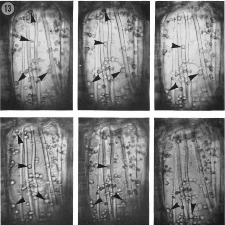
to show nothing but random Brownian movement. However, time lapse photography shows that movement of some of the larger organelles does occur (Fig. 13). The movement is not continuous, but it is persistently directional. It is extremely slow in comparison with that observed in other famiHar cells, amounting at the maximal rate to no more than 0.1 μ per second, in comparison with the 3-5 μ per second recorded by Sabnis and Jacobs (1967) for the green alga Caulerpa, and up to 60 μ per second measured in Nitetta (Kamiya, 1959). Moreover, what is observed is not a uniform movement of particular volumes of cytoplasm, but the slow erratic progression of individual organelles. It is therefore rather more akin, both in char
acter and velocity, with the orientational movements of chloroplasts (Zurzychi, 1962).
The organelles marked in Fig. 13 happen to show movement all in the same general direction, toward the face of the cell directed toward the anther loculus, and there is no indication of a return stream in this focal plane. The movement is thus polarized in the same direction as the thickening bars, but beyond this there would seem to be no particular association: one plastid may be sliding along a bar, one is certainly moving between, and others pass across the thickening bands. The movement may reflect a slow rotation of the cytoplasm, but it would be difBcult to interpret the observations as revealing pathways of streaming associated specifically with the pattern of secondary wall growth, and this in turn perhaps means that there is no special correlation with the disposition of micro- tubules. However, one observation on the endothecium may offer some slight support for a relationship between the presence of micro- tubules and the slow movement of plastids in the peripheral film of cytoplasm: in cells growing isodiametrically following colchicine treatment, the plastids show no detectable directional movement at all, and remain clustered around the nucleus (Fig. 12).
There is some conflict among other accounts of cytoplasmic move
ment and microtubule distribution in plants. O'Brien and Thimann (1966) found no association in epidermal coleoptile cells, and Nagai and Rebhun ( 1966 ) similarly observed no connection in the actively streaming cells of Niteïïa; in both of these studies the authors showed that microtubules were not present in moving cytoplasm, and implicated a cytoplasmic microfilament as the structure more likely to be concerned with streaming. However, in Caulerpa, Sabnis
PATTERN I N P L A N T CELL WALLS 133
FIG. 13. Time lapse sequence of a living endotheciai cell during the period of secondary wall growth, showing the movement of plastids. The three micrographs of the upper row were taken at intervals of 3-5 minutes, reading from left to right.
Those of the lower row were taken after a lapse of 35 minutes, and are again at intervals of 3-5 minutes reading from left to right. The marked bodies, likely all to be plastids, are those showing most conspicuous movement in this focal plane.
The maximum rate of directional plastid movement observed in this cell did not exceed 0.1 μ per second. X ca. 495.
and Jacobs (1967) concluded that microtubules did have a direct or indirect influence on cytoplasmic streaming, suggesting that the microtubules serve some form of "cytoskeletal" function, providing either the framework or delimiting the areas where the motive force responsible for streaming is generated.
134 J . HESLOP-HARRISON
CYTOPLASMIC STRUCTURES ASSOCIATED WITH WALL PATTERNING:
MEMBRANES
There are now several examples known of correlation between features of the wall and the disposition of elements of the endo- plasmic reticulum (ER) in adjoining cytoplasmic layers. Associations of this type are seen during the formation of plasmodesmata in the developing cell plate. Small tubular elements of the ER persist be
tween daughter cells of a division as the new wall develops, later to be enclosed by the growing plasmalemmas to give a tube within a tube, the form of the mature plasmodesmata (Porter and Machado, 1960; Frey-Wyssling et al, 1964; Hepler and Newcomb, 1967).
While the distribution of plasmodesmata may be essentially random in some walls, in others it is commonly patterned, as in pit fields (Fig. 1). Porter and Machado (1960) pointed out that such patterns must depend in turn upon the presence of patterns in the ER lattice which develops first across the spindle equator. It may be added that where this occurs there must be a coordination between the layers of ER in the two daughter cells, indicating again cell interaction in the determination of pattern.
Components of the ER may be seen intervening in a related pro
cess, namely the formation of pores in the sieve plates, the per
forated walls between neighboring sieve tube elements in the phloem.
The events were described first by Esau et al. (1962) in Cucurbita maxima. In the young undifferentiated sieve elements the sieve plate is smooth and unpitted, with a normal plasmalemma and scattered single plasmodesmata. As differentiation begins, sheets of ER become apposed in localized areas to the plasmalemma over future sieve plates, and these areas are the pore sites. Beneath the apposed sheets of ER, callose is deposited, while over the sites not occupied by ER cellulose is added to the wall. As development continues the callose plates thicken until they appear partly embedded in the wall.
This process occurs in the cells on each face of the future sieve plate, and the callose platelets on the two faces are coincident—localized, seemingly, around the sites of plasmodesmata originally traversing the young wall. The apposed ER continues to extend laterally at each pore site, and the associated plates of callose increase in diameter.
In consequence, the intervening bars of cellulose become narrower, although each thickens concomitantly in the dimension at right angles to the plasmalemma. Ultimately a perforation forms in the
PATTERN IN P L A N T CELL WALLS 135 center of each pore site, and the callose on the two surfaces of the sieve plate becomes continuous through the pores. The observations of Esau et al. (1962) were made on KMn04 fixed material, but the association of ER with the growing callose plugs has been observed in glutaraldehy de-osmium tetroxide fixed tissues by E. H. Newcomb (personal communication), and with similarly fixed material North- cote and Wooding (1965) have been able to demonstrate how the erosion of the wall beneath the callose plugs is mediated by strands of ER expanding in the plane of the middle lamella.
Striking examples of relationships between wall patterning and ER distribution have been recorded in developing microspores. The often elaborate sculpturing of the pollen exine raises many questions of importance for patterning processes in general, and the onto- genetic facts merit treatment in some detail. The four microspores resulting from each meiotic division in the anther remain in the tetrad configuration within a thick callose wall. This wall is partly derived from that of the parent meiocyte, and partly synthesized after the meiotic divisions. The essential features of the final sculp
turing of the pollen grain wall are established while the microspores are still enclosed in the tetrad wall, and the patterning process begins very soon after the separation of the cytoplasms at the end of meiosis II. The first step is the formation of individual spore walls, the primexines, within the common callose wall. These walls are patterned almost from their inception. They consist of a diffuse microfibrillar matrix material, apparently cellulose ( Heslop-Harrison, 1968a ), which thins out locally in the vicinity of prospective pores or furrows, and is traversed between these sites by radially directed rods of material of different electron density, the probacula. These two features, pore sites and probacula, are the main elements contributing to the final pattern of the pollen grain.
During subsequent development, a continuous layer is formed by outgrowth from the bases of the probacula, the foot layer, or nexine 1. In lily, the heads of the probacula become linked to form a kind of continuous balustrade, and the distribution of the probacula then becomes reflected in the raised reticulum, which is the most con
spicuous feature of the mature grain (Fig. 14). In other species, platelike outgrowths form between the heads of the probacula, pro
ducing a roof over the matrix material of the primexine; this kind of development gives the so-called tegillate exine.
All this initial development occurs within the tetrad wall, and the
136 J. HESLOP-HARRISON
FIG. 14. Ripe pollen grains within an anther of Lilium longiflorum, at the time of natural dehiscence. The position of the corpus, C, is marked on one grain.
The grains marked S are sterile, but still show the characteristic exine patterning.
Stereoscan electron micrograph. X ca. 470.
microspores are wholly isolated from each other and parental tissue throughout by the continuous callose sheath. The characteristic material of the mature exine has been named sporopoUenin by Zetzsche; it has been partly typified chemically by Shaw and Yeadon
(1966) as possessing a lignin-like component and a lipid fraction which gives as its most characteristic breakdown products simple mono- and dicarboxylic acids with an apparent maximum of 16 car
bon atoms. The most striking property of sporopoUenin is its extreme resistance to chemical and biological degradation, the property that accounts for the persistence of taxonomically recognizable pollen in fossil deposits. During the early emergence of pattern in the tetrad, no compounds with the characteristic properties of sporopoUenin are synthesized, but a material with some of its qualities later invades
PATTERN IN PLANT CELL WALLS 137 the probacula, which then become résistent to acetolysis (Heslop- Harrison, 1968a). The spores are liberated from the tetrads by the rapid dissolution of the callose wall. In the period immediately following release new materials are added to the patterned component of the former primexine, which acquires the chemical properties of sporopollenin proper. The matrix of the primexine is shredded during the release period, although a residuum may persist in the exine cavities. Two other wall layers are laid down around the spore before maturation, the riexine 2, also of sporopollenin, and the intine, of cellulose. Neither of these walls shows special patterning, except insofar as this is imposed by their conformation with the patterned part of the exine.
Summarizing exine ontogeny, we see that the pattern is first de
veloped within a sheath of callose, while the microspores are in com
plete isolation. Pattern is defined first in a wall, the matrix of which is cellulose, by the localized formation of apertures and probacula.
Through the development of interconnections between probacula, the patterned component takes on an independent identity. After this stabilization, the ensheathing callose wall is eliminated, and the material of the patterned part of the primexine is translated pro
gressively into the sporopollenin of the mature exine. As this occurs, the cellulose matrix material of the original primexine is dispersed.
An association of the ER with pore or furrow sites during the early pattern-determining period is now well established for some species.
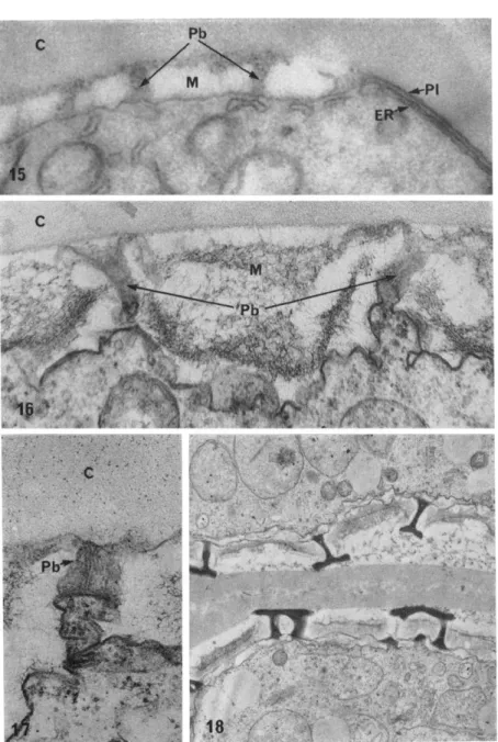
In Silène pendula, for example, before extensive growth of the pri
mexine matrix has occurred, plates of ER become apposed to par
ticular regions of the plasmalemma, giving the conspicuous triple membrane configuration seen in Fig. 15 ( Heslop-Harrison, 1963a).
These regions are the prospective pore locations, and no matrix material is deposited over them, so that the plasmalemma persists in contact with the callose tetrad wall. Later, a single, unpatterned layer of sporopollenin is deposited over these sites. Essentially the same process has been described in the definition of the germinal aperture region of the pollen of Zea mays (Skvarla and Larson, 1966), and of the furrow regions of the tricolpate grain of Helleborus foetidus (Godwin and Echlin, 1968).
What links can be traced between the role of the ER in locating the aperture sites of pollen grains and in the establishment of sieve plate pattern? In each, the close apposition of a surface of the ER
138 J . HESLOP-HARRISON
FIG. 15. Surface region of a microspore of Silène pendula, still within the meiotic tetrad. On the right, the locus of a pore may be seen. The plasmalemma
PATTERN IN PLANT CELL WALLS 139
to the plasmalemma excludes cellulose formation over the outer face of the plasmalemma; in the microspore the plasmalemma remains in contact with the callose mother cell surrounding the tetrad, and at the sieve plate the association of ER with the plasmalemma seems to bring about the localized formation of callose at the expense of an existing wall. I have suggested elsewhere ( Heslop-Harrison, 1966) that the close association of ER and plasmalemma would exclude the apposition of microtubules, as it would also of dictyosome vesicles concerned with the transfer of wall materials. This might indicate a stencil-like function for the ER. The situation can hardly be so simple with the sieve plate, however, where the positioning of the ER seems to be related to localized synthesis of callose and probably erosion of cellulose; here an enzymatic function is indicated.
Elements of the ER have been implicated also in the localization of the probacula which, as we have seen, determine the other major features of the pollen exine pattern. In the young microspores of Silène pendula (Heslop-Harrison, 1963a,b) and Zea mays (Skvarla and Larson, 1966), tubules or ribbons of ER are conspicuously associated with the basal ends of the probacula as they develop across the matrix of the primexine. Here again it might be supposed that the relationship is connected with some special local synthesis, since the material of the probacula difiFers chemically from that of the primexine matrix.
(PI) is directly apposed to the callose ( C ) of the tetrad wall; below it lies a plate of endoplasmic reticulum (ER). To the left, the plasmalemma abuts the primex
ine, consisting of matrix material (M) and the radially arranged probacula (Pb).
With this KMn04 fixation, the primexine matrix appears homogeneously electron transparent. X ca. 41,000.
FIG. 16. Very early phase of primexine formation within the tetrad of Lilium longijiorum. The callose tetrad wall (c) appears homogeneously granular. The matrix material of the primexine ( M ) has a loose fibrillar texture; there is evidence to suggest it is cellulosic. The probacula ( Pb ) rise from radially directed cytoplas- mic eminences. X ca. 78,000.
FIG. 17. Detail of probacula, same developmental phase as in Fig. 16. A col
umn of parallel lamellae arise from the cytoplasmic eminence and traverse the matrix of the primexine. X ca. 85,000.
FIG. 18. Later developmental stage in the tetrad of Lilium longijiorum, show
ing consolidation of the probacula, which now contain material with some of the properties of sporopollenin. X ca. 14,000.
Electron micrographs of Figs. 16-18 are by Dr. H. G. Dickinson.
140 J. HESLOP-HARRISON
Not all studies of early microspore ontogeny have revealed an association between the ER and the disposition of probacula in the primexine (e.g., Echlin and Godwin, 1968). Moreover, it is becoming clear that the young probaculum is not merely an amorphous rod.
When pattern is first emerging in the primexine of lily, the probacula appear as lamellated structures rising from the plasmalemma ( Figs. 16 and 17 ), and there is some indication that ribosomes, often in polysome configurations,- cluster in the vicinity (Heslop-Harrison, 1968b). It is conceivable that the lamellae are lipoprotein, in which case the probacula would represent something very like extensions of the cytoplasm in these very early developmental stages. A link would then be possible with the observations of Rowley (1967), who found that sporopollenin was formed on lamellae, "of unit membrane di
mensions," during the growth of the nexine 2 in microspores of Anthurium. In lily, the lamellation of the probacula later disappears (Fig. 18).
It is obvious that the relationship between the patterned part of the primexine—represented initially by these probacula—and the matrix component is of some interest. Which "determines" which, if determination of one by the other is indeed involved? Is the patterning process the exsertion of the probacula, the matrix material merely filling the intervening gaps? Or is it that the cellulose of the matrix is laid down as a negative pattern, the material of the probacula penetrating where gaps are left? Some unification of pattern-de
termining processes might be at hand could it be asserted that, as in secondary thickening in the endothecium, microtubules are con
cerned in establishing the conformation of the matrix as the comple
mentary template. However, there is no indication that the matrix is laid down in a controlled manner with regularly oriented micro- fibrils, nor of any relationship with patterns of microtubules in ad
jacent cytoplasm, so the possibility seems excluded, at least so far as current evidence goes.
Nevertheless, it seems that there still may be some room for micro- tubule participation in the pattern-determining process in the prim
exine. Long ago, Beer (1911) observed what he termed "kinoplastic radiations" from the nucleus in developing microspores of Ipomoea caerulea, and suggested that these might be related to the determi
nation of wall pattern during the tetrad stage. Recently, Echlin and Godwin (1968) have recorded comparable structures in Helleborus
PATTERN IN PLANT CELL WALLS 141 foetidus, and have pointed to the possibility that microtubules may play some direct part in determining probacular distribution, pre
sumably by guiding dictyosome vesicles.
There is an aspect of microsporogenesis which merits some mention here because it bears upon the general question of nuclear control of wall pattern determination. The first patterned wall, the primexine, is formed around an isolated haploid cell. Is the patterning con
trolled by the spore nucleus, or is it executed in accordance with programs handed 'on from the parental diploid cell? A case can be made for control by the spore nucleus on the grounds largely of propinquity and the very fact that the process does go through in isolation ( Heslop-Harrison, 1963b). Yet the matter is by no means straightforward. If haplophase control is involved, then some examples of pattern-segregation in the tetrad would be expected; none is known (Godwin, 1968). We may note also that pertinent evidence comes from polyad formation in aneuploid plants with abnormal chromosome segregation. In these, quite small fragments of cytoplasm with micronuclei often round up in the callose wall of the polyad and begin some of the preliminaries, at least, of spore wall formation.
Certainly these cells must be genetically deficient, yet the fact does not preclude the expression of a pattern-determining process. This may suggest that the determinants are already present in the cytoplasm of the sporocyte before the cleavage at cytokinesis. It may not be without significance here that the cytoplasm of the meiocyte, which undergoes a phase of ribosome elimination during prophase ( Macken
zie et al., 1967), becomes carved up during this same period into membrane-bounded enclaves which are transmitted through the meiotic divisions (Heslop-Harrison, 1968b; Dickinson and Heslop- Harrison, in preparation), and distributed among; the spores. Would it strain credibility too much to suppose that these form a vehicle for the transfer of selected fragments of morphogenetic information including that related to wall pattern?
It may be noted that the general idea of transmitted determinants is not new; Sinnott (1960) for example noted that various specific types of wall differentiation in somatic tissues seem "to be related to specific cell lineages, almost as though 'determiners' were being parcelled out at each division." However, there is a great deal of difference between the partitioning of morphogenetic information between four segments of the same original cytoplasm and th§
142 J . HESLOP-HARRISON
initiation of a tendency carried on through several generations of cell increase. In the first case the operation is once and for all, and no replication of the determinants would be demanded; in the second, replication would be needed if the information were not to be diluted out.
The conclusion from argument along these lines seem obvious enough. Although in some special circumstances, exemplified by the developing spore, the execution of wall pattern need not necessarily be governed by the cell concerned, in most cases control is bound to be located at least in part in the cell undergoing change. It is difficult to envisage this as meaning anything other than control through the agency of gene action, unless the intervention of a plasmagene system be accepted.
CONSPECTUS
The recognition of cytoplasmic elements that comport themselves as though they were connected in some way with the establishment of features of the cell wall certainly marks a step forward in our understanding of the genesis of wall patterning. However, there are many gaps to be bridged before the full chain of causation can be said to have been elucidated. It is perhaps in regard to the role of the microtubule that we may hope to see early advance, particularly in its apparent control of microfibril disposition. The colchicine ex
periments seem to show that the case here is not merely post hoc, ergo propter hoc; and what these experiments indicate is that the microtubules is not concerned with cellulose synthesis as such, but specifically with localizing where synthesis shall occur, and orienting the microfibrils produced. How spatial control of this kind can be exerted is not yet apparent. As we have seen, the microfibril is formed outside the plasmalemma. Presumably the process is one of tip growth, as has been graphically shown in extracellular microfibril production by the bacterium, Acetobacter xylinum (Colvin and Dennis, 1964).
Frei and Preston ( 1961 ) have argued from the way microfibrils form lamellae in certain algal walls that growth must occur in some depth, and Preston (e.g., 1964) has suggested how this might take place.
When the cytoplasm is withdrawn from the walls of algae such as Chaetomorpha by plasmolysis, files of granules remain adherent on the inner surface of the wall, oriented either along existing micro
fibrils, or at right angles to them. Individual microfibrils terminate
PATTERN IN P L A N T CELL WALLS 143 within the granules, indicating that these bodies may be concerned with end synthesis. Preston's suggestion is that the granules, presumed to be enzyme-containing, are in three-dimensional close packing, and that during microfibril synthesis the cellulose chains grow forward, pass through a granule and abut upon the next, which is then stimu
lated to take over the synthesis. In this scheme neither microfibril nor granule moves, but the direction of propagation of the microfibril will depend upon how the granules are themselves arranged, or along which of the several lines of contact points between the granules synthesis proceeds.
The freeze-etching technique has revealed adherent granules com
parable with those described by Preston on the outer surfaces of the plasmalemmas of yeast and the algae Chlorella and Cyanidium ( Moor and Miihlethaler, 1963; Mühlethaler, 1967). Fibrils extend from some of these particles, suggesting that they are indeed concerned with cellulose synthesis. In Chlorella there are indications that the particles become detached from the plasmalemma, and move to the outer sur
face where the microfibrils are synthesized across a layer of matrix material, presumed itself to be noncellulosic.
There are higher plant cells where evidence already exists for con
trolled growth of cellulose microfibrils remote from the plasmalemma.
Setterfield and Bayley (1957, 1959) and Beer and Setterfield (1958) have shown that in collenchymatous cell the outer longitudinal ribs, which have microfibrils oriented axially rather than transversely as in the inner wall, continue to thicken as the cells grow. Autoradiography shows that cellulose precursors are incorporated through the depth of the wall in these cells, not solely at the plasmalemma. Considering this case, Preston (1964) has pointed out that end-synthesizing particles might continue to lay down microfibrils in any layer of the wall—given the substrate—following the pattern already determined by the exist
ing microfibrils.
In what ways, then, might the microtubule be concerned in affecting spatial aspects of microfibril orientation on the remote side of the plasmalemma? Two possibilities suggest themselves: that they might have a function in determining where substrate shall be added to the wall, and that they might play some part in controlling at least the initial disposition of Preston's particles, if indeed bodies of this kind are involved in the tip-propagation of microfibrils in the cell walls of higher plants.
144 J . HESLOP-HARRISON
There is evidence for the participation of microtubules in the first of these roles. Microtubules do seem to serve in some way to steer dictyosome-derived vesicles containing wall precursors to the vicinity of the cell plate during cytokinesis ( Pickett-Heaps and Northcote, 1966; Hepler and Newcomb, 1967), and it may be surmised that they undertake a similar function during secondary wall thickening, where vesicles are often seen to be much more numerous in the tracts of cor
tical microtubules than elsewhere in the cytoplasm (E. H. Newcomb, personal communication).
Currently, any comment on the possible second role can only be speculative. Particles of the type envisaged by Preston have yet to be observed in higher plant cells and shown to have distributions cor
related with microtubules; but should their presence be proven, we may perhaps see the hint of a mechanism. Presumably the enzyme particles would be synthesized in the cell and passed through the plas- malemma. Were their preferential aggregation over particular parts of the wall determined by the presence of microtubules, and were they to be arranged in linear fashion below or between microtubules before extrusion, their pattern on the outer surface of the plasmalemma would be correlated with the alignment of the microtubules, and this would be reflected again in microfibril orientation once cellulose synthesis began. This kind of mechanism would seem more appealing than that proposed by Roelofson (1965), who suggested that the microtubules might exert an influence on the direction of microfibril growth through some form of electrical field effect. In any event, it is probably well within the range of current fine-structural technique to establish the main point, whether particles of the type described by Preston do exist in higher plant cells.
When we turn to consider the localization and orientation of the microtubules themselves, it seems that we are dealing with phenomena common to animal and plant cells. In his contribution to this sym
posium, Dr. Tilney has given some account of the now massive body of evidence indicating that microtubules perform a function like that postulated years ago by Needham for a "cytoskeleton," establishing asymmetries in the cell, in respect to shape, internal structure, and the movement of other cell components. Porter (1966) has suggested that for such a role to be discharged it must be supposed that the development and elimination of microtubule populations occur accord
ing to a series of rather strict programs, related presumably to special
PATTERN IN PLANT CELL WALLS 145 initiating sites and points of anchorage. There is a challenge to dis
cover what these sites are in plant cells, which lack obvious counter
parts to centrioles and other initiating structures. The properties of the nuclear envelope may be worth special attention here, since there are now several reports of microtubules emanating radially from its sur
face during periods of morphogenetic activity in the cell.
There is also the outstanding problem of what determines the intra- cellular disposition of the membranes of the ER insofar as these are concerned in wall.patterning processes. Without doubt much of the ER is in a highly labile condition in the cell (Whaley et al, 1964);
and it is only necessary to view a leaf cell of Elodea, or time-lapse films of dividing cells, to become convinced that it must be subject to much disturbance and movement. Yet the persistent association of some membrane surfaces with wall features like the sieve plate pores or the microspore apertures shows that there are stable areas. Mostly these appear to be near the cell surface, in the same, presumably gel
like, layer of cytoplasm inhabited by microtubules during their associa
tion with wall patterning. It seems that the preferential disposition of plates of ER in this cortical region is often controlled in part at least by cellular interactions, and by the microenvironment of individual cells when they are not in contact. This point has been stressed espe
cially by Echlin and Godwin ( 1968 ) in relation to the role of the ER in defining the positions of the furrows of the pollen grain of Helle- borus foetidus. They suggested that the contact geometry within the tetrad may provide the initial orienting stimulus, since there is some evidence that a furrow may be initiated at the point of closest ap
proach of the four microspores. There is a similar relationship between spore patterning and the overall geometry of the tetrad in lily ( Heslop- Harrison, 1968b).
The inescapable requirement for any explanation of wall patterning taking into account genetic control has already been noted: it must, ultimately, show us how base sequences in DNA come to be expressed in patterns of dimensions many orders of magnitude greater, executed in polysaccharides and other compounds. An elucidation of the roles played by microtubules and cytoplasmic membranes in wall growth would clarify some of the mechanics of the process, leaving a gap in our understanding of the immediately antecedent patterning events.
Yet there are indications of how the matter may be managed at this level Sometimes when we contemplate biological pattern it is difficult
146 J . HESLOP-HARRISON
to imagine how great a part of it could arise from physical causes, and correspondingly easy to slip into accepting that genetical control extends down to all detail. Yet this is obviously not so. Once more taking lily pollen as an example: Fig. 14 shows us that the genetic community among the grains in an anther is expressed in a general theme of patterning; for the detail, the execution is almost infinitely variable. This surely tells us that control must be exerted only at cer
tain strategic points, in determining what shall associate with what and in what order, and in mapping out, in interaction with the cell environment, where the greatest probabilities will lie for association to begin. After this, physical processes akin to crystallization, or the formation of standing waves as envisaged in Turing's now classical model (1952), must take over to complete the space-filling operation.
From what is known of the dynamics of microtubules and cytoplasmic membranes it would seem that they do have the kind of controlled lability to be expected of entrepreneurs in activities of this kind. In certain cells at least, pools of structural units do exist, ready to partici
pate in cycles of polymerization and depolymerization as conditions determine (Whaley et at, 1964; Porter, 1966). Evidently response could be both to changes in the microenvironment of the cell and to the time-related appearance of new gene products in the metabolic arena, factors which could interact to initiate patterning processes expressed in first tangible form in the disposition of tubules and membranes.
At this time it is probably easiest for us to think along these lines, particularly since we have become aware of the existence of cytoplas
mic "blue prints" for some types of wall patterning. But there is no compelling reason a priori for supposing that the execution of pattern
ing in the wall necessarily requires the setting up of precursor forms within the plasmalemma of the cell. It is not excluded that pattern should also emerge from the folding and aggregation of secreted pro
teins at sites remote from the protoplast, within the fabric of the wall.
ACKNOWLEDGMENTS
My thanks are due to Mr. H. G. Dickinson, who is responsible for the electron micrographs of Figs. 6-8 and 16-18; and to Engis Equipment Company, of Mor
ton Grove, Illinois, the U. S. agents for the Cambridge Instruments Stereoscan electron microscope, and particularly to Mr. F. Rossi of Engis Equipment. I am also grateful to Dr. E. H. Newcomb for Fig. ls and for helpful discussion of some
PATTERN IN P L A N T CELL WALLS 147
of the topics touched upon in the text; he is not, of course, responsible for what
ever may prove erroneous or tendentious.
Original research was supported by the Science Research Council and the Wisconsin Alumni Research Association.
REFERENCES
BEER, R. (1911). Studies in spore development. Ann. Botany (London) 25, 1 9 9 - 214.
BEER, M., and SETTERFIELD, G. (1958). Fine structure in thickened primary walls of collenchyma cells, of celery petioles. Am. J. Botany 45, 571-580.
BORISY, G. G., and TAYLOR, E. W. (1967a). The mechanism of action of colchi- cine. Binding of colchicine-Ή to cellular protein. / . Cell Biol. 34, 525-533.
BORISY, G. G., and TAYLOR, E. W. (1967b). The mechanism of action of colchi- cine. Colchicine binding to sea urchin eggs and the mitotic apparatus. J. Cell Biol 34, 535-548.
COLVIN, J. R., and DENNIS, D. T. (1964). The shape of the tips of growing bac
terial cellulose microfibrils and its relationship to the mechanism of cellulose biosynthesis. Can. J. Microbiol. 10, 763-767.
CRONSHAW, J., and BOUCK, G. B. (1965). The fine structure of differentiating xylem elements. J. Cell Biol. 24, 415-431.
ECHLIN, P., and GODWIN, H. (1968). Ultrastructure and ontogeny of pollen in Helleborus foetidus L. II. Pollen grain development through the callose special wall stage. /. Cell Set. 3, 175-186.
ESAU, K., CHEADLE, V. I., and RISLEY, E. B. (1962). Development of sieve plate pores. Botan. Gaz. 123, 233-243.
FREI, E., and PRESTON, R. D. (1961). Cell wall organization and wall growth in the filamentous green algae Cladophora and Chaetomorpha. I. The basic struc
ture and its formation. Proc. Roy. Soc. B154, 70-94.
FREY-WYSSLING, A. (1948). "Submicroscopic Morphology of Protoplasm and Its Derivatives." Elsevier, Amsterdam.
FREY-WYSSLING, A., LOPEZ-SÂEZ, J. F., and MÜHLETHALER, K. ( 1964 ). Formation
and development of the cell plate. /. Ultrastruct. Res. 10, 422-431.
GODWIN, H. (1968). The origin of the exine. New Phytologist, in press.
HEPLER, P. K., and NEWCOMB, E. H. (1963). The fine structure of tracheary xylem elements arising by re-differentiation of parenchyma in wounded Coleus stem. J. Exptl. Botany 14, 496-503.
HEPLER, P. K., and NEWCOMB, E. H. (1964). Microtubules and fibrils in the cyto
plasm of Coleus cells undergoing secondary wall deposition. /. Cell Biol. 20, 529-533.
HEPLER, P. K., and NEWCOMB, E. H. (1967). Fine structure of cell plate forma
tion in the apical meristem of Phaseolus roots. /. Ultrastruct. Res. 19, 498-513.
HESLOP-HARRISON, J. (1963a). Ultrastructural aspects of differentiation in sporo- genous tissue. Symp. Soc. Exptl. Biol. 17, 315-340.
HESLOP-HARRISON, J. (1963b). An ultrastructural study of pollen wall ontogeny in Silène pendula. Grana Palynol. 4, 7-24.
HESLOP-HARRISON, J. (1966). Morphogenesis at the sub-cellular level. In "Trends
148 J . HESLOP-HARRISON
in Plant Morphogenesis" ( E . Cutter, ed.), pp. 127-139. Longmans, Green, New York.
HESLOP-HARRISON, J. ( 1968a ). Wall development within the microspore tetrad of Lilium longiflorum. Can. J. Botany in press.
HESLOP-HARRISON, J. (1968b). Pollen wall development. Science 161, 230-237.
JUNIPER, B. E. (1959). The surfaces of plants. Endeavour 18, 20-25.
KAMIYA, N. (1959). Protoplasmic streaming. In "Handbuch der Protoplasma
forschung ( L . V. Heilbrun and F . Weber, eds.), Vol. 8. Springer, Berlin.
LAMPORT, D. T. A. (1965). The protein component of primary cell walls. Advan.
Botan. Res. 2, 151-213.
LEDBETTER, M. C , and PORTER, K. (1963). A "microtubule" in plant fine struc
ture. / . Cell Biol. 19, 239-250.
LEVAN, A. (1939). Cytological phenomena connected with the root swelling caused by growth substances. Hereditas 25, 87-96.
LEVAN, A. ( 1942 ). The macroscopic colchicine effect—a hormone action? Heredi- tas 28, 244-245.
MACKENZIE, A., HESLOP-HARRISON, J., and DICKINSON, H. G. ( 1 9 6 7 ) . Elimination
of ribosomes during meiotic prophase. Nature 215, 997-999.
MOLLENHAUER, H. H., W H A L E Y , W . G., and L E E C H , J. H. ( 1 9 6 1 ) . A function of
the Golgi apparatus in outer root cap cells. / . Ultrastruct. Res. 5, 193-200.
MOOR, H., and MÜHLETHALER, K. (1963). Fine structure in frozen etched yeast cells. / . Cell Biol. 17, 609-628.
MÜHLETHALER, K. (1967). Ultrastructure and formation of plant cell walls. Ann.
Rev. Plant Physiol. 18, 1-24.
NAGAI, R., and REBHUN, L. I. (1966). Cytoplasmic microfilaments in streaming Nitella cells. / . Ultrastruct. Res. 14, 571-589.
NORTHCOTE, D. H., and PICKETT-HEAPS, J. D. (1966). A function of the Golgi apparatus in polysaccharide synthesis and transport in the root cap cells of wheat. Biochem. J. 98, 159-167.
NORTHCOTE, D. H., and WOODING, F . B. P. (1965). Development of sieve tubes in Acer pseudoplatanus. Troc. Roy. Soc. B163, 524-537.
O'BRIEN, T. P., and THIMANN, K. V. (1966). Intracellular fibres in oat coleoptile cells and their possible significance in cytoplasmic streaming. Proc. Natl. Acad.
Set. U. S. 56, 888-894.
PICKETT-HEAPS, J. D. (1967). The effect of colchicine on the ultrastructure of dividing plant cells, xylem wall differentiation, and distribution of cytoplasmic microtubules. Develop. Biol. 15, 206-236.
PICKETT-HEAPS, J. D., and NORTHCOTE, D . H. (1966). Relationship of cellular organelles to the formation and development of the plant cell wall. / . Exptl.
Botany 17, 20-26.
PORTER, K. R. ( 1966 ). Cytoplasmic microtubules and their functions. Ciba Found.
Symp. Principles Bimolecular Organ, pp. 308-345. Little, Brown, New York.
PORTER, K. R., and MACHADO, R. D. (1960). Studies on the endoplasmic reticulum.
IV. Its form and distribution during mitosis in cells of onion root tip. / . Biophys.
Biochem. Cytol. 7, 167-180.