Contents lists available atScienceDirect
Vascular Pharmacology
journal homepage:www.elsevier.com/locate/vph
Systematic analysis of different pluripotent stem cell-derived cardiac myocytes as potential testing model for cardiocytoprotection
J. Pálóczi
a,1, Á. Szántai
a,1, J. Kobolák
b, I. Bock
b, E. Ruivo
a, B. Kiss
a,e, R. Gáspár
a, J. Pipis
c, I. Ocsovszki
a, Z. Táncos
b, A. Fehér
b, A. Dinnyés
b,d, Z. Onódi
e, R. Madonna
f,g, P. Ferdinandy
a,c,e,h, A. Görbe
a,c,e,h,⁎aCardiovascular Research Group, Department of Biochemistry, University of Szeged, 6720 Hungary
bBiotalentum Ltd., Gödöllő, 2100 Hungary
cPharmahungary Group, Szeged, 6722 Hungary
dMolecular Animal Biotechnology Laboratory, Szent István University, Gödöllő, 2100 Hungary
eMTA-SE System Pharmacology Research Group, Department of Pharmacology and Pharmacotherapy, Semmelweis University, Budapest, 1085 Hungary
fInstitute of Cardiology, Department of Surgical, Medical and Molecular Pathology and Critical Area Medicine, University of Pisa, 56124 Pisa
gInternal Medicine, Cardiology Division, University of Texas Medical School in Houston, Houston, Texas
hCardiovascular Research Group, Department of Pharmacology and Pharmacotherapy, University of Szeged, 6720, Hungary
A R T I C L E I N F O Keywords:
Cell culture In vitro
Cardiocytoprotection Drug screening
Induced pluripotent stem cell Embryoid body
A B S T R A C T
Introduction:Stem cell-derived cardiac myocytes are potential sources for testing cardiocytoprotective molecules against ischemia/reperfusion injuryin vitro.
Materials and methods:Here we performed a systematic analysis of two different induced pluripotent stem cell lines (iPSC 3.4 and 4.1) and an embryonic stem cell (ESC) line-derived cardiac myocytes at two different de- velopmental stages. Cell viability in simulated ischemia/reperfusion (SI/R)-induced injury and a known cardi- ocytoprotective NO-donor, S-nitroso-n-acetylpenicillamine (SNAP) was tested.
Results:After analysis of full embryoid bodies (EBs) and cardiac marker (VCAM and cardiac troponin I) positive cells of three lines at 6 conditions (32 different conditions altogether), we found significant SI/R injury-induced cell death in both full EBs and VCAM+ cardiac cells at later stage of their differentiation. Moreover, full EBs of the iPS 4.1 cell line after oxidative stress induction by SNAP was protected at day-8 samples.
Conclusion:We have shown that 4.1 iPS-derived cardiomyocyte line could serve as a testing platform for car- diocytoprotection.
1. Introduction
There is still no cardioprotective drug on the market after more than 3 decades of intensive research [1], therefore, search for therapeutic agents to alleviate myocardial ischemia/reperfusion injury is of great importance [9,38].
In preclinical drug testing,in vitrocardiac myocyte-based platforms developed from cardiomyoblast lines, neonatal or adult primary car- diomyocytes are frequently and traditionally used experimental tools in screening for cardioprotective drugs [38]. Despite their advantages for pharmaceutical applications, these assays face severe challenges, in- cluding limited proliferation capacity, uncontrolled stress during cell isolation, low throughput nature and poor predictability towardsin vivo
efficacy [49]. Additionally, taking into account of 3R (replacement, reduction and refinement) principles, a more appropriate cell source is highly desirable [21].
Embryonic stem cells (ESC) with human origin offer a virtually unlimited number of cells capable of differentiating towards cardiac lineage [50], however, ethical and immunological concerns sur- rounding human ESCs impede their clinical or research application [48]
as well. The discovery of induced pluripotent stem cells (iPSC) by the reprogramming of somatic cells has provided a new alternative plur- ipotent stem cell (PSC)-derived cell systems without the above- mentioned limitations [41,42,53]. IPS cells can be derived from human somatic cells; therefore, they can be promisingin vitroplatform and also tool for cell therapy as well [7,22,37,39]. Although, iPSC-derived
https://doi.org/10.1016/j.vph.2020.106781 Received 17 July 2020; Accepted 13 August 2020
⁎Corresponding author at: MTA-SE System Pharmacology Research Group, Department of Pharmacology and Pharmacotherapy, Semmelweis University, Budapest H-1089, Hungary.
E-mail address:gorbe.aniko@med.semmelweis-univ.hu(A. Görbe).
1These authors contributed equally to this work.
Available online 19 August 2020
1537-1891/ © 2020 The Authors. Published by Elsevier Inc. This is an open access article under the CC BY-NC-ND license (http://creativecommons.org/licenses/BY-NC-ND/4.0/).
T
cardiac myocytes have been used forin vitrodrug safety testing, there is no data on their application for modelling ischemia/reperfusion injury and testing cardioprotection.
In contrast to the encouraging results and the enormous potential of iPSC-derived cardiomyocytes, numerous technical obstacles need to be overcome. These include the development of efficient and reliable protocols for differentiation and strategies for cardiomyocyte selection [18,27]. Application potential of iPSC-derived cardiac myocyte in heart regeneration and repair is still limited in clinical trials, where the protection of them in the unfavourable ischemic myocardial micro- environment will be indispensable [28]. Therefore, characterization of iPSC-derived cells in a simulated ischemia/reperfusion test platform and testing potential cardioprotective candidate compounds for is- chemic heart diseases has a great importance.
Thus, in this study we performed a systematic analysis of 2 different mouse iPSC lines and an embryonic stem cell line at two stages of cardiac differentiation, with and without oxidative stress conditions, respectively. In each series full embryoid bodies and cardiac marker positive cell were implemented to study the response of cells to simu- lated ischemia/reperfusion (SI/R) and cardiocytoprotection of NO- donor SNAP.
2. Materials and methods
The investigation conforms to the Guide for the care and use of laboratory animals published by the US National Institutes of Health (NIH publication No. 85-23, revised 1996) and was approved by the Ethics Committee at the University of Szeged.
Materials for the cell culture and sample preparation, unless otherwise indicated were purchased from Thermo Fischer Scientific Inc.
(Carlsbad, USA).
2.1. Cell culture
The HM1 mouse ESCs [36] at passage 19, was kindly provided by Roslin Institute, UK. The iPS 3.4 and iPS 4.1 mouse iPSC lines were provided by Prof Roger Pedersen (University of Cambridge, UK). The ESC and iPSC lines were cultured and maintained under feeder-free conditions in KOSR + 2i media (KnockOut™ DMEM supplemented with 15% KnockOut™ Serum Replacement, 1 mM Na-Pyruvate, 0.1 mM non- essential amino acids, 0.1 mM 2-mercaptoethanol, 4 mM GlutaMAX™, 50 U/ml penicillin/streptomycin, 2000 U/ml Leukemia Inhibitory Factor (LIF) (Millipore), 5 μg/ml Insulin (Sigma), 1 μM of the mitogen- activated protein kinase inhibitor PD0325901 (StemGent) and 3 μM of the glycogen synthase kinase-3 inhibitor CHIR99021 (StemGent) [12].
When in culture, the cells were maintained at 37 °C humidified air Fig. 1.Cardiac differentiation and experimental protocol of simulated ischemia and reperfusion in mouse ESC- or iPSC-derived embryoid bodies.
(A) Pluripotent stem cells of mouse origin (ESC or iPSCs) were dissociated and EBs were generated in hanging drops. After three days the formed EBs were further differentiated to generate cardiomyocytes by culturing them individually in 24-well plates for 8 and 16 days. EBs were characterized by immunocytochemistry (ICC) and RT-qPCR at day 8 or 16. The experiment was also carried out under oxidative stress condition induced by 24 h H2O2treatment before differentiation.
(B) EBs were exposed to 150 min hypoxia followed by 120 min reperfusion at day 8 or day 16. Cells were treated with vehicle or NO-donor SNAP during simulated ischemia. Cell viability of full EBs was assessed by propidium iodide staining. Cell viability of VCAM-1 or cTnI positive cells was evaluated after immunolabelling with flow cytometry.
containing 5% CO2in incubator and passaged every third day using 0.25% trypsin-EDTA.
2.2. In vitro cardiac differentiation
For cardiac differentiation, embryoid bodies (EBs) were generated using the Hanging Drop method. Briefly, one day before EB plating the 2i media was changed to LIF-free basic ESC media (DMEM-GlutaMax, 15% (v/v) ESC qualified FCS (fetal calf serum, SLI Ltd), 0.1 mM non- essential amino acids, 0.1 mM 2-mercaptoethanol, 50 U/ml penicillin/
streptomycin). Single cell suspensions were obtained by dissociating iPSCs with 0.25% trypsin-EDTA (day 0). The cells were diluted in 4 × 10⁴cells/ml and 20 μl droplets (800 cells/droplet) were pipetted onto the lid of low attachment Petri dish in differentiation medium (basic ESC medium without LIF). The cells were allowed to aggregate using gravity by inverting the lid over the dish. On day 3, individual EBs were plated into a well of a 24-well plate containing coverslips coated with 0.1% gelatin. Differentiation medium was changed every second day, the number of contracting cardiomyocytes was con- tinuously observed.
2.3. Oxidative stress assay
One series of iPSCs were treated for 24 h by supplementing the media with 0.01 μM or 0.05 μM H2O2(Sigma) for oxidative stress in- duction. After 24 h of treatment stem cells were harvested and differ- entiated as described above. The differentiation was monitored after 8 and 16 days (Fig. 1, panel A).
Beating rates were calculated at day 8 and 16, by counting the beating EBs on a 24-well plate. In one experiment three parallels (n= 72) were counted and four independent experiments were ana- lysed.
2.4. Immunocytochemistry
Samples were fixed with 4% PFA (30 min, RT), then permeabilized with 0.1% Triton-X 100, and 0.1% bovine serum albumin (BSA)-con- taining PBS. Cells were screened for the expression of cardiac differ- entiation markers by incubation with the primary antibodies against:
Nkx2–5, Troponin T, VCAM-1, Phalloidin (labelling of F-actin) and Desmin (overnight, 4 °C). Samples were then incubated with the ap- propriate fluorescent secondary antibodies (1 h, RT). For counter- staining DAPI (20 min, RT) was used. The details of antibodies and dilutions are listed in Table 1. The cells were observed under fluor- escent microscope equipped with 3D imaging module (AxioImager system with ApoTome, controlled by AxioVision 4.8.1 Microscope software; Carl Zeiss).
2.5. Reverse transcription-qPCR (RT-qPCR) analysis
Total RNA was isolated from cells using RNeasy Plus Mini Kit (Qiagen). SuperScript VILO cDNA Synthesis Kit was used according to the manufacturer's instructions.
Primers for genes (see Table S1) were designed using the Primer3 software [34]. To avoid interfering secondary DNA structures mFOLD software was utilized [55], and the specificity of the primers was ver- ified by Primer-BLAST [52]. 6 selected genes andGapdhas reference were used for RT-qPCR analysis (Table 2). Each RT-qPCR reaction contained 5 ng RNA equivalent cDNA template, 400 nM of each primer and 50% SYBR Green JumpStart Taq ReadyMix (Sigma Aldrich) in a total volume of 15 μl. PCR reactions were set up using QIAgility liquid handling robot and were performed on a Rotor-Gene Q cycler (Qiagen).
The cycling parameters were as follows: 94 °C for 3 min initial dena- turation followed by 40 cycles of 95 °C for 5 s, 60 °C for 15 s and 72 °C for 30 s. Melting curve analysis and agarose gel electrophoresis con- firmed the specificity of the primers. Data of three replicates were analysed using the Relative Expression Software Tool 2008 V2.0.7 [30].
2.6. Simulated ischemia/reperfusion (SI/R) treatment of mouse ESC- and iPSC-derived cardiomyocytes
The protocol of SI/R experiments was based on Gorbe et al. [14], whereas both mouse ESC-derived and iPSC-derived embryoid bodies (EBs) on either day-8 or -16 of differentiation were tested under nor- moxia or were subjected to SI (Fig. 1, panel B).
In the normoxic groups, the growth medium of cells was replaced with a normoxic solution (in mM: NaCl 125, KCl 5.4, NaH2PO41.2, MgCl20.5, HEPES 20, glucose 15, taurine 5, CaCl21, creatine 2.5, BSA 0.1%, pH 7.4, 310 mOsm/l) [20] and cells were incubated under 95%
air and 5% CO2tension (150 min, 37 °C). Regarding SI groups, EBs were exposed to hypoxia (0,04% O2, 5CO2, 95% N2) by incubating the cells in hypoxic solution (in mM: NaCl 119, KCl 5.4, MgSO41.3, NaH2PO4 1.2, HEPES 5, MgCl2 0.5, CaCl2 0.9, Na-lactate 20, BSA 0.1%, 310 mOsm/l, pH = 6.4) [20] and were kept in the multigas incubator (150 min, 37 °C).
The experimental setup consisted the following groups: 1, normoxic (covered by normoxic solution); 2, simulated ischemia (vehicle) and 3, simulated ischemia supplemented with SNAP (S-nitroso-N-acetyl-D,L- penicillamine, 1 μM). All treatments were followed by 120 min simu- lated reperfusion in growth medium and superfusion with 95% air and 5% CO2at 37 °C. At the end of SI/R experiments, cell death of EBs was assessed by using propidium iodine (PI) assay combined with PI + digitonin staining (total cell count). In another set of EBs, the ratio of cell death was also determined using Live/Dead assay after SI/R followed by immunolabelling of cardiac markers and flow cytometric analysis (Fig. 1, panel B).
In the second set of experiments, all procedures were performed on accelerated cardiac differentiation (H2O2 induced) EB series (Fig. 1, panels A and B).
2.7. Assessment of cell death in full embryoid bodies
For the assessment of cell death in full EBs, PI viability assay was performed in combination with total cell count determination (PI + digitonin staining). Briefly, medium of EBs was removed and cells were washed twice with warm D-PBS (Sigma). Then cells were Table 1
Antibodies used for immunocytochemistry.
Antibody Dilution Company (CatN)
Primary antibodies Anti-VCAM-1 1:250 Abcam (ab134047)
Anti-Nkx2-5 1:100 Santa Cruz Biotechnology (sc-14033)
Anti-cardiac Troponin T 1:200 Abcam (ab33589)
Anti-CD106 1:200 BioLegend (BZ-105710)
Anti-Desmin 1:100 DAKO (M0760)
Secondary antibodies Alexa Fluor 488 donkey anti-mouse IgG 1:2000 Thermo Fisher Scientific Inc. (A-21202)
Alexa Fluor 488 donkey anti-rabbit IgG 1:2000 Thermo Fisher Scientific Inc. (A-21206)
Other DY-554-Phalloidin 1:500 Dyomics (554-33)
incubated with PI solution (50 μM) and plates were kept in a dark place (7 min, RT). The fluorescence intensity of each EB was detected by using a fluorescent plate reader. As for the PI + digitonin assay, PI (50 μM) and digitonin (500 μM) were added into the wells in order to estimate total cell number in each well. Cells were then incubated with PI + digitonin solution (7 min, RT). Then fluorescence intensity was measured at 544em/620ex. Background fluorescence intensity (dye control) was subtracted from the fluorescence intensity of each well after staining, and the average intensity of each group was plotted.
2.8. Assessment of cell death by flow cytometry
For the determination of cardiac cell death in EBs, Live/Dead assay was used (Life technologies, L10120) Reagents were prepared ac- cording to the manufacturer's instruction. At the end of simulated re- perfusion, cells were washed twice with warm D-PBS, then they were incubated Live/Dead reagent in dark (30 min, RT). Subsequently, cells were washed again with D-PBS and processed towards flow cytometry.
2.9. Immunolabelling of VCAM-1 (CD106) positive cell population of EBs for flow cytometry
EBs were detached by adding collagenase type IV solutions (0.125 mg/ml) into the wells, because of avoiding destruction of cell surface marker. Cell suspensions were collected into Eppendorf tubes and centrifuged at 400×g(5 min, 4 °C). Then cells were resuspended and incubated with Alexa Fluor 488 anti-mouse CD106 (also known as vascular cell adhesion molecule 1 or VCAM-1) antibody (BioLegend BZ- 105710) at the dilution of 1:200 (1 h, 4 °C). After the immunolabelling of VCAM-1, cells were washed and centrifuged at 400×gfor (5 min, 4 °C) then they were resuspended in PBS containing 2% paraf- ormaldehyde (PFA) and fixed for 10 min at room temperature.
Subsequently, cells were washed with PBS and centrifuged again at 400×g(5 min, 4 °C). The supernatant was discarded and the cells were resuspended in 1 ml PBS for flow cytometry. Investigations were carried out by using a PARTEC TM FACS analyser. For each experiment, 20,000 events were counted and analysed in the appropriate size range. Gating strategy was chosen according to the unstained controls. Besides CD106 positive events, dead cells were also counted and analysed.
2.10. Immunolabelling of cTnI positive cell population of EBs for flow cytometry
The detection of troponin I, cardiac (cTnI) positive cells required the same steps as it was mentioned above. Regarding the digestion step, trypsin solution (0.25%) was used to detach EBs. After the supernatant was removed, the cells were resuspended and fixed in 2% PFA solution
(10 min, RT). After the washing step with PBS, samples were incubated with Anti-Cardiac Troponin I (cTnI) antibody (abcam, ab19615) at 1:100 dilution (overnight, 4 °C). Primary anti-cTnI antibodies were di- luted in PBS solution containing Triton-X (1%). On the following day, cells were washed with PBS and centrifuged at 400×g (5 min, 4 °C).
Then cells were resuspended and incubated with secondary antibody (goat anti-mouse-TRITC) at a dilution of 1:500 (30 min, RT).
Subsequently, cells were washed with PBS. The supernatant was dis- carded and cell pellets were resuspended in PBS. The cTnI positive fraction of EBs was analysed by flow cytometry as described above. The ratio of dead cells was also counted and analysed in the same manner.
For CD106 and cTnI co-staining studies, samples were immunolabelled for CD106, then following fixation, cells were incubated with primary anti-cTnI antibodies and processed as described above.
2.11. Statistical analysis
Results are expressed as mean ± SEM. One-way analysis of var- iance (ANOVA) followed by Tukey post-hoc tests were used to analyse differences in mean values between the experimental groups.
Differences were considered significant atp< 0.05.
3. Results
3.1. Characterization of cardiac-differentiated ESCs and iPSCs
To obtain myocardial cells for the investigation of ischemia and reperfusion, we have performed directed differentiation of a mouse ESC- and two iPSC-lines. The cultures were first characterized with comparative immunocytochemistry at two time points of cellular dif- ferentiation (day 8 and 16). The fluorescent signal of each cardiac marker (NKX2-5, Troponin T, VCAM-1 and F-actin labelled with Phalloidin) was detected by fluorescent microscopy in all the three lines. The expression of cardiac markers was elevated from day 8 to 16 (Fig. 2), which demonstrates the progress of cardiac differentiation.
Although the early cardiac precursor marker NKX2-5 was more pro- minent in day 8 samples, it was still observable in several cells at day 16. In line with this the later cardiac markers Troponin T and VCAM-1 were more prominent in day 16 samples, however no major difference was detected among the examined iPSC lines (Fig. 2). The cardiac specificity of VCAM-1 was verified by Desmin counterstaining in mouse ESC-derived embryoid bodies at day-8 and 16. Both cardiac markers showed higher expression at later stage of differentiation (day-16) and showed co-expression at both differentiation stages (Supplementary Fig. S1). To further investigate the cardiac specificity of VCAM-1, an- other series of experiments was performed by fluorescence-activated cell sorting (FACS). Mouse ESC-derived cells from HM1 strain Table 2
Primers used for RT-qPCR analysis.
Symbol Description Gene ID Fwd and Rvs primer PCR product (bp)
Nkx2-5 NK2 homeobox 5 18091 ACACCCACGCCTTTCTCAGT 74
AGGTCCCCAGACGCCA
Myh6 Myosin, heavy polypeptide 6, cardiac muscle, alpha 17888 CTACGCCTTCGTCTCTCAGG 80
AGGCACTATCAGTGGCCAAG
Flt1 FMS-like tyrosine kinase 1 14254 CCTGTCACCACAATCACTCC 172
GAGTCAGCCACCACCAATGT
Pecam1 Platelet/endothelial celladhesion molecule 1 18613 CGATTGTAGCCACCTCCAAG 132
TGTTGACCATGATGCTACTGG
Notch1 Notch1 18128 ACTGCGAGAACAACACACCT 104
GAAGCCAGGTGGACACAGAC
Vwf Von Willebrand factor 22371 GGCAAGAGAATGAGCCTGTC 122
TAGAGTCCTTGGGAGGCGTA
Gapdh⁎ Glyceraldehyde-3-phosphate dehydrogenase 14433 AATGTGTCCGTCGTGGATCT 79
CCTGCTTCACCACCTTCTTG
⁎ The reference gene used in the study.
underwent flow cytometric analysis to determine the ratio of VCAM-1+
and cTnI+ cells at day 8 and 16. We found a notable increase in number of VCAM-1+/cTnI+ cells in parallel with the stage of differ- entiation (32% vs. 71%), indicating the high cardiac specificity of VCAM-1 (Supplementary Fig. S2). Further validation of cardiac speci- ficity of VCAM-1 was performed in premature hearts of mouse embryos.
3.2. Validation of cardiac specificity of VCAM-1 in premature hearts Ultrasound imaging of post-coital female Bl6 mice was applied to reveal pregnancy and follow up embryonic development (Supplementary Figs. S3 and S4). At day 12.5 of mouse embryonic development (E12.5), mice were anesthetized by isoflurane with an initial rate of 5% and a maintained by 2%. The abdominal cavity was opened and the uterus was excised and put into phenol red free DMEM.
Mouse embryos were gently isolated by removing the remnant part of uterus, the decidua and the yolk sac. Finally, the amniotic sac was re- moved as well. Premature hearts were carefully isolated and ventricles were collected into Eppendorf tubes containing DMEM supplemented with collagenase IV (0,125 mg/ml). Then ventricles were digested at 37 °C with gentle agitation for 20–30 min. Then cells were centrifuged at 400×gfor 5 min at 4 °C. Pellets were resuspended and incubated in PBS solution containing 2% bovine serum albumin supplemented with Alexa Fluor 488 anti-mouse CD106 antibody at the dilution of 1:200 for 1 h at 4 °C. After a subsequent washing step, cells were centrifuged again at 400×gfor 5 min at 4 °C then resuspended in 2% PFA solution and fixed for 10 min at room temperature. Subsequently, cells were washed and centrifuged at 400×gfor 5 min at 4 °C. Supernatant was discarded and cell pellets were resuspended in 1 ml PBS for analysis.
Flow measurements were carried out by using a flow cytometer Fig. 2.Comparative immunocytochemical characterization of strain HM1; iPS 3.4 and iPS 4.1 at stage day 8 and 16.
Fluorescence images are shown with fluorescence signals for DAPI (blue), cardiac marker (NKX2–5, Troponin T, VCAM-1) (green) and Phalloidin (red) labelled F- actin immunoreactivity. Scale bars = 50 μM. (For interpretation of the references to colour in this figure legend, the reader is referred to the web version of this article.)
(Supplementary Fig. S3). First, we evaluated the specificity of VCAM1 during in vivo cardiac differentiation. To examine the specificity of VCAM1 expression, we performed flow cytometry analysis on prenatal cardiac cell population of collected hearts of E12.5 mouse embryos. The VCAM1+ subpopulation in embryonic hearts resulted in 52% of the intact cell population (see Supplementary Fig. S4, panel D).
3.3. Cell death of ESC- and iPSC-derived EBs and cardiac cells after simulated ischemia/reperfusion injury
To investigate the ischemic tolerance of mouse ESC- and iPSC-de- rived cardiomyocytes, EBs were subjected to 150 min simulated ischemia followed by 120 min reperfusion (SI/R). One of the SI groups was administered with cytoprotective NO-donor SNAP to test whether SNAP can attenuate the detrimental effects of SI/R in our model. Cell viability assays were performed immediately after reperfusion to de- termine the ratio of dead cells.
While tendency to increased cell death was observed only for ESC- derived cells in the vehicle-treated simulated ischemic group of full EBs on day-8, on day-16 the reduction was significant for ESC and iPS 3.4 lines (Fig. 3A, B). A similar tendency was observed in viability of VCAM-1+ cells both at day 8 and 16, (Fig. 3C, D), however, there were no significant differences between normoxic vs SI and SI vs SI treated SNAP groups. SI/R did not influence cell viability in cTnI+ populations of both age cohorts (Supplementary Fig. S5). In summary, full EBs re- sponded to SI/R injury at their later stage of differentiation, meanwhile cytoprotective effect of SNAP was not observed in any tested series
(Fig. 3A).
3.4. Effect of oxidative stress on cardiac differentiation
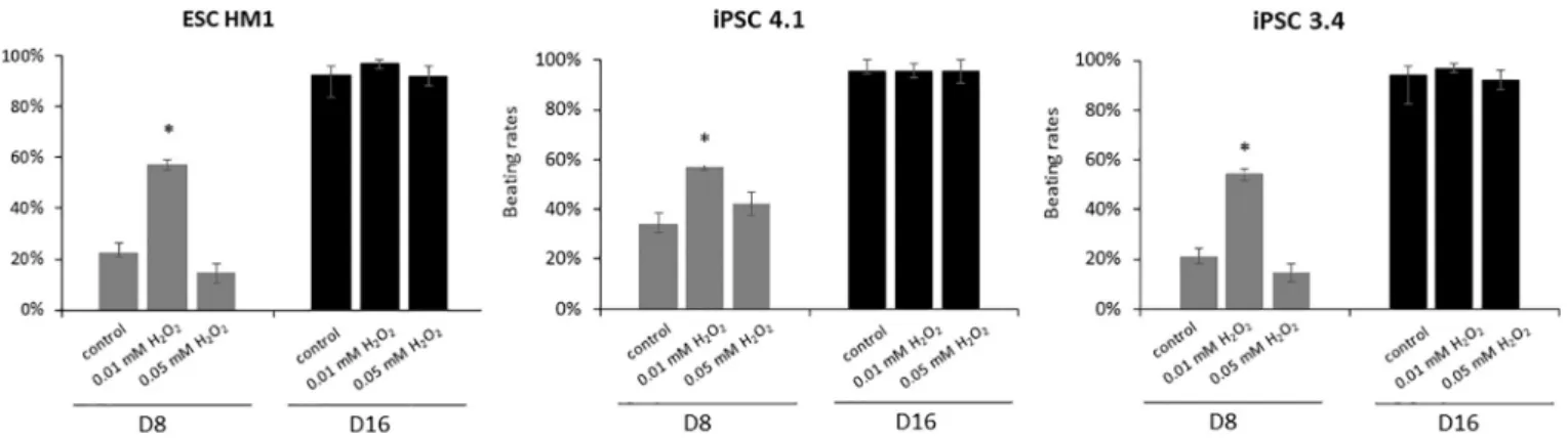
In a further experiment, we investigated if oxidative stress accel- erates the process of cardiac differentiation. First of all, the beating rate of EBs were compared as a sign of cardiac differentiation. As a result, the H2O2induced oxidative stress increased the number of beating EBs on a concentration dependent manner in all the three cell lines (Fig. 4).
Interestingly, the 0.01 μM H2O2treatment significantly increased the number of beating EBs on day 8, while this difference was not sig- nificant when 0.05 μM concentration was used. On day 16 this effect could not be further observed as the beating rate of all control and oxidative stress treated groups reached the plateau (Fig. 4).
Next, we investigated if the expression of cardiac tissue-specific genes reflects the differences observed for the beating rate. Expression of early cardiac lineage markerNkx2-5showed the highest increase at day 8 with both H2O2concentrations in all cell lines, while at day 16 it decreased but was still significantly upregulated (Fig. 5). Myosin heavy chain 6 (Myh6) showed a robust increase in both time points and its peak was as well at day 8. Furthermore, we observed significant dif- ferences between the cell lines; the H2O2treatment could induce the Myh6 expression in both iPSC lines, but not in the ESC line (Fig. 5). For platelet/endothelial cell adhesion molecule 1 (Pecam1) a relatively stable expression was observed throughout differentiation (Supple- mentary Fig. S6). The expression of FMS-like tyrosine kinase 1 (Flt1) and Notch1 increased with the progression of the differentiation in all Fig. 3.Effect of simulated ischemia/reperfusion injury on cell viability of mouse ESC- or iPSC-derived embryoid bodies.
Embryoid bodies (A, B) and VCAM-1+ (C, D) subpopulation of mouse ESC- or iPSC-derived cells were subjected to simulated ischemia (SI) and reperfusion at day 8 and 16 of differentiation. Cells were treated with vehicle or NO-donor SNAP during SI. Reperfusion was followed by cell viability assay. Data are normalized to normoxic control and expressed as mean ± SEM;⁎p< 0.05 one-way ANOVA, followed by Tukey’s multiple comparisons test compared to normoxic group,
#p< 0.05 one-way ANOVA, followed by Tukey’s multiple comparisons test compared to SI + vehicle treated group,n= 10–12. Flow cytometric data obtained from three independent measurements.
lines, which was attenuated by the oxidative stress treatment. Von Willebrand factor (Vwf1) showed a similar trend, except on day 16 when the oxidative stress treatments resulted a misregulation (Sup- plementary Fig. S6).
3.5. Cell death of accelerated differentiation ESC- and iPSC-derived EBs and cardiac cells after simulated ischemia/reperfusion injury
When oxidative stress was applied before differentiation, SI/R sig- nificantly induced cell death in iPS4.1 full EBs, which was abolished with application of NO-donor cardioprotective SNAP at day 8. In ESC and iPS3.4-derived full EBs at day 8 showed only slight tendency to that pattern (Fig. 6A). In contrast, increased cell death was shown at day 8 for the group of VCAM-1+ cells of all lines (Fig. 6C). At day 16, in ESC and iPS3.4-derived full EBs SI/R resulted significant cell death, but not in VCAM-1 positive lines (Fig. 6B, D). The presence of cytoprotective SNAP was able to reduce excessive cell death only in VCAM-1+ iPS4.1 cells at day 8 (Fig. 6).
4. Discussion
In the present study, we aimed to establish an ESC and iPSC-derived cardiac myocyte-basedin vitrodrug screening platform which is able to assess the ischemic tolerance of differentiating cardiac cells. This is the first systematic analysis of stem cell-derived cardiac myocytes to SI/R induced cell death and cardiocytoprotection. After analysis of full EBs and cardiac marker positive cells of three cell lines at 6 conditions (32 different conditions altogether), we conclude that the severity of SI/R injury-induced cell death increased in both full EBs and VCAM+ car- diac cells at a later stage of their differentiation. Moreover, full EBs of the iPS 4.1 cell line after accelerated differentiation at Day 8 was protected by SNAP, showing that this setting could serve as a testing platform for cardiocytoprotection.
Differentiated cardiomyocyte-based platforms have an emerging role in modelling of cardiac tissue and developing drug screening assays for pharmaceutical research [21]. The primary neonatal cardiomyo- cytes may be used for SI/R models [11,13], and adult cardiac myocytes are appropriate for studying individual cells [10,40]. However, extra- cellular matrix proteins are required for maintenance and function of Fig. 4.Effect of oxidative stress on the number of beating EBs.
Mouse ESCs and iPSCs were treated for 24 h by supplementing the culture media with 0.01 or 0.05 μM H2O2or untreated (control). Pluripotent cells were dissociated and EBs were generated in hanging drops. After three days the formed EBs were further differentiated to generate cardiomyocytes by culturing them individually in 24-well plates for 8 and 16 days. The number of beating EBs were counted at day 8 and day 16. Data is presented in the % of the total number of plated EBs (beating rates) (mean ± SEM;⁎p< 0.05). Data was collected from 4 independent experiments.
Fig. 5.Expression of cardiac marker genes in EBs after oxidative stress.
Mouse ESCs and iPSCs were treated for 24 h by supplementing the culture media with 0.01 μM (grey bars) or 0.05 μM H2O2(black bars) or kept without oxidative stress treatments (white bars). Furthermore, cells were differentiated towards cardiomyocytes and their gene expression pattern was evaluated by RT-qPCR on day 8 and 16. Values are expressed as relative expression (mean ± SEM) normalized to the day 0 control (⁎⁎p≤ 0.05;⁎p≤ 0.1 calculated using Relative Expression Software Tool).
adult cardiomyocytes which may influence the cell viability during si- mulated ischemia-reperfusion [49], and the isolation procedure as well the maintenance of cell cultures are complicated [47]. Embryo-derived and reprogrammed pluripotent stem cell lines including ESCs and iPSCs have been used in combination with biomaterials for tissue engineering [42,43]. The genotypic and phenotypic features of primary and ESC- derived cardiac myocytes are very similar [3]. It is well known, that the cardiovascular progenitor cells (CVPCs) segregate from the mesoderm compartment in the early phase of development. CVPCs are multipotent cells that express Nkx2-5, Flk-1, Isl1and c-KIT, and give rise to en- dothelial, smooth muscle and cardiac precursor cells (CPCs) as well.
CPCs are the ones which are capable of differentiating towards cardi- omyocytes and still express NKX2-5 representing an early and selective marker of this precursor population. CPCs also express GATA4 protein, a zinc finger transcription factor, which has a role in the activation of genes related to myocardial differentiation. Further specification and differentiation of CPCs is marked by the expression of functional sar- comere- and chamber-specific cardiac proteins (α- and β–MHC, MLC- 2v, ANF) [32,46]. When mouse iPSCs are differentiated in hanging drops the differentiation follows the above-mentioned lineage. How- ever, the efficiency and percentage of different subpopulations can vary in LIF-free conditions without the administration of extra growth fac- tors and cytokines [25,26,44]. The utilization of ESC-derived cardio- myocytes would be suitable for drug screening; our work group pre- viously successfully derived cardiomyocytes from mouse ESC, and also demonstrated pharmacological cardioprotection in the HM1 line-de- rived cardiac myocyte-based SI/R model [14,28]. However, ethical considerations limit the utilization of human ESC-derived platforms.
IPSC-derived cardiomyocyte platforms can overcome these ethical concerns, but little is known about iPSC-derived cardiomyocytes and
their behaviour in ischemic conditions.
We characterized the cardiomyocytes with comparative im- munocytochemistry at two time points of cellular differentiation (day 8 and 16) after the directed differentiation of a mouse ESC- and iPSC- lines. The expression of early cardiac marker NKX2-5 and later marker Troponin T demonstrated the progress of cardiac differentiation.
VCAM-1 was also included to our study as it was shown previously that this protein is a potent cell surface marker for robust, efficient and scalable purification of cardiomyocytes from human ESC and iPSCs [45]. In accordance with this, mouse ESC-derived cardiomyocytes can be isolated with almost perfect purity and viability, based on their ex- pression of VCAM-1 and these cells were furthermore suitable forin vitro culture [31]. Recently, the proliferation of mouse ESC-derived cardiomyocytes could be enhanced through VCAM-1 signalling, leading to an increase in functional myocardial cells in bioengineered tissue [16]. Nevertheless, there is limited information available on the ex- pression of this gene in iPSC-derived cardiomyocytes. Accordingly, we aimed to investigate the cardiac specificity of VCAM-1 in these cells.
The results have shown that VCAM-1 were more prominent in day 16 samples in all the examined ESC and iPSC lines, which confirmed the previous results. The cardiac specificity of VCAM-1 in mouse ESC-de- rived EBs was demonstrated by immunocytochemistry and FACS. We also tested VCAM-1 expression in premature hearts of mouse embryos.
This allowed us to enrich mature cardiomyocyte population of our samples (VCAM-1+ group of cells) in the further experiments. Based on the literature, primary cardiomyocytes differentiated from mouse PSCs are expressing VCAM-1, which are not representing fully matured adult cardiac cells. Referring to the work of Pontén et al. [31], mouse PSC derived VCAM-1 positive cardiomyocytes belongs to both atrial and ventricular compartments and could not be distinguished based on this Fig. 6.Effect of simulated ischemia/reperfusion injury on cell viability of mouse ESC- or iPSC-derived embryoid bodies induced by 24 h H2O2treatment during differentiation.
Embryoid bodies (A, B) and VCAM-1+ (C, D) subpopulation of mouse ESC- or iPSC-derived cells were subjected to simulated ischemia (SI) and reperfusion at day 8 and 16 of differentiation. Cells were treated with vehicle or NO-donor SNAP during SI. Reperfusion was followed by cell viability assay. Data are normalized to normoxic control and expressed as mean ± SEM;⁎p< 0.05 one-way ANOVA, followed by Tukey’s multiple comparisons test compared to normoxic group,
#p< 0.05 one-way ANOVA, followed by Tukey’s multiple comparisons test compared to SI + vehicle treated group,n= 10–12. Flow cytometric data obtained from three independent measurements.
marker. Although VCAM-1 is reported to be present on multipotent progenitor cells (MPPs) as well (Together with Flt3) during haemato- poiesis, due to the different differentiation pathways of lymphoid and myeloid lineages (reviewed by Lai and Kondo [19]), the activation of VCAM-1 expression on cardiac progenitors is the result of distinct processes.
In the next set of experiments, we assessed and compared cardio- myocyte-based platforms derived from a mouse ESC and two different iPSC cell lines in SI/R conditions. We have shown that both mouse ESC- and iPSC-derived full EBs are more sensitive to hypoxia at an advanced stage of cardiac differentiation, as an overall tendency towards reduced tolerance to hypoxia at day 16 compared to day 8 was observed. It was previously shown that murine and human iPSC-derived cardiomyocytes are particularly sensitive to hypoxia and glucose deprivation [4,54].
Cardiac constructs derived from human iPSc subjected to simulated ischemia-reperfusion were protected following ischemic pre- conditioning [5]. The viability patterns of VCAM-1+ cells were similar to that of the full EBs, as excessive cell death was observed at day 16 compared to day 8.
The cardiac differentiation can be accelerated with temporary ap- plication of oxidative stress. It seems that upon preceding oxidative stress treatment the VCAM-1+ fraction of cardiomyocytes become more vulnerable to the forthcoming ischemia/reperfusion (SI/R)-in- duced injury as they have decreased viability already at day 8. In contrast, day 16 VCAM-1+ cardiomyocytes tolerate the SI/R better.
The possible explanation of this observation may be the time dependent expression of VCAM-1, which shows rapid upregulation in early stage of cardiomyocyte differentiation, then it slows down by time [6].
Reactive oxygen species (ROS) is generated during normal cellular metabolism, however the overproduction of ROS can induce oxidative stress [8]. Besides its toxic effect, it is considered as signalling molecule as well [15,33]. It was previously reported that ROS promotes differ- entiation towards both cardiomyogenic and vascular cell lineages in mouse and human ESCs [17,35]. Free radicals have a distinct role in the proliferation and differentiation of stem cells in a concentration and time dependent manner. Even at very low concentration, ROS has a prominent effect on differentiation; our data clearly show that only slight changes in concentration of ROS produced by H2O2was able to influence gene expression pattern as well as the differentiation. In ad- dition, the gene expression of major cardiac-, endothelial and smooth muscle markers as well clearly reflects the effect of oxidative stress on the cardiac commitment. RT-qPCR experiments have shown that H2O2 treatment significantly increased the expression of the selected cardiac, smooth muscle and epithelial marker genes. The demanding increase in the structural protein cardiacMyh6showed that an intensive cardiac differentiation occurs in the EBs, which was furthermore promoted by the oxidative stress treatment in a dose dependent manner. This was in accordance with the observed increase in the beating rate which is a morphological attribute of successful cardiac differentiation. One pos- sible explanation for these findings is that by the H2O2, unsaturated fatty acids are oxidized, leading to an increased eicosanoid level which promote the differentiation of pluripotent cells [51]. In conclusion, although we observed some cell line dependent difference, the lower dose of H2O2(0.01 μM) has a triggering effect on cardiac tissue for- mation. It accelerated the differentiation process and resulted earlier maturation.
Cardiocytoprotection induced by NO-donor SNAP was also tested in our SI/R models. Exogenous nitric oxide is cardioprotective in various myocardial ischemia models. SNAP had a significant cytoprotective effect against ischemia-reperfusion in primary neonatal, mouse and human ESC-derived cardiomyocytes [13,14,28]. However, in our ex- periments SNAP had only a tendency towards cytoprotective effect in mouse ESC-derived EBs at day 8 and no protection at day 16 or in any other groups including iPSC-derived EBs. A protective effect of NO- donor SNAP was detected only in the VCAM-1+ fraction of iPS 4.1 line when it was previously subjected to oxidative stress. In contrast, NO-
donor nicorandil had protective effects in dystrophin-deficient iPSC- derived cardiac model via NO-cGMP and KATP-channel dependent me- chanism [2]. The causes of limited effect in iPSC-based cell platform are not clearly understood; the expression level of cGMP-PKG changes during cardiac differentiation in ESC-derived cardiac myocytes [23], but little is known about the downregulation of NO-cGMP pathway in iPSC-derived cardiomyocytes. However, the observed cytoprotection by SNAP in the identical ESC-based platform may highlight the important differences between the differentiation of ESC- and iPSC-derived car- diac myocytes.
5. Limitations
PSC-derived cardiac myocytes partially mimic the embryonic stage, however, several adult-stage genes are also expressed [24]. Accord- ingly, in our present study using mouse embryonic and iPS cell-derived cardiac myocytes in EB culture, the expression of VCAM-1 showed early cardiac differentiation stage and the expression of MYH6 and Troponin- T showed mature phenotype. Although the present 3D EB culture did not provide a clean cardiac myocyte lineage differentiation, it is widely accepted that 3D cultures can mimic better the original tissue en- vironment ([29]). Moreover, human PSC models may provide data of more translational value but its significantly higher cost and less ca- pacity limits its widespread use. In spite of the several limitations de- scribed above, we believe that the present mouse iPS-based assay pro- vides a fast and cost effective model to study certain cellular processes at a fast and cost effective way.
6. Conclusion
In summary, this is the first systematic analysis of stem cell-derived cardiac myocytes to SI/R induced cell death and cardiocytoprotection.
After analysis of full EBs and cardiac marker positive cells of three cell lines at 6 conditions (32 different conditions altogether), we conclude that mouse iPSC differentiation is comparatively similar to the ESC- based methods. However, from the experimental results and the pub- lished data it seems that although the pluripotency of mouse ESCs and iPSCs are very similar, and the differentiation protocols used are exactly the same, their differentiation capacity can differ. Besides, a high in- terline variability was detected in the efficiency of cardiac differentia- tion. Moreover, iPSC derived cardiomyocytes were proved to be less mature than ECS or foetal tissue derived counterparts. The severity of SI/R injury-induced cell death increased in both full EBs and VCAM+
cardiac cells at later stage of their differentiation. Moreover, full EBs of the iPS 4.1 cell line after accelerated differentiation at day 8 was pro- tected by SNAP, showing that this setting could serve as a testing platform for cardiocytoprotection.
Data availability
The datasets generated during and/or analysed during the current study are available from the corresponding author on reasonable re- quest.
Funding
This work was supported from EU FP7 projects (EpiHealth, HEALTH-2012-F2-278418; EpiHealthNet, PITN-GA-2012-317146).
OTKAPD-106001; ‘National Excellence Program’ (TÁMOP-4.2.4.A/2- 11/1-2012-0001) and National Research, Development and Innovation Office of Hungary (National Heart Program, NVKP-16-1-2016-0017), GINOP-2.3.2-15-2016-00040. A.G. held a “János Bolyai Fellowship”
from the Hungarian Academy of Sciences (2012–15). Higher Education Institutional Excellence Programme of the Ministry of Human Capacities in Hungary, within the framework of the Therapeutic Development thematic programme of the Semmelweis University.
Research Excellence Programme of the National Research, Development and Innovation Office of the Ministry of Innovation and Technology in Hungary (TKP/ITM/NKFIH). R.M. was supported by grants from Incyte S.r.l. and Ministero dell'Istruzione, Università e Ricerca Scientifica (549901_2020_Madonna_Ateneo). J.P. held an
"Apáczai Fellowship" from the European Union and State of Hungary, within the framework of National Excellence Program (TÁMOP- 4.2.4.A/2-11/1-2012-0001).
Author contributions
János Pálóczi developed all conditions for FACS analysis and im- munocytochemical staining and prepared figures and ultrasound images. Ágnes Szántai performed Calcein and PI viability assays and handled mouse breading pairs, analysed data. Bernadett Kiss performed plate reader measurements. Ernesto Ruivo performed live/dead viabi- lity assay and performed embryo isolation. Renáta Gáspár performed simulated ischemia/reperfusion testing. Judit Pipis performed simu- lated ischemia/reperfusion testing and digestion assays. Imre Ocsovszki performed FACS analysis. Zsófia Onódi analysed data, prepared sup- plemental figures and wrote manuscript. István Bock performed the mRNA isolation, qPCRs and data analysis. Anita Fehér implemented the oxidative stress assays. Zsuzsanna Táncos performed the ICC staining.
Julianna Kobolák performed mouse ESC and iPSC culture and cell dif- ferentiation, analysed data, supervised the experiments and wrote the manuscript. Anikó Görbe planned, analysed and supervised the ex- periments and wrote the manuscript. Rosalinda Madonna, András Dinnyés and Péter Ferdinandy approved the manuscript. All the authors contributed to the editing. All authors read and approved the final version of the manuscript.
Declaration of Competing Interest
Author Kobolák, Julianna; Bock, István; Fehér, Anita; Dinnyes, Andras was employed by the company Biotalentum Ltd. Author Anikó Görbe, Péter Ferdinandy and Judit Pipis was employed by the company Pharmahungary Group Ltd. The remaining authors declare that the research was conducted in the absence of any commercial for financial relationships that could be construed as a potential conflict of interest.
Acknowledgments
We are grateful for the Roslin Institute, UK for providing the HM1 mouse ESC line, and Prof Roger Pedersen (University of Cambridge, Cambridge, UK) for providing the iPS 3.4 and iPS 4.1 mouse iPSC lines.
We are thankful to the technical help of Marianna Tóth with mouse ESC culture, Karolina Szczesna for antibody testing, Zsuzsanna Lajtos, Tokio Hiura with simulated ischemia/reperfusion testing.
Appendix A. Supplementary data
Supplementary data to this article can be found online athttps://
doi.org/10.1016/j.vph.2020.106781.
References
[1] D.J. Hausenloy, D. Garcia-Dorado, H.E. Botker, S.M. Davidson, J. Downey, F.B. Engel, et al., Novel targets and future strategies for acute cardioprotection:
position paper of the European Society of Cardiology Working Group on Cellular Biology of the Heart, Cardiovasc. Res. 113 (6) (2017) 564–585,https://doi.org/10.
1093/cvr/cvx049.
[2] M.Z. Afzal, M. Reiter, C. Gastonguay, J.V. McGivern, X. Guan, Z.D. Ge, et al., Nicorandil, a nitric oxide donor and ATP-sensitive potassium channel opener, protects against dystrophin-deficient cardiomyopathy, J. Cardiovasc. Pharmacol.
Ther. 21 (6) (2016) 549–562,https://doi.org/10.1177/1074248416636477.
[3] H. Baharvand, M. Hajheidari, R. Zonouzi, S.K. Ashtiani, S. Hosseinkhani, G.H. Salekdeh, Comparative proteomic analysis of mouse embryonic stem cells and neonatal-derived cardiomyocytes, Biochem. Biophys. Res. Commun. 349 (3) (2006)
1041–1049,https://doi.org/10.1016/j.bbrc.2006.08.151.
[4] A. Brodarac, T. Saric, B. Oberwallner, S. Mahmoodzadeh, K. Neef, J. Albrecht, et al., Susceptibility of murine induced pluripotent stem cell-derived cardiomyocytes to hypoxia and nutrient deprivation, Stem Cell Res Ther 6 (2015) 83,https://doi.org/
10.1186/s13287-015-0057-6.
[5] T. Chen, G. Vunjak-Novakovic, Human tissue-engineered model of myocardial ischemia-reperfusion injury, Tissue Eng. Part A (2018),https://doi.org/10.1089/
ten.TEA.2018.0212.
[6] V.C. Chen, J. Ye, P. Shukla, G. Hua, D. Chen, Z. Lin, et al., Development of a scalable suspension culture for cardiac differentiation from human pluripotent stem cells, Stem Cell Res. 15 (2) (2015) 365–375,https://doi.org/10.1016/j.scr.2015.08.002.
[7] J.C. Del Alamo, D. Lemons, R. Serrano, A. Savchenko, F. Cerignoli, R. Bodmer, et al., High throughput physiological screening of iPSC-derived cardiomyocytes for drug development, Biochim. Biophys. Acta 1863 (7 Pt B) (2016) 1717–1727, https://doi.org/10.1016/j.bbamcr.2016.03.003.
[8] J. Egea, I. Fabregat, Y.M. Frapart, P. Ghezzi, A. Gorlach, T. Kietzmann, et al., European contribution to the study of ROS: a summary of the findings and prospects for the future from the COST action BM1203 (EU-ROS), Redox Biol. 13 (2017) 94–162,https://doi.org/10.1016/j.redox.2017.05.007.
[9] P. Ferdinandy, D.J. Hausenloy, G. Heusch, G.F. Baxter, R. Schulz, Interaction of risk factors, comorbidities, and comedications with ischemia/reperfusion injury and cardioprotection by preconditioning, postconditioning, and remote conditioning, Pharmacol. Rev. 66 (4) (2014) 1142–1174,https://doi.org/10.1124/pr.113.
008300.
[10] M.I. Garcia, A. Karlstaedt, J.J. Chen, J. Amione-Guerra, K.A. Youker,
H. Taegtmeyer, et al., Functionally redundant control of cardiac hypertrophic sig- naling by inositol 1,4,5-trisphosphate receptors, J. Mol. Cell. Cardiol. 112 (2017) 95–103,https://doi.org/10.1016/j.yjmcc.2017.09.006.
[11] R. Gaspar, M. Pipicz, F. Hawchar, D. Kovacs, L. Djirackor, A. Gorbe, et al., The cytoprotective effect of biglycan core protein involves Toll-like receptor 4 signaling in cardiomyocytes, J. Mol. Cell. Cardiol. 99 (2016) 138–150,https://doi.org/10.
1016/j.yjmcc.2016.08.006.
[12] M. Gertsenstein, L.M. Nutter, T. Reid, M. Pereira, W.L. Stanford, J. Rossant, et al., Efficient generation of germ line transmitting chimeras from C57BL/6N ES cells by aggregation with outbred host embryos, PLoS ONE 5 (6) (2010) e11260, ,https://
doi.org/10.1371/journal.pone.0011260.
[13] A. Gorbe, Z. Giricz, A. Szunyog, T. Csont, D.S. Burley, G.F. Baxter, et al., Role of cGMP-PKG signaling in the protection of neonatal rat cardiac myocytes subjected to simulated ischemia/reoxygenation, Basic Res. Cardiol. 105 (5) (2010) 643–650, https://doi.org/10.1007/s00395-010-0097-0.
[14] A. Gorbe, Z.V. Varga, J. Paloczi, S. Rungarunlert, N. Klincumhom, M.K. Pirity, et al., Cytoprotection by the NO-donor SNAP against ischemia/reoxygenation injury in mouse embryonic stem cell-derived cardiomyocytes, Mol. Biotechnol. 56 (3) (2014) 258–264,https://doi.org/10.1007/s12033-013-9704-2.
[15] K.K. Griendling, D. Sorescu, B. Lassegue, M. Ushio-Fukai, Modulation of protein kinase activity and gene expression by reactive oxygen species and their role in vascular physiology and pathophysiology, Arterioscler. Thromb. Vasc. Biol. 20 (10) (2000) 2175–2183.
[16] T. Iwamiya, K. Matsuura, S. Masuda, T. Shimizu, T. Okano, Cardiac fibroblast-de- rived VCAM-1 enhances cardiomyocyte proliferation for fabrication of bioengi- neered cardiac tissue, Regen. Ther. 4 (2016) 92–102.
[17] A.-R. Ji, S.-Y. Ku, M.S. Cho, Y.Y. Kim, Y.J. Kim, S.K. Oh, et al., Reactive oxygen species enhance differentiation of human embryonic stem cells into mesendodermal lineage, Exp. Mol. Med. 42 (3) (2010) 175.
[18] M. Kawamura, S. Miyagawa, K. Miki, A. Saito, S. Fukushima, T. Higuchi, et al., Feasibility, safety, and therapeutic efficacy of human induced pluripotent stem cell- derived cardiomyocyte sheets in a porcine ischemic cardiomyopathy model, Circulation 126 (11 Suppl 1) (2012) S29–S37,https://doi.org/10.1161/
CIRCULATIONAHA.111.084343.
[19] A.Y. Lai, M. Kondo, T and B lymphocyte differentiation from hematopoietic stem cell, Semin. Immunol. 20 (4) (2008) 207–212,https://doi.org/10.1016/j.smim.
2008.05.002.
[20] X. Li, F.R. Heinzel, K. Boengler, R. Schulz, G. Heusch, Role of connexin 43 in is- chemic preconditioning does not involve intercellular communication through gap junctions, J. Mol. Cell. Cardiol. 36 (1) (2004) 161–163.
[21] R. Madonna, F.B. Engel, S.M. Davidson, P. Ferdinandy, A. Gorbe, J.P. Sluijter, et al., Stem cell aging and age-related cardiovascular disease: perspectives of treatment by ex-vivo stem cell rejuvenation, Curr. Drug Targets 16 (8) (2015) 780–785.
[22] T. Magdy, A.J.T. Schuldt, J.C. Wu, D. Bernstein, P.W. Burridge, Human induced pluripotent stem cell (hiPSC)-derived cells to assess drug cardiotoxicity: opportu- nities and problems, Annu. Rev. Pharmacol. Toxicol. 58 (2018) 83–103,https://doi.
org/10.1146/annurev-pharmtox-010617-053110.
[23] K. Mujoo, J.S. Krumenacker, Y. Wada, F. Murad, Differential expression of nitric oxide signaling components in undifferentiated and differentiated human em- bryonic stem cells, Stem Cells Dev. 15 (6) (2006) 779–787,https://doi.org/10.
1089/scd.2006.15.779.
[24] C.L. Mummery, J. Zhang, E.S. Ng, D.A. Elliott, A.G. Elefanty, T.J. Kamp, Differentiation of human embryonic stem cells and induced pluripotent stem cells to cardiomyocytes: a methods overview, Circ. Res. 111 (3) (2012) 344–358,https://
doi.org/10.1161/CIRCRESAHA.110.227512.
[25] G. Nair, E. Abranches, A.M. Guedes, D. Henrique, A. Raj, Heterogeneous lineage marker expression in naive embryonic stem cells is mostly due to spontaneous differentiation, Sci. Rep. 5 (2015) 13339,https://doi.org/10.1038/srep13339.
[26] J. Nussbaum, E. Minami, M.A. Laflamme, J.A. Virag, C.B. Ware, A. Masino, et al., Transplantation of undifferentiated murine embryonic stem cells in the heart: ter- atoma formation and immune response, FASEB J. 21 (7) (2007) 1345–1357,
https://doi.org/10.1096/fj.06-6769com.
[27] H. Okano, M. Nakamura, K. Yoshida, Y. Okada, O. Tsuji, S. Nori, et al., Steps toward safe cell therapy using induced pluripotent stem cells, Circ. Res. 112 (3) (2013) 523–533,https://doi.org/10.1161/CIRCRESAHA.111.256149.
[28] J. Paloczi, Z.V. Varga, A. Apati, K. Szebenyi, B. Sarkadi, R. Madonna, et al., Exogenous nitric oxide protects human embryonic stem cell-derived cardiomyo- cytes against ischemia/reperfusion injury, Oxidative Med. Cell. Longev. 2016 (2016) 4298945,https://doi.org/10.1155/2016/4298945.
[29] F. Pampaloni, E.G. Reynaud, E.H. Stelzer, The third dimension bridges the gap between cell culture and live tissue, Nat. Rev. Mol. Cell Biol. 8 (10) (2007) 839–845,https://doi.org/10.1038/nrm2236.
[30] M.W. Pfaffl, G.W. Horgan, L. Dempfle, Relative expression software tool (REST) for group-wise comparison and statistical analysis of relative expression results in real- time PCR, Nucleic Acids Res. 30 (9) (2002) e36.
[31] A. Pontén, S. Walsh, D. Malan, X. Xian, S. Schéele, L. Tarnawski, et al., FACS-based isolation, propagation and characterization of mouse embryonic cardiomyocytes based on VCAM-1 surface marker expression, PLoS ONE 8 (12) (2013) e82403.
[32] K. Rajala, M. Pekkanen-Mattila, K. Aalto-Setala, Cardiac differentiation of plur- ipotent stem cells, Stem Cells Int. 2011 (2011) 383709,https://doi.org/10.4061/
2011/383709.
[33] S.G. Rhee, S.W. Kang, W. Jeong, T.S. Chang, K.S. Yang, H.A. Woo, Intracellular messenger function of hydrogen peroxide and its regulation by peroxiredoxins, Curr. Opin. Cell Biol. 17 (2) (2005) 183–189,https://doi.org/10.1016/j.ceb.2005.
02.004.
[34] S. Rozen, H. Skaletsky, Primer3 on the WWW for general users and for biologist programmers, in: S. Misener, S.D. Krawetz (Eds.), Bioinformatics Methods and Protocols SE - 20, 132 ed, Humana Press DA, 1999, pp. 365–386 (LA – English).
[35] H. Sauer, M. Wartenberg, Reactive oxygen species as signaling molecules in car- diovascular differentiation of embryonic stem cells and tumor-induced angiogen- esis, Antioxid. Redox Signal. 7 (11−12) (2005) 1423–1434.
[36] J. Selfridge, A.M. Pow, J. McWhir, T.M. Magin, D.W. Melton, Gene targeting using a mouse HPRT minigene/HPRT-deficient embryonic stem cell system: inactivation of the mouse ERCC-1 gene, Somat. Cell Mol. Genet. 18 (4) (1992) 325–336.
[37] D. Sinnecker, K.L. Laugwitz, A. Moretti, Induced pluripotent stem cell-derived cardiomyocytes for drug development and toxicity testing, Pharmacol. Ther. 143 (2) (2014) 246–252,https://doi.org/10.1016/j.pharmthera.2014.03.004.
[38] J.P. Sluijter, G. Condorelli, S.M. Davidson, F.B. Engel, P. Ferdinandy,
D.J. Hausenloy, et al., Novel therapeutic strategies for cardioprotection, Pharmacol.
Ther. 144 (1) (2014) 60–70,https://doi.org/10.1016/j.pharmthera.2014.05.005.
[39] A.S. Smith, J. Macadangdang, W. Leung, M.A. Laflamme, D.H. Kim, Human iPSC- derived cardiomyocytes and tissue engineering strategies for disease modeling and drug screening, Biotechnol. Adv. 35 (1) (2017) 77–94,https://doi.org/10.1016/j.
biotechadv.2016.12.002.
[40] K. Suzuki, S. Kostin, V. Person, A. Elsasser, J. Schaper, Time course of the apoptotic cascade and effects of caspase inhibitors in adult rat ventricular cardiomyocytes, J.
Mol. Cell. Cardiol. 33 (5) (2001) 983–994,https://doi.org/10.1006/jmcc.2001.
1364.
[41] K. Takahashi, K. Okita, M. Nakagawa, S. Yamanaka, Induction of pluripotent stem cells from fibroblast cultures, Nat. Protoc. 2 (12) (2007) 3081–3089,https://doi.
org/10.1038/nprot.2007.418.
[42] K. Takahashi, S. Yamanaka, Induction of pluripotent stem cells from mouse em- bryonic and adult fibroblast cultures by defined factors, Cell 126 (4) (2006) 663–676,https://doi.org/10.1016/j.cell.2006.07.024.
[43] J.A. Thomson, J. Itskovitz-Eldor, S.S. Shapiro, M.A. Waknitz, J.J. Swiergiel, V.S. Marshall, et al., Embryonic stem cell lines derived from human blastocysts, Science 282 (5391) (1998) 1145–1147.
[44] S. Ueno, G. Weidinger, T. Osugi, A.D. Kohn, J.L. Golob, L. Pabon, et al., Biphasic role for Wnt/beta-catenin signaling in cardiac specification in zebrafish and em- bryonic stem cells, Proc. Natl. Acad. Sci. U. S. A. 104 (23) (2007) 9685–9690, https://doi.org/10.1073/pnas.0702859104.
[45] H. Uosaki, H. Fukushima, A. Takeuchi, S. Matsuoka, N. Nakatsuji, S. Yamanaka, et al., Efficient and scalable purification of cardiomyocytes from human embryonic and induced pluripotent stem cells by VCAM1 surface expression, PLoS ONE 6 (8) (2011) e23657.
[46] P. Van Vliet, S.M. Wu, S. Zaffran, M. Pucéat, Early Cardiac Development: A View From Stem Cells to Embryos, (2012).
[47] K.B. Walsh, T.C. Rich, Z.J. Coffman, Development of a high-throughput assay for monitoring cAMP levels in cardiac ventricular myocytes, J. Cardiovasc. Pharmacol.
53 (3) (2009) 223–230,https://doi.org/10.1097/FJC.0b013e31819b5479.
[48] K.C. Wollert, H. Drexler, Clinical applications of stem cells for the heart, Circ. Res.
96 (2) (2005) 151–163,https://doi.org/10.1161/01.RES.0000155333.69009.63.
[49] E.A. Woodcock, S.J. Matkovich, Cardiomyocytes structure, function and associated pathologies, Int. J. Biochem. Cell Biol. 37 (9) (2005) 1746–1751,https://doi.org/
10.1016/j.biocel.2005.04.011.
[50] C. Xu, S. Police, N. Rao, M.K. Carpenter, Characterization and enrichment of car- diomyocytes derived from human embryonic stem cells, Circ. Res. 91 (6) (2002) 501–508.
[51] O. Yanes, J. Clark, D.M. Wong, G.J. Patti, A. Sánchez-Ruiz, H.P. Benton, et al., Metabolic oxidation regulates embryonic stem cell differentiation, Nat. Chem. Biol.
6 (6) (2010) 411–417.
[52] J. Ye, G. Coulouris, I. Zaretskaya, I. Cutcutache, S. Rozen, T.L. Madden, Primer- BLAST: a tool to design target-specific primers for polymerase chain reaction, BMC Bioinformatics 13 (2012) 134,https://doi.org/10.1186/1471-2105-13-134.
[53] J. Yu, M.A. Vodyanik, K. Smuga-Otto, J. Antosiewicz-Bourget, J.L. Frane, S. Tian, et al., Induced pluripotent stem cell lines derived from human somatic cells, Science 318 (5858) (2007) 1917–1920,https://doi.org/10.1126/science.1151526.
[54] Y. Zhang, D. Wang, K. Cao, M. Chen, X. Yang, Y. Tao, Rat induced pluripotent stem cells protect H9C2 cells from cellular senescence via a paracrine mechanism, Cardiology 128 (1) (2014) 43–50,https://doi.org/10.1159/000357423.
[55] M. Zuker, Mfold web server for nucleic acid folding and hybridization prediction, Nucleic Acids Res. 31 (13) (2003) 3406–3415.