1114
|
wileyonlinelibrary.com/journal/aas Acta Anaesthesiol Scand. 2020;64:1114–1119.Received: 17 November 2019
|
Revised: 22 April 2020|
Accepted: 22 April 2020 DOI: 10.1111/aas.13617O R I G I N A L A R T I C L E
Determination of adequate positive end-expiratory pressure level required for carbon dioxide homeostasis in an animal model of infant laparoscopy
Jacob Karlsson
1,2| Gergely H. Fodor
3| Andre dos Santos Rocha
3|
Na Lin
3| Walid Habre
3,4| Mats Wallin
1,5| Magnus Hallbäck
5| Ferenc Peták
6| Per-Arne Lönnqvist
1,2© 2020 The Acta Anaesthesiologica Scandinavica Foundation. Published by John Wiley & Sons Ltd
1Anestesi-ochIntensivvårdsavdelningen, Department of Physiology and Pharmacology (FYFA), C3, Eiksson I Lars Group – Section of Anesthesiology and Intensive Care, Karolinska Institute, Stockholm, Sweden
2Pediatric Perioperative Medicine and Intensive Care, Karolinska University Hospital Eugenivägen 23, Stockholm, Sweden
3Unit for Anaesthesiological Investigations, Department of Anesthesiology
Pharmacology Intensive Care and Emergency Medicine, University of Geneva, Geneva, Switzerland
4Pediatric Anesthesia Unit, Geneva Children’s Hospital, Geneva, Switzerland
5Maquet Critical Care AB, Solna, Sweden
6Departmenet of Medical Physics and Informatics, University of Szeged, Szeged, Hungary
Correspondence Jacob Karlsson, Anestesi- ochIntensivvårdsavdelningen, C3, Eriksson I Lars, PA Lönnqvist Group—
Section of Anesthesiology and Intensive Care, Department of Physiology and Pharmacology (FYFA), Karolinska Institute, 171 76 Stockholm, Sweden.
Email: jacob.karlsson.1@ki.se Funding information
This work was supported by the Karolinska Institute and partially by grants from Maquet Critical Care AB, Solna Sweden, and the regional agreement on medical training and research (ALF) between Stockholm County Council and the Karolinska Institute.
Background: Capnoperitoneum provides a ventilatory challenge due to reduction in end-expiratory lung volume and peritoneal carbon dioxide absorption in both children and adults. The primary aim of this controlled interventional trial was to determine the positive end-expiratory pressure (PEEP) level needed to ensure for adequate carbon dioxide clearance and preservation of carbon dioxide homeostasis in an experimental model of infant laparoscopy. The secondary aim was to evaluate potential effects on cardiac output of PEEP and abdominal pressure level variations in the same setting.
Methods: Eight chinchilla bastard rabbits were anesthetized and mechanically venti- lated. Intra-abdominal pressures were randomly set to 0, 6, and 12 mm Hg by carbon dioxide insufflation. Carbon dioxide clearance using volumetric capnography, arterial blood gas data, and cardiac output was recorded, while PEEP 3, 6, and 9 cmH2O were applied in a random order.
Results: A PEEP of 9 cmH2O showed restoration of carbon dioxide clearance without causing changes in arterial partial pressure of carbon dioxide and bicarbonate and with no associated deterioration in cardiac output.
Conclusion: The results promote a PEEP level of 9 cmH2O in this model of infant cap- noperitoneum to allow for adequate carbon dioxide removal with subsequent preser- vation of carbon dioxide homeostasis. The use of high PEEP was not associated with any decrease in cardiac output.
1 | INTRODUCTION
Laparoscopic surgery is becoming increasingly common even in neonates and infants.1 The abdominal insufflation of carbon diox- ide (CO2) that is necessary for adequate surgical visualization does, however, produce a double challenge with regards to the ventilation.
First, the patient is subjected to a peritoneal CO2 absorption that will require an increase in CO2 excretion by the lungs to preserve normocapnea.2,3 Second, the increase in intra-abdominal pressure (IAP) will cause an upward displacement of the diaphragm that will reduce end-expiratory lung volume, which in turn will negatively influence ventilation and make adequate CO2 removal more diffi- cult.4 In addition to this, maintaining stable CO2 levels is particularly important in neonates and infants, were the CO2 absorption seen during capnoperitoneum has been shown to proportionally increase with smaller age and size,2 and fluctuations in PaCO2 is a main factor in the regulation of cerebral blood flow and thus a key component in preventing of neurological injury.5-7
An increased respiratory rate and/or an increased driving pres- sure, resulting in larger tidal volumes and minute ventilation, are the most frequently used option to increase ventilatory CO2 removal in the setting of capnoperitoneum.2,5 However, these actions are rel- atively poor in preserving lung volume.6,7 A potentially more effec- tive measure to preserve lung volume is the application of positive end-expiratory pressure (PEEP) and, thus, to apply a correct PEEP during laparoscopic surgery in children can be hypothesized as being a potentially more suitable action in this context. However, it is cur- rently not known which PEEP level may be the most appropriate in this setting. Available recommendations regarding PEEP levels are few and variable but PEEP levels of approximately 5 cmH2O have been described.8 Furthermore, using excessive PEEP may cause unwanted reductions in cardiac output, by reducing venous return, and thereby interfere with hemodynamic stability and adequate gas exchange.
The primary aim of this experimental study was therefore to characterize the PEEP level needed to assure for adequate CO2 clearance (VCO2) and preservation of arterial CO2 partial pressure (PaCO2), in a pediatric model mimicking laparoscopic surgery. A sec- ondary aim was to estimate cardiac output (CO), by transpulmonary thermodilution, to monitor any potential repercussions in CO of the increased PEEP and intra-abdominal pressure levels.
2 | METHODS
2.1 | Animal preparation
The study was performed at the Unit for Anaesthesiological Investigations, Department of Anesthesiology, Pharmacology and Intensive Care, University of Geneva, Geneva, Switzerland.
The experimental protocol was approved by the institutional eth- ics committee (registration number GE/4/17) for experimental re- search of the University of Geneva and animal welfare committee of
the Canton of Geneva, Switzerland (Chairperson Marjolaine Philit), February 2, 2017. The rabbit model was chosen since the weight span and lung volumes of the animals are within the range of normal human neonates and small infants with mature lungs. The use of this model in the context of mechanical ventilation has previously been reported by our research group.9,10
Eight adult chinchilla rabbits (median weight of 3.7 kg, range 3.6-3.9 kg), purchased from the animal farm of the University of Geneva, received pre-medication with intramuscular injection of xylazine (3 mg kg–1) and ketamine (15 mg kg–1). Anesthesia was then induced by iv injection of propofol (2 mg kg–1) via an ear vein. The animals were subsequently tracheostomized and an endotracheal tube (4 mm i.d., Portex®; Smiths Medical) was inserted for airway management. The jugular vein and carotid artery were cannulated for pressure measurements and blood sampling. The animals were mechanically ventilated in volume-control mode (tidal volume 7 mL/
kg) using an inspired oxygen fraction of 0.4, and the respiratory rate was adjusted to maintain end-tidal normocapnia (5.5-6.0 kPa).
Anesthesia was maintained with a continuous intravenous infu- sion of propofol (10 mg kg–1 h–1) and fentanyl (50 μg kg–1 h–1) via the ear vein. All animals were given a continuous crystalloid infu- sion (Ringer-acetate) for volume replacement (4 mL kg–1 h–1), and atracurium (0.5-1.0 mg kg–1h–1) was administered for muscle relax- ation after confirming an adequate anesthetic and analgesic depth.
An intra-abdominal CO2 insufflation device (Electronic Endoflator;
Karl Storz) was placed in the abdominal cavity through a small air- tight incision for regulation of IAP. Airway pressure, heart rate, and rectal temperature were measured and stored on a computer at a sampling rate of 1 kHz via an analog/digital interface converter (AD Instruments, PowerLab model 8/35 and LabChart 7).
2.2 | Volumetric capnography and arterial blood gases
Carotid arterial blood gas samples were taken for measurement of PaCO2 and HCO3- using a blood gas analyzer (Abbott i-STAT 1 Handheld Blood Gas Analyzer).
VCO2 was estimated using volumetric capnography with data from standard measurements of end-tidal CO2 concentration with a mainstream infrared CO2 sensor (Capnostat-3, Respironics Inc) as previously described.11,12 Mean VCO2 values recorded over 1 min- ute were used for further statistical analysis (on average 30 VCO2 values).
Editorial Comment
The use of laparoscopic techniques is increasing in pedi- atric patients. This experimental study, in a rabbit laparos- copy model, shows that a PEEP of 9 cm H2O is optimal to counteract negative effects of abdominal CO2 insufflation.
2.3 | Assessment of Cardiac output using transpulmonary thermodilution
Cardiac output (CO) was monitored by using a transpulmonary thermodilution technique (PiCCO; PULSION Medical Systems, Feldkirchen, Germany). A 3-ml thermal indicator bolus (<8°C) was injected in the right atrium via the central venous catheter for each measurement.13 Due to the large number of measurements in each subject, single dilutions rather than average of triplets were used.
2.4 | Study protocol
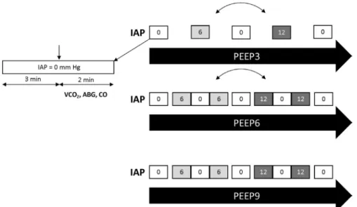
The rabbits were allowed a 15 minutes stabilization period before starting the study protocol. To standardize lung volume, a recruit- ment maneuver with PEEP 3 cmH2O and 14 mL/kg tidal volume was performed while applying two consecutive periods of 10 seconds inspiratory hold.10 This recruitment procedure was performed after surgical preparation and prior to each established PEEP level.
Figure 1 depicts the order of the experimental protocol. Baseline measurements were first performed at an IAP of 0 mm Hg and a PEEP of 3 cmH2O. After this, IAP was elevated to either 6 or 12 mm Hg in random order and recordings were repeated twice after return- ing to IAP 0 mm Hg. Another randomization was done for the PEEP level, which was either 6 or 9 cmH2O and the whole procedure was repeated. The overall sequence was randomized prior to the study, whereas the sequence was not individually randomized for each animal. VCO2, PaCO2, HCO3-, and CO recordings were performed 3 minutes after the target IAP level was established. The time frame for each measurement block was approximately 2 minutes.
Following completion of the protocol, the animals were killed with an overdose of iv sodium thiopental (100 mg/kg).
2.5 | Statistical analysis
Data were checked for normal distribution with Shapiro-Wilk K2 test. Data are presented as mean ± standard deviation (SD).
A priori and based on previous studies,9,10 we calculated a sample size of 7 animals in order to detect significant differences in VCO2, PaCO2, HCO3-, and CO for the interventions (power 80%, alpha error 0.05).
Two-way repeated measure analysis of variation with subse- quent Bonferroni multiple comparison tests was used to detect sig- nificant changes in the parameters measured by considering PEEP level and IAP as within-subject factors. P < .05 was considered to indicate statistical significance.
GraphPad Prism (version 7.0 for Windows; GraphPad Software) was used for all statistical calculations and Microsoft Excel for Mac 2011 version 14.5.7 for data handling (Microsoft Corp.).
3 | RESULTS
All 8 animals survived the interventions associated with the proto- col. Data from one rabbit is missing for PEEP 9 cmH2O due to techni- cal sampling problems.
No significant changes in VCO2 were found between IAP 0, 6, and 12 mm Hg at PEEP 3. When increasing IAP from 0 to 6, a signif- icant increase in VCO2 was observed both at PEEP 6 and 9 (P < .05).
Increasing IAP from 6 to 12 did not induce any further differences in VCO2 at any PEEP level. At PEEP 6 and 9, VCO2 was significantly higher for IAP 6 and 12 compared with the same intra-abdominal pressures at PEEP 3.
No significant alteration of PaCO2 was observed when increas- ing IAP from 0 to 6 to 12 at PEEP 9. However, a significant increase
F I G U R E 1 Scheme of the experimental protocol. Carbon dioxide elimination (VCO2), arterial blood gas (ABG), and cardiac output (CO) measurements were performed at positive end-expiratory pressure (PEEP) levels of 3, 6, and 9 cmH2O while intra-abdominal pressure (IAP) of 0, 6, and 12 mmHg was randomly maintained by capnoperitoneum. After the measurements, lung volume estimations associated with another study were made
in PaCO2 was seen for both PEEP 3 and 6 when IAP was increased from 0 to 6 (P < .05). Increase in IAP from 6 to 12 did not cause any further significant changes in PaCO2 at PEEP 6 and 9. For baseline values, PaCO2 was slightly higher at PEEP 9 than PEEP 3 (P < .05).
At PEEP 9, no significant changes in HCO3-were seen between the different IAP levels. HCO3- was at all times significantly lower at PEEP 9 when compared with PEEP 3 and 6.
No significant changes in CO were seen for PEEP 3 in response to increased IAP. For PEEP 6, CO was significantly higher at IAP6 than IAP 0. A similar difference in CO was seen between IAP 0 and 6 for PEEP 9 and also between IAP 0 and 12 for PEEP 9. At PEEP 6 and 9, CO was significantly higher than PEEP 3 for IAP 6. For PEEP 9, CO was significantly higher than PEEP 3 also at IAP 12.
The changes in VCO2, PaCO2, HCO3-, and CO for each PEEP level and IAP step are shown in Figure 2. Additional blood gas data not associated with the aim of this study are presented in Appendix S1.
4 | DISCUSSION
The present study used an experimental model that mimics lapa- roscopic surgery in infants to investigate the effect of PEEP and various intra-abdominal pressures on VCO2, PaCO2, HCO3-, and CO.
The results indicate that the PEEP level needs to be adjusted to a minimum of 9 cmH2O during abdominal insufflations of CO2 to allow for adequate CO2 removal and maintaining stable arterial CO2 ten- sion without any associated increase in HCO3-. This effect of PEEP 9 was obtained with no subsequent impairment in CO when compared with PEEP 3 and 6.
As mentioned above, the use of laparoscopic techniques is steadily increasing in the pediatric population.1 Although the tech- nique provides benefits for the patient and offers optimal conditions for the surgeon, this is partly achieved at the expense of adequate CO2 removal and thus provides the anesthesiologist with a substan- tial challenge with regards to choice of ventilation strategy. Various
F I G U R E 2 Ventilation and hemodynamic parameters. Mean ± SD. PEEP, positive end-expiratory pressure; IAP, intra-abdominal pressure;
PaCO2, arterial partial pressure of carbon dioxide; HCO3-, bicarbonate level; VCO2, tidal elimination rate of carbon dioxide; CO, cardiac output by transpulmonary thermodilution. *P < .05 vs IAP0 within a PEEP, §P < .05 vs IAP6 within a PEEP, #P < .05 vs PEEP 3 within an IAP,
$P < .05 vs PEEP 6 within an IAP
ventilation approaches have been considered to counteract the del- eterious effects of laparoscopy on lung volume. The application of appropriate PEEP level is the most suitable means to avoid reduced lung volumes, atelectasis formation, and subsequent gas exchange deterioration.14 Therefore, the results of the present study provide new and interesting information on how to ventilate neonates and small children who undergo laparoscopic surgery.
4.1 | CO
2clearance and preservation of carbon dioxide homeostasis
A prerequisite for CO2 homeostasis during a period of increased CO2 load is that the lung is in a state that allows for increased CO2 removal. Although PEEP 3 was capable of achieving adequate arte- rial CO2 tension at normal IAP, it was found to be insufficient to allow for increased VCO2 in response to the CO2 load associated with IAP 6 and 12. PEEP 6 performed appropriately in this aspect at IAP 6 and appeared still partly effective at IAP 12. PEEP 9 per- formed well both at IAP 6 and 12 thus indicating that a PEEP level of 9 appears to be the most suitable with regards to maintaining CO2 homeostasis in this particular setting. Furthermore, even if the differences in absolute numbers of PaCO2 were relatively small between the altered PEEP levels for all IAP, this appears to be at the expense of activation of the bicarbonate buffer system.15 This is reflected by the significant increase in HCO3- seen at PEEP 3 and 6, whereas a stable and at all times significantly lower HCO3- was seen at PEEP 9.
The ability to increase VCO2 in response to external CO2 load seen at PEEP 6 and 9 is potentially mediated through decreased shunting. We suggest that this then is caused by a larger end-ex- piratory lung volume, with an increased part of the lung taking part in effective gas exchange. This will in turn indicate a more open and homogenous lung at the higher PEEP levels.
4.2 | Study limitations
Since animal data cannot be immediately transposed to human ba- bies, the use of exactly PEEP 9 cannot instantly be recommended in the clinical scenario of pediatric capnoperitoneum. However, our data indicate that an inadequate PEEP will impede CO2 elimination as CO2 is insufflated and IAP becomes raised. The inadequate VCO2 resulting from low PEEP is probably even more likely to cause further increases in PaCO2 if the insufflation period is prolonged beyond the relatively short 3 minutes used in this study. Thus, to provide a situation of unrestricted CO2 elimination by the lungs in this clinical setting, a higher PEEP than usually used appears advisable.
The assessment of CO in this study is associated with certain limiting issues regarding absolute values. The method should ideally be based on triplicate injection of ice-cold saline but in this study, it was based on a single injection. The rationale for this is that the small animals otherwise would have been exposed to clearly excessive
volume load, both regarding animal well-being and potentially af- fecting CO per se. Additionally, the injected volume was reduced to only 3 milliliter instead of the customary 10 milliliters, a fact that potentially can result in a certain overestimation of CO.16 However, our data are in line with what has previously been reported in this setting.17 Since the aim of assessing CO in this study only was to demonstrate that PEEP increases are not associated with reduced CO, the absolute value of CO in this setting is of less importance.
Thus, the fact that CO did not change during the various changes in PEEP and IAP is still indicative of stable hemodynamics throughout the various experimental manipulations.
5 | CONCLUSION
In conclusion, PEEP 9 cm H2O was found to be the most effective PEEP to ensure optimal CO2 clearance without fluctuations in PaCO2 at intra-abdominal pressures of 6 and 12 mm Hg. Furthermore, PEEP 9 could be used without being associated with any decrease in car- diac output compared to lower PEEP levels.
ACKNOWLEDGMENTS
Assistance with the study: The laboratory faculty and staff of the Unit for Anaesthesiological Investigations, Dept. of Acute Medicine, University of Geneva, Geneva, Switzerland
CONFLIC TS OF INTEREST
Jacob Karlsson received an unrestricted research grant from Maquet Critical Care AB. Mats Wallin and Magnus Hallbäck are employed by Maquet Critical Care AB. The other authors declare no conflicts of interest.
AUTHORS’ CONTRIBUTIONS
Study design, data collection, and analysis: J.K, G.F, A.R, N.L, W.H, M.W, F.P, and PA.L. Manuscript writing and critical revision of manu- script: J.K, G.F, A.R, W.H, M.W, M.H, F.P, and PA.L.
ORCID
Jacob Karlsson https://orcid.org/0000-0003-2277-0092 Gergely H. Fodor https://orcid.org/0000-0002-4736-4966 Andre dos Santos Rocha https://orcid.org/0000-0001-6038-8500 Walid Habre https://orcid.org/0000-0002-6521-5091
Mats Wallin https://orcid.org/0000-0003-1492-0089 Ferenc Peták https://orcid.org/0000-0001-6249-9327 Per-Arne Lönnqvist https://orcid.org/0000-0002-8586-1429
REFERENCES
1. Chan IH, Tam PK. Laparoscopic inguinal hernia repair in infants and chil- dren: state-of-the-art technique. Eur J Pediatr Surg. 2017;27:465-471.
2. McHoney M, Corizia L, Eaton S, et al. Carbon dioxide elimination during laparoscopy in children is age dependent. J Pediatr Surg.
2003;38(1):105-110; discussion-10.
3. Pacilli M, Pierro A, Kingsley C, Curry JI, Herod J, Eaton S.
Absorption of carbon dioxide during laparoscopy in children
measured using a novel mass spectrometric technique. Br J Anaesth.
2006;97(2):215-219.
4. Kim K, Jang DM, Park JY, Yoo H, Kim HS, Choi WJ. Changes of di- aphragmatic excursion and lung compliance during major laparo- scopic pelvic surgery: a prospective observational study. PLoS ONE.
2018;13(11):e0207841.
5. Truchon R. Anaesthetic considerations for laparoscopic surgery in neonates and infants: a practical review. Best Pract Res Clin Anaesthesiol. 2004;18(2):343-355.
6. Levene M. Minimising neonatal brain injury: how research in the past five years has changed my clinical practice. Arch Dis Child.
2007;92(3):261-265.
7. Pryds O, Edwards AD. Cerebral blood flow in the newborn infant.
Arch Dis Child Fetal Neonatal Ed. 1996;74(1):F63-F69.
8. SurekaSK PN, Mittal V, Kapoor R, et al. Safe and optimal pneumo- peritoneal pressure for transperitoneal laparoscopic renal surgery in infant less than 10kg, looked beyond intraoperative period: A prospective randomized study. J Pediatr Urol. 2016;12(5):281.e1-.
e7.
9. Albu G, Wallin M, Hallbäck M, et al. Comparison of static end-expiratory and effective lung volumes for gas exchange in healthy and surfactant-depleted lungs. Anesthesiology.
2013;119:101-110.
10. Albu G, Petak F, Zand T, Hallbäck M, Wallin M, Habre W. Lung volume assessments in normal and surfactant depleted lungs:
agreement between bedside techniques and CT imaging. BMC Anesthesiol. 2014;14:64.
11. Suarez-Sipmann F, Bohm SH, Tusman G. Volumetric capnography:
the time has come. Curr Opin Crit Care. 2014;20(3):333-339.
12. Tolnai J, Fodor GH, Babik B, et al. Volumetric but not time capnog- raphy detects ventilation/perfusion mismatch in injured rabbit lung.
Front Physiol. 2018;9:1805.
13. Babik B, Balogh AL, Sudy R, Ivankovitsne-Kiss O, Fodor GH, Petak F. Levosimendan prevents bronchoconstriction and adverse
respiratory tissue mechanical changes in rabbits. Am J Physiol Lung Cell Mol Physiol. 2017;313(5):L950-L956.
14. Trachsel D, Svendsen J, Erb TO, von Ungern-Sternberg BS.
Effects of anaesthesia on paediatric lung function. Br J Anaesth.
2016;117(2):151-163.
15. Brackett NC, Cohen JJ, Schwarts WB.
Carbondioxidetitrationcurveofnormalman. Effectofincreasingde greesofacutehypercapniaonacid-baseequilibrium. N Engl J Med.
1965;272:6-12.
16. Garofalo NA, Teixeira-Neto FJ, Rodrigues JC, Cerejo SA, Aguiar A, Becerra-Velásquez DR. Comparison of transpulmonary thermodi- lution and calibrated pulse contour analysis with pulmonary artery thermodilution cardiac output measurements in anesthetized dogs.
J Vet Intern Med. 2016;30(4):941-950.
17. Maarek J-M, Holschneider DP, Harimoto J, Yang J, Scremin OU, Rubinstein EH. Measurement of cardiac output with indocy- anine green transcutaneous fluorescence dilution technique.
Anesthesiology. 2004;100(6):1476-1483.
SUPPORTING INFORMATION
Additional supporting information may be found online in the Supporting Information section.
How to cite this article: Karlsson J, Fodor GH, dos Santos Rocha A, et al. Determination of adequate positive end- expiratory pressure level required for carbon dioxide homeostasis in an animal model of infant laparoscopy. Acta Anaesthesiol Scand. 2020;64:1114–1119. https://doi.
org/10.1111/aas.13617