The Cardiac Pacemaker Story — Fundamental Role of the Na + /Ca 2+
Exchanger in Spontaneous Automaticity
Zso´fia Kohajda1,2†, Axel Loewe3†, Noe´mi To´th2, Andra´s Varro´1,2*and Norbert Nagy1,2
1MTA-SZTE Research Group of Cardiovascular Pharmacology, Hungarian Academy of Sciences, Szeged, Hungary,
2Department of Pharmacology and Pharmacotherapy, Faculty of Medicine, University of Szeged, Szeged, Hungary,
3Institute of Biomedical Engineering, Karlsruhe Institute of Technology (KIT), Karlsruhe, Germany
The electrophysiological mechanism of the sinus node automaticity was previously considered exclusively regulated by the so-called “funny current”. However, parallel investigations increasingly emphasized the importance of the Ca2+-homeostasis and Na+/ Ca2+ exchanger (NCX). Recently, increasing experimental evidence, as well as insight through mechanisticin silico modeling demonstrates the crucial role of the exchanger in sinus node pacemaking. NCX had a key role in the exciting story of discovery of sinus node pacemaking mechanisms, which recently settled with a consensus on the coupled-clock mechanism after decades of debate. This review focuses on the role of the Na+/Ca2+
exchanger from the early results and concepts to recent advances and attempts to give a balanced summary of the characteristics of the local, spontaneous, and rhythmic Ca2+
releases, the molecular control of the NCX and its role in the fight-or-flight response.
Transgenic animal models and pharmacological manipulation of intracellular Ca2+
concentration and/or NCX demonstrate the pivotal function of the exchanger in sinus node automaticity. We also highlight where specific hypotheses regarding NCX function have been derived from computational modeling and require experimental validation.
Nonselectivity of NCX inhibitors and the complex interplay of processes involved in Ca2+
handling render the design and interpretation of these experiments challenging.
Keywords: Na+/Ca2+exchanger, pacemaking, sinus node, automaticity, Ca2+-handling
Abbreviations:AP, action potential; APD, action potential duration; ATP, adenosine triphosphate; Ca2+/CA, Ca2+-cation antiporter; Ca2+i, intracellular Ca2+concentration; CaM, calmodulin; CaMKII, Ca2+/calmodulin-dependent protein kinase;
cAMP, cyclic adenosine monophosphate; CICR, Ca2+-induced Ca2+release; CL, cycle length; CRU, Ca2+- release unit; DD, diastolic depolarization; DDR, diastolic depolarization rate; EC50, half maximal effective concentration; ENCX, NCX equilibrium potential; GPCR, G-protein coupled receptor; hESC-CM, human embryonic stem cell-derived cardiomyocyte;
IC50, half maximal inhibitory concentration; ICaL, L-type Ca2+-current; ICaT, T-type Ca2+-current; If, funny-current; IK1, inward rectifier potassium current; IK(Ca): small-conductance Ca2+-activated K+current; IKr, rapid delayed rectifier potassium current; IKs, slow delayed rectifier potassium current; Ito, transient outward potassium current; LCR, local Ca2+releases;
LDCaE, late diastolic Ca2+elevation; LLFS model, Loewe–Lutz–Fabbri–Severi model; M-clock, membrane clock; MDP, maximal diastolic potential; Na+i, intracellular Na+concentration; NCX, Na+/Ca2+exchanger; NO, nitrogen-monoxide; PDE, phosphodiesterase; PKA, protein kinase A; PKC, protein kinase C; PLB, phospholamban; PP1, protein phosphatase 1; PP2A, protein phosphatase 2; RyR, ryanodine receptor; SERCA, sarcoplasmic reticulum Ca2+ATPase; SN, sinus node; SNC, sinus node cell; SR, sarcoplasmic reticulum; Vm, transmembrane potential.
Edited by:
Esther Pueyo, University of Zaragoza, Spain Reviewed by:
Sanjay Ram Kharche, University of Western Ontario, Canada Edward Lakatta, National Institutes of Health (NIH), United States Tatiana M. Vinogradova, National Institutes of Health (NIH), United States Matteo Elia Mangoni, Centre National de la Recherche Scientifique (CNRS), France
*Correspondence:
Andra´s Varro´ varro.andras@med.u-szeged.hu
†These authors have contributed equally to this work
Specialty section:
This article was submitted to Cardiovascular and Smooth Muscle Pharmacology, a section of the journal Frontiers in Pharmacology Received:01 October 2019 Accepted:01 April 2020 Published:28 April 2020 Citation:
Kohajda Z, Loewe A, To´th N, Varro´ A and Nagy N (2020) The Cardiac Pacemaker Story—Fundamental Role of the Na+/Ca2+Exchanger in Spontaneous Automaticity.
Front. Pharmacol. 11:516.
doi: 10.3389/fphar.2020.00516
doi: 10.3389/fphar.2020.00516
INTRODUCTION
Few areas of cardiac cellular electrophysiology had more intense debate over the years than ‘what makes our hearts beat'—the electrophysiology of the sinus node (SN). In the last two decades, we could witness the struggle of two competing oscillator concepts explaining SN automaticity: in essence, the funny- current (If) and the Ca2+-induced depolarization of the membrane potential (Vm) via Na+/Ca2+ exchanger (NCX, INaCa). Year by year, several studies were published supporting both theories. Thus, the exciting exploration of SN pacemaking mechanisms has been full of debates for over a half century.
Numerous mechanisms have been suggested, tested, refuted, accepted, tested, and retested again, updated and after intense point–counterpoint public debates finally settled at a delicate balance known as the coupled-clock mechanism that includes both membrane and sarcoplasmic reticulum (SR) linked contributions. The general field of SN pacemaking (Lakatta et al., 2010; Maltsev et al., 2014; DiFrancesco, 2020) and computational modeling thereof has been excellently reviewed elsewhere (Wilders, 2007;Li et al., 2013). In this review, we focus on the role of NCX function on SN pacemaking and aim to give a balanced view from the very early to recent experimental findings and accompanying computational models.
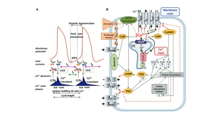
The SNCs show a characteristic slow diastolic depolarization (DD) starting from the maximal diastolic potential (MDP) and ending at the action potential (AP) threshold of about−40 mV.
This threshold is called takeoff potential (Kharche et al., 2011).
At the takeoff potential, the course of the AP changes qualitatively from slow DD to the AP upstroke (Figure 1A). If, NCX, and ICaT(T-type Ca2+-current) are mainly active before (at Vmmore negative than the takeoff potential), whereas ICaL(L- type Ca2+-current) and IKr (rapid delayed rectifier potassium current) are mainly active thereafter (at more positive Vm), while NCX is active throughout the entire AP cycle. Early studies suggested a major role of a decaying delayed rectifier potassium current (IKr) (Noble, 1962) and the nonselective, cAMP- dependent and hyperpolarization activated current [funny- current, If, (DiFrancesco, 1991)] in the development of DD.
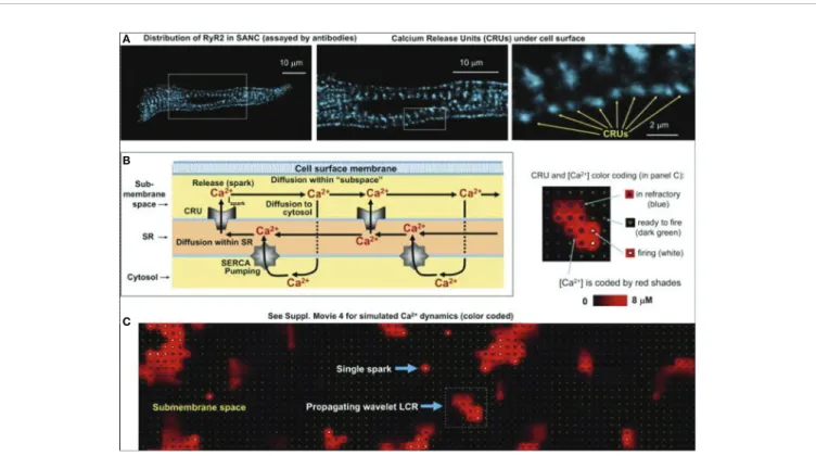
This mechanism, governed by transmembrane ion channels with Hodgkin–Huxley kinetics was termed as“membrane-clock”(M- clock). Later studies identified rhythmic, spontaneous locally propagating subsarcolemmal Ca2+ releases (LCR) generated by the SRviaryanodine receptors (RYRs) during the DD (Figure 1B) (Huser et al., 2000;Bogdanov et al., 2001;Monfredi et al., 2013). The LCRs activate the forward mode of the Na+/Ca2+
exchanger generating inward current that contributes to the DD (Bogdanov et al., 2001). Under certain experimental conditions,
A B
FIGURE 1 |The coupled-clock pacemaker system of the sinoatrial node. Schematic illustration of the currents and functional interactions between the two oscillator subsystem contributing to the SN AP(A). The interplay of the regulatory molecules comprising the coupled-clock pacemaker system is illustrated in(B). Membrane clock (M-clock) of sinoatrial node pacemaker cells comprises the major surface membrane currents: L-type Ca2+channels (ICaL), T-type Ca2+channels (ICaT), delayed rectifier K+channels (IK), hyperpolarization activated funny channels (If), Na+/Ca2+exchanger (NCX), and Na+/K+ATPase (INaK). Regulatory factors that affect the function of proteins of both clocks are indicated with different colors: molecules with red letters couple the Ca2+clock to the M-clock; factors in orange regulate both the M-clock and Ca2+clockviathe GPCR pathway. Interrupted lines refer to inhibitory effects. AC, adenylate-cyclase; CaM, calmodulin; cAMP, cyclic adenosine monophosphate; CaMKII, Ca2+/calmodulin-dependent protein kinase II; GPCR, G-protein coupled receptor; PDE, phosphodiesterase; PKA, protein kinase A; PLB, phospholamban; RyR, ryanodine receptor; SERCA, sarcoplasmic reticulum Ca2+ATPase 2a.
and at least temporarily, this mechanism can operate independently of the M-clock, therefore it was termed as
“Ca2+-clock”(Maltsev et al., 2006). Many of the effects of the LCRs can be represented by their influence on the Ca2+
concentrations in the different compartments of the cell as done in most computational models of SNCs. After intense and long debate, several studies provided evidence that the M- clock and Ca2+-clock are functionally coupled, converging to the concept of a coupled-clock system (Figure 1B). The coupling is based on numerous voltage-, time- and Ca2+-dependent mechanisms (Maltsev and Lakatta, 2009; Lakatta et al., 2010) including an important role of the L-type Ca2+ current that
“resets” and “refuels” the Ca2+-clock (Maltsev and Lakatta, 2009) (Figure 1). Both ICaL and NCX have important roles in the two clock-like subsystems, i.e. they contribute to both the membrane voltage-clock and the Ca2+-clock (Figure 1B).
THE ROLE OF NCX IN THE Ca
2+FLUX BALANCE
Na+/Ca2+exchangers were identified as members of the Ca2+/cation antiporters (CaCA) superfamily, as the exchanger carries three Na+ in exchange for one Ca2+. Thus, the exchanger is electrogenic and affects the transmembrane voltage. The first data regarding the exchange ratio of NCX was published by Reuter and Seitz describing the Na+/Ca2+exchange as neutral,i.e., changing two Na+ions for one Ca2+(Reuter and Seitz, 1968). Later, in squid axon experiments, it became clear that the NCX function is electrogenic, with a suggested stoichiometry of 4:1 (Mullins and Requena, 1979).
Chapman and Tunstall (1980)suggested a 3:1 ratio in the heart muscle (Chapman and Tunstall, 1980). Nowadays, the 3:1 Na+/Ca2+
stoichiometry is clearly supported by several experimental studies and generally accepted as the functional exchanger ratio (Bers and Ginsburg, 2007). The electrogenic operation of the Na+/Ca2+
exchanger results in an ion current with one net elementary charge in each transport cycle. The mode in which the exchanger operates is defined by the relationship of Vm and the NCX equilibrium potential (ENCX), where ENCX is determined by the transmembrane Na+ and Ca2+ gradients (ENCX = 3ENa − 2ECa).
When the difference between Vmand the equilibrium potential is positive, the exchanger acts in the reverse mode; however Ca2+
extrusion is favored when Vmis more negative than the equilibrium potential (Bers, 2002). Depending on the mode of action (forward vs. reverse), the Na+/Ca2+exchanger can generate either inward or outward current, it can depolarize or repolarize the cell membrane, respectively.
The activity of the exchanger has complex intra- and extracellular regulatory molecules: Among the intracellular factors, the Ca2+i, ATP, phosphatidylinositol-di-phosphate-2 (PIP2), NO and extracellular factors such as monovalent cations, PKC activators, and agents that induce proteolysis or protein phosphorylation can activate the exchanger (Ballard and Schaffer, 1996;Bers, 2001;Bers, 2008). Otherintracellular factors like Na+i, H+, bivalent and trivalent cations, and protein dephosphorylation are inhibitory factors of NCX function
(Ballard and Schaffer, 1996; Bers, 2001; Bers, 2008).
Modulatory proteins of NCX are PKA, PKC, PP1, and PP2A (Ballard and Schaffer, 1996; Bers, 2001; Bers, 2008). Based on previous experimental results, the modulatory effect of the cAMP-PKA pathway on the NCX function is controversial.
Several studies have shown PKA-related activation of the Na+/ Ca2+exchanger with forskolin and isoproterenol (Iwamoto et al., 1995;Iwamoto et al., 1996;Iwamoto et al., 1998); however, other studies do not demonstrate such effects. Latest studies concluded that the NCX function is not modified by PKA activation (Ginsburg and Bers, 2005).
Three mammalian NCX isoforms have been identified: NCX1 is expressed in the heart, kidney, and brain, having the most extended spectrum of expression among the isoforms, NCX2 is present in the brain, and NCX3 is expressed in the brain and skeletal muscle (Nicoll et al., 1990; Reilly and Shugrue, 1992;
Kofuji et al., 1994;Li et al., 1994;Nicoll et al., 1996).
PHARMACOLOGY OF THE Na
+/Ca
2+EXCHANGER
Since NCX plays a crucial role under physiological circumstances in the maintenance of Ca2+flux balance and SN pacemaking as well as in pathological settings, e.g., in arrhythmia generation (delayed afterdepolarizations, Ca2+overload), selective inhibition of the exchanger became one of the most intensive research areas demanding the development of properly selective agents. In this chapter, we summarize the NCX inhibitors from the early nonspecific substances to novel, highly selective agents.
Nonspeci fi c Inhibitory Substances
Divalent (Co2+, Sr2+, Mg2+, Cd2+, Ba2+, Mn2+) and trivalent (La3+, Nd3+, Tm3+, Y3+) cations have a nonspecific inhibitory effect on the NCX current (Wakabayashi and Goshima, 1981;Trosper and Philipson, 1983) resulting from direct action on the NCX1 isoform or by replacing Ca2+ions at the transport sites. It is important to note that these cations are nonselective blockers, even the most commonly used Ni2+ has nonspecific effects. Ni2+ is used in concentrations between 1 and 10 mM and inhibits the reverse mode rather than the forward mode of NCX.
“ Selective ” Benzyloxyphenyl Derivative Inhibitors
Some benzyloxyphenyl derivatives such as KB-R7943 (2-[2-[4-(4- nitrobenzyl-oxy)phenyl]ethyl]isothio-urea-methanesulfonate, carbamidothioic acid) were developed as novel NCX inhibitors with promising blocking potency; however experimental studies reported that KB-R7943 exerts complex nonspecific effects on other transmembrane ion currents including ICaL, INa, IKr. The IC50values of KB-R7943 are 3.35 ± 0.82 μM on the forward mode and 4.74 ± 0.69 μM on the reverse mode (Birinyi et al., 2005).
SEA-0400 (2-(4-(2,5-difluorobenzyloxy)phenoxy)-5-ethoxy aniline) has improved selectivity compared to KB-R7943.
However, an ICaLinhibition of approximately 20% was reported (Birinyi et al., 2005). The reverse and forward modes were equally
inhibited, having IC50values of 108 ± 18 nM and 111 ± 43 nM, respectively (Birinyi et al., 2005).
Novel NCX Inhibitors With Increased Selectivity
Due to the lack of selective NCX inhibitors, the exact role of the exchanger in SN pacemaking, cardiac arrhythmogenesis, and Ca2+-handling could not be investigated directly. Efforts were made continuously to develop a potent, selective inhibitor to clarify the role of this highly complex transport system in cardiac function. Recently, two novel NCX blocking compounds were identified: ORM-10103 and ORM-10962.
The selectivity of both agents was intensively tested by (Jost et al., 2013) and (Kohajda et al., 2016). While ORM-10103 has improved selectivity compared to previous blockers, it still exhibits minor nonspecific effects of inhibiting IKrat 3mM. The estimated EC50 values of ORM-10103 at 1mM concentration for the forward and reverse modes of the NCX are 800 and 960 nM, respectively. In contrast, the EC50 values of ORM-10962 on the inward and outward NCX current are 55 and 67 nM, respectively. The inhibitory effect of ORM-10962 is highly selective with high efficacy on the NCX current and no effect on the L-type Ca2+ current, peak and late sodium current, sodium-potassium pump, and all the repolarizing potassium currents (IKr, IKs, Ito(transient outward potassium current), IK1(inward rectifier potassium current)) (Kohajda et al., 2016).
The effect of selective NCX-inhibition on SN pacemaking by using 1 mM ORM-10962 was also investigated recently (Kohajda et al., 2020).
NCX Activators
Probably the most important activating factor for NCX is Ca2+
itself. The activation is strongly promoted by elevated Na+iand increased by higher pacing frequency (Ginsburg et al., 2013).
Pharmacological selective activation of the forward NCX mode is expected to facilitate Ca2+ extrusion. The utility of a possible NCX activator compound is controversial, since the facilitation of the forward NCX function could be useful under Ca2+overload;
however the arrhythmogenic inward current would also be increased, especially in ventricular cells. A possible increase of the reverse mode, at the same time, could largely modulate this effect. In SNCs, activation of the forward NCX mode may accelerate the firing rate without b-adrenergic activation and may decrease intracellular Ca2+. However, currently there is no selective NCX activator compound available. Li+ is known to stimulate NCX, but its effect on NCX1 is markedly smaller than on NCX2 and NCX3 (Nicoll et al., 1990;Iwamoto et al., 1998).
Several papers also claimed that NCX1 is sensitive to redox agents (Reeves et al., 1986) (Amoroso et al., 2000; Santacruz- Toloza et al., 2000). A combination of a reductant (GSH, DTT, Fe2+) and an oxidant (Fe3+, H2O2) factor is required to enhance NCX. Diethylpyrocarbonate increased Na+-dependent Ca2+
uptake (Ottolia et al., 2002). Several peptides (concanavalin-A and insulin) were also shown to activate the exchanger (Gupta et al., 1986;Makino et al., 1988).
DISCOVERY AND CHARACTERIZATION OF THE ROLE OF NCX IN SN
PACEMAKING
Pioneer Studies Regarding the Role of NCX in SN Pacemaking
Thefirst experimental results regarding NCX function in SNCs were published by Brown et al. (Brown et al., 1984). They suggested that the slow inward current is composed of two different types of inward currents: a fast component, which they attributed to a channel-gated mechanism (ICa,f) and a slow component: NCX triggered by the release of stored intracellular Ca2+ (INaCa). Satoh et al. (Satoh, 1993) provided experimental evidence that caffeine applied in the bath solution at concentrations of 1 to 10 mM caused a frequency decrease and arrhythmias in isolated rabbit cells. This work did not discuss the ionic currents (e.g., NCX) in detail but demonstrated that cellular Ca2+overload was induced during exposure of SNCs to caffeine (Satoh, 1993). However, considering the complex effects of caffeine especially if it is applied in the bath solution makes the interpretation extremely difficult. In the same year, Zhou and Lipsius strengthened the initial suggestions of NCX's role in SN automaticity by using latent pacemaker cells isolated from cat atrium (Zhou and Lipsius, 1993). They demonstrated that 1 μM ryanodine notably decreased the spontaneous pacemaking activity but did not stop automaticity, which led to the conclusion that a ryanodine sensitive component contributes to but is not essential for pacemaking. In line with these results, Rigg and Terrar (1996)found that 2 μM ryanodine and 100 μM CPA applied to guinea pig SNCs substantially decreased the spontaneousfiring rate (29 ± 2% and 37 ± 6% respectively). They concluded that both agents markedly reduce the transient amplitude and the diastolic Ca2+levels causing various changes in the Ca2+ dependent mechanisms and could affect the contribution of NCX. Similar conclusions were drawn one year later byLi et al. (1997)using ryanodine on rabbit isolated SNCs.
The first computational pacemaking model of mammalian SNCs including NCX was proposed byNoble and; Noble (1984) and later extended byWilders et al. (1991)(Noble and Noble, 1984;
Wilders et al., 1991). Another early SN model comprising NCX was proposed by Rasmusson et al. in 1990 (Rasmusson et al., 1990) for bullfrog SNCs. However, in their model parametrization, NCX was small and while modulating the MDP, it had little influence on the AP, which they attributed to Ca2+ buffering by myoplasmic proteins. In contrast, the Demir et al. (1994) rabbit model comprised a larger inward NCX (peak −96 pA) during the first third of the pacemaker potential, which then declined slowly through the remainder of the cycle (Demir et al., 1994). The model by Dokos et al. (1996) was mainly driven by an inward background Na+current (Dokos et al., 1996). Upon complete block of their NCX formulation, which reproduced the saturation characteristics at high Ca2+i and negative potential low Ca2+i, automaticity ceased. The Dokos et al. NCX formulation was later on integrated in a rabbit SNC model byKurata et al. (2002)in which it contributed significantly to DD (Kurata et al., 2002). However,
NCX current amplitude decreases during DD in the Kurata et al.
model (Figure 2E) in contrast to later models introduced below [e.g., (Maltsev and Lakatta, 2009)] where INaCaincreases before ICaL
is strongly activated. The NCX time course during the spontaneous APs of different early SNC models is shown inFigure 2. For the models available in the CellML repository,Figure 2also shows their response to complete NCX block (red lines). The Demir et al. model exhibited CLs as during control after 5 s, i.e., continued to spontaneously generate APs in the absence of NCX (data not shown), the Dokos et al. and Kurata et al. models did not exhibit sustained pacemaking behavior upon complete NCX block. The effect of clamping the intracellular (and in the Kurata et al. model also the subspace) Ca2+concentration to the initial value (i.e., value during early DD) is shown inFigure 2as well (yellow lines). Under these conditions, pacemaking ceased in the Dokos et al. model; the CL was shortened in the Demir et al. model, whereas it was prolonged in the Kurata et al. model.Table 1 compares all SN models comprising a NCX formulation mentioned in this paper.
Almost all NCX formulations originate from either DiFrancesco and Noble (1985)orMatsuoka and Hilgemann (1992).
In 1998,Ju and Allen (1998)studied the potential role of Ca;2
+-dynamics in the spontaneousfiring rate of APs in sinus venosus of cane toad and provided quantitative data regarding the magnitude of the NCX current during the SN AP. They measured 20–27 pA NCX current in early diastole (Ca2+i: 250– 300 nM) and 12 pA (Ca2+i: 200 nM) in late diastole in toad isolated sinus venosus cells. The authors emphasized that the net inward current (i.e., total current) required during DD to generate a pacemaker potential is less than 1 pA (DiFrancesco, 1993):
I=CmdVm
dt = 0:02V
s 35 pF= 0:7 pA (1) Therefore, quite small relative changes of NCX current (as a response of Ca2+ichange) can cause substantial changes in the firing rate.Hagiwara and Irisawa (1989)reported 1 pA/pF NCX current density at Vmof−40 mV in the presence of 500 nM Ca2+i
(Hagiwara and Irisawa, 1989). Wilders et al. (1991), in a numerical model, demonstrated that the NCX current had an amplitude of 0.64 pA/pF under similar conditions (Wilders et al., 1991). However, one should keep in mind that the absolute value of NCX current amplitudeper sedoes not exclusively define the contribution of the exchanger to DD. The current integral, i.e.
the total charge transferred through the membrane and its relation to other, concurrently active currents, determines the role for pacemaking, meaning that larger NCX current amplitude does not necessarily imply larger contribution (cf.Figure 3).
Ju and Allen (1998)also reported that BAPTA and ryanodine (2 μM) caused frequency reduction until the cells stopped beating. However, this happened after 30 min when other nonspecific actions and spontaneous current decline could not be ruled out. They also demonstrated that decrease of external Na+caused a rise of Ca2+idue to reverse mode NCX. Application of Na+-free solution resulted in a rapid rise in Ca2+ithat exceeded the preceding systolic Ca2+ilevel. This rapid rise of Ca2+iand the generation of an inward current when intracellular [Ca2+] is elevated are characteristics of the activation of Na+/Ca2+
exchange (Allen et al., 1983; Ju and Allen, 1998). The authors also demonstrated that the exchanger current seemed to be close to a linear function of Ca2+i.Bufo marinus pacemaker cells do
A B C D E
FIGURE 2 |Time-independent background and transporter currents including NCX during spontaneous pacemaking in early SN models: (Wilders et al., 1991)(A), (Demir et al., 1994)(B), (Dokos et al., 1996)(C), (Zhang et al., 2000) [(D); central model], and (Kurata et al., 2002)(E). Top 3 rows reproduced fromKurata et al.
(2002)with permission. Bottom row: effect of complete NCX block (red line) and clamping intracellular Ca2+to the initial value (yellow line). As the Kurata et al. model comprised a separate subspace Ca2+concentration, this was clamped as well. The Wilders et al. model was not available in the CellML repository, the Zhang model did not exhibit sustained pacemaking behavior in the version deposited in the CellML repository.
not contain any If(Ju et al., 1995). Therefore, Ifdecrease can be ruled out as a contamination of the experiments. They conclude that NCX has a crucial role in setting the resting heart rate, and the actual level of Ca2+i has great importance for indirectly setting the APfiring rate.
Discovery and Characterization of Local Ca
2+Release (LCR) Events: The Role of NCX in SN Automaticity
A seminal study was published by Huser et al. in 2000 (Huser et al., 2000). By using voltage clamp and confocalfluorescence microscopy in cat atrial spontaneously beating cells, they discovered an increase of Ca2+i in the last third of DD just before the action potential depolarization, due to local release of Ca2+from the SR. As small dose (25–50 μM) of NiCl2suppressed the local release, thus they suggested the T-type Ca2+channels triggered subsarcolemmal Ca2+sparks, which in turn stimulated the inward NCX to depolarize the pacemaker potential to threshold.
One year later, the Lakatta group (Bogdanov et al., 2001) further analyzed the spontaneous Ca2+ releases (local Ca2+
releases, LCR,Figure 4) in isolated rabbit sinoatrial cells. Their findings indicate that the pre-AP releases are locally propagating Ca2+ waves, resulting from ryanodine-sensitive Ca2+-induced
TABLE 1 |Comparison of model features for the 21 SNC models described in this review. Only studies that introduced new models or markedly advanced existing models are listed.
Model Species NCX formulation Dynamic intracellular
sodium/calcium concentrations
Separate subscarcolemmal space formulation
If IKs
Noble and Noble, 1984 mammalian DiFrancesco and Noble, 1985 no/yes no yes no
Rasmusson et al., 1990 bullfrog Fischmeister and Vassort, 1981 yes/yes yes no no
Wilders et al., 1991 rabbit Noble and Noble, 1984 yes/yes no yes no
Demir et al., 1994 rabbit DiFrancesco and Noble, 1985 yes/yes yes yes no
Dokos et al., 1996 mammalian Matsuoka and Hilgemann,
1992
yes/yes no yes no
Zhang et al., 2000 rabbit Demir et al., 1994 no/no no yes yes
Kurata et al., 2002 rabbit Dokos et al., 1996 yes/yes yes yes yes
Garny et al., 2003 rabbit Zhang et al., 2000 no/no no yes yes
Silva and Rudy, 2003 derived from guinea pig ventricular cells
Luo and Rudy, 1994 yes/yes no no no
Maltsev et al., 2004 rabbit Kurata et al., 2002 yes/yes yes yes yes
Kurata et al., 2005 derived from human ventricular cells
Priebe and Beuckelmann, 1998 yes/yes no no yes
Maltsev and Lakatta, 2009
rabbit Kurata et al., 2002 no/yes yes yes no
Chandler et al., 2009 human Courtemanche et al., 1998 yes/yes no yes yes
Imtiaz et al., 2010 rabbit Kurata et al., 2002 yes/yes yes yes yes
Kharche et al., 2011 mouse Kurata et al., 2002 yes/yes yes yes yes
Severi et al., 2012 rabbit Kurata et al., 2002 yes/yes yes yes yes
Maltsev and Lakatta, 2013
rabbit/human-like Maltsev and Lakatta, 2009 no/yes yes yes no
Yaniv et al., 2013b rabbit Maltsev and Lakatta, 2009 no/yes yes yes no
Stern et al., 2014 rabbit Maltsev and Lakatta, 2009 no/yes 3D yes no
Fabbri et al., 2017 human Severi et al., 2012 no/yes yes yes yes
Lyashkov et al., 2018 rabbit Maltsev and Lakatta, 2009 no/yes yes yes yes
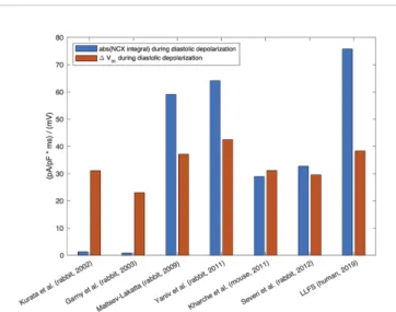
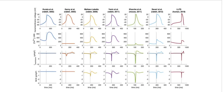
FIGURE 3 |Absolute value of the integral of signed NCX during the DD (MDP to AP takeoff) (blue, unit: ms * pA/pF≡mV), in relation to the difference in maximum diastolic Vmand takeoff potential for computational models of different species: rabbit (Kurata et al., 2002;Garny et al., 2003;Maltsev and Lakatta, 2009;Yaniv et al., 2011;Severi et al., 2012), mouse (Kharche et al., 2011), and the human Loewe–Lutz–Fabbri–Severi (LLFS) model (Loewe et al., 2019a;Loewe et al., 2019b).
Ca2+release (CICR). In turn, the negative chronotropic effect of ryanodine is the result of disappearance of localized pre-AP Ca2+
releases. Substituting Na+ for Li+to inhibit NCX caused sinus arrest leading to the conclusion that Ca2+release-NCX cross-talk has crucial importance in pacemaking. The authors provided a possible scenario for the current interactions underlying spontaneous automaticity: after reaching the maximal diastolic potential, Ifactivates to depolarize the membrane and activates ICaTand ICaLcausing LCR from the SR, which in turn activates NCX and the inward current augments to activate the remaining L-type Ca2+channels. These results were interpreted such that SR Ca2+release has a pivotal impact in defining the actualfiring rate and inactivation of either the RyR or NCX activity lead to frequency slowing or abolishment.
The pivotal role of the strong interaction between the Ca2+
concentration in the subspace and VmviaNCX for spontaneous depolarizations in SNCs was numerically reproduced and mechanistically underpinned by Maltsev et al. (2004) in a biophysical modeling study in 2004 based on an extension of the Kurata et al. (2002)model (Kurata et al., 2002). In 2009, Maltsev and Lakatta proposed a coupled oscillator model incorporating aspects of both a membrane and a Ca2+ clock, which yielded rhythmic spontaneous depolarization over a wider range of parameters of the currents involved in the M-clock (ICaL, If) (Maltsev and Lakatta, 2009). By modulating SR Ca2+
pumping rate, the coupled-clock yielded robust pacemaking in the range of 1.8 to 4.6 Hz, which is wider than could be achieved by modulation of M-clock properties alone.Kurata et al. (2012) performed stability and bifurcation analyses on this coupled- clock model and found that the most important model parameters for generation of cyclic depolarization (equilibrium
point instability) are the SR Ca2+pumping rate, NCX density as well as ICaL conductance (Kurata et al., 2012). However, the model system does not exhibit cyclic intracellular Ca2+
oscillations when Vmis clamped (independent of the clamped value), i.e., arguing against an independent Ca2+ clock, thus underlining the coupled nature of the pacemaking mechanism.
In line with this finding, Maltsev and Lakatta (2009)observed damped oscillations when clamping the transmembrane voltage to −65 mV (at Pup = 20 mM/s) (Maltsev and Lakatta, 2009).
However, sustained oscillations were observed at Pup= 40 mM/s.
The NCX was a major contributor to the observed equilibrium point in the study byKurata et al. (2012).
Vinogradova et al. explored in 2004 whether the LCRs require previous membrane depolarization as was suggested so far (Vinogradova et al., 2004). Experimental measurements on isolated rabbit SNCs as well as numerical modeling revealed threefindings i) the LCRs during the DD do not require previous membrane depolarization; ii) the LCRs are rhythmic and generate rhythmic inward current movements; iii) the LCR periods are highly correlated with the spontaneous CLs.
In contrast to the increasing body of evidence, Sanders et al.
claimed in 2006 that the contribution of NCX to spontaneous depolarization does not exclusively depend on the intact SR function in guinea pig SNCs (Sanders et al., 2006). They demonstrated that low Na+ solution abolished spontaneous firing in the presence of CPA, an inhibitor of SR uptake. On the other hand, if NCX would be entirely a consequence of SR Ca2+release, the CPA should have a similar effect as a low Na+ solution. However, the SR inhibition only slowed the heart rate, while low Na+ abolished spontaneous pacemaking within one beat, with concomitant decrease of Ca2+i. However, considering
A
B
C
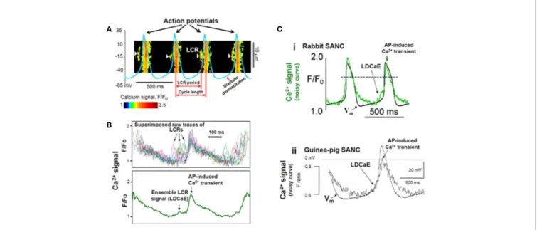
FIGURE 4 |Confocal line scanning image of Ca2+-signal with superimposed spontaneous AP traces recorded in rabbit SNCs show DD, distribution of LCRs (white arrows), LCR period, and CL(A). Confocal microscopic image of LCRs are depicted in [(B), upper panel], ensemble LCR signal or Late Diastolic Ca2+Elevation (LDCaE) that precedes Ca2+-transient are created from temporal average of LCRs [(B), lower panel]. LDCaE in single SNC of rabbit [(C), panel i] and guinea-pig [(C), panel ii] is shown. LCR: local Ca2+release. From (Maltsev et al., 2014) with permission.
the rather complex effects of reduced Na+solution, the results could not unequivocally support the authors' hypothesis. The application of KB-R7943 to inhibit NCX has to be considered problematic because of its nonselectivity.
Using high-speed Ca2+ imaging, Yaniv et al. (2013) could show that the coupled clocks also control beat-to-beat regulation of the spontaneous beating rate (Yaniv et al., 2013b). Acute application of low concentrations of caffeine (2 to 4 mM) caused Ca2+ release from the SR and subsequently led to increased spontaneous beating ratesviamembrane depolarization caused by increased NCX activation. This effect could be reproduced mechanistically in an extended version of the Maltsev–Lakatta computational model (Yaniv et al., 2013b). Toward this end, Severi et al. (2012)proposed an update of the Maltsev–Lakatta rabbit SNC model incorporating a coupled-clock with Ca2
+-handling (including NCX) and refined current formulations based on more recent experimental data (Severi et al., 2012). On the other hand, the Severi et al. model could not predict the effect of acute caffeine injection described above and complete block of Iflead to cessation of spontaneous activity. Simulations including the effects of 1 μM isoproterenol [causing, among other, a 25%
increase of SR Ca2+pumping as inMaltsev and Lakatta (2010) (Maltsev and Lakatta, 2010)], yielded a 28.2% rate increase in the Severi et al. model in good agreement with the experimentally observed increase of 26.3 ± 5.4% byBucchi et al. (2007b).
In 2013, Sirenko et al. confirmed the presence of LCR in SNCs (Sirenko et al., 2013). The authors compared the spontaneous Ca2+releases in permeabilized (i.e., in the absence of functional ion channels) rabbit SNvs.ventricular cells. They found that in the presence of similar physiological Ca2+ concentrations, the SNCs generate large rhythmic spontaneous Ca2+ releases in contrast to ventricular cells in which they found Ca2+ sparks with small amplitude and stochastic nature, as was originally described (Cheng et al., 1993). This ability of SNCs was associated with more abundant SERCA, reduced extent of phospholamban (PLB), and increased Ca2+-dependent phosphorylation of PLB and RyR.
In 2013, Yaniv et al. found new evidence regarding the membrane and Ca2+ clock coupling (Yaniv et al., 2013a).
Application of the If-inhibitor ivabradine (3 μM) reduced the APfiring rate, partially by inhibiting Ifand by decreasing the SR Ca2+content in isolated SNCs. This means that despite the fact that ivabradine has no direct effect on intracellular Ca2+cycling and does not exert effects on surface membrane channels other than If, it still suppresses the SR Ca2+load and the LCR events.
These novelfindings further supported the hypothesis that cross- talk between the membrane and Ca2+ clock regulates SN automaticity.
Sirenko et al. (2016) analyzed in detail the electrochemical Na+/Ca2+ gradients in the pacemaker function of the NCX (Sirenko et al., 2016). They used a combination of numerical modeling and experimental rabbit approaches (by using the Na/K inhibitor digoxigenin) to determine how the coupling of Na+and Ca2+ electrochemical gradients regulates pacemaking. Minimal increase of Na+i (5%) enhanced the LCRs, shortened the CL,
which was interpreted as an extension of normal chronotropic response. Further increase of Na+i by up to 15 mM caused reduction of LCRs, large CL variability as a consequence of Vm- Ca2+clock uncoupling. In parallel with the biphasic CL changes, the NCX current also showed biphasic alterations: initially the INCXincreased due to the high amplitude LCRs, then declined as ENCX reduced. The authors claimed that ENa, ECa, and ENCX
tightly regulate the SN automaticity via influencing several ion channels and components of the SN clocks.
Stern et al. (2014) proposed a computational model with subcellular spatial resolution that allows studying intracellular Ca2+release propagation patterns that was employed inMaltsev et al. (2017)to classify LCRs and provide insight into mechanisms increasing robustness of pacemaking at various SERCA pumping rates. Consistent with previous studies, they found a major role of NCX in the fight-or-flight response: faster SR Ca2+ pumping prepared the cell for stronger (higher amplitude) and more synchronous LCRs (more subcellular Ca2+ wave collisions) in the next cycle, which elicited a higher NCX current.
Torrente et al. in 2016 studied the role of the predominant isoform of L-type Ca2+channels (Cav1.3) in the generation of the LCRs and Ca2+idynamics in SNCs from wild-type and Cav1.3 knockout mice (Torrente et al., 2016). This work challenged the results of previous experimental work (under voltage-clamp conditions with Vm below the activation of Cav1.3 (Vinogradova et al., 2004) and in membrane-permeabilized SNCs, without the influence of any plasma membrane ion channels (Sirenko et al., 2013) claiming that LCRs are clearly spontaneous in nature and entirely independent from the M- clock. Cav1.3 deficiency significantly impaired Ca2+idynamics by reducing the frequency of LCR events and preventing synchronization. The results of Torrente et al. suggest that the local increase of Ca2+igenerated by Cav1.3 Ca2+current induces RyR opening, thus supporting the coupling between membrane depolarization and SR Ca2+release. Thus Cav1.3 channels can be inducers of LCR generation in the late DD. Considering this mechanism, Cav1.3 could stimulate the NCX-mediated depolarizing current to reach the threshold potential necessary to trigger an AP. Although LCRs are likely triggered by Cav1.3- mediated ICaLrelease in mouse SNCs (Torrente et al., 2016), LCR generation has different mechanisms in pacemaker cells of various species. As mentioned before, Vinogradova et al.
showed that LCRs in rabbit SNCs do not require a change of Vm(Vinogradova et al., 2004). Instead, LCRs are spontaneous in nature and occur in permeabilized SNCs. Takimoto et al. showed that the a1D Ca2+ channel mRNA is expressed in rat atrium (Takimoto et al., 1997). This observation was confirmed in mouse and human cells by Mangoni et al. (Mangoni et al., 2003). However, it remains unclear whethera1DCa2+channels are present and functional in rabbit SNCs since expression at the protein level has not been verified (Qu et al., 2005).
Huser et al. found that T-type Ca2+channels can also have a role in the generation of LCRs since in cat atrial pacemaker cells, LCRs are triggered by voltage-dependent activation of T-type Ca2+channels (Huser et al., 2000).
The Role of Phosphorylation in SN Pacemaking and the Fight-or-Flight Response
A crucial feature of the SN is the capability of adapting thefiring rate to the momentary requirements of the body.b-adrenergic activation stimulates the cAMP-PKA cascade and phosphorylates several components of the Ca2+ handling machinery and transmembrane ion channels. As a consequence, higher frequency and magnitude of LCRs provide enhanced drive for NCX, thus accelerating DD (Vinogradova et al., 2005;
Vinogradova and Lakatta, 2009).
CaMKII
In 2000, Vinogradova et al. reported a crucial role of CaMKII in SN pacemaking (Vinogradova et al., 2000). Selective inhibition of CaMKII activity (by autocamtide 2-inhibitory peptide, and KN- 93/KN-92) reduced the pacemaker frequency in rabbit SNCs, furthermore, larger doses of the compounds completely terminated SN automaticity. The authors also demonstrated that CaMKII activity is driven by subsarcolemmal Ca2+
movements and located in the vicinity of L-type Ca2+channels.
Therefore, the vital role of CaMKII could be based on the modulation of ICaL via subsarcolemmal Ca2+ changes (Vinogradova et al., 2000).
In 2011, Gao and Anderson showed that CaMKII can support heart rate increase independent ofb-adrenergic stimulation (Gao et al., 2011). Furthermore, CaMKII is required forb-adrenergic response and Ca2+-based mechanism of pacemaking.
Li et al. demonstrated that CaMKII inhibition reduced phosphorylation of RyR and PLB, decreased the LCR size, and increased the LCR period (Li et al., 2016). As a consequence, SN firing rate was reduced. These results led the authors to the conclusion that high basal CaMKII activation ultimately regulates the pacemaker function via phosphorylation of Ca2+
handling proteins.
cAMP-PKA-PDE
The Lakatta group in 2008 demonstrated considerably high basal activity of phosphodiesterase (PDE) enzyme, which restricts local RyR Ca2+ release during DD via reduction of cAMP PKA-dependent protein phosphorylation to keep the basal spontaneous SNC firing under control (Vinogradova et al., 2008). Also in 2008, Younes et al. demonstrated in isolated SNCs that the adenylate-cyclase is activated in the entire physiological concentration range of intracellular Ca2+and the cAMP-PKA axis activation drives SR Ca2+release, which in turn activates AC (Younes et al., 2008). This feed-forward“fail-safe” system has a crucial role in maintaining the normal rhythm of SNCs.
In 2011, Liu et al. studied the role of the AC-cAMP-PKA-Ca2+
signaling cascade in mouse SNCs, and they demonstrated that the Ca2+handling proteins are abundantly expressed, and LCRs were also detected in skinned cells. They showed that inhibition of intrinsic PKA activity reduces PLB phosphorylation and prolongs the LCR period causing reduction in the SNCs firing rate. The PDE inhibition resulted in the opposite effects:
increased PLB phosphorylation, shortened LCR period, and accelerated firing. In the same study the authors also demonstrated that b-adrenergic activation requires intact Ca2+
handling, indicating a pivotal role of AC-cAMP-PKA-Ca2+
signaling cascade in maintaining the normal automaticity in mouse SNCs (Liu et al., 2011).
Vinogradova et al. provided data regarding the synergistic role of PDE3/PDE4 activity in rabbit SNCs in 2018. Individual inhibition of PDE3 or PDE4 caused a moderate increase in the beating rate (20vs.5%, respectively) (Vinogradova et al., 2018a).
However, parallel block of both caused a 45% increase of pacemaking rate, indicating a synergistic effect. Furthermore, dual inhibition enhanced the LCR numbers and size, reduced the SR refilling time and LCR period due to decrease of cAMP/PKA phosphorylation (Vinogradova et al., 2018b). An amplification of local RyR Ca2+ release activates augmented NCX current at earlier times leading to an increase in the DD rate and spontaneous SANC beating rate. When RyRs were disabled by ryanodine, PDE inhibition failed to amplify local Ca2+ releases and increase NCX current. As a result, there was no increase in the DDR and spontaneous SN beating rate (Vinogradova et al., 2018b). In this context, Vinogradova et al. showed in the same year that PDE3/PDE4 modulation of SNCs is self-adaptive,i.e., full functional effect is achieved only when both PDEs are inhibited. Such inhibition will lead to an elevation of the local level of cAMP and PKA phosphorylation. The authors claimed that local cAMP signals may have greater importance in the regulation of spontaneous automaticity in SNCs than the‘global cAMP' (Vinogradova et al., 2018a).
Autonomic Modulation
In 2002, Vinogradova et al. elucidated the Ca2+ release during DD as the specific link between b-adrenergic stimulation and increasedfiring rate in rabbit SNCs (Vinogradova et al., 2002).
They demonstrated that 3 μM ryanodine abolished the effect of 0.1 μM isoproterenol. In the presence of ryanodine, the b- adrenergic stimulation failed to alter the slope of DD, firing rate, and subsarcolemmal Ca2+ releases, whereas the ICaL
amplitude exerted clear b-adrenergic stimulation-induced increase. In 2006, Bogdanov et al. described how application of agents affecting the timing or amplitude of LCRs (ryanodine, BAPTA, nifedipine or isoproterenol) caused immediate changes of spontaneous beating rate, which could be traced back to changes of local NCX using mechanistic computational modeling (Bogdanov et al., 2006). Similarly, PKA-dependent phosphorylation affected LCR spatiotemporal synchronization and therefore modulated beating rate mediated by altered NCX (Vinogradova et al., 2006).
Himeno et al. (2008)studiedb1-adrenergic stimulation in a computational model of guinea pig SNCs with large IKsin which they described the role of NCX (Himeno et al., 2008). While activation of IKs during the AP preceding the b1-adrenergic stimulation was negligibly small, IKs counterbalanced the increase in ICaL and NCX, which otherwise compromised the positive chronotropic effect of theb1-adrenergic stimulation by elongating the APD in their model.
Lyashkov et al. (2009) investigated the mechanism of muscarinic-receptor stimulation on heart rate reduction (Lyashkov et al., 2009). They found that carbachol (at IC50) reduced the number and size of LCRs and lengthened the LCR period with concomitant decrease of the beating rate. Numerical modeling indicated that cholinergic-modulation of the beating rate is integrated into the Ca2+ cycling via LCR-mediated function of the NCX. The authors concluded that in the presence of low doses of carbachol, the muscarinic activation induced beating rate reduction is mediated by suppression of cAMP-PKA-Ca2+signaling, while IK(Ach)activation contributes only under higher carbachol concentrations.
In the same year, Maltsev and Lakatta (2010) published a modeling study demonstrating how G protein-coupled receptor modulation of spontaneous SNC beating rate is not only mediated by effects on membrane currents but also by effects on the SR Ca2+pumping rate, which in turn affect diastolic NCX (Maltsev and Lakatta, 2010). Also in 2010, Gao et al.
demonstrated in healthy canine hearts that complete or almost complete Ifblock attenuated but did not eliminate the positive chronotropy of isoproterenol suggesting that Ifis not the only target of the b-adrenergic response (Gao et al., 2010). Their results also support the notion of spontaneous Ca2+ releases during late DD, which was sensitive to isoproterenol. In their experiments, 2 μM ryanodine caused a 14% decrease in pacemaker frequency but dramatically decreased the effect of isoproterenol. They also provided evidence that ryanodine has no direct effect on If. The authors concluded that both voltage and Ca2+dynamics have a role in the pacemaker mechanism.
In the same year, Vinogradova and Lakatta demonstrated that the LCR period and the spontaneous SN cycle is tightly regulated by the SR refilling time in rabbit SNCs (Vinogradova et al., 2010).
Therefore, phosphorylation/dephosphorylation of the SERCA regulator protein PLB has also critical roles in setting the actual CL of the SN automaticity especially during b- adrenergic stimulation.
In a theoretical analysis of the relative importance of the membrane and Ca2+oscillators,Imtiaz et al. (2010)could show that increased SR Ca2+uptake leads to i) hyperpolarized MDP viaa reduction of NCX due to lower Ca2+i(Imtiaz et al., 2010).
This means that due to lower Ca2+the smaller inward current through NCX enables more negative MDP. ii) The more negative Vmincreases the magnitude of Ifand ICaTduring early diastole.
iii) The enhanced Ca2+entry during DD increases intracellular Ca2+and CICR causing a higher NCX current.
Transgenic Modi fi cations of NCX
In 2013, several studies using transgenic NCX knockout mice were published. First, Gao et al. used cardiac specific,incomplete NCX1 knockout mice where the baseline sinus frequency was completely identical between control and NCX1-deleted mutants (Gao et al., 2013). However, they found that NCX1 deletion only partially impaired the isoproterenol effect indicating that Ifis also required. The authors concluded that the NCX has a minor role in maintaining the normal heart rate but NCX1 deletion dramatically enhanced the effects of ivabradine on isoproterenol-
induced automaticity indicating that NCX and If mutually contribute in the fight-or-flight response. Since the genetic knockout of NCX was incomplete, one cannot rule out the possibility that the remaining NCX may be able to provide enough current to fulfill its role in maintaining the Ca2+balance.
In response to that paper, Maltsev et al. in 2013 provided rabbit SN numerical simulations (Maltsev et al., 2013) to address some unanswered questions byGao et al. (2013). The authors extended their simulation with new stabilization of NCXvialocal control of CICR. They argued that the Ca2+released from the RyR is able to recruit the neighboring RyRs producing Ca2+
wavelets (LCRs) having amplitudes larger than the Ca2+sparks and smaller than global Ca2+transients. Since the NCX extrudes Ca2+from the vicinity of RyRs it attenuates the spread of Ca2+
leading to weaker recruitment. However, when NCX is genetically decreased (i.e. incomplete NCX1 knockout), the Ca2+ spread and recruitment are enhanced providing larger Ca2+ and driving force for the remaining NCX molecules.
These results indicate that NCX has a crucial role under the normal heart rate, and a small fraction (i.e.20%) of NCX is able to produce sufficient current under DD. However, when NCX expression is low, its capacity is fully used for Ca2+ extrusion under rest and there is no more support for frequency increase underb-adrenergic response.
Herrmann et al. in 2013 used inducible and SN specific Cre transgenic mice lacking NCX1 selectively in SN pacemaking cells (Herrmann et al., 2013). The NCX1 was genetically ablated in a temporally controlled and tissue selective manner. The animals exerted severe bradycardia and large variability with arrhythmias leading to the conclusion that NCX has a role to maintain normal rhythm. The caffeine induced transient decay was markedly slowed (3.6% of control) in cpNCXKO cells indicating that in SNCs, almost the entire trans-sarcolemmal Ca2+ extrusion is carried by NCX. At the same time, it is an interesting finding that the Ca2+ transient amplitude was decreased and the decay was slowed while the SR Ca2+content was unaffected. The authors claimed that the NCX1 deleted cells are not able to compensate the NCX1 deletion. They argue for a fundamental role of NCX in pacemaking even under resting conditions, and the normal pacemaking function is not possible without proper NCX function.
Groenke et al. found that in NCX1 knockout mice (Cre/LoxP system), there is no indication of spontaneous atrial depolarizations on the ECG and the animals exerted junctional escape rhythm (Groenke et al., 2013). Furthermore, isolated cells did not show spontaneous automaticity despite the presence of If
and intact Ca2+stores. They claim that NCX has a crucial role in normal pacemaking. They observed LCRs but no Ca2+transients in NCX deleted cells. They also conclude that Ifis not sufficient to depolarize the cell membrane if NCX is completely lacking, interestingly not even if isoproterenol was added. Torrente et al. in 2015 created an atrial-specific NCX knockout mouse where NCX was totally eliminated from the atria including the SN (Torrente et al., 2015). These cells lacked spontaneous APs despite intact If. In contrast, the intact tissue showed spontaneous burst–pause type depolarizations. The authors conclude that in
the absence of NCX-mediated depolarizations, ICaLand Ifare able to initiate AP burst where Ca2+ is gradually accumulated and finally terminated burst activity via Ca2+-dependent inactivation or by other Ca2+ activated processes such as small-conductance Ca2+-activated K+ currents (Torrente et al., 2017).
In 2016, Choi et al. observed regular oscillations of inward currents in voltage-clamped human embryonic stem cell-derived cardiomyocytes, which exhibit spontaneous APs when not being voltage clamped (Choi et al., 2016). The oscillatory Ca2+releases could be eliminated by blocking NCX (with 1 μM SN-6 and 10 μM KB-R7943) but not by blocking If(with 3 mM ivabradine).
Their observations suggest that in these hESC-CMs, the M-clock and the voltage clock act as two redundant pacemaker mechanisms which can work independently.
Kaese et al. (2017)used a homozygously overexpressed NCX mouse model and found that the basal heart rate is not affected, suggesting that NCX is not a crucial factor in setting the resting heart rate (Kaese et al., 2017). In contrast, b-adrenergic stimulation elicited higher heart rates in NCX-overexpressing mice, suggesting more inward NCX current which enhances the speed of DD.
Between 2003 and 2011 several genetically manipulated mice studies with deficient If function were published where autonomic rate modulation was preserved (Ludwig et al., 2003;
Herrmann et al., 2007;Harzheim et al., 2008;Hoesl et al., 2008;
Baruscotti et al., 2011). These results further indicate the lack of an essential and irreplaceable role of Ifin SN pacemaking and emphasize that Ifworks together with its“teammates”within the coupled-clock system (Lakatta and Maltsev, 2012)
AP Ignition Model as the Most Current SN Pacemaking Mechanism
In 2018, a new model was proposed by the Lakatta group extending SN electrophysiology by an integrated regulatory mechanism including close interactions of LCRs, NCX, and ICaLcoupled by a positive feedbackviadiastolic CICR and DD acceleration (Lyashkov et al., 2018). In contrast to previous theories arguing for a role of LCRs during the late DD, the AP ignition model suggests that early LCRs (ICaL-independent) activate inward NCX, which defines the ignition onset. If, and ICaT also contribute to early depolarization to reach the ICaL
threshold. The physiological role of the low-voltage activation threshold L-type Ca2+channel isoform (Cav1.3) may have more importance to regulate the early ignition phase. However, the details of the specific contribution of Cav1.3 require further investigation. The ICaL-mediated Ca2+-influx recruits more LCRs via diastolic CICR, and this positive feedback further increases NCX. It was also shown that ICaT also has a role in the diastolic CICR, similarly as early results suggested (Huser et al., 2000). However, previously ivabradine and ryanodine were considered to cause AP CL lengthening by different mechanisms (i.e.ivabradine by Ifinhibition and ryanodine by takeoff potential depolarization). The underlying mechanism of ryanodine induced decrease in the firing rate could be driven by the marked decrease in DDR, which was observed in isolated
rabbit sinoatrial node cells by Vinogradova et al. (2002). The AP ignition model brings them common ground: regardless of whether the M-clock (in case of ivabradine) or the Ca2+-clock (in case of ryanodine) is perturbed, it will indirectly affect the other clock via the coupling mechanism. As a coupling, similar responses happened, the ignition potential become depolarized, and time-to-ignition phase becomes longer. This provides delay in clock-coupling by CICR delay.
The Role of NCX in Human SNCs in Comparison to Other Species
In contrast to the significant amount of experimental data on the role of NCX in pacemaking derived from animal experiments (mostly isolated rabbit SNCs), data from human SNCs or tissue are scarce. Considering the vast difference in the main quantity of interest, the beating rate, between commonly used laboratory animals and humans (mouse: 500 bpm, rabbit: 300 bpm, dog:
100 bpm, human: 60 bpm), it however cannot be assumed that the details of pacemaking mechanisms and most importantly the delicate balance of competing effects can be transferred from animal models to human in general. Indeed, it has been shown that computational models of SNCs of different species (mouse, rabbit, human) react markedly different to changes of e.g.
extracellular Ca2+ concentration (Loewe et al., 2019b). Given the potential relevance of this alteration regarding sudden bradycardic death in dialysis patients (Loewe et al., 2019a), species-dependent investigations are desirable. As a particular example of interspecies differences, IKrhas been reported to be absent in porcine SNCs and IKs to be negligibly small in rabbit SNCs during rest, while both are present in guinea pig SNCs (Satoh, 2003). Moreover, SN AP duration is markedly different between species (mouse: 80 ms, rabbit: 200 ms, human: 300 ms;
approximate values) as are other electrophysiological parameters such as AP upstroke velocity, MDP, and DDR (Opthof, 2001).
Regarding the role of NCX, its importance in terms of relative contribution to Ca2+ extrusion differs between species as well:
28% in rabbit compared to 7% in rat, i.e., a 4-fold difference (Bers, 2002).
One of the few experiments using human SNCs was performed by Tsutsui et al. (2018). The authors show that similarly to animal models, spontaneous rhythmic LCRs generated by the Ca2+clock are coupled to electrogenic surface membrane molecules to initiate rhythmic APs, and that Ca2+- cAMP-protein kinase A (PKA) signaling has a regulatory role in the clock coupling in human SNCs (Tsutsui et al., 2018). In the absence of strong coupling between the oscillator subsystems, it was shown that SNCs fail to trigger spontaneous APs and only exhibit disorganized LCRs that were unable to activate the M- clock (Tsutsui et al., 2018). In summary, the study by Tsutsui et al. provides evidence that also in humans it is the coupled- clock mechanism that fundamentally drives SNC pacemaking and provides hints for mechanisms underlying SN dysfunction (Tsutsui et al., 2018).
To highlight interspecies differences and provide an overview of the characteristics of the role of NCX in different models, basic in silicoexperiments were performed.
![FIGURE 2 | Time-independent background and transporter currents including NCX during spontaneous pacemaking in early SN models: (Wilders et al., 1991) (A), (Demir et al., 1994) (B), (Dokos et al., 1996) (C), (Zhang et al., 2000) [(D); central model], and (](https://thumb-eu.123doks.com/thumbv2/9dokorg/796691.37734/5.892.67.824.638.977/independent-background-transporter-currents-including-spontaneous-pacemaking-wilders.webp)
![FIGURE 6 | Change of CL [absolute (A) and relative (B)], t dia,early (C), t dia,late (D) and APD (E) of computational models of different species (compare Figure 4) in response to gradual NCX block.](https://thumb-eu.123doks.com/thumbv2/9dokorg/796691.37734/13.892.80.821.91.529/figure-change-absolute-relative-computational-different-species-response.webp)