Physiology International, Volume 106 (4), pp. 368–378 (2019) DOI: 10.1556/2060.106.2019.34
The importance of ST elevation in V
2–4ECG leads in athletes
Zs Major1, R Kirschner2, N Medvegy3, K Kiss2, GM Török2, G Pavlik4, G Simonyi5, Zs Komka6, M Medvegy2
1Institute of Physical Education and Sport Sciences, University of Nyíregyháza, Nyíregyháza, Hungary
2Department of Cardiology, Fl´or Ferenc Hospital of Pest County, Kistarcsa, Hungary
3Department of Marketing, KRKA Hungary Ltd., Budapest, Hungary
4Department of Health Sciences and Sports Medicine, University of Physical Education, Budapest, Hungary
5Metabolic Center, St. Imre University Teaching Hospital, Budapest, Hungary
6The Heart and Vascular Center, Semmelweis University, Budapest, Hungary
Received: August 26, 2019 Accepted: October 30, 2019
Background:Early repolarization in the anterior ECG leads (ERV2–4) is considered to be a sign of right ventricular (RV) remodeling, but its etiology and importance are unclear.Methods:A total of 243 top-level endurance-trained athletes (ETA; 183 men and 60 women, weekly training hours: 15–20) and 120 leisure-time athletes (LTA; 71 men and 49 women, weekly training hours: 5–6) were investigated. The ERV2–4sign was evaluated concerning type of sport, gender, transthoracic echocardiographic parameters, and ECG changes, which can indicate elevated RV systolic pressure [left atrium enlargement (LAE), right atrium enlargement (RAE), RV conduction defect (RVcd)].Results:Stroke volume and left ventricular mass were higher in ETAs vs. LTAs in both genders (p<0.01). Prevalence of the ERV2–4sign was significantly higher in men than in women [p=0.000, odds ratio (OR)=36.4] and in ETAs than in LTAs (p=0.000).
The highest ERV2–4prevalence appeared in the most highly trained triathlonists and canoe and kayak paddlers (OR=13.8 and 5.2, respectively). Within the ETA group, the post-exercise LAE, RAE, and RVcd changes developed more frequently in cases with than without ERV2–4(LAE: men:p<0.05, females:p<0.005; RAE: men:p<0.05, females:
p<0.005; RVcd: N.S.). These post-exercise appearing LAE, RAE, and RVcd are associated with the ERV2–4sign (OR=4.0, 3.7, and 3.8, respectively).Conclusions:According to these results, ERV2–4develops mainly in male ETAs due to long-lasting and repeated endurance training. The ERV2–4sign indicates RV’s adaptation to maintain higher compensatory pulmonary pressure andflow during exercise but its danger regarding malignant arrhythmias is unclear.
Keywords:pulmonary artery pressure, early repolarization, exercise ECG, endurance athletes, right ventricular adaptation
Introduction
Early repolarization (ER)–a concave-shaped ST elevation–is a frequently described ECG change. It has been known for 50 years; nevertheless, its significance has not been clarified yet. ER may have anterolateral and/or inferolateral manifestation (1,33), but in athletes, it appears most frequently in the V2–4anterior precordial leads (ERV2–4). The prevalence of ERV2–4in athletes ranges from 35% to 91% depending on its definition (12,16). The etiology of ERV2–4 is unclear. The role of exercise is unquestionable, but impact of drugs (e.g., cocaine) was also presumed (16). Cardiac magnetic resonance examinations proved that there is no ischemic mechanism in the manifestation of ER (29).
Corresponding author: Dr. Gábor Simonyi MD, PhD, FAHA
Metabolic Center, St. Imre University Teaching Hospital, Tétényi út 12-16, H-1115 Budapest, Hungary Phone: +36 1 464 87 03; Fax: +36 1 464 87 18; E-mail:bmbel3@gmail.com
Some experts assume that (1) ERV2–4is a harmless ECG alteration; (2) however, others agree that ERV2–4may reflect right ventricular (RV) remodeling and may have causal role in malignant arrhythmias.
(1) Studies dealing with ECG changes in athletes considered ERV2–4(and some other ECG changes) as a physiological phenomenon depending on gender (they were less frequent in women), type of sports, and level as well as duration of training. These ECG changes were explained by increased parasympathetic tone, RV, septum and left ventricular (LV) hypertrophy, right atrium enlargement (RAE), and left atrium enlargement (LAE) (5,16,36). The prevalence of ERV2–4also depends on the total burden of exercise; therefore, data vary considerably (22,35).
(2) ERV2–4 may also be dangerous because of its close relationship with Brugada syndrome and arrhythmogenic RV cardiomyopathy (ARVC). It is well-known that Brugada syndrome may cause life-threatening ventricular tachyarrhythmias. Both ERV2–4 and Brugada syndrome respond similarly to pharmacologic agents, neuromodulation, and exercise (15, 17). A similar mechanism is supposed for ERV2–4 and Brugada ECG abnormalities, i.e., the plateau of epicardial action potential is lower than that of the endocardium leading to ST-segment elevation on the electrocardiogram (37). ARVC is another syndrome with predilection to malignant arrhythmias and it has been found to be part of the cause of sudden cardiac death (SCD) of athletes (3,24).
The role of RV alteration in the etiology of athletes’ SCD has just recently been identified. ARVC-like histological alterations (RV dysplasia and remodeling) may be developed by endurance sports (18–20,30,38). RV dilatation has also been demonstrated in endurance-trained male athletes (6,21). RV and right atrium dimensions were significantly greater in endurance-trained athletes than in strength-trained athletes and controls (8). The underlying cause of the morphological alterations of the RV may be the elevation of pulmonary artery pressure (PAP) during exercise. This elevation of PAP correlates with the time spent with highly intensive training (7,31). During exercise, the mean PAP (mPAP) depends primarily on the bloodflow (Q), but also on pulmonary vascular resistance (PVR) and left atrium pressure (LAP): mPAP=(PVR×Q)+LAP (8,20,26,32). Among trans- thoracal echocardiography (TTE) parameters, the LV stroke volume (SV) correlates best with systolic PAP (7,8). It has been demonstrated that during exercise the systolic PAP can be significantly increased even up to 80 mmHg accompanied by temporary enlargement of the RV. It has also been proven that elevated PAP and RV diameters return to the basic state some minutes after the termination of the exercise (6,19,26).
SCD of athletes is a rare clinical event (0.5–1/100,000 athletes/year), but it is 2–4 times more frequent than in the sedentary members of the corresponding age group (14).
It is interesting that the prevalence of SCD in female athletes is 5–20 times rarer than in male ones (2,14,23). The cause of this difference is unclear; one of the supposed causes may be milder LV hypertrophy due to the regulatory effect of estrogens in women (9,28).
All publications dealing with ER agree that ERV2–4needs further investigation to clarify its predilection to malignant arrhythmias just like its origin and hemodynamic importance (12,16).
We intended to find an explanation for the etiology and hemodynamic impact of the ERV2–4phenomenon. For this reason, the prevalence of ERV2–4has been studied in different endurance sports in male and female athletes separately. We analyzed the relationship of the ERV2–4sign with other ECG changes and TTE parameters indicating elevated PAP.
The importance of ST elevation in athletes 369
Materials and Methods
The 12-lead ECG, exercise ECG and TTE records of 243 top-level Caucasian endurance-trained athletes (ETA; weekly training: 15–20 h) and 120 Caucasian leisure-time athletes (LTA; weekly training: 5–6 h) were investigated. The following ETAs participated in the study: triathletes (54 men/18–35 years, mean age: 25 years and 22 women/18–33 years, mean age: 20 years), canoe and kayak paddlers (79 men/18–35 years, mean age: 24 years and 18 women /18–30 years, mean age: 21 years), soccer players (35 men/18–32 years, mean age: 24 years), handball players (15 men/20–35 years, mean age: 28 years), and water polo players (20 women /18–30 years, mean age: 22 years). LTAs: 71 men/18–35 years, mean age: 29 years and 49 women/
18–35 years, mean age: 27 years. From the above athletes, only triathletes practice a typical endurance-type sport, but the other investigated sports also involved high levels of endurance.
ECGs were implemented by a Cardiosys H-01 recorder (Experimetria Ltd., Budapest, Hungary) in supine position. ECG changes at rest (1 day after last training) and following exercise (using Bruce protocol) were matched. The age-predicted maximal heart rate (HR) was targeted. For standardization of the interpretation, exercise ECGs were analyzed when the HR fell between 100 and 120 bpm.
TTE examinations were conducted using a Philips HD15 ultrasound (Amsterdam, Netherlands) equipment with a 2.5–3.5 MHz-s transducer (Bothell, WA, USA) on the day of exercise ECG. Apical four-chamber and parasternal long-axis views were acquired according to the guidelines of the American Society of Echocardiography.
ECG and TTE examinations were carried out at Semmelweis University, Faculty of Physical Education and Sport Sciences between 2009 and 2014.
The ECG and TTE characteristics analyzed had earlier been described as potential signs of ARVC, elevated PAP, parasympathetic activity, malignant arrhythmias, or LV hypertrophy (7).
ECG alterations were defined in this study as follows:
ERV2–4: ST elevation exceeding 2 mm at least in two adjacent leads of V2–4was accepted (in the literature, ST elevation exceeding 0.1 mV is already defined as ER) (4).
RAE –(=P pulmonale): sign of right atrium pressure and/or volume overload. It was accepted when the positive P wave was higher than 2.5 mV in leads II–III–aVF.
LAE–(=P terminal force): sign of LAP and/or volume overload: the P wave in lead V1
was wider than 0.04 s and more negative than 0.1 mV.
RVcd–: it should be highlighted that the presence of terminal r′restricted only to lead V1
was defined as slow RV conduction without any block. The term of incomplete Right Bundle Branch Block was used if r′ was present in leads V1and V2, and r′ exceeded 0.2 mV, and the duration of QRS was less than 0.12 s (25).
Right-axis deviation: if the amplitude of the R wave in lead III is greater than that in lead II.
QTc: QT corrected to HR according to Bazett’s formula.
HR abnormalities: sinus bradycardia, bradyarrhythmias (e.g., conduction abnormalities, sino-auricular, and atrio-ventricular blocks), and tachyarrhythmias.
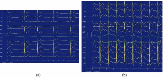
Resting ECG with sign of ERV2–4and post-exercise ECG with signs of RAE, LAE, and RVcd of a 20-year-oldfirst-class male kayak paddler are demonstrated in Fig. 1.
TTE alterations analyzed:
SVc: corrected LV SV to body surface area.
LVMc: LV mass (LVM) corrected to body surface area (LVMc=LVM/body surface area3/2) (27).
E/A ratio: the ratio between the peak velocities of the early and late phases of the diastolic filling.
EF: ejection fraction assessed by the modified Simpson’s rule formula.
Statistical analysis
Statistical analysis was carried out using SigmaStat 2.03 and SPSS 15.0 for Windows (SPSS Inc., Chicago, IL, USA). The relationship between ERV2–4and types of sport, ECG, and exercise ECG signs as well as TTE parameters was investigated by logistic regression,χ2test, and analysis of variance.
Results and Discussion The results are summarized in TablesI–III.
TableIdemonstrates the appearance of ERV2–4in different ETA groups and in the LTA group. The LV hypertrophy of these groups is characterized by LVMc. SVcis associated with systolic PAP.
The SVc and LVMc were significantly higher in ETAs than in LTAs both in men (p<0.01 andp<0.01, respectively) and in women (p<0.01 and p<0.01, respectively).
The prevalence of ERV2–4was significantly higher in ETAs than in LTAs (p=0.000).
The highest prevalence appeared in triathlonists [odds ratio (OR)=13.8, pertaining confi- dence interval (CI)=2.5–75.8; p=0.002] and canoe and kayak paddlers (OR=5.2, CI=1.5–18.1;p=0.009).
The prevalence of ERV2–4 was significantly higher in male ETAs+LTAs than in female ETAs+LTAs (OR=36.4, CI=8.8–149;p=0.000).
TableIIdemonstrates LV TTE parameters and exercise-induced ECG signs (which can indicate elevated PAP) as a function of the presence of the ERV2–4sign.
ETAs with ERV2–4 sign compared to LTAs was linked to significantly higher LV parameters (LVMc: in both gendersp<0.01; SVc: males p<0.001, females p<0.05) and
Fig. 1.Resting ECG record (a) with sinus brady-arrhythmia and ST elevation in V2–4and post-exercise ECG record (b) with signs of RAE, LAE, and RVcd of a 20-year-oldfirst-class male kayak paddler
The importance of ST elevation in athletes 371
TableI.STelevation(earlyrepolarization)inECGleadsV2–4(ERV2–4)invarioussports SportsGenderTraininghoursHeight(cm)Weight(kg)N
TTEECG SVc(ml/m2)LVMc(g/m2)ERV2–4(%) Kayaker–canoeistsMale18±5184±684±77955±1096±1889 Female19±5169±466±71848±788±1222 TriathletesMale18±7179±768±105454±897±1689 Female15±7171±755±52242±781±1545 HandballplayersMale20±2193±6102±81552±586±873 Female–––0––– SoccerplayersMale16±2182±779±93547±784±1271 Female–––0––– WaterpoloplayersMale–––0––– Female18±6176±672±92045±679±1125 AllETAsmean/sumMale18±4183±680±818352±893±1484 Female17±6172±664±76045±682±1232 LTAMale6±3177±874±13714282±1417 Female5±3167±668±9493568±92 TTE:transthoracicechocardiographic;N:numberofcases;SVc:leftventricularstrokevolumecorrectedtobodysurfacearea;LVMc:leftventricularmusclemasscorrectedtobody surfacearea;ETA:endurance-trainedathletes;LTA:leisure-timeathletes
TableII.Transthoracicechocardiographic(TTE)parametersandECGchangesinthefunctionofthepresenceofERV2–4sign GenderTraining hoursN TTERestingECGPost-exerciseECG SVc(ml/m2)LVMc(g/m2)HR(bpm)LAE(%)RAE(%)RVcd(%)LAE(%)RAE(%)RVcd(%) ETAwith ERV2–4
Male17±415450135545131586033 Female17±319471116332053637411 ETA without ERV2–4
Male16±42948131533017242114 Female17±2414210757221724275 LTA without ERV2–4
Male6±35939105661032010105 Female5±348368870601725172 ETA:endurance-trainedathlete;LTA:leisure-timeathlete;N:numberofcases;SVc:leftventricularstrokevolumecorrectedtobodysurfacearea;LVMc:leftventricularmuscle masscorrectedtobodysurfacearea;ERV2–4:STelevationinleadsV2–4;HR:heartrate;LAE:leftatrialenlargement;RAE:rightatrialenlargement,RVcd:rightventricular conductiondisturbances
The importance of ST elevation in athletes 373
higher rate of exercise-induced ECG alterations (LAE: in malesp<0.005, in females N.S.;
RAE: in malesp<0.005, in femalesp<0.05; RVcd: in malesp<0.05, in females: N.S.).
Within ETA individuals, LVMc and SVc did not differ significantly between groups with or without ERV2–4, but the rate of exercise-induced ECG alterations was higher in the ERV2–4group vs. the group without ERV2–4(LAE: in malesp<0.05, in femalesp<0.005;
RAE: in malesp<0.05, in females p<0.005; Rvcd: N.S.).
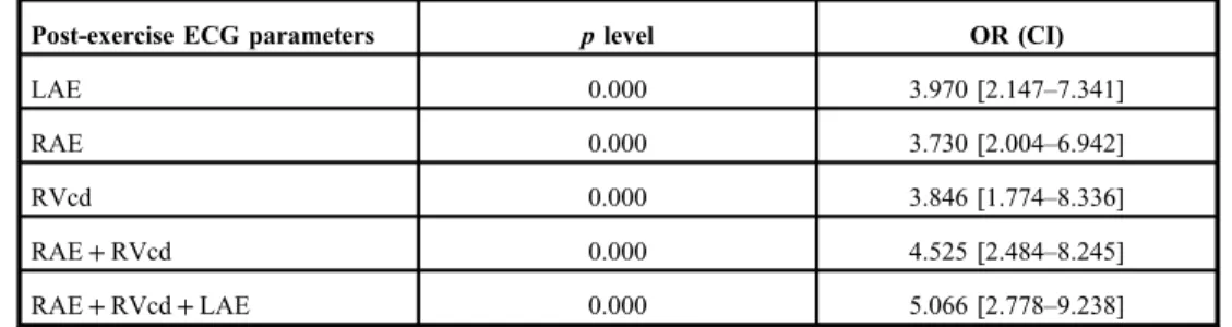
Table III demonstrates the statistical relationships between ERV2–4 and exercise- induced ECG alterations indicating elevated PAP during exercise.
The results of some observations are not demonstrated in the previous tables, but they are worth mentioning.
ER in the inferior-lateral leads exceeding 0.2 mV ST-segment elevation occurred only in 12 subjects vs. 173 subjects presenting ERV2–4in this study population.
Right-axis deviation was observed in eight cases, whereas left-axis deviation above 0°developed in six cases. R/S wave ratio in lead V1could not be evaluated owing to small R-wave amplitudes, which depended on the position of the V1electrode.
Negative T-waves were seen in leads V2–4in 10 athletes (2 kayaker-canoeists/1 man;
8 triathletes/7 men). These negative T-wave alterations were mentioned in the literature mostly in Afro-American athletes. Their 13% incidence was not considered pathological (10, 34). In our 10 cases showing this morphology, positive but not tall T-waves were apparent during exercise. The value of QTc was not pathologic either at rest or during exercise in this population. The resting HR was significantly lower (47 vs. 55/min) and the LV hypertrophy was slightly more pronounced in our 10 cases with negative T-waves vs. all ETA cases. The“not pathological”attribution of this ECG alteration is not entirely correct in our opinion; rather, it is more appropriate to state that no evidence has been found to date for negative consequences in athletes having this ECG alteration.
Arrhythmias were also present in this study population. Premature ventricular contrac- tions (PVCs) appeared during exercise only in subjects with ERV2–4(6 cases). PVCs were always of RV origin. Repetitive non-sustained (4–16 beats) ventricular runs with left bundle branch block morphology (i.e., with RV origin) developed following exercise in one triathlonist woman.
Bradyarrhythmias like SA-block, 1st or 2nd degree AV block showed up in 14 cases at rest.
Table III.Relationships (OR) between ERV2–4and post-exercise ECG changes indicating elevated pulmonary artery pressure
Post-exercise ECG parameters plevel OR (CI)
LAE 0.000 3.970 [2.147–7.341]
RAE 0.000 3.730 [2.004–6.942]
RVcd 0.000 3.846 [1.774–8.336]
RAE+RVcd 0.000 4.525 [2.484–8.245]
RAE+RVcd+LAE 0.000 5.066 [2.778–9.238]
LAE: left atrial enlargement; RAE: right atrial enlargement, RVcd: right ventricular conduction disturbances;
OR: odds ratio; CI: confidence interval
QTc was always in normal range, and there was no difference in its value either among different sports or according to the presence of ERV2–4.
There was no difference in blood pressure between groups of various sports.
Systolic and diastolic function (EF, E/A) determined at rest have been found to be normal in all groups, and there was no significant difference between groups of various sports.
Many researchers reported frequent occurrence of ERV2–4, RAE, LAE, and RVcd in ETAs (5,11,12,31,36). RVcd and RAE are directly related to high RV pressure and may be associated with RV and atrium wall strain. LAE indirectly refers to elevated PAP, whereas PAP also involves LAP. It is due to that under extreme endurance exercise, LV hypertrophy may lead to temporary relative compliance failure and elevated LV end diastolic pressure indicated by LAE. Although the sensitivity of these ECG alterations is not high and their occurrence in athletes is regarded not pathological, their appearance during exercise may indicate elevated PAP. In this study, it was shown that ECG signs of elevated PAP occur more frequently in those ETAs whose ERV2–4persists.
The physiological mechanism of the ERV2–4sign and its relationship with exercise were not clear and its clinical relevance was still debated in the relevant publications. According to this study, ERV2–4could be the consequence of repetitive and sustained PAP elevation due to endurance sports. The causes of PAP elevation during exercise are increased systemic blood flow (which exceeds the decrease of PVR), as well as increasing left atrial pressure, i.e., PAP elevation is a compensatory response during exercise (20,26). This is due to the prevalence of the ERV2–4alteration is high particularly in male endurance athletes (it is rare in women athletes and in non-athletes), in whom LV hypertrophy is much more explicit (characterized by greater LVMcand SVc) compared to women and non-athletes. The role of LV hypertrophy in elevated PAP may be twofold: on the one hand, males are able to reach higher exercise levels generating greater bloodflow; on the other hand, LV hypertrophy may lead to higher LAP and consequently to higher pulmonary post-capillary pressure.
Our interpretation for the earlier supposed ERV2–4etiologies is the followings:
The role of parasympathetic tone has been suggested (16). According to our opinion, the parasympathetic tone does not explain the ERV2–4, because in ETA individuals, the low HR (indicating parasympathetic tone) does not differ between groups with or without ERV2–4.
Interventricular septum hypertrophy has also been mentioned as the possible etiology of the ERV2–4(16). Our interpretation is the following: although high-level physical exercise could result in both septal hypertrophy and ERV2–4, nevertheless, septal hypertrophy in asymmetric septum hypertrophy disease does not cause ERV2–4(13).
The role of cocaine has also been brought up (16). We did not investigate the effect of cocaine, but we agree with this idea, since cocaine may constrict small vessels, thus promoting the elevation of PAP. Therefore, the use of cocaine for endurance athletes means an increased risk.
Our examinations highlight the fact that the ECG parameters of elevated PAP persist for different periods. LAE and RAE rapidly regress. They are found on the resting ECG only rarely in ETAs in contrast with their high occurrence rate during exercise. These data prove that RAE and LAE ECG signs can develop due to atrium wall strain without atrium enlargement. The RVcd persists longer. About half of RVcd was also shown at rest 1 day after the last training. ERV2–4is also reversible, but it persists for weeks. We did not see ERV2–4 even in ETAs who had stopped sport activity some months earlier. This study
The importance of ST elevation in athletes 375
confirms the regression capability of these parameters. They are related to temporary pressure overload and wall strain.
In conclusion, we found that ERV2–4alteration and some post-exercise ECG parameters (LAE, RAE, and RVcd) were more frequent in ETAs (mainly in the highest trained triathlonists and canoe and kayak paddlers) and males than in LTAs and women.
The higher level of LV hypertrophy in the ETA population (proved by higher LVMcon TTE) can result in greater bloodflow and can contribute to elevated PAP. We concluded that ERV2–4may develop due to long lasting and repetitive elevated PAP, and the increase of PAP during exercise is more frequent in case of ERV2–4. Therefore, the ERV2–4indicates that the RV is able to maintain the greater compensatory pulmonary flow during exercise with higher PAP.
We can hypothesize that athletes during exercise without RV remodeling and ERV2–4 are not able to reach high PAP, and they are threatened by acute RV failure during extreme exercise (e.g., marathon).
On the other hand, publications suppose that ERV2–4 (as a sign of RV remodeling) can mean a higher risk of malignant arrhythmias because of more morphological and electrophysiological similarities with Brugada syndrome and ARVC. It may be the cause why SCD in athletes is 2–4 times more frequent than in the corresponding age group. We found ERV2–4less frequent in women, which may explain the lower prevalence of SCD in female athletes.
Limitation of the study We did not perform TTE examination during physical exercise.
REFERENCES
1. Cappato R, Furlanello F, Giovinazzo V, Infusino T, Lupo P, Pittalis M, Foresti S, De Ambroggi G, Ali H, Bianco E, Riccamboni R, Butera G, Ricci C, Ranucci M, Pelliccia A, De Ambroggi L: J wave, QRS slurring, and ST elevation in athletes with cardiac arrest in the absence of heart disease. Marker of risk or innocent bystander?
Circ. Arrhythm. Electrophysiol. 3, 305–311 (2010)
2. Chandra N, Bastiaenen R, Papadakis M, Sharma S: Sudden cardiac death in young athletes. Practical challenges and diagnostic dilemmas. J. Am. Coll. Cardiol. 61, 1027–1040 (2013)
3. Corrado D, Basso C, Schiavon M, Thiene G: Screening for hypertrophic cardiomyopathy in young athletes.
N. Eng. J. Med. 339, 364–369 (1998)
4. Corrado D, BiffiA, Basso C, Pelliccia A, Thiene G: 12-lead ECG in the athlete: physiological versus pathological abnormalities. Br. J. Sports Med. 43, 669–676 (2009)
5. Corrado D, Pelliccia A, Heidbuchel H, Sharma S, Link M, Basso C, BiffiA, Buja G, Delise P, Gussac I, Anastasakis A, Borjesson M, Bjørnstad HH, Carrè F, Deligiannis A, Dugmore D, Fagard R, Hoogsteen J, Mellwig KP, Panhuyzen-Goedkoop N, Solberg E, Vanhees L, Drezner J, Estes NA 3rd, Iliceto S, Maron BJ, Peidro R, Schwartz PJ, Stein R, Thiene G, Zeppilli P, McKenna WJ; Section of Sports Cardiology, European Association of Cardiovascular Prevention and Rehabilitation: Recommendations for interpretation of 12-lead electrocardiogram in the athlete. Eur. Heart J. 31, 243–259 (2010)
6. D’Andrea A, Caso P, Scarafile R, Salerno G, De Corato G, Mita C, Di Salvo G, Allocca F, Colonna D, Caprile M, Ascione L, Cuomo S, Calabr´o R: Biventricular myocardial adaptation to different training protocols in competitive master athletes. Int. J. Cardiol. 115, 342–349 (2007)
7. D’Andrea A, Naeije R, D’Alto M, Argiento P, Golia E, Cocchia R, Riegler L, Scarafile R, Limongelli G, Di Salvo G, Citro R, Caso P, Russo MG, Calabrò R, Bossone E: Range in pulmonary artery systolic pressure among highly trained athletes. Chest 139, 788–794 (2011)
8. D’Andrea A, Riegler L, Golia E, Cocchia R, Scarafile R, Salerno G, Pezzullo E, Nunziata L, Citro R, Cuomos S, Caso P, Di Salvo G, Cittadini A, Russo MG, Calabr´o R, Bossom E: Range of right heart measurements in top- level athletes: the training impact. Int. J. Cardiol. 164, 48–57 (2013)
9. Deschamps AM, Murphy E, Sun J: Estrogen receptor activation and cardioprotection in ischaemia reperfusion injury. Trends Cardiovasc. Med. 20, 73–78 (2010)
10. Di Paolo FM, Schmied C, Zerguini YA, Junge A, Quattrini F, Culasso F, Dvorak J, Pelliccia A: The athlete’s heart in adolescent Africans: an electrocardiographic and echocardiographic study. J. Am. Coll. Cardiol. 59, 1029–1036 (2012)
11. Drezner JA, Ackerman MJ, Anderson J, Ashley E, Asplund CA, Baggish AL, Börjesson M, Cannon BC, Corrado D, DiFiori JP, Fischbach P, Froelicher V, Harmon KG, Heidbuchel H, Marek J, Owens DS, Paul S, Pelliccia A, Prutkin JM, Salerno JC, Schmied CM, Sharma S, Stein R, Vetter VL, Wilson MG: Electrocar- diographic interpretation in athletes: the‘Seattle Criteria’. Br. J. Sports Med. 47, 122–124 (2013)
12. Drezner JA, Fischbach P, Froelicher V, Marek J, Pelliccia A, Prutkin JM, Schmied CM, Sharma S, Wilson MG, Ackerman MJ, Anderson J, Ashley E, Asplund CA, Baggish AL, Börjesson M, Cannon BC, Corrado D, DiFiori JP, Harmon KG, Heidbuchel H, Owens DS, Paul S, Salerno JC, Stein R, Vetter VL: Normal electrocardiographic findings: recognising physiological adaptations in athletes. Br. J. Sports Med. 47, 125–136 (2013) 13. Engler RL, Smith P, LeWinter M, Gosink B, Johnson A: The electrocardiogram in asymmetric septal
hypertrophy. Chest 75, 167–73 (1979)
14. Ferreira M, Santos-Silva PR, de Abreu LC, Valenti VE, Crispim V, Imaizumi C, Filho CF, Murad N, Meneghini A, Riera ARP, de Carvalho TD, Vanderlei LCM, Valenti EE, Cisternas JR, Filho OFM, Ferreira C: Sudden cardiac death athletes: systematic review. Sports Med. Arthrosc. Rehabil. Ther. Technol. 2, 19–26 (2010) 15. Gussak I, Antzelevitch C: Early repolarization syndrome: clinical characteristics and possible cellular and ionic
mechanisms. J. Electrocardiol. 33, 299–309 (2000)
16. Gussak I, George S, Bojovic B, Vajdic B: ECG phenomena of the early ventricular repolarization in the 21 century. Indian Pacing Electrophysiol. J. 8, 149–157 (2008)
17. Kawata H, Noda T, Yamada Y, Okamura H, Satomi K, Aiba T, Takaki H, Aihara N, Isobe M, Kamakura S, Shimizu W: Effect of sodium-channel blockade on early repolarization in inferior/lateral leads in patients with idiopathic ventricularfibrillation and Brugada syndrome. Heart Rhythm. 1, 77–83 (2012)
18. La Gerche A, Burns AT, Mooney DJ, Inder WJ, Taylor AJ, Bogaert J, Maclsaac AI, Heidbüchel H, Prior DL:
Exercise-induced right ventricular dysfunction and structural remodelling in endurance athletes. Eur. Heart J. 33, 998–1006 (2012)
19. La Gerche A, Claessen G, Dymarkowski S, Voigt JU, De Buck F, Vanhees L, Droogne W, Van Cleemput J, Claus P, Heidbuchel H: Exercise-induced right ventricular dysfunction is associated with ventricular arrhythmias in endurance athletes. Eur. Heart J. 3, 1998–2010 (2015)
20. La Gerche A, Heidbüchel H, Burns AT, Mooney DJ, Taylor AJ, Pfluger HB, Inder WJ, MacIsaac AI, Prior DL:
Disproportionate exercise load and remodeling of the athlete’s right ventricle. Med. Sci. Sport Exerc. 43, 974–981 (2011)
21. Major Zs, Csajági E, Kneffel Zs, Kováts T, Szauder I, Sid´o Z, Pavlik G: Comparison of left and right ventricular adaptation in endurance-trained male athletes. Acta Physiol. Hung. 102, 23–33 (2015)
22. Major Zs, Kneffel Zs, Sid´o Z, Komka Zs, T´oth M, Pavlik G: Repolarization changes in elite athletes (in Hungarian). Sportorvosi Szemle. 52, 10–15 (2011)
23. Marijon E, Tafflet M, Celermajer DS, Dumas F, Perier MC, Mustafic H, Toussaint FF, Desnos M, Rieu M, Benameur N, Le Heuzey JY, Empana JP, Jouven X: Sports-related sudden death in the general population.
Circulation 24, 672–681 (2011)
24. Maron BJ, Doerer JJ, Haas TS, Tierney DM, Mueller FO: Sudden death in young competitive athletes. Analysis of 1866 death in the United States, 1980–2006. Circulation 119, 1085–1092 (2009)
25. Medvegy M, Antal´oczy Z: Clinical significance of incomplete right bundle branch block (in Hungarian). Orv.
Hetil. 134, 1525–1528 (1993)
26. Naeije R, Vanderpool R, Dhakal BP, Saggar R, Saggar R, Vachiery JL, Lewis GD: Exercise-induced pulmonary hypertension: physiological basis and methodological concerns. Am. J. Respir. Crit. Care Med. 187, 576–583 (2013)
27. Pavlik G, Olex´o Z, Osváth P, Sid´o Z, Frenkl R: Echocardiographic characteristics of male athletes of different age. Br. J. Sports Med. 35, 95–99 (2001)
28. Pedram A, Razandi R, Aitkenhead M, Levin ER: Estrogen inhibits cardiomyocyte hypertrophy in-vitro:
antagonism of calcineurin-related hypertrophy through induction of MCIP1. J. Biol. Chem. 280, 26339–26348 (2005)
The importance of ST elevation in athletes 377
29. Savard P, Cohen D, Lepeschkin E, Cuffin BN, Madias JE: Magnetic measurement of S-T and Q-T segment shifts in humans. Part I: early repolarization and left bundle branch block. Circ. Res. 53, 264–273 (1983) 30. Sharma S, Merghani A, Mont L: Exercise and the heart: the good, the bad, and the ugly. Eur. Heart J. 36,
1445–1453 (2015)
31. SofiF, Capalbo A, Pucci N, Giuliattini J, Condino F, Alessandri F, Abbate R, Gensini GF, Califano S:
Cardiovascular evaluation, including resting and exercise electrocardiography, before participation in competi- tive sports: cross sectional study. BMJ. 337, a346 (2008)
32. Teske AJ, Prakken NH, De Boeck BW, Velthuis BK, Martens EP, Doevendans PA, Cramer MJ: Echocardio- graphic tissue deformation imaging of right ventricular systolic function in endurance athletes. Eur. Heart J. 30, 969–977 (2009)
33. Tikkanen JT, Junttila MJ, Anttonen O, Aro AL, Luttinen S, Kerola T, Sager SJ, Rissanen HA, Myerburg RJ, Reunanen A, Huikuri HV: Early repolarization: electrocardiographic phenotypes associated with favorable long- term outcome. Circulation 123, 2666–2673 (2011)
34. Venteuolo CE, Klinger JR: Management of acute right ventricular failure in the intensive care unit. Ann. Am.
Thorac. Soc. 11, 811–822 (2014)
35. Wilson MG, Sharma S, Carré F, Charron P, Richard P, O’Hanlon R, Prasad SK, Heidbuchel M, Brugada J, Salah O, Sheppard M, George KP, Whyte G, Hamilton B, Chalabi M: Significance of deep T-wave inversions in asymptomatic athletes with normal cardiovascular diagnostic conundrum. Br. J. Sports Med. 46, 51–58 (2012) 36. Wu J, Stork TL, Perron AD, Brady WJ: The athlete’s electrocardiogram. Am. J. Emerg. Med. 24, 77–86 (2006) 37. Yan GX, Lankipalli RS, Burke JF, Musco S, Kowey PR: Ventricular repolarization components on the
electrocardiogram. J. Am. Coll. Cardiol. 42, 401–409 (2003)
38. Zaidi A, Sharma S: Arrhythmogenic right ventricular remodeling in endurance athletes: Pandora’s box or Achilles’heel? Eur. Heart J. 36, 1955–1957 (2015)