SEMMELWEIS EGYETEM DOKTORI ISKOLA
Ph.D. értekezések
2390.
RUPPERT MIHÁLY
Szív- és érrendszeri betegségek élettana és klinikuma
című program
Programvezető: Dr. Merkely Béla, egyetemi tanár Témavezető: Dr. Radovits Tamás, egyetemi docens
The effect of pressure overload and pressure unloading of the left ventricle on cardiac structure and function
PhD thesis
Mihály Ruppert, M.D.
Doctoral School of Basic and Translational Medicine Semmelweis University
Supervisor: Tamás Radovits M.D., Ph.D
Official reviewers: József Kaszaki M.D., Ph.D.
Anikó Görbe M.D., Ph.D.
Head of the Complex Examination Committee:
Tivadar Tulassay, M.D., D.Sc.
Members of the Complex Examination Committee:
Attila Patócs, M.D., D.Sc.
Péter Andréka, M.D., Ph.D.
Budapest
2020
1
Table of contents
1. List of abbreviations ... 5
2. Introduction ... 7
2.1. Clinical conditions associated with pressure overload of the left ventricle ... 8
2.2. Pressure overload-induced pathological myocardial remodeling ... 9
2.2.1. Increased systolic wall stress: the main driving force of pressure overload- induced pathological remodeling ... 9
2.2.2. Pressure overload-induced structural alterations... 10
2.2.2.1. Myocardial hypertrophy ... 10
2.2.2.2. Myocardial fibrosis ... 11
2.2.3. Molecular markers of pressure overload-induced pathological remodeling: reactivation of the fetal gene program... 12
2.2.4. Pressure overload-induced functional alterations ... 14
2.2.4.1. Left ventricular systolic function in chronic pressure overload ... 14
2.2.4.2. Left ventricular diastolic function in chronic pressure overload ... 15
2.3. Pressure unloading-induced myocardial reverse remodeling ... 17
2.3.1. Pressure unloading-induced structural alterations... 17
2.3.1.1. Regression of myocardial hypertrophy ... 17
2.3.1.2. Regression of myocardial fibrosis ... 18
2.3.2. Pressure unloading-induced molecular alterations ... 19
2.3.3. Pressure unloading-induced functional alterations ... 20
2.3.3.1. Recovery of left ventricular systolic function after pressure unloading . 20 2.3.3.2. Recovery of left ventricular diastolic function after pressure unloading 20 2.4. Sex differences in pressure overload-induced pathological remodeling and pressure unloading-induced reverse remodeling ... 23
2.4.1. Effect of sex on structural alterations ... 23
2.4.1.1. Sex-associated differences in myocardial hypertrophy ... 23
2.4.1.2. Sex-associated differences in myocardial fibrosis... 24
2.4.2. Effect of sex on molecular alterations ... 25
2.4.3. Effect of sex on left ventricular function ... 25
2.4.3.1. Sex-associated differences in left ventricular systolic function ... 25
2
2.4.3.2. Sex-associated differences in left ventricular diastolic function ... 26
2.5. Animal models of pressure overload-induced remodeling and pressure unloading- evoked reverse remodeling ... 27
3. Objective ... 28
4. Methods ... 29
4.1. Animals ... 29
4.2. Animal models ... 29
4.2.1. Rat model of pressure overload-induced myocardial remodeling ... 29
4.2.2. Rat model of pressure unloading-induced reverse myocardial remodeling .. 30
4.3. Study protocols ... 31
4.3.1. Study 1: Longitudinal assessment of pressure overload-induced structural and functional alterations of the left ventricle in male rats ... 31
4.3.2. Study 2: Investigating the effects of myocardial reverse remodeling from early- versus late-stage left ventricular hypertrophy in male rats ... 32
4.3.3. Study 3: Investigation of sex differences during the development of pressure overload-induced left ventricular hypertrophy. ... 34
4.4. Echocardiography ... 35
4.5. Pressure-volume analysis ... 36
4.6. Morphometry and tissue conservation ... 37
4.7. Left ventricular histology ... 38
4.8. Left ventricular gene expression analysis ... 38
4.9. Left ventricular protein expression analysis ... 40
4.10. Statistical analysis ... 42
4.10.1. Study 1... 42
4.10.2. Study 2... 42
4.10.3. Study 3... 43
5. Results ... 45
5.1. Longitudinal assessment of pressure overload-induced structural and functional alterations of the left ventricle ... 45
5.1.1. Echocardiography... 45
5.1.2. Pathological hypertrophy and fibrosis markers ... 46
3
5.1.3. Left ventricular function... 48
5.1.3.1. Arterial loading ... 48
5.1.3.2. Load-dependent systolic parameters ... 48
5.1.3.4. Ventricular-arterial coupling ... 55
5.1.3.5. Diastolic parameters ... 55
5.2. Investigating the effects of myocardial reverse remodeling from early- versus late-stage left ventricular hypertrophy in male rats ... 57
5.2.2. Effect of early and late debanding on pathological hypertrophy markers .... 59
5.2.3. Effect of early and late debanding on reactive myocardial fibrosis ... 61
5.2.4. Effect of early and late debanding on hemodynamic parameters ... 63
5.2.4.1. Arterial loading and meridional wall stress ... 63
5.2.4.2. Load-dependent systolic parameters ... 64
5.2.5.3. Load-independent contractility parameters ... 68
5.2.5.4. Ventriculo-arterial coupling ... 69
5.2.5.5. Diastolic parameters ... 70
5.3. Investigation of sex differences during the development of pressure overload- induced left ventricular hypertrophy ... 72
5.3.1. Effect of sex on the temporal development of left ventricular hypertrophy . 72 5.3.2. Sex differences in pathological hypertrophy and fibrosis markers ... 73
5.3.2. Characteristic sex-related functional differences ... 77
5.3.2.1. Arterial loading ... 77
5.3.2.2. Load-dependent systolic parameters ... 77
5.3.2.3. Load-independent contractility parameters ... 80
5.3.2.4. Ventricular-arterial coupling ... 81
5.3.2.5. Diastolic parameters ... 82
6. Discussion ... 84
6.1. Longitudinal assessment of pressure overload-induced structural and functional alterations of the left ventricle ... 84
6.1.1. Structural and molecular alterations during the development and progression of pressure overload-induced left ventricular hypertrophy ... 84
6.1.2. Characterization of left ventricular systolic function and contractility during the development and progression of pathological hypertrophy ... 85
4
6.1.3. Characterization of LV diastolic function during the development and
progression of pathological hypertrophy... 86
6.2. Investigating the effects of myocardial reverse remodeling from early- versus late-stage left ventricular hypertrophy ... 87
6.2.1. Regression of myocardial hypertrophy after early and late debanding ... 87
6.2.2. Regression of reactive myocardial fibrosis after early and late debanding ... 88
6.2.3. Effect of complete versus incomplete structural reverse remodeling on left ventricular systolic and diastolic function ... 89
6.2.3.1. Recovery of left ventricular systolic function after early and late debanding ... 89
6.2.3.2. Recovery of left ventricular diastolic function after early and late debanding ... 90
6.3. Sex differences during the development of pressure overload-induced left ventricular hypertrophy ... 91
6.3.1. Sex differences in myocardial hypertrophy and fibrosis ... 92
6.3.2. Sex differences in left ventricular systolic function under pressure overload ... 92
6.3.3. Sex differences in left ventricular diastolic function under pressure overload ... 93
6.4. Limitations ... 94
7. Conclusion ... 96
8. Summary ... 97
9. Összefoglalás ... 98
10. Bibliography ... 99
11. Bibliography of the candidate’s publications ... 120
11.1. Publications related to the dissertation ... 120
11.2. Publications not related to the dissertation ... 121
12. Acknowledgements ... 128
5
1. List of abbreviations
AB: aortic banding AH: arterial hypertension ANP: atrial natriuretic peptide ANOVA: analysis of variance AVR: aortic valve repair AS: aortic stenosis
AWTd: anterior wall thickness in diastole BNP: B-type natriuretic peptide
CD: cardiomyocyte diameter CHF: chronic heart failure
CMR: cardiovascular magnetic resonance cGMP: cyclic guanosine monophosphate CO: cardiac output
CTGF: connective tissue growth factor DBP: diastolic blood pressure
dP/dtmax:maximal systolic pressure increment dP/dtmin:maximal diastolic pressure decrement
dP/dtmax-EDV: slope of the maximal systolic pressure increment-end diastolic volume relationship
Ea: arterial elastance ECM: extracellular matrix
EDPVR: slope of the end-diastolic pressure-volume relationship ER-β: estrogen receptor-β
ESPVR: slope of the end-systolic pressure-volume relationship GAPDH: glyceraldehyde 3-phosphate dehydrogenase
HF: heart failure
HW/TL: heart weight-to-tibial length IHD: ischemic heart disease
LGE: late gadolinium enhancement LV: left ventricle
LVEDD: left ventricular end-diastolic diameter
6 LVEDP: left ventricular end-diastolic pressure LVEDV: left ventricular end-diastolic volume LVH: left ventricular hypertrophy
LVESD: left ventricular end-systolic diameter LVESP: left ventricular end-systolic pressure LVESV: left ventricular end-systolic volume LW/TL: lung weight-to-tibial length
MAP: mean arterial pressure MMP: matrix metalloproteinase MHC: myosin heavy chain
mRNA: messenger ribonucleic acid NP: natriuretic peptide
PCR: polymerase chain reaction pGC: particulate guanylate cyclase PO: pressure overload
PRSW: preload recruitable stroke work P-V: pressure-volume
SBP: systolic blood pressure SHR: spontaneous hypertensive rat SV: stroke volume
SW: stroke work
TAC: transverse aortic constriction
TAVI: transcatheter aortic valve implantation Tau: active relaxation time constant
TL: tibial length
VAC: ventriculo-arterial coupling
7
2. Introduction
In the twenty-first century, heart failure (HF) represents the most rapidly growing cardiovascular condition worldwide (1). Per definitionem, the term of HF defines a clinical syndrome when characteristic symptoms (e.g. breathlessness, ankle swelling and fatigue) are present, and the development of these typical symptoms are related to the structural and/or functional abnormalities of the heart (2). According to the current reports, the estimated prevalence of HF reaches 26 million (3). The projections for 2030 are even more alarming due to the aging trend of the human population (4). Besides the high patient prevalence, chronic HF (CHF) is associated with significant patient morbidity and mortality, with the 5-year mortality rate approaching 50% (5). Patients with CHF have a substantially worse quality of life when compared to both healthy individuals and patients suffering from other chronic health conditions (4). Finally, the financial cost of HF-associated medical treatment is substantial. In 2010, the estimated cumulative cost of HF was 39.2 billion American dollars in the United States (5).
Therefore, despite the advances in CHF management, the continuing high prevalence of this condition coupled with high mortality rate, poor quality of life and severe financial burden for the health care system warrants further therapeutical improvements.
A wide range of pathological conditions can lead to the development of CHF. Among them, myocardial ischemia, primary cardiomyopathies (due to genetic background) and secondary cardiomyopathies (due to toxic, infectious or immunological reasons) are responsible for the majority of HF cases (1). It is noteworthy that most forms of ischemic heart disease ([IHD] e.g. following an extensive myocardial infarction) and primary/secondary cardiomyopathies are associated with irreversible myocardial insult.
Therefore, in this patient population heart transplantation often remains the only curative medical therapy.
Besides the above-mentioned etiologies, chronic hemodynamic overload (pressure overload [PO] in case of arterial hypertension [AH], aortic stenosis [AS] and volume overload [VO] in case of aortic/mitral regurgitation) remains one of the main contributors to HF development (1). Of particular interest, a great body of scientific evidence supports the notion that termination of hemodynamic overload could result in a state of reverse remodeling with substantial improvement in cardiac structure and function (6).
Furthermore, due to the recent technical advances in the field of interventional cardiology,
8
patients with AS (via transcatheter aortic valve implantation [TAVI]) can receive definitive medical therapy without undergoing open heart surgery (7). Thus, the unique opportunity for successful medical interventions has brought hemodynamic overload- induced HF into the focus of interest.
Parallel to the clinical investigations, substantial efforts have been made in the field of experimental research to better understand the (patho)physiology of hemodynamic overload-induced remodeling and unloading-evoked reverse remodeling. Due to the extremely high prevalence of AH and AS in Western societies, the majority of preclinical studies has focused on PO-induced HF and pressure unloading-evoked myocardial reverse remodeling. These investigations have revealed important findings which might serve as a basis for the optimization of current medical therapies. However, there is still a considerable number of unsolved questions, which warrants further preclinical experimentations.
2.1. Clinical conditions associated with pressure overload of the left ventricle In case of PO, the afterload (load that the heart has to pump against during systole) of the left ventricle (LV) is increased. The two most common clinical conditions, which are associated with increased afterload of the LV are AH and AS.
Arterial hypertension. AH is generally defined as a sustained systolic blood pressure (SBP) at least 140 mmHg and/or diastolic blood pressure (DBP) at least 90 mmHg (8).
Based on the latest surveys, the estimated global prevalence of AH was 1.13 billion in 2015 (9). Another terrifying fact that the prevalence of AH is exponentially increasing by age (10). Therefore, due to the aging trend of the Western population, the epidemiological burden of AH is expected to progress in the following years.
Aortic stenosis. AS refers to the pathophysiological state when the outflow tract of the LV is narrowed. AS can be divided into the following subcategorizes: supravalvular, valvular and subvalvular. In the present thesis, AS will refer to valvular AS. Data from the Euro Heart Survey has revealed that AS is the most common single native valvular left-sided heart disease in Europe (11, 12). Besides its high prevalence, the significance of AS is given by the fact that approximately 67,500 aortic valve replacement (AVR) surgeries are performed every year in the USA alone (13). Similar to AH, degenerative
9
AS also typically affects the elderly. Correspondingly, the prevalence of AS increases by age, reaching almost 10% in people over 80 years (14).
2.2. Pressure overload-induced pathological myocardial remodeling
Under sustained PO the heart undergoes extensive remodeling, which affects both the myocardium, the extracellular matrix (ECM), and the intramyocardial coronary arteries (15, 16). The remodeling process has some initial advantageous components that promotes the early adaptation of the ventricles to the increased hemodynamic loading conditions. However, in the long term the PO-associated molecular, cellular and histological alterations become maladaptive and contribute to the progression of HF. It is also important to point out that the remodeling process which can be observed in physiological states (e.g. regular exercise-induced or pregnancy-evoked myocardial remodeling) is characteristically different from chronic PO-induced remodeling (17).
Therefore, the terminology of pathological remodeling is used to distinguish hemodynamic overload-induced maladaptive alterations from other physiological processes.
2.2.1. Increased systolic wall stress: the main driving force of pressure overload- induced pathological remodeling
In case of PO, the primary stimulus of pathological remodeling is increased wall tension (mechanical load) on the level of the individual cardiac cells (18). Traditionally, Laplace’s law has been used to describe wall tension. According to Laplace’s equation (T=P*r/2h), wall tension (T) is directly related to ventricular pressure (P) and ventricular radius (r) and inversely related to ventricular wall thickness (h) (19). In case of PO (AS, AH), systolic ventricular pressure is elevated leading to increased wall stress. Of particular interest, cardiac cells possess specific sensors (the so called mechano-sensors), which make them able to convert the mechanical stimulus (increased systolic wall tension) into specific biochemical signals (20). These initial biochemical signals further activate distinct intracellular signal transduction pathways that eventually lead to pathological remodeling. The main characteristics of PO-induced pathological remodeling on the structural, molecular and functional level is summarized in the following sections.
10
2.2.2. Pressure overload-induced structural alterations 2.2.2.1. Myocardial hypertrophy
The most fundamental structural finding in case of chronic LV PO is myocardial hypertrophy. Cardiac hypertrophy is broadly defined as an increase in heart mass (21).
Since cardiomyocytes are terminally differentiated cells with very low ability for proliferation, cardiomyocyte hypertrophy rather than cardiomyocyte hyperplasia underpins the increase in heart mass (22).
It is noteworthy to mention that cardiomyocyte hypertrophy manifests promptly after induction of PO. Accordingly, the development of myocardial hypertrophy can be already detected 2 days after PO induction under standard laboratory conditions (23). These findings call attention to the fact that cardiomyocytes are extremely sensitive to an increase in afterload.
According to the conventional interpretation of Laplace’s law, the increase in cardiomyocyte size is a compensatory reaction as it reduces systolic wall tension through the thickening of the LV wall. However, this long-standing hypothesis has been reconsidered, as it was demonstrated that normalization of wall stress may not be necessary for the adaptation to sustained hemodynamic overload (24). On the contrary, the Framingham Heart study has yielded unequivocal evidence that LV myocardial hypertrophy (LVH) increases the risk for cardiovascular morbidity and mortality (25).
Hence, to date, PO-induced LVH is generally considered to be deleterious.
On the subcellular level, the hypertrophic response of the cardiomyocytes consist of altered gene expression, intensified rate of protein synthesis and organization of the synthesized contractile proteins into sarcomeres (21). In case of PO-evoked remodeling, the freshly created sarcomeric units are placed parallel within the cells, which subcellular localization mostly increases the width of the cardiomyocytes (21). Consistent with this ultrastructural pattern, clinical and experimental investigations have reported that PO- induced remodeling is predominantly associated with thickening of the ventricular walls with no or only moderate chamber dilatation (26, 27). Consequently, concentric LVH is most often observed during PO-evoked pathological remodeling.
11 2.2.2.2. Myocardial fibrosis
Another hallmark of PO-induced structural remodeling is myocardial fibrosis (28).
Myocardial fibrosis generally manifests later than cardiomyocyte hypertrophy, representing a maladaptive structural milestone in the progression of PO-induced LVH to decompensated HF. Importantly, myocardial fibrosis can be subcategorized into replacement and reactive fibrosis (including interstitial and perivascular fibrosis) (29).
Chronic PO-induced remodeling is predominantly associated with reactive fibrosis.
Nevertheless, at advanced stages of PO-induced LVH, replacement fibrosis also manifests (30).
Replacement fibrosis. Replacement (or reparative) fibrosis occurs following macroscopic (the most typical pathophysiological situation is that takes place after myocardial infarction) or microscopic myocardial injury and aims to replace the irreversibly damaged cardiac tissue (31). At severe stages of PO-induced HF, different forms of cell death become activated (e.g. apoptosis (32)), which collectively result in substantial loss of myocardial tissue. Hence, at advanced stages of PO-induced HF, replacement fibrosis is often observed (33).
Reactive interstitial fibrosis. In contrast to replacement fibrosis, reactive fibrosis is the consequence of direct stimulation of fibroblasts without cellular injury (31). Fibroblast stimulation in PO involves complex pathomechanisms. Increased mechanical stress (enhanced systolic wall stress) has been implicated as one of the main pathological stimuli (34). Importantly, fibroblasts have mechano-sensors embedded in their extracellular membrane similar to cardiomyocytes (35). Therefore, fibroblasts are also capable to directly sense the physical stimuli of increased wall stress, converting it to pro-fibrotic biochemical signals. Furthermore, increased wall stress also determines fibroblast function indirectly via cardiomyocyte-fibroblast cross-talk. In addition inflammation, oxidative stress and mast cell infiltration/degranulation have been also suggested to contribute to the development of interstitial fibrosis in PO-induced HF (29). All of these mechanisms eventually promote the transformation of fibroblasts into myofibroblasts (36). These transformed cells are mainly responsible for the excessive synthesis of different ECM components (e.g. distinct types of collagens, fibronectin) (29). However, it must be noted that PO-induced ECM remodeling does not simply implicate the accumulation of distinct matrix proteins in the extracellular space, but includes
12
simultaneous degradation of important scaffold proteins. Of particular interest, these pathological degrading processes disrupt the integrity of the cellular-ECM network, leading to substantial alterations in LV geometry. The molecular background for the degradation process is supported by a group of distinct enzymes, the so-called matrix metalloproteinases (MMP). Under physiological conditions, the biological action of MMPs is tightly regulated on the transcriptional, translational and posttranslational levels (37). In contrast, in advanced stages of PO-induced HF, both the expression, the activation and the inhibition of MMPs become disturbed, leading to an uncontrolled breakdown of important ECM components and consequently LV dilatation (38).
Reactive perivascular fibrosis. Perivascular fibrosis is also an important pathogenic feature of PO-induced LVH and HF (39). This is underpinned by the fact that in certain clinical (e.g. in AH) and experimental (e.g. abdominal aortic banding [AB]) forms of PO, hypertension is also present in the coronary artery system. In these cases, the elevated blood pressure in the small intramyocardial arteries induces inflammation in the arterial walls (40). This inflammatory reaction evokes the transmigration of macrophages into the perivascular space. The accumulated macrophages subsequently activate fibroblasts located around the arteries, eventually inducing reactive perivascular fibrosis.
2.2.3. Molecular markers of pressure overload-induced pathological remodeling:
reactivation of the fetal gene program
During fetal development the LV is associated with a specific gene expression profile termed as the “fetal gene program” (41). Importantly, under physiological conditions, this characteristic gene expression pattern cannot be observed after birth. However, in case of PO-induced pathological LVH the gene expression profile of the postnatal heart resembles that of the fetal gene program (42, 43). The most widely known features of the fetal gene program are the switch of the myosin heavy chain (MHC) isoforms and the increased ventricular expression of the atrial natriuretic peptide (ANP).
MHC isoform switch. Myosin II is a motor protein that is required for muscle contraction in myocytes. Myosin II is a hexameric enzyme, consisting of two MHCs and four myosin light chains (44). The MHC has an actin-binding site and an ATP hydrolysis site. Based on the enzymatic activity of the ATP hydrolysis site, two isoforms of MHC can be distinguished. The α-MHC isoform has the higher, while the β-MHC isoform has
13
the lower actomyosin ATPase activity (44). During fetal development the ratio of β/α- MHC expression is relatively high in the ventricles. However, this ratio substantially decreases after birth. It is noteworthy that the isoform switch occurs to a different extent in rodents and humans. Accordingly, α-MHC makes up 90% of the total MHC in the postnatal rodent ventricles (45). On the contrary, β-MHC remains the dominant isoform in “adult” human ventricles. The discrepancy between humans and rodents might be explained by the robust differences in heart rate (HR), with rats on average demonstrating 6-7 times higher HR than humans (46). Despite the inter-species differences, the relative ratio of β/α-MHC has been found to increase in both humans and rodents in PO-induced pathological LVH and HF. Due to the high baseline expression of β-MHC, the increment of β/α-MHC is mainly the consequence of the reduction of α-MHC in humans (44, 45).
On the contrary, intensified expression of β-MHC and decreased expression of α-MHC equally contribute to the altered β/α-MHC ratio among rodents (47).
Ventricular ANP expression. Natriuretic peptides (NPs) are endocrine factors with natriuretic, diuretic and vasorelaxant effects on the cardiovascular system (42, 48). Three main types of NPs exist, including ANP, B-type NP (BNP) and C-type NP. ANP gene expression is observed in the fetal LV (49). However, the expression of ANP is restricted to the atria in the postnatal heart under physiological conditions. In contrast, during the development of congestive CHF, robust elevations in mRNA levels of ANP have been documented in ventricular samples (49, 50). Of particular interest, recent experimental studies have discovered that NPs mediate anti-hypertrophic paracrine/autocrine signaling in the myocardium. In more details, cardiomyocytes have NP receptors that are coupled with the particulate guanylate cyclase (pGC) enzyme. In case of ANP/BNP binding, the pGC enzyme catalyzes the formation of the second messenger cyclic guanosine monophosphate (cGMP) (42). The elevated cytoplasmic cGMP content potentiates the activity of the enzyme protein kinase G, which subsequently downregulates many pro- hypertrophic signaling pathways. Thus, although ANP is a member of the fetal gene program, its increased expression during chronic LV PO also represents an inbuilt break system of cardiac hypertrophy (51).
14
2.2.4. Pressure overload-induced functional alterations
The in vivo assessment of LV function under sustained PO is technically challenging.
The difficulty arises due to the fact that the robust increment in afterload makes the conventional load-dependent parameters inappropriate to characterize in vivo LV function in pathological conditions associated with sustained PO. Hence, to adequately investigate alterations of LV function in PO-induced LVH and HF, the confounding effect of different loading conditions has to be eliminated. To date, invasive pressure-volume (P-V) analysis represents the “gold standard” technique to assess distinct aspects of in vivo LV function independently from loading conditions (52, 53). Therefore, our knowledge concerning the alterations in systolic and diastolic LV function during the development of PO-induced LVH and HF are mainly derived from P-V analysis data.
2.2.4.1. Left ventricular systolic function in chronic pressure overload
Numerous research groups have aimed at measuring systolic function in PO-induced LVH. These investigations have consistently reported that LV systolic function is maintained (ejection fraction [EF] is preserved) at a relatively early stage of PO-induced pathological remodeling but deteriorates at advanced stages (EF is reduced). Of particular interest, it has been demonstrated that the time point at which decompensation of systolic function takes place is dependent on the severity of the PO. Accordingly, in rodent models with extremely elevated LV PO (e.g. severe aortic banding [AB]), rapid deterioration of LV systolic function is frequently observed (54). In contrast, in animal models with relatively moderate LV PO (e.g. spontaneous hypertensive rat [SHR] strain), a substantially longer “compensated” phase precedes the development of systolic HF (55).
In clinical practice, the majority of patients with AH and AS demonstrate preservation of LV global systolic function for a relatively long period of time.
Previous clinical and experimental studies using P-V analysis have noted that the load- independent indices of LV contractility are markedly increased at the early stage of PO- induced LVH (47, 56, 57). Based on these findings, it has been postulated that augmentation of LV contractility represents an early compensatory reaction that enables the LV to counterbalance the increased arterial afterload. To describe the interaction between the contractile state of the LV and the afterload of the connecting arterial system, ventriculo-arterial coupling (VAC) is traditionally assessed by P-V analysis. VAC can be
15
defined as the ratio of arterial elastance ([Ea], an integrative parameter of arterial afterload) and LV contractility (measured by the slope [Ees] of the end-systolic pressure- volume relationship [ESPVR]) (58, 59). In case of PO, Ea is increased. However, the increment in LV contractility (Ees) is proportional to the elevation of afterload (Ea) during the early stage of PO-induced LVH. As a result, VAC is maintained and systolic function (EF) is preserved (57).
In contrast, during the transition of compensated LVH to systolic HF, a mismatch develops between LV contractility and the afterload of the connecting arterial system (impaired VAC). Previous experiments have yielded controversial findings, whether decrement in LV contractility or increment in arterial elastance underlies the contractility- afterload mismatch during the progression of PO-induced LVH to systolic HF (60-62).
As such, further studies utilizing P-V analysis are warranted to better understand the decompensation of systolic function in PO-induced LVH.
2.2.4.2. Left ventricular diastolic function in chronic pressure overload
Diastolic dysfunction is a characteristic feature of PO-induced HF. Of particular interest is the observation that impairment of diastolic function frequently precedes the deterioration of systolic function in case of PO-induced LVH. Hence, many HF patients with long-lasting PO exhibits diastolic dysfunction but preserved systolic performance (63). Importantly, data from invasive P-V analysis has revealed that both the active and the passive components of diastolic function are impaired in PO-induced HF.
In order to describe ventricular relaxation independently from loading conditions, the active relaxation time constant (Tau) is often calculated by the use of P-V analysis data (52). In patients with AS, marked prolongation of Tau has been repeatedly observed, indicating severely impaired active relaxation (64, 65). These findings have been precisely replicated by numerous animal models of PO-induced HF (47, 57, 66, 67).
Evaluation of the passive diastolic properties of the LV by P-V analysis relies on the calculation of LV end-diastolic pressure (LVEDP) and the slope of the end-diastolic P-V relationship (EDPVR) (52). Both of these indices have been found to be greatly increased in patients with AS as well as in a wide range of different animal models with sustained LV PO (56, 64, 65, 68).
16
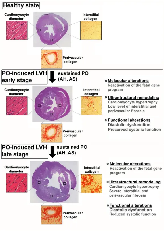
Figure 1. Summary of pressure overload-induced pathological myocardial remodeling. The early stage of pressure overload (PO)-induced left ventricular myocardial hypertrophy (LVH) is associated with cardiomyocyte hypertrophy and low level of interstitial and perivascular fibrosis on the ultrastructural level. At this stage, diastolic dysfunction is often present, however systolic function is preserved. At later stages, further accumulation of interstitial and perivascular collagen occurs. In addition, systolic function also deteriorates. AH: arterial hypertension, AS: aortic stenosis, PO: pressure overload. Cross sectional whole heart pictures are adapted from Ruppert et al. (2018) Front Physiol, 9:1869.(68)
17
2.3. Pressure unloading-induced myocardial reverse remodeling
In recent years, a growing body of clinical data has provided evidence that termination of the pathological insult of sustained PO (e.g. by AVR/TAVI in case of AS or by antihypertensive medications in case of AH) induces the regression of LVH. This process is termed myocardial reverse remodeling and involves the restoration of the physiological LV phenotype on the molecular, ultrastructural, geometrical as well as functional level (69). In the following sections, different aspects of pressure unloading-induced myocardial reverse remodeling are summarized.
2.3.1. Pressure unloading-induced structural alterations 2.3.1.1. Regression of myocardial hypertrophy
Clinical and experimental studies have shown that myocardial hypertrophy remains reversible even after a long period of sustained PO (70, 71). Correspondingly, substantial reduction in LV mass has been documented after different types of pressure unloading therapies. Furthermore, the regression of myocardial hypertrophy after TAVI/AVR or antihypertensive therapy has been found to follow a characteristic time course. Based on these reports, termination of the pathological stimulus of sustained PO leads to a rapid regression of LVH (6, 72-78). Of particular interest is the observation that the majority of LV mass reduction (approximately two-thirds of the total mass regression) occurs during this early phase (within the first 18 months). The initial phase is subsequently followed by a second more chronic phase, which continues over several years after the correction of the primary hemodynamic abnormality (73, 79).
Beyond the clinical investigations, animal models have been also utilized to investigate the temporal regression of LVH under laboratory conditions. These preclinical studies have reported that decrement in myocardial hypertrophy takes place already within 3 days of PO termination (80, 81). Furthermore, data from animal experiments have confirmed that the prompt regression of LVH is followed by an additional chronic phase under standard laboratory circumstances as well (70).
Furthermore, numerous studies have sought to determine the extent to which the hypertrophied myocardium may recover after a successful pressure unloading therapy.
These investigations have found that LV mass almost returns to the healthy controls’ level
18
in case of a long follow-up period after AVR (72, 73). Nevertheless, it has to be noted that a “restitutio ad integrum” generally does not occur. One possible explanation for the incomplete regression of LVH is that pressure unloading therapies often fail to completely alleviate the increased PO. For example, in case of AVR, the prosthetic valve itself may create a slight stenosis compared to the absolutely healthy aortic valves (73). Therefore, a relatively low level of PO may persist even after successful surgical interventions. The residual increment in PO might impede the complete normalization of myocardial hypertrophy.
In addition to the macroscopic findings, ample amount of data has provided evidence that reduced cardiomyocyte size (assessed by myocardial fiber diameter or cross sectional area) is mainly responsible for the decrement of LV mass on the microscopic level. This observation is corroborated by the analysis of endomyocardial biopsies obtained from AS patients who underwent AVR (72). Moreover, the detailed histopathological analysis of the whole LV myocardium in different animal models has come to the same conclusion (82, 83).
2.3.1.2. Regression of myocardial fibrosis
Regression of myocardial fibrosis after pressure unloading is generally considered to be a less efficient process compared to the regression of cardiomyocyte hypertrophy.
Importantly, as previously mentioned, myocardial fibrosis can be subcategorized into replacement and reactive (including perivascular and interstitial) fibrosis. Recent investigations have come to the conclusion that these two forms of myocardial fibrosis differ in their plasticity.
Replacement fibrosis. In vivo assessment of myocardial replacement fibrosis (also known as focal fibrosis) is carried out by late gadolinium enhancement (LGE) cardiovascular magnetic resonance imaging (CMR) (84). Thus, majority of the available clinical research data concerning the regression of replacement fibrosis after pressure unloading therapy is derived from LGE CMR reports. These studies have revealed that replacement fibrosis persists after AVR (6, 30, 85). The irreversible nature of replacement fibrosis stands in parallel with the current concept of its development. Accordingly, as also mentioned above, replacement fibrosis is believed to substitute for the place of dead
19
cardiac cells. Considering that cardiomyocytes have very low proliferative ability, the permanent nature of replacement fibrosis after pressure unloading is an expected finding.
Reactive interstitial fibrosis. The non-invasive, in vivo evaluation of interstitial fibrosis has not been feasible for a long period of time. Nevertheless, recent advances in CMR imaging technology has made the quantification of interstitial fibrosis possible (75, 86). Data from current CMR studies along with prior endomyocardial biopsies have revealed that interstitial fibrosis is plastic and able to regress on the long term (6, 85, 87).
However, the time course of interstitial fibrosis regression has been reported to be much slower than that of cardiomyocyte hypertrophy. As a consequence, at a relatively early time point after pressure unloading, when a substantial reduction in cardiomyocyte size (diameter/cross sectional area) has already taken place, the amount of interstitial fibrotic tissue often remains unchanged (88, 89). Accordingly, the percent of interstitial fibrosis has been reported to even increase at an intermediate stage of myocardial reverse remodeling (64, 65). In contrast, at later stages (8-10 years after AVR) the amount of extracellular collagen decreases, and hence the percentage of interstitial fibrosis reduces as well (65). However, the extent to which interstitial fibrosis regresses after pressure unloading therapy is still under intense investigation. Results from experimental studies indicate that complete regression of pre-established interstitial fibrosis does not occur, even in the long term follow-up (90).
Reactive perivascular fibrosis. Currently, detection of perivascular fibrosis is only accomplished by histological analysis. However, the possibility for carrying out myocardial biopsies in AS patients after pressure unloading therapy is limited. Hence, our knowledge concerning the reversibility of perivascular fibrosis is mainly derived from animal studies. These investigations have revealed that perivascular fibrosis displays similar regression to interstitial fibrosis after pressure unloading (91).
2.3.2. Pressure unloading-induced molecular alterations
Due to the fact that LV mRNA/protein expression analysis requires myocardial tissue collection, molecular features of myocardial reverse remodeling has been mainly investigated in relevant animal models. These preclinical experiments have provided compelling evidence that the regression of LVH after pressure unloading therapy is accompanied by robustly reduced fetal gene expression (70, 80). Interestingly, prior
20
research has also indicated that normalization of the fetal gene program might even precede the structural and functional recovery of the hypertrophied and failing myocardium (92).
Furthermore, recent findings have called attention to the fact that those molecular alterations that occur during myocardial reverse remodeling are not merely the reverse of those that cause LVH and HF (92). Correspondingly, characteristically different gene expression profiles have been observed during PO-induced remodeling and pressure unloading-evoked reverse remodeling (93). Hence, it seems possible that from a molecular point of view the reverse-remodeled heart is not simply the halfway mark between PO-induced LVH and the healthy state, but it rather represents a distinct third category.
2.3.3. Pressure unloading-induced functional alterations
2.3.3.1. Recovery of left ventricular systolic function after pressure unloading Most of the clinical studies have found that removal of the chronic PO induces a rapid improvement in systolic function (94-96). Correspondingly, it has been suggested that enhancement of LV systolic performance after pressure unloading therapy does not necessarily require a complete normalization of LV structural abnormalities (especially fibrosis) (70). These observations raise the possibility that the termination of the increased afterload might be sufficient to achieve a prompt improvement in LV systolic function without substantial changes in LV contractility. However, a common limiting factor for the above mentioned investigations is that LV systolic function has been assessed only by load-dependent methods. Hence, more sophisticated hemodynamic characterization by P-V analysis is warranted to gain a deeper insight into the rapid recovery of LV systolic function after pressure unloading.
2.3.3.2. Recovery of left ventricular diastolic function after pressure unloading Contrary to systolic dysfunction, amelioration of PO-induced diastolic dysfunction occurs in a slow fashion and usually continues over several years following pressure unloading. Interestingly, clinical experience suggests that the two aspects of diastolic function, namely passive filling and active relaxation, differ in their reversibility after pressure unloading (65).
21
Passive filling. Villari et al. have found that myocardial stiffness follows very precisely the changes of interstitial fibrosis in patients with AS undergoing pressure unloading therapy (64). As previously mentioned, the percent of interstitial fibrosis may even be increased at a relatively early time point after AVR. At this particular stage, Villari et al. have observed that myocardial stiffness was also further impaired. In contrast, at later stages (8-10 years after AVR) the resorption of interstitial fibrosis implied the normalization of myocardial stiffness (64, 65).
Active relaxation. On the contrary, in the same investigations a significant improvement in the active relaxation time constant (Tau) was documented already at an intermediate time point after pressure unloading therapy (64). Furthermore, a rapid (2 weeks after surgery) recovery in active relaxation was also demonstrated by Ikonodimis et al. Of particular interest, in the later echocardiographic follow-up study, patients undergoing AVR had experienced a significant improvement in myocardial relaxation early after surgery, at a time point when regression of LV wall thicknesses has not yet occurred (79).
These clinical findings suggest that improvement of passive filling is immensely dependent on the regression of structural abnormalities (mainly fibrosis). On the other hand, clinical data indicate that normalization of active relaxation might require less structural reverse remodeling. However, experimental results are not available to support these phenomena since the effect of complete versus incomplete structural reverse remodeling on diastolic function has not yet been investigated under experimental conditions.
22
Figure 2. Summary of pressure unloading-induced myocardial reverse remodeling. Pressure unloading induces a rapid regression of cardiomyocyte hypertrophy leading to substantial reduction in left ventricular mass. However, reactive interstitial and perivascular myocardial fibrosis often persists at a relatively early stage after pressure unloading. From a functional point of view, the early phase of reverse remodeling is characterized by improved systolic function but impaired diastolic function. In contrast, at later stages, reactive myocardial fibrosis also regresses, resulting in the amelioration of diastolic function. AVR: aortic valve replacement, TAVI:
transcatheter aortic valve implantation. Cross sectional whole heart pictures are adapted from Ruppert et al. (2018) Front Physiol, 9:1869.(68)
23
2.4. Sex differences in pressure overload-induced pathological remodeling and pressure unloading-induced reverse remodeling
Sex differences have been increasingly recognized in various frequent cardiovascular disorders, such as in IHD and hemodynamic overload-induced HF. Importantly, sex- related discrepancies have been found to affect both the pathophysiology, the clinical manifestation and the epidemiology of cardiovascular pathologies (97, 98). Recent investigations have shed light on the molecular basis, which might be responsible for the observed differences between men and women. These subcellular mechanisms involve sex chromosomes-derived genetic differences, diversity in epigenetic regulation (due to characteristic microRNA expression) and the effects of different sex steroid hormones (98). Although, all of the three proposed mechanisms may be involved, the majority of previous investigations have focused on the role of sex steroid hormones (mainly estrogen). The observation, which drew attention to the potential cardioprotective effects of estrogen was that differences associated with female sex disappear in women at the postmenopausal stage. In good agreement with the clinical findings, preclinical studies have reported that sex differences in hemodynamic overload-induced HF diminish in female rats after ovariectomy (99). In contrast, exogenous estrogen administration in ovariectomized female rats restores the characteristic sex-related differences in PO- induced HF (100). Further research has also identified that the regulatory effects of estrogen are mainly mediated via the nuclear estrogen receptor β (ER-β) in PO-evoked LVH (101-103). The interaction between estrogen and ER-β induces alterations in myocardial gene expression, which fundamentally affects the hypertrophied LV phenotypes in females. In the following sections, sex-related differences in PO-induced LVH are briefly summarized.
2.4.1. Effect of sex on structural alterations
2.4.1.1. Sex-associated differences in myocardial hypertrophy
Women with long-lasting AH or AS, as well as female rats with sustained PO are prone to develop thicker ventricular walls, smaller LV chamber dimensions and a more concentric form of LVH compared to male counterparts (104-109). Recent experiments have proposed that the characteristic morphological and geometrical differences might be the consequence of a set of pathological cellular processes which occur in males but not
24
in females during the progression of PO-induced LVH. Accordingly, relatively early after PO-induction, concentric LVH is observed in both genders. However, during the progression of chronic cardiovascular conditions, continuous cardiac cell loss and ECM remodeling takes place in males but not in females, resulting in chamber dilatation and eccentric geometry (110, 111). In contrast, female sex and estrogen-mediated signaling yield protection against apoptosis and MMP upregulation (111, 112), and prevents chamber dilation.
Recent clinical experiments also aimed at investigating potential sex-related discrepancies in the regression of LVH after pressure unloading therapy. Petrov and his colleagues found that LV end-diastolic diameter (LVEDD) decreased in female but not in male patients 3 days after AVR (111). In contrast, Dobson et al. have reported no sex- related differences in LV mass regression during a 6-month long follow-up period (76).
Similarly, Stangl et al. have also observed that reduction of LV mass occurred to a similar extent in both genders 3 month after AVR/TAVI (113). In contrast, antihypertensive therapies have demonstrated a less efficient regression of LV mass in female patients compared to their male counterparts (108, 114). Unfortunately, no animal studies to date have been conducted on this field to confirm or contradict the mentioned clinical findings.
2.4.1.2. Sex-associated differences in myocardial fibrosis
Sex differences in myocardial fibrosis have been widely documented by numerous preclinical and clinical investigations during the development of PO-induced LVH.
Accordingly, examination of endomyocardial biopsies from AS patients have consistently revealed more severe collagen accumulation in men compared to women (109, 115). The histological findings have been further supported by gene expressional analysis, reporting substantially higher mRNA levels of pro-fibrotic genes in LV speciments from male AS patients compared to their female counterparts (111). Besides the histological examinations, current CMR studies have reported considerable differences in myocardial fibrosis between men and women under sustained PO. Importantly, in these particular studies, the newly developed CMR imaging modalities have allowed investigators to differentiate between replacement and reactive fibrosis. The separate quantification of replacement and reactive interstitial fibrosis has revealed that both types of myocardial fibrosis are increased to a greater extent in men compared to women despite an equal
25
increment in afterload (116). Of particular interest, animal models of LV PO have also confirmed that male sex is associated with more maladaptive ECM remodeling under standard laboratory conditions (101, 102).
To date, only a few studies have analyzed the potential influential effect of sex on the regression of myocardial fibrosis following pressure unloading. Among them, Dobson et al. has reported that myocardial fibrosis regressed only in female but not in male patients after AVR during a 6 month long follow-up period (76). However, further preclinical research is warranted to test whether female sex is indeed associated with a more plastic form of myocardial fibrosis.
2.4.2. Effect of sex on molecular alterations
The adaptation of the LV to chronic PO seems to differ on the molecular level as well between the two genders. Accordingly, in a TAC-induced pathological LVH model, PO evoked more severe upregulation of the fetal gene program (β-MHC and ANP) in male rats compared to their female counterparts (117). Furthermore, detailed analysis of the LV transcriptome has identified specific sex-dependent regulation of gene expression.
These investigations have revealed that the transcription of genes associated with mitochondrial function and fatty acid oxidation were more robustly suppressed in males compared to females. In contrast, genes encoded ribosomal proteins and ECM components were more intensely upregulated in male rodents compared to female ones (118).
2.4.3. Effect of sex on left ventricular function
2.4.3.1. Sex-associated differences in left ventricular systolic function
Sex is a major determinant of LV systolic function during the development of PO- induced LVH. Accordingly, better EF and fractional shortening have been consistently reported in female patients with chronic AH or AS compared to male patients (105, 106, 108, 109, 119). Furthermore, it has been also suggested that females might experience a greater improvement in EF after pressure unloading compared to their male counterparts (113). Besides the clinical findings, animal models of PO-induced LVH have confirmed that following a common compensated stage, deterioration of LV systolic function occurs in male but not in female rodents under standard laboratory conditions (104, 117).
26
However, a major limitation of the beforementioned clinical and preclinical investigations is that none have assessed in vivo LV contractility independently from loading conditions.
2.4.3.2. Sex-associated differences in left ventricular diastolic function
Besides the prominent sex-related differences in LV systolic function, hemodynamic characterization of male and female AS patients have indicated that distinct discrepancies might exist in diastolic function as well.
Active relaxation. Villari and his colleagues have observed the prolongation of the active relaxation time constant (Tau) in both genders with chronic PO (109). According to their findings, deterioration of active ventricular relaxation occurred to an equal extent in both men and women.
Passive filling. In contrast to active relaxation, major differences have been documented in passive filling between the two sexes. Correspondingly, impairment of myocardial stiffness was only detected in men but not in women patients with AS (109).
Furthermore, LVEDP was found to be elevated in male rodents, while no significant alteration could be detected in LVEDP in their female counterparts (101, 104). Thus, clinical and experimental evidence suggests that male sex is associated with a more severe impairment of LV passive diastolic properties under sustained PO. This latter functional finding is in good agreement with the ultrastructural differences between the two genders, with more severe ECM remodeling having been reported in male patients and experimental animals.
27
2.5. Animal models of pressure overload-induced remodeling and pressure unloading-evoked reverse remodeling
In the past years, various animal models of PO-induced pathological remodeling have been introduced. Among them, aortic constriction-induced PO (by surgically constricting the ascending [AAC], the transverse [TAC] or the abdominal [AB] aorta), essential hypertension-induced PO (e.g. the SHR strain or the Dahl salt sensitive rat strain), renal hypertension-induced PO (e.g. 5/6 nephrectomy) and genetically modified rodent strains (e.g. rats overexpressing the mouse renin gene) are the most widely utilized (120-123).
All of these experimental approaches have proven to be valuable tools in studying the temporal development of PO-induced LVH and its progression to symptomatic HF.
Nevertheless, surgical models have the advantage that the pathological trigger of PO (the aortic constriction) can be removed by a second minimal invasive operation. Therefore, these models provide a unique opportunity to study not only PO-induced remodeling but also pressure unloading-evoked reverse remodeling under standard laboratory conditions.
Importantly, prior investigation utilizing AB and debanding rodent models have reported that these experimental approaches reliably recapitulate those alterations that occur in human patients after pressure unloading therapy (68, 70, 80, 83, 88, 89, 92, 124-128).
Hence, these particular animal models could indeed serve as an adequate tool to address those scientific questions regarding myocardial reverse remodeling that cannot be investigated in a clinical scenario.
28
3. Objective
A considerable body of clinical evidence has accumulated to support the notion that in case of PO-induced HF, termination of the pathological stimulus (by antihypertensive medications in case of AH or AVR/TAVI in case of AS) could results in a state of reverse remodeling with substantial improvement in LV structure and function. Nevertheless, it has been also recognized that not all the patients experience the same extent of functional and structural improvement following pressure unloading therapy. Hence, efforts have been taken to identify factors which might influence the regression of LVH. These recent studies have raised the possibility that the time point of medical interventions (performing pressure unloading at early versus late stages of PO-induced LVH) and the sex of the patients might determine the success of the reverse remodeling process (70, 76, 92, 111).
Considering that many confounding factors (differences in comorbidities, age and medications) can limit the overall impact of the clinical observations, further experimental studies utilizing animal models are warranted.
Based upon that, the aims of the present studies were:
1. To establish a rat modelof mechanical PO-induced pathological remodeling and pressure unloading-evoked reverse remodeling with microsurgical techniques.
2. To explore timeline alterations in LV structure and function during the development of PO-induced LVH and its progression to HF in male rats.
3. To investigate the effects of myocardial reverse remodeling from early versus late stages of PO-induced LVH in male rats.
4. To study the effect of sex on different aspects of LV structure and function during the development of PO-induced LVH.
29
4. Methods
4.1. Animals
The investigation conformed to the EU Directive 2010/63/EU and the Guide for the Care and Use of Laboratory Animals used by the US National Institutes of Health (NIH Publication No.85–23, revised 1996). The experiments were approved by the ethics committee of the Land Baden-Württemberg for Animal Experimentation (G-94/15 and G-198/16). The studies were interpreted in accordance with the ARRIVE (Animals in Research: Reporting in Vivo Experiments) guidelines (129). The animals were kept under standard conditions (22±2ºC with 12h light/dark cycles) and were allowed access to laboratory rat diet and water ad libitum during the whole experimental period. Before performing any type of surgeries, the rats underwent a one-week long acclimatization period.
4.2. Animal models
4.2.1. Rat model of pressure overload-induced myocardial remodeling
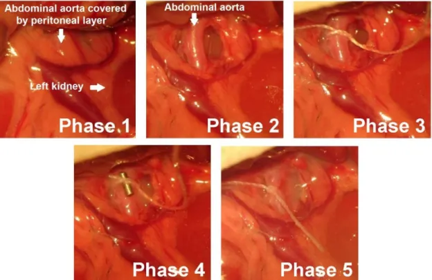
In order to induce PO of the LV, the previously described method of AB was established in our laboratory with slight modifications (61). In brief, the surgery consisted of the following steps. General anesthesia was induced in male and female Sprague- Dawley rats (5-6 weeks-old; 160-180g; Janvier, France) with 5% of isoflurane gas in a chamber and maintained by inhalation from a connected tube with 1.5–2% of isoflurane in O2. The animals were placed in a supine position on an automatic heating pad to maintain the core temperature at 37°C, measured via a rectal probe. The abdominal hair was removed and an antiseptic solution was applied to disinfect the skin on the surgical area. The surgery was performed in midline laparotomy under sterile conditions. After the intestinal tract was gently placed aside, the peritoneal layer was dissected in order to gain access to the retroperitoneal space. The abdominal aorta between the right renal artery and the superior mesenteric artery was carefully cleaned from the surrounding connective tissue. A blunted 22-gauge needle was utilized to constrict the aorta above the right renal artery. After AB was completed, the intestines were placed back to the abdominal cavity and the abdominal muscle layer was sutured in single interrupted fashion. Finally, the skin wound was closed by applying surgical clips. Following surgery,
30
analgesia was provided by subcutaneously administered buprenorphine in the dose of 0.05mg/kg. Furthermore, 1ml physiological saline was also applied subcutaneously to avoid post-operative hypovolemia. Sham-operated animals were subjected to the same surgical procedure, except the aortic constriction (n=28).
Figure 3. Abdominal aortic banding. Photos were taken at different phases during the surgical procedure of abdominal aortic banding. Phase 1: After a midline laparotomy, the intestines were taken aside and the peritoneal layer was visualized. Phase 2: By dissecting the peritoneal layer the abdominal aorta was identified. Phase 3: A 2.0 silk suture was placed around the abdominal aorta at the suprarenal level. Phase 4: A blunted 22-gauge needle was taken between the anterior surface of the aorta and the ligature. Phase 5: The ligature around both the needle and the aorta was tied. Finally, the needle was carefully removed, leaving a constriction on the abdominal aorta with a size of the external diameter of a 22-gauge needle.
4.2.2. Rat model of pressure unloading-induced reverse myocardial remodeling After 6 weeks of AB (early debanding) or 12 weeks of AB (late debanding) a group of animals underwent a second minimal invasive surgery, when the aortic constriction was removed from the abdominal aorta (debanding). During this second surgery, general anesthesia was provided by isoflurane inhalation (5% for induction of anesthesia and 2- 3% for maintenance of anesthesia). Rats were placed on a heating pad in a supine position.
The surgical area was cleaned by removing the abdominal hair and disinfecting with an
31
antiseptic solution. A midline laparotomy was performed in the line of the scar from the previous surgery. The intestines were then localized in a way, that their position allowed us to visualize the abdominal aorta. The previously placed aortic constriction was identified and it was cleaned from the surrounding fibrotic tissue. The narrowing suture was carefully cut and removed from the abdominal aorta. The abdominal muscle and the skin layer were sutured in the single interrupted fashion. To prevent postoperative pain and dehydration, buprenorphine (in the dose of 0.05mg/kg) and physiological saline (1.5ml) was injected subcutaneously before the animals regained awareness.
4.3. Study protocols
The study protocols are summarized in Figure 4, Figure 5 and Figure 6.
4.3.1. Study 1: Longitudinal assessment of pressure overload-induced structural and functional alterations of the left ventricle in male rats
In the AB model, LV PO is evoked to a moderate level, which results in a relatively slow disease progression. Based on previous literature data, the time points of week 6, 12 and 18 were chosen to detect characteristic alterations at early and advanced stages of PO-induced HF. Accordingly, after AB or sham operations, the rats were randomized into the following 6 experimental groups:
Sham-wk6 group (n=9): after sham operation the rats were followed-up for 6 weeks;
AB-wk6 group (n=13): after AB the rats were followed-up for 6 weeks;
Sham-wk12 group (n=9): after sham operation the rats were followed-up for 12 weeks;
AB-wk12 group (n=13): after AB the rats were followed-up for 12 weeks;
Sham-wk18 group (n=10): after sham operation the rats were followed-up for 18 weeks;
AB-wk18 group (n=13): after AB the rats were followed-up for 18 weeks, respectively.
32
Figure 4. Schematic illustration of the experimental protocol of Study 1. At baseline, the rats underwent abdominal aortic banding ([AB]; n=39) or sham operation (n=28) and were subsequently randomized into 6 groups. In the Sham-wk6 (n=9) and AB-wk6 (n=13) groups, pressure-volume (P-V) analysis and tissue collection were performed 6 weeks after AB/sham operations. In the Sham-wk12 (n=9) and AB-w12 (n=13) groups, P-V analysis and tissue collection were performed 12 weeks after AB/sham operations. In the Sham-wk18 (n=10) and AB- w18 (n=13) groups, P-V analysis and tissue collection were performed 18 weeks after AB/sham operations. Furthermore, repetitive echocardiographic measurements at baseline, week 3, 6, 9, 12, 15 and 18 were also carried out in the Sham-wk18 and AB-wk18 groups.
4.3.2. Study 2: Investigating the effects of myocardial reverse remodeling from early- versus late-stage left ventricular hypertrophy in male rats
Similar to Study 1, the AB model was utilized to induce different stages of PO-induced LVH in Study 2 as well. However, in this investigation two additional groups were introduced to study the regression of LVH from early and late stages of LVH. In these experimental groups, a second minimal invasive surgery was performed to remove the aortic constriction at week 6 (early debanded) and week 12 (late debanded). Both debanded groups were followed up for 6 weeks. Accordingly, Study 2 consisted of the following 8 groups:
Sham-wk6 group (n=10): after sham operation the rats were followed-up for 6 weeks;
AB-wk6 group (n=10): after AB the rats were followed-up for 6 weeks;
Sham-wk12 group (n=10): after sham operation the rats were followed-up for 12 weeks;
33
AB-wk12 group (n=11): after AB the rats were followed-up for 12 weeks;
Sham-wk18 group (n=9): after sham operation the rats were followed-up for 18 weeks;
AB-wk18 group (n=13): after AB the rats were followed-up for 18 weeks, respectively.
Early debanded group (n=14); these rats underwent AB operation, after week 6 the banding suture was removed and the rats were followed up until week 12;
Late debanded group (n=15): these rats underwent AB operation, after week 12 the banding suture was removed and the rats were followed up until week 18.
Figure 5. Schematic illustration of the experimental protocol of Study 2. At baseline, the rats underwent abdominal aortic banding ([AB]; n=63) or sham operation (n=29) and were subsequently randomized into 8 groups. The Sham-wk6 (n=10), AB-wk6 (n=10) groups were followed-up for 6 weeks. The Sham-wk12 (n=10), AB-wk12 (n=11) groups were followed-up for 12 weeks, while the Sham-wk18 (n=9), AB-wk18 (n=13) were followed-up for 18 weeks. In the early debanded group (n=14) the aortic constriction was removed after week 6, while in the late debanded group (n=15) the narrowing suture was removed after week 12.
34
4.3.3. Study 3: Investigation of sex differences during the development of pressure overload-induced left ventricular hypertrophy.
In this particular study, male and female rats underwent AB to investigate sex-related differences in the adaptation to chronic PO. Age- and sex-matched sham operated animals served as controls. Considering that sex differences were suggested to develop by time, functional and structural characterization of PO-induced LVH was carried out at a relatively early (week 6) and late (week 12) time points as well after PO induction.
Accordingly, the following experimental groups were used in Study 3:
Male sham-week6 group (n=8): male rats underwent sham operation and were followed- up for 6 weeks
Male AB-week6 group (n=8): male rats underwent AB and were followed-up for 6 weeks;
Female sham-week 6 group: female rats underwent sham operation and were followed- up for 6 weeks
Female AB week 6 group (n=7): female rats underwent AB and were followed-up for 6 weeks;
Male sham week 12 group (n=8): male rats underwent sham operation and were followed- up for 12 weeks;
Male AB week 12 group (n=10): male rats underwent AB and were followed-up for 12 weeks;
Female sham week 12 group (n=8): female rats underwent sham operation and were followed-up for 12 weeks;
Female AB week 12 group (n=7): female rats underwent AB and were followed-up for 12 weeks.