Transplantation Facilitates Aortic Stiffening
Bernadett Bordaa, György Lázára, Árpád Kormányosb, Péter Domsikb, Anita Kalaposb, Edit Szederkényia, Csaba Lengyelc, Tamás Várkonyic, Csilla Keresztesd, Tamás Forsterb, and Attila Nemesb,*
aDepartment of Surgery, Faculty of Medicine, Albert Szent-Györgyi Clinical Center, University of Szeged, Szeged, Hungary;b2nd Department of Medicine and Cardiology Centre, Faculty of Medicine, Albert Szent-Györgyi Clinical Center, University of Szeged, Szeged, Hungary;c1st Department of Medicine, Faculty of Medicine, Albert Szent-Györgyi Clinical Center, University of Szeged, Szeged, Hungary; anddDepartment for Medical Translation and Communication, Faculty of Medicine, Albert Szent-Györgyi Clinical Center, University of Szeged, Szeged, Hungary
ABSTRACT
Background. Following kidney transplantation (KT), new-onset diabetes mellitus (NODM) is one of the most common complications. NODM usually occurs early after KT, and is diagnosed according to the general guidelines relevant for general diabetes mellitus patients. Arterial stiffness is a surrogate marker of cardiovascular risk.
According to the literature, a successful KT has only limited and late beneficial effects on aortic elastic properties. The present study aimed to assess whether NODM has any additive value on the worsening of echocardiography-derived aortic elastic properties in transplanted patients.
Methods. We have included 28 nondiabetic post-KT patients in the study, older than 18 years (mean age: 48.2 6.9 years; 13 men, 15 women). After an oral glucose tolerance test, 8 patients were diagnosed with NODM, and their results were compared to 23 age-, sex-, and risk factor-matched controls (mean age: 54.911.0 years; 9 men, 14 women). All post-KT patients and matched controls underwent a complete transthoracic 2-dimensional Doppler echocardiography, together with an assessment of echocardiographic aortic elastic properties. The assessments included aortic strain, aortic distensibility, and aortic stiffness index.
Results. Aortic elastic properties showed alterations in post-KT patients compared to matched controls (aortic strain: .084.039 vs .057.032,P < .05; aortic distensibility:
2.361.09 cm2/dynes 10e6vs 1.831.18 cm2/dynes 10e6,P¼.07; aortic stiffness index:
7.15 3.58 vs 11.2 6.1, P < .05). Further deterioration in the aortic stiffness index (14.87.6 vs 9.684.88,P< .05) was detected in the presence of NODM.
Conclusions. NODM following successful KT facilitates aortic stiffening.
N
EW-ONSET diabetes mellitus (NODM) is one of the most common complications following kidney trans- plantation (KT). Untreated NODM negatively influences allograft function and further increases the risk of cardio- vascular diseases[1]. This issue was addressed in the 2003 Consensus Guidelines developed by the American Diabetes Association. A diagnosis of NODM is a threat to the renal allograft, as well as carrying the same short- and long-term implications of type 2 diabetes mellitus (DM) seen in the general population. NODM usually occurs early after KT,and is usually diagnosed according to the guidelines relevant to general diabetic patients. According to this, NODM is proven if the fasting blood glucose level is7 mmol/L, or,
*Address correspondence to Attila Nemes, MD, PhD, DSc, FESC, 2nd Department of Medicine and Cardiology Center, Medical Faculty, Albert Szent-Györgyi Clinical Center, University of Szeged, H-6725 Szeged, Semmelweis Street 8, Hungary, P.O.
Box 427. Tel: þ36-62-545220; Fax: þ36-62-544568. E-mail:
nemes.attila@med.u-szeged.hu ª2019 The Authors. Published by Elsevier Inc. This is an open
access article under the CC BY-NC-ND license (http://
creativecommons.org/licenses/by-nc-nd/4.0/).
230 Park Avenue, New York, NY 10169
0041-1345/19 https://doi.org/10.1016/j.transproceed.2019.04.009
Transplantation Proceedings,51, 1239e1243 (2019) 1239
in the case of a properly administered oral glucose tolerance test, a blood glucose level of11.1 mmol/L 2 hours after the oral administration of 75 g glucose [2]. The general principle behind standardized NODM incidence reporting is that the diagnostic criteria should reflect what is used in the general population[3]. Hopefully, these developments will allow for a more consistent reporting of NODM in the future, leading to a more exact estimation of the incidence.
Arterial stiffness is a surrogate marker of cardiovascular risk[4]. According to the literature, KT has only limited and late beneficial effects on aortic elastic properties [5]. Our study aimed to assess whether NODM has any additional beneficial effects on the worsening of echocardiography- derived aortic elastic properties in KT patients.
PATIENTS AND METHODS
Our study was carried out at the Department of Surgery, University of Szeged, Szeged, Hungary. It included 28 patients who had un- dergone a cadaver KT within at least 1 year. All patients were above the age of 18 years (mean age: 48.26.9 y; 13 men, 15 women).
Eight patients were diagnosed with NODM by oral glucose toler- ance test. Their results were compared to those of 23 control sub- jects (mean age: 54.911.0 y; 9 men, 14 women), matched with regard to age, sex, and risk factors. Both the transplant patients and the matched controls underwent a complete transthoracic 2- dimensional (2D) Doppler echocardiography, extended by the assessment of echocardiographic aortic elastic properties, which included aortic strain, aortic distensibility, and aortic stiffness index (ASI)[6]. Neither the KT patients nor the controls had experienced a cardiovascular event in their medical history or had cardiovascular symptoms at the time of examination. DM was defined according to the American Diabetes Association and World Health Organiza- tion criteria[7,8]. Hypertension was defined as either a systolic or a diastolic elevation of blood pressure (>140/90 mm Hg). Hyper- cholesterolemia was defined as a total cholesterol level of >5.0 mmol/L or current treatment with lipid-lowering medications (flu- vastatin). Informed consent was obtained from each KT patient and control subject. The institutional human research committee approved the protocol, which conformed with the ethical guidelines of the 1975 Declaration of Helsinki.
Biochemical Measurements
Blood samples were drawn by venipuncture to assess routine blood parameters after 8 hours of fasting.
Immunosuppressive Therapy
The following immunosuppressive treatment was used in post-KT patients according to the guidelines:
Tacrolimus (Tac) is a macrolide molecule that has strong lipophilicity. The initial daily regimen of Tac was 2 doses of .10 mg/kg per day; the target blood trough level was 10e15 ng/mL for 6 weeks, and 5e10 ng/mL after week 6.
Cyclosporin-A (CsA) is a lipophilic, hydrophobic, cyclic peptide composed of 11 amino acids. The initial regimen of CsA was 2 doses of 4e5 mg/kg per day; the target blood level was 1300e1600 ng/mL in month 1, 900e1300 ng/mL in months 2 and 3, 750e950 ng/mL in months 4e6, and 700 ng/mL afterwards (blood levels were determined).
A steroid was administered intravenously (500 mg) on day 0 in the operating room, immediately before restoring bloodflow to the graft; 125 mg was administered intravenously on day 1, 32 mg orally on day 2, 24 mg orally on day 3, 16 mg orally on days 4e28, 12 mg orally each day during month 2, 8 mg orally each day during month 3, and 4 mg orally during months 4e6. The dose was further decreased in problem-free cases.
Mycophenolate mofetil was given in two 1 g doses per day, and was reduced by 50% or 75% in the event of gastrointestinal symptoms or leucopenia.
Blood Pressure Measurement
Systolic and diastolic blood pressures (SBP and DBP, respectively) were measured with a sphygmomanometer using an inflatable cuff and a mercury manometer positioned on the left arm in supine position after 10 min of rest. Appearance of a tone (first) and disappearance (fifth) of Korotkoff sounds corresponded with SBP and DBP. No stimulant consumption (caffeine) was allowed for 30 minutes prior to the blood pressure measurements. Three consec- utive blood pressure measurements were averaged and used as blood pressure data[9].
2D Doppler Echocardiography
Transthoracic imaging and measurements were carried out using a Toshiba Artida echocardiography system (Toshiba Medical Systems, Tokyo, Japan) with a 1e5 MHz PST-30SBP phased-array transducer.
While in the left lateral decubitus position, all patients underwent a complete 2D Doppler echocardiographic study. Left ventricular di- ameters, volumes and ejection fractions, left atrial diameter, and aortic dimensions respecting the cardiac cycle were measured ac- cording to the recent guidelines[10]. All echocardiographic mea- surements were averaged from 3 beats. The degree of valvular regurgitation was visually quantified, and valvular stenosis was excluded by color Doppler echocardiography according to clinical practice. Mitral inflow E/A was measured by pulsed Doppler.
Assessment of Echocardiographic Aortic Elastic Properties Systolic and diastolic ascending aortic diameters (SD and DD, respectively) were recorded during M-mode echocardiography at a level of 3 cm above the aortic valve in the parasternal long-axis view according to the literature[9]. SD and DD were measured at the time of maximum aortic anterior motion and at the peak of the QRS complex, respectively. The following aortic elastic indices were calculated:
Aortic strain¼(SDeDD) / DD
Aortic stiffness index (b) ¼ ln (SBP / DBP) / [(SD-DD) / DD], where ln is the natural logarithm.
Aortic distensibility¼2 x (SD-DD) / [(SBP-DBP)DD].
Statistical Analysis
Continuous variables are expressed as meanstandard deviation, whereas dichotomous variables are expressed as absolute and relative frequencies (percentages). Continuous variables were compared with the unpaired Studentttest, while categorical vari- ables were examined using the c2 test and Fisher exact test. A Pearson correlation coefficient was calculated featuring the nu- merical correlation. Statistical tests were 2-sided, and a Pvalue
<.05 was considered to be statistically significant. Statistical ana- lyses were carried out by using MedCalc software (MedCalc, Ostend, Belgium).
RESULTS
Clinical Data, Medications and Laboratory Findings
There were 20 patients in the non-NODM group and 8 patients in the NODM group. The clinical data and subse- quent medications are presented in Table 1. All post-KT patients received a triple combination that consisted of calcineurin inhibitor (23 patients Tac, 5 patients CsA), mycophenolate mofetil, and steroids. Only 5 of the 8 NODM patients required antidiabetic treatment: 3 of them were put on insulin, and 2 were administered oral antidia- betics. Three patients were not given any treatment, as their abnormal glucose metabolism was identified only during the examination. All post-KT patients received antihypertensive treatment with double (calcium channel blocker and angiotensin-converting enzyme inhibitors) or triple combi- nation (calcium channel blocker, angiotensin-converting enzyme inhibitors, andbblocker). The results of the labo- ratory tests showed higher serum urea (17.410.4 mmol/L vs 8.6 2.9 mmol/L, P< .05), creatinine (209.6 104.2 mmol/L vs 121.7 30.1mmol/L, P< .05), fasting glucose (7.61.5 mmol/L vs 5.5.7 mmol/L,P< .05), and HbA1c (6.9 .8% vs 5.7 .4%, P < .05), as well as a lower
estimated glomerularfiltration rate (35.323.9 vs 57.3 15.9,P< .05) in NODM post-KT patients compared with nondiabetic post-KT cases.
2D Echocardiography Data
The results of the routine 2D echocardiographic studies are presented in Table 2. None of the KT patients and their matched controls showed a grade 1 or higher mitral or tricuspid regurgitation, or had a significant valvular stenosis.
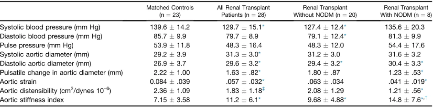
Blood Pressure, Aortic and Aortic Distensibility Data Results are presented inTable 3. Aortic elasticity properties showed alterations in post-KT patients, and further im- pairments were detected in the presence of NODM.
DISCUSSION
Arterial stiffness is an age-related process, a shared conse- quence of several disorders, including DM and chronic kidney disease (CKD)[11,12]. Different cardiovascular pa- rameters, including arterial stiffness, have been shown to have a prognostic value in CKD[12]. KT is a widely used Table 1. Relevant Clinical Data in the Study Groups
Matched Healthy Controls (n¼23)
All KT Patients (n¼28)
KT Patients Without NODM (n¼20)
KT Patients With NODM (n¼8)
Clinical data
Age (y) 54.911.0 48.26.9 46.16.4 53.64.8*
Male sex (%) 9 (39) 13 (46) 8 (40) 5 (63)
Hypertension (%) 17 (74) 22 (79) 15 (75) 7 (88)
Hypercholesterolemia (%) 6 (26) 6 (21) 4 (20) 2 (25)
Diabetes mellitus (%) 4 (17) 8 (29) 0 (0) 8 (100)*,†
Weight (kg) 89.218.2 75.513.6 74.711.0 77.419.6
Height (m) 162.66.2 168.68.6 169.18.8 167.48.6
Medication
b-blocker (%) 4 (17) 12 (43) 7 (35) 5 (63)†
ACE inhibitor or ARB (%) 10 (44) 18 (64) 13 (65) 5 (63)
Ca-antagonist (%) 4 (17) 8 (29) 4 (20) 4 (50)
Antidiabetics (%) 3 (13) 5 (18) 0 (0) 5 (63)†
Statin (%) 3 (13) 11 (39) 7 (35) 4 (50)†
Abbreviations: ACE, angiotensin-converting enzyme; ARB, angiotensin-receptor blocker; Ca, calcium; NODM, new-onset diabetes mellitus following kidney transplantation.
*P< .05 vs renal transplant without NODM.
†P<.05 vs matched controls.
Table 2. 2D Doppler Echocardiographic Parameters in the Examined Groups Matched Controls
(n¼23)
All Renal Transplant Patients (n¼28)
Renal Transplant Without NODM (n¼20)
Renal Transplant With NODM (n¼8)
LA diameter (mm) 38.13.2 39.94.9 39.35.3 41.53.6
LV-EDD (mm) 46.24.0 53.15.1* 52.45.1* 54.85.2*
LV-ESD (mm) 29.33.9 31.74.1* 31.44.0 32.54.5
IVS (mm) 10.01.2 11.0.9* 10.80.8* 11.51.5*
LV-PW (mm) 9.71.1 11.01.2* 10.91.0* 11.51.5*
LV-EF (%) 65.26.4 68.45.2 68.75.2 67.95.7
E/A ratio .95.25 .97.26 1.04.25 .75.14*,†
Abbreviations: E/A, ratio of E and A transmitralflow velocities; EDD, end-diastolic; EF, ejection fraction; ESD, end-systolic; IVS, interventricular septum; LV, left ventricular; NODM, new-onset diabetes mellitus following kidney transplantation; PW, posterior wall.
*P< .05 vs matched controls.
†P<.05 vs renal transplant without NODM.
method of treating CKD[13]. NODM is a well-known and relatively common entity in KT recipients. Some risk factors have been identified as predating the transplant, and could be used for risk-stratification and to implement the proper strategies for risk-reduction [14]. Several clinical studies have shown that serum creatinine and estimated glomerular filtration rate levels are higher in NODM patients[15,16].
These results were confirmed by thefindings of our study.
We detected significant differences between the nondiabetic and NODM post-KT patients with regard to the renal lab- oratory parameters. NODM was also found to be associated with decreased graft survival and an increased risk for the development of cardiovascular disease[17].
There are several ways to assess arterial stiffness, including pulse wave analysis and the measurement of pulse wave ve- locity (PWV) by tonometry, oscillometry, or sonography[9].
However, there is another method, several aortic elasticity properties can be calculated by imaging (for instance, by echocardiography) using aortic dimensions with respect to the cardiac cycle and forearm blood pressure values[9]. It has been demonstrated that the noninvasively evaluated ASI is comparable to invasive methods and achieves a high degree of accuracy [18]. A recent study has demonstrated an improvement of aortic elastic properties including echocar- diographic ASI after KT[5]. The most significant changes of these indices can be observed only between years 1e3 after KT. This continuous and prolonged improvement process suggests a reverse remodeling. In our study, ASI 1 year after the operation was significantly higher in KT patients compared to matched controls and was proven to be more pronounced in patients who developed NODM. In our study, ASI was proven to be higher compared to thefindings of others in KT patients [5]. The PWV was determined by pressure tracing over the carotid and femoral arteries.
Another study found that ASI was significantly higher in transplant recipients with NODM compared to transplant patients without NODM[19]. These results are confirmed by Kato et al, who used brachial-ankle PWV measurements in their assessment[20]. In these studies, determinants for PWV have been identified as an increase in SBP and recipients’age.
They conform with ourfindings, which also demonstrated
higher aortic stiffness values in NODM patients [19e20].
These results could be explained by DM-related vascular al- terations and endothelial dysfunction, and they could suggest the importance of regular control of carbohydrate meta- bolism in KT patients. Multidisciplinary care of the kidney recipients should be managed by a common team, and a minimum of 1 clinical control is required every year. Further studies are warranted to examine whether echocardiographic ASI has a real prognostic value in KT patients.
ACKNOWLEDGMENTS
We wish to thank the Hungarian Diabetes Association and Novo Nordisk for their joint grant in 2017.
REFERENCES
[1] Coiso FG, Kudva Y, van der Velde M, et al. New onset hy- perglycemia and diabetes are associated with increased cardiovas- cular risk after kidney transplantation. Kidney Int 2005;67:2415e21.
[2] American Diabetes Association. Diagnosis and classification of diabetes mellitus. Diabetes Care 2010;33:S62e9.
[3] Davidson J, Wilkinson A, Dantal J, et al. International expert panel: New-onset diabetes after transplantation: 2003 International consensus guidelines. Proceedings of an international expert panel meeting. Barcelona, Spain, 19 February 2003. Transplantation 2003;75:S3e24.
[4] Laurent S. Arterial stiffness: intermediate or surrogate endpoint for cardiovascular events? Eur Heart J 2005;26:1152e4.
[5] Zapolski T, Furmaga J, Jaroszynski A, et al. The reverse remodeling of the aorta in patients after renal transplantationdthe value of aortic stiffness index: prospective echocardiographic study.
BMC Nephrol 2017;18:33.
[6] Townsend RR. Arterial stiffness in CKD: A review. Am J Kidney Dis 2019;73:240e7.
[7] American Diabetes Association. American Diabetes Associ- ation home page.www.diabetes.org. [Accessed 21.11.18].
[8] World Health Organization. Diabetes.www.who.int/diabetes/
en. [Accessed 21.11.18].
[9] Nemes A, Geleijnse ML, Forster T, et al. Echocardiographic evaluation and clinical implications of aortic stiffness and coronary flow reserve and their relation. Clin Cardiol 2008;31:304e9.
[10] Lang RM, Badano LP, Mor-Avi V, et al. Recommendations for cardiac chamber quantification by echocardiography in adults:
an update from the American Society of Echocardiography and the European Association of Cardiovascular Imaging. Eur Heart J Cardiovasc Imaging 2015;16:233e70.
Table 3. Blood Pressure and 2D Echocardiography-derived Aortic Elastic Properties in Groups Examined Matched Controls
(n¼23)
All Renal Transplant Patients (n¼28)
Renal Transplant Without NODM (n¼20)
Renal Transplant With NODM (n¼8)
Systolic blood pressure (mm Hg) 139.614.2 129.715.1* 127.412.4* 135.620.3
Diastolic blood pressure (mm Hg) 85.79.9 79.78.9 79.112.4* 81.39.9
Pulse pressure (mm Hg) 53.911.8 48.316.4 48.312.0 54.417.6
Systolic aortic diameter (mm) 29.23.9 31.33.0* 31.23.0 31.63.2
Diastolic aortic diameter (mm) 26.93.7 29.63.2* 29.43.2* 30.43.3*
Pulsatile change in aortic diameter (mm) 2.221.00 1.63.82* 1.80.87 1.23.53*
Aortic strain 0.084.039 .057.032* .063.034 .041.019*
Aortic distensibility (cm2/dynes 10e6) 2.361.09 1.831.18‡ 2.081.29 1.21.56*
Aortic stiffness index 7.153.58 11.26.1* 9.684.88* 14.87.6*,†
Abbreviation: NODM, new-onset diabetes mellitus following kidney transplantation.
*P< .05 vs matched controls.
†P<.05 vs renal transplant without NODM.
‡P¼.07 vs matched controls.
[11] Prenner SB, Chirinos JA. Arterial stiffness in diabetes mellitus. Atherosclerosis 2015;238:370e9.
[12] Chen SC, Huang JC, Su HM, et al. Prognostic cardiovas- cular markers in chronic kidney disease. Kidney Blood Press Res 2018;43:1388e407.
[13] Sarnak MJ, Levey AS, Schoolwerth AC, et al. Kidney dis- ease as a risk factor for development of cardiovascular disease: a statement from the American Heart Association Councils on Kid- ney in Cardiovascular Disease, High Blood Pressure Research, Clinical Cardiology, and Epidemiology and Prevention. Circulation 2003;108:2154e69.
[14] Alagbe SC, Voster A, Ramesar R, Swanepoel CR. New- onset diabetes after transplant: incidence, risk factors and outcome.
S Afr Med J 2017;107:791e6.
[15] Borda B, Szederkényi E, Lengyel C, et al. Functional and histopathological changes in renal transplant with new onset dia- betes and dyslipidemia. Transplant Proc 2011;43:1254e8.
[16] Gámán G, Sárváry E, Gelley F, et al. New-onset diabetes mellitus and the analysis of dipeptidyl-peptidase-4 after liver transplantation. Transplant Proc 2014;46:2177e80.
[17] Sezer S, Erkmen Uyar M, Tutal E, et al. New-onset diabetes and glucose regulation are significant determinants of left ventric- ular hypertrophy in renal transplant recipients. J Diabetes Res 2015;2015:293896.
[18] Stefanadis C, Stratos C, Boudoulas H, Kourouklis C, Toutouzas P. Distensibility of the ascending aorta: comparison of invasive and non-invasive techniques in healthy men and in men with coronary artery disease. Eur Heart J 1990;11:990e6.
[19] Opazo Saez A, Kos M, Witzke O, Kribben A, Nürnberger J.
Effect of new-onset diabetes mellitus on arterial stiffness in renal transplantation. Transpl Int 2008;10:930e5.
[20] Kato K, Matsuhisa M, Ichimaru N, et al. The impact of new- onset diabetes on arterial stiffness after renal transplantation.
Endocr J 2008;55:677e83.