T h e R i g h t H e a r t
I n t e r n a t i o n a l N e t w o r k ( R I G H T- N E T )
Rationale, Objectives, Methodology, and Clinical Implications
Francesco Ferrara,
MD, PhDa, Luna Gargani,
MD, PhDb, William F. Armstrong,
MDc, Gergely Agoston,
MD, PhDd, Antonio Cittadini,
MD, PhDe, Rodolfo Citro,
MD, PhDa, Michele D ’ Alto,
MD, PhDf, Antonello D ’ Andrea,
MD, PhDf, Santo Dellegrottaglie,
MD, PhDg,h, Nicola De Luca,
MD, PhDi, Giovanni Di Salvo,
MD, PhDj, Stefano Ghio,
MDk,
Ekkehard Grünig,
MDl, Marco Guazzi,
MD, PhDm, Jaroslaw D. Kasprzak,
MD, PhDn,
Theodore John Kolias,
MDc, Gabor Kovacs,
MD, PhDo,p, Patrizio Lancellotti,
MD, PhDq,r, Andrè La Gerche,
MD, PhDs, Giuseppe Limongelli,
MD, PhDf,t, Alberto Maria Marra,
MDu, Antonella Moreo,
MDv, Ellen Ostenfeld,
MD, PhDw,
Francesco Pieri,
MDx, Lorenza Pratali,
MD, PhDb, Lawrence G. Rudski,
MDy, Rajan Saggar,
MDz,aa, Rajeev Saggar,
MDab, Marco Scalese,
PhDb,
Christine Selton-Suty,
MDac, Walter Serra,
MD, PhDad, Anna Agnese Stanziola,
MD, PhDae,
Damien Voilliot,
MD, PhDaf, Olga Vriz,
MD, PhDag,
Robert Naeije,
MD, PhDah, Eduardo Bossone,
MD, PhDai,*
a Heart Department, University Hospital of Salerno, Salerno, Italy;b Institute of Clinical Physiology–C.N.R., Pisa, Italy; c Division of Cardiovascular Medicine, University of Michigan Medical Center, Ann Arbor, MI, USA;dDepartment of Family Medicine, University of Szeged, Szeged, Hungary;e Department of Translational Medical Sciences, University Federico II, Naples, Italy;f Department of Cardiology, University of Campania
“Luigi Vanvitelli”, Naples, Italy;gDivision of Cardiology, Ospedale Medico-Chirurgico Accreditato Villa dei Fiori, Acerra, Naples, Italy;hZena and Michael A. Wiener Cardiovascular Institute, Marie-Josee and Henry R.
Kravis Center for Cardiovascular Health, Icahn School of Medicine at Mount Sinai, New York, NY, USA;
i Hypertension Research Center “CIRIAPA”, Federico II University, Napoli, Italy;j Imperial College, London, UK;k Fondazione IRCCS, Policlinico San Matteo, Pavia, Italy;l Centre for Pulmonary Hypertension, Thorax- clinic, Heidelberg University Hospital, Heidelberg, Germany;m Heart Failure Unit, Cardiopulmonary Labora- tory, University Cardiology Department, IRCCS Policlinico San Donato University Hospital, Milan, Italy;
n Bieganski Hospital, Medical University of Lodz, Lodz, Poland;o Department of Internal Medicine, Division of Pulmonology, Medical University of Graz, Graz, Austria;p Ludwig Boltzmann Institute for Lung Vascular Research, Graz, Austria;qDepartment of Cardiology, University of Lie`ge Hospital, GIGA Cardiovascular Sci- ences, Liege, Belgium;r Gruppo Villa Maria Care and Research, Anthea Hospital, Bari, Italy;s Baker Heart and Diabetes Institute, Melbourne, Australia;t Institute of Cardiovascular Sciences, University College of London, London, UK; u IRCCS S.D.N., Naples, Italy; v Cardiovascular Department, Niguarda Hospital, Milan, Italy;
w Department of Clinical Sciences Lund, Clinical Physiology, Ska˚ne University Hospital, Lund, Sweden;
x Department of Heart, Thorax and Vessels, Azienda Ospedaliero Universitaria, Florence, Italy;y Azrieli Heart Center and Center for Pulmonary Vascular Diseases, Jewish General Hospital, McGill University, Montreal, Quebec, Canada; z Lung and Heart-Lung Transplant Program, David Geffen School of Medicine, UCLA, Los Angeles, CA, USA;aa Pulmonary Hypertension Program, David Geffen School of Medicine, UCLA, Los Angeles, CA, USA;ab Lung Institute Banner University Medical Center-Phoenix, University of Arizona, Phoenix, AZ, USA;
Heart Failure Clin 14 (2018) 443–465 https://doi.org/10.1016/j.hfc.2018.03.010
heartfailure.theclinics.com
RATIONALE
Exercise stress testing of the pulmonary circulation to detect early-stage pulmonary vascular disease (PVD) or any cardiac condition associated with an increase in pulmonary venous pressure was part of the hemodynamic work-up in the early years of cardiac catheterization.1 After years of doubt entertained due to insufficient knowledge of the limits of normal, variable methodologies, and limited validation, the advent of noninvasive exer- cise imaging of the pulmonary vasculature and car- diac function generated renewed interest.2–7 Currently, there is emerging consensus to define exercise-induced pulmonary hypertension (PH) by the presence of a resting mean pulmonary artery pressure (mPAP) less than 25 mm Hg and of mPAP greater than 30 mm Hg at peak exercise, with total pulmonary vascular resistance (PVR) of more than 3 Wood units (WU).8,9However, these criteria are based on a limited number of studies with a mixture of invasive and noninvasive ap- proaches and variable protocols. On the other hand, only limited information is available about right ventricular (RV) function indices during exer- cise testing of the pulmonary circulation,10,11 so that the added value of these indices to
measurements of the pulmonary circulation re- mains undefined. Thus, more methodologically robust noninvasive exercise stress tests of the pul- monary circulation and the right heart are needed.
Transthoracic Doppler echocardiography (TTE) is the noninvasive method of choice because it is part of daily cardiology practice, is flexible, and relatively cheap, and is implemented anyway in the diagnostic work-up of any suspicion of PH.12–15Current preliminary experience with exer- cise TTE for detection or diagnosis of early PVD or left heart conditions with increased pulmonary venous pressure is promising.16–18 However, the exact clinical relevance of abnormal responses in healthy patients with a known increased risk of developing PH and right heart failure remains un- clear. The current state of knowledge based on re- ported exercise TTE studies of the pulmonary circulation and the RV in different populations are presented inTables 1–3.19–64Thus, the available literature shows great disparities in sample sizes (from n 58 to n 5113), exercise protocols (leg press, cycle, or treadmill ergometry), timing of measurements, selection of variables of interest, and different work rates (ranging from 237 WU up to 17550 WU). Therefore, the need for stan- dardization is evident. For this purpose, as recently
ac Department of Cardiology, University Hospital of Nancy, France;ad Cardiology Unit, Surgery Department, University Hospital of Parma, Italy;ae Department of Respiratory Diseases, Monaldi Hospital, University “Fed- erico II”, Naples, Italy; af Centre Hospitalier Lune´ville, Service de Cardiologie, Lune´ville, France; ag Heart Centre, King Faisal Specialist Hospital and Research Centre, Riyadh, Saudi Arabia;ah Free University of Brussels, Brussels, Belgium;ai Cardiology Division, Heart Department, “Cava de’ Tirreni and Amalfi Coast” Hospital, Uni- versity of Salerno, Salerno, Italy
* Corresponding author. Via Pr. Amedeo, 36, Lauro, Avellino 83023, Italy.
E-mail address:ebossone@hotmail.com
=
KEYWORDS
Right heartPulmonary circulationPulmonary hypertensionExercise doppler echocardiography
KEY POINTS
Exercise Doppler echocardiography has been implemented for applications beyond coronary ar- tery disease detection, but with a great variability of protocols to assess early stage pulmonary vascular disease and/or left heart failure.
The RIGHT heart international NETwork (“RIGHT-NET”) is a large prospective clinical and echocar- diography observational multicenter study.
Aims of the RIGHT-NET: a) define limits of normal in right heart function and pulmonary circulation hemodynamics during exercise in a large cohort of healthy individuals and elite athletes; b) inves- tigate the impact of abnormal responses on clinical outcome in individuals with overt or at risk of developing pulmonary hypertension.
argued, a series of methodological requirements can be determined from sound physiologic princi- ples and previously available experience8,9,16:
Exercise must be dynamic (cycling, running) because resistive or static exercise (weight lifting, handgrip) is associated with increases in cardiac output (CO) too small to obtain a meaningful range of pulmonary vascular pressure-flow relationships.35
Body position does not matter because the same slope of mPAP as a function of CO, maximum oxygen uptake (VO2), and maximum CO are estimated during incremental cardiopulmonary exercise testing in supine, semirecumbent or upright positions. Thus the body position allowing for the TTE approach can be used safely.33,65
Table 1
Pulmonary pressure response to exercise in normal subjects and athletes
Subjects:
Gender (M, F) Age (y)
Baseline sPAP or mPAPa (mm Hg)
Peak sPAP or mPAPa(mm Hg) Normal Subjects
Himelman et al,191989c 12: 1 F, 11 M 27–68 224 317 Oelberg et al,201998g 10: 4 F, 6 M 52.310.9 178 198
Bossone et al,211999e 14: 14 M 18.90.9 9 21
CI 95%:9–19
Gru¨nig et al,222000c 11: 11 M 3711 274 363
Kiencke et al,232008c 9/— 323 173 —
Gru¨nig et al,242009c 191: 91 F, 100 M 3210 20.45.3 35.55.4 Mahjoub et al,252009d 70: 36 F, 34 M 4816 274 519 Mo¨ller et al,262010c 88: 49 F, 30 M 18.33.5 21.83.6 39 (17–63) Argiento et al,272010f 25: 12 F, 13 M 3614 195 4611 La Gerche et al,282010c 15/2 F/13 M 386 21.63.8 47.06.5 D’Alto et al,292011c 88: 78 F, 10 M 55.312.4 20.63.7 25.93.3 Argiento et al,302012f 124: 62 M, 62 F 3713 15.52.6 Ma 36.05.9 Ma
15.12.9 Fa 30.57.2 Fa Lalande et al,312012f 24: 6 F, 18 M 256 233 615
Simaga et al,322015d 30 Blb — 168a 34.76.2a
30 Whb 16.62.3a 38.55.5a
Forton et al,332016g,f,c 30: 15 F, 15 M 232 15.41.2a,g 34.23.6a,g 15.51.3a,f 34.33.8a,f 15.50.8a,c 34.33.2a,c Faoro et al,342017f 38: 5 F, 33 M 386 17.72a,sea level 44.94.4a,sea level
202.8a,altitude 47.64.9a,altitude Motoji et al,352017f 26: 14 F, 12 M 223 15.81a 28.83.2a Athletes
Bossone et al,211999e 26: 26 M 20.31.7 21 41
CI 95%:21–41 La Gerche et al,282010d 40/4 F/36 M 368 21.53.8 60.712.4
Bidart et al,362007e 15/— 38.7 19.4 54.8
Abbreviations:Bl, black; F, female subject; M, male subject; sPAP, systolic PAP; Wh, white; —, not available.
amPAP (mm Hg) was calculated as 0.6sPAP12.
b30 black subjects (age 276 M, 256 F) of sub-Saharan descent and 30 matched by age, sex, and body size European white subjects (age 276 M, 278 F).
cExercise protocol: supine.
dExercise protocol: semisupine.
eExercise protocol: recumbent.
fExercise protocol: semirecumbent.
gExercise protocol: upright bicycle.
Table 2
Pulmonary artery pressure response to exercise Doppler echocardiography in subjects with high risk for pulmonary hypertension
Author, Year Age (y)
Baseline sPAP (mm Hg)
Peak sPAP (mm Hg) Associated Disease
Subjects: Gender Lung disease
Himelman et al,19 1989g
COPD 32–80 4620 8330
36: 15 F, 21 M 224 (ctrl) 317
Rodrı`guez et al,37 2017
COPD 689 3127 5729
81: 15 F, 66 M Congenital heart disease
Oelberg et al,20 1998k
Asymptomatic ASD 52.911.2 318 5110
10: 4 F, 6M 178 (ctrl) 198
Mo¨ller et al,26 2010g
ASD and VSD 17.53.3 20.75.3 37 (24–76)
44: 25 F, 19 M 21.83.6 (ctrl) 39 (17–63)
Ait Ali et al,38 2014g
Operated Fallot 26.211.3 4922.4 79.434.2
123: 41 F, 82 M Brenner et al,39
2015g
HA dwellers PFO (n518)
54.210.3 24.45.3 (HA PFO) 49.99.6 HA dwellers no PFO
(n539)
49.610.7 24.83.6 (HA no PFO)
40.39.1 Van Riel et al,40
2015
ASD, VSD, PDA, other 43.214.5 27.55.2 40.26.6
76: 50 F, 26 M 37.17.9 59.38.5
CMS
Stuber et al,41 2010g
CMS subjects 4713 30.38 (CMS) 56.419 (CMS)
30: — 25.44.5 (ctrl) 39.88 (ctrl)
Groepenhoff et al,422012g
CMS subjects (13:
13 M)
503 26 ± 2(CMS) 56 ± 4(CMS)
HA dwellers (15: 6 F, 9 M)
412 23 ± 1(HA) 42 ± 3(HA)
L (15: 6 F, 9 M) 353 20 ± 1(L)sea level 31 ± 2(L)sea level Pratali et al,43
2013g
CMS subjects (n546) 5110 306 (CMS) 5012 (CMS) HA dwellers (n540) 488 275 (HA) 388 (HA) HAPE-S
Gru¨nig et al,22 2000g
HAPE-S 458 284 5511
9: — 274 (ctrl) 363
Kiencke et al,23 2008g
HAPE-S 332 194 236
10: — NA 173 (ctrl) 115
Relative of iPAH Gru¨nig et al,22
2000g
Relatives of iPAH cases
NA 244 (nr) 373 (nr)
52: — 233 (ar) 5611(ar)
Gru¨nig et al,24 2009g
Relatives of iPAH cases
3716 20.75.4 39.55.6
291: 125 F, 166 M 20.45.3 (ctrl) 35.55.4
Connective tissue disease Collins et al,44
2006l
Scleroderma 53.912.0 248 3812
51: 49 F, 2 M NA
Alkotob et al,45 2006l
Scleroderma 5112 258 398
65: 56 F, 9 M NA
Steen et al,462008l Scleroderma NA 34.511.5 51.4
54: 51 F, 3 M
(continued on next page)
Measurements should be made during, not after, the exercise stress because too-fast postexercise recovery of vascular pressures and flows normally occurs within minutes.30,66 Exercise stress should be incremental, with stepwise increase in workload allowing for measurements in quasi-steady-state VO2, after 2 to 5 minutes of stabilization at each step and increments in workload individ- ually tailored to obtain at least 3 (but preferably 5) pressure-flow coordinates in an exercise duration of less than 12 to 15 minutes.4,5,9 It is important to estimate all the components of
PVR, thus pulmonary artery pressure (PAP),
wedged PAP (PAWP), and CO. Using workload or VO2as surrogates for CO greatly decreases the accuracy and precision of pulmonary vascular function as defined by mPAP–CO rela- tionships. The relationships between CO, VO2, and workload are near-linear but with a pla- teauing at high levels of exercise, and predic- tion equations based on linear regression also fail due to the considerable variability of CO at any given level of workload or VO2.30,33 Noninvasive exercise stress testing of the pulmo- nary circulation using TTE is acceptable because exercise measurements are probably accurate.
Table 2 (continued)
Author, Year Age (y)
Baseline sPAP (mm Hg)
Peak sPAP (mm Hg) Associated Disease
Subjects: Gender Reichenberger
et al,472009g
Scleroderma 5411 238 4011
33: 31 F, 2 M NA
Kovacs et al,48 2010g
Connective tissue disease
5411 275a 5510a
52: 42 F, 10 M 233b 298b
D’Alto et al,29 2011g
Systemic sclerosis 51.821.5 26.25.3 36.98.7
172: 155 F, 17 M 20.63.7 (ctrl) 25.93.3
Gargani et al,49 2013h
Systemic sclerosis 5813 — TRV 332 cm/s
164: 150 F, 14 M Range 185–533
Gru¨nig et al,50 2013g
PAH, CTPEH 5416 6417 9825
124: 87 F, 37 M Codullo et al,51
2013g
Systemic sclerosis 55.213 23.78.1e 33.112.6e
170: 153 F, 17 M NA 29.55.5f 47.712.2f
Voilliot et al,52 2014h
Systemic sclerosis 543 257 4614
45: 34 F, 11 M
Nagel et al,532015i Systemic sclerosis 5814 25.67.3c 49.912.7c 76: 64 F, 12 M 1.80.2 52.018.0d 83.9 18.9d Kovacs et al,54
2017j
Systemic sclerosis 51.311.5m 25.0 (22.0–27.0)m 43.211.7m 58: 56 F, 2 M 55.311.6n 25.0 (22.8–30.0)n 49.510.7n Bold type values indicate mPAP (mm Hg) calculated as 0.6sPAP12.
Abbreviations:ar, abnormal response to exercise; ASD, atrial septal defect; CMS, chronic mountain sickness; COPD, chronic obstructive pulmonary disease; ctrl, controls; HA, high altitude; HAPE-S, high altitude pulmonary edema suscep- tible; iPAH, idiopathic pulmonary arterial hypertension; nr, normal response to exercise; L, lowlanders; NA, not available;
PDA, patent ductus arteriosus; PFO, patent foramen ovale; VSD, ventricular septal defect.
aExercise sPAP greater than 40 mm Hg.
bExercise sPAP less than 40 mm Hg, peak VO2less than 75%.
cNo PH group of 54 subjects (mPAP <25 mm Hg).
d22 subjects with manifest PH (mPAP >25 mm Hg).
eSubjects (n5164) with complete follow-up who did not develop PH.
fSubjects (n56) who did develop PH.
gExercise protocol: supine.
hExercise protocol: semisupine.
i Exercise protocol: recumbent.
j Exercise protocol: semirecumbent.
kExercise protocol: upright bicycle.
l Exercise protocol: treadmill.
m Exercise protocol: baseline examination.
nExercise protocol: follow-upw4 y after their baseline examination.
The reliability of exercise TTE of the pulmonary cir- culation is still under discussion, although average responses and derived limits of normal seem to agree very well with those obtained during a right heart catheterization.4,5,48 Bland-Altman analysis of PAP, PAWP, and CO measured at rest by TTE versus right heart catheterization show almost no bias, indicating excellent accuracy; however, limits of agreement are sometimes wide, indicating possible problems of insufficient precision for indi- vidual decision-making.67Echocardiographic esti- mates of PAP from the maximum velocity of tricuspid regurgitation (TR) compared with inva- sively measured PAP during exercise have recently also been shown by Bland-Altman analysis to be associated with only minimal bias, demonstrating acceptable accuracy; however, limits of agreement were broad, indicating limited precision.11,68 Furthermore, the agreement between TTE and invasive measures of PAP during upright exercise is good among the subset of patients with high- quality TR Doppler signal.68There still is a concern that TTE might underestimate CO during exer- cise.11This can probably be overcome by intensive training of dedicated operators. Even so, a 5%
underestimation remains, showing concomitant measurements of CO by the Innocor device (by rebreathing of nitrous oxide and sulfur hexafluoride) and TTE of the left ventricular outflow tract in 10 subjects reported by Forton and colleagues.33
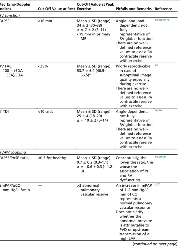
At this stage, knowledge regarding limits of normal of exercise TTE indices of RV function is limited to measurements of tricuspid annular plane systolic excursion (TAPSE), tricuspid annulus S0, and stroke volume (SV), along with estimates of systolic PAP (sPAP) in 90 healthy young adults (45 male subjects).10 Changes from rest to maximum workload were (D) 4 to 10 mm for TAPSE, 6 to 14 cm per second for S0, 12 to 57 mm Hg for sPAP, 0 to 96 mL for SV, and 0.6 plus or minus 0.3 (1.3 0.4–0.7 0.2) mm/mm Hg for TAPSE/sPAP.
THE RIGHT HEART INTERNATIONAL NETWORK
The Right Heart International Network (RIGHT- NET) is a large prospective observational multi- center clinical study, including resting and exercise TTE performed at different European Table 3
Pulmonary pressure response to exercise Doppler echocardiography in left heart diseases and valvular heart diseases
Author, Year
Subjects,
Gender (M, F) Age (y)
Baseline sPAP (mm Hg)
Peak sPAP (mm Hg) Heart Failure
Lancellotti et al,552003d Survivors 89: 60 M 6511 2610 4418
Nonsurvivors 9: 6 M 699 229 4816
Tumminello et al,562007c 46: — 6610 3111 5218
Ennezat et al,572008d 104: 29 F, 75 M 5412 299 4418
Marechaux et al,582008d 85: 21 F, 64 M 5713 278 4318
Bandera et al,592014d 136a: 50 F, 86 M 6411 — —
Group Ab36: 20 F 6710 3717 6119
Group Bb100: 30 F 6112 3314 5118
Guazzi et al,602016d 97: 20 F, 67 M 6411 3716 5918
Degenerative Asymptomatic Mitral Regurgitation (at least moderate)
Magne et al,612010d 78: 34 F, 44 M 6113 3911 6217
Kusunose et al,622013 196: 70 F, 126 M 5613 398 5613
Asymptomatic Severe Aortic Stenosis
Lancellotti et al,632012d 105: 53 F, 62 M 719 388 6216 Asymptomatic Mitral Stenosis
Brochet et al,642011c 48: 32 F, 16 M 5114 365 687
aThe underlying diseases were heart failure with reduced (n554, 40%) or preserved ejection fraction (n58, 6%), history of stable coronary artery disease (n518, 13%), high-risk subjects with hypertrophic cardiac remodeling (n533, 24%), hypertrophic or restrictive cardiomyopathy (n55, 4%), and mitral or tricuspid valvular regurgitation (n518, 13%).
bDoxygen consumption (VO2)/Dwork rate: flattening (group A), not flattening (group B).
cExercise protocol: semisupine.
dExercise protocol: tilting bicycle.
and American centers (ClinicalTrials.govidentifier:
NCT03041337).
Aims
The aims of this study are
1. To evaluate the feasibility of exercise TTE for the noninvasive assessment of the right heart and pulmonary circulation in a large cohort of healthy subjects, elite athletes, and subjects with overt or at risk of PH.
2. To explore the physiologic spectrum of re- sponses of the right heart and pulmonary circu- lation during an optimally standardized exercise TTE in a large cohort of healthy sub- jects, elite athletes, and subjects with overt PH or at risk of PH.
3. To systematically compare the morphologic and functional behavior of the right heart and pulmonary circulation to exercise in subjects with normal versus abnormal responses, and their clinical correlations.
4. To investigate the prognostic impact of an abnormal right heart and pulmonary circulation response to exercise during a long-term follow- up.
Methods
Study population
It is anticipated that the study population will comprise approximately 3000 subjects (18 years old): 500 healthy subjects, 500 elite athletes, and about 2000 subjects with overt PH or at risk of PH.
Healthy subjects Healthy volunteers (or subjects for work ability assessment) with no structural heart disease on TTE and without a history of any cardiovascular disease and/or any systemic dis- eases known to affect the cardiovascular system will be prospectively recruited. Exclusion criteria will be systemic arterial hypertension, diabetes mellitus, coronary artery disease, significant (at least moderate) valvular heart disease, congenital heart disease, history of congestive heart failure, cardiomyopathies, sinus tachycardia, atrial fibrilla- tion or flutter, use of illicit drugs, medical therapy with cardioactive drugs, chronic excessive alcohol consumption, elite athletes, pregnancy, obesity (body mass index30 kg/m2), pulmonary disease, renal disease, hepatic disease, significant endo- crine alterations, cancer, and inadequate echocar- diographic image quality.69
Elite athletes Cardiac adaptations in athletes mainly depend on the characteristics, intensity, and cumulative duration of training protocols, with a dose-effect relation.70,71 In particular,
isotonic (dynamic) exercise is associated with a substantial increase in CO and reduction in periph- eral vascular resistance; therefore, endurance training mainly results in volume overload.
Conversely, isometric (static) exercise is charac- terized by less increase in CO and by a transient increase in peripheral resistance; therefore, its training is characterized by a pressure overload.71 In this study, protocol activity levels will be assessed by questionnaire, and subjects will be asked to describe and quantify exercise during a typical week during the preceding months. All the athletes will have been trained intensively for 15 to 20 hours per week for at least 4 years.72Based on their training protocol and the type of sports ac- tivity, they will be categorized into 2 groups:
endurance-trained athletes and strength-trained athletes.72–74Endurance-trained athletes (long-dis- tance and middle-distance swimming or running, cycling, rowing) will be defined as subjects actively engaged in endurance sports competition, sub- jected to intensive aerobic isotonic dynamic exer- cise at incremental workloads of 70% to 90% of maximal heart rate. In particular, they should have performed 3 hours per day of incremental long- distance swimming (7000 m per day divided into a series of 400–800 m) or 3 hours per day of long- distance running or cycling, and only 2 hours per week of weight-lifting at a low workload.70On the other hand, the strength-trained athletes group will include top-level competitive athletes (weight- lifting, martial arts, windsurfing) who should have undergone an aerobic isometric static exercise at incremental workloads of 40% to 60% of maximal heart rate. Their training protocol should have included 3 hours per day of short-distance running and/or 4 hours per day of weight-lifting at a high workload.72–74
Patients with overt pulmonary hypertension or at risk of pulmonary hypertension Patients with overt or at risk of PH will be classified according to European guidelines.12
1. Pulmonary arterial hypertension (PAH): idio- pathic, heritable, healthy relatives of patients with PAH, healthy carriers of mutations of bone morphogenetic receptor 2 gene, associated with connective tissue disease, portal hyperten- sion, schistosomiasis, congenital heart disease 2. PH due to left heart disease: left heart valve dis-
ease, heart failure with reduced or preserved ejection fraction, congenitally acquired inflow or outflow tract obstruction, and congenital cardiomyopathies
3. PH due to chronic lung diseases and/or hypoxia: chronic altitude exposure, chronic
obstructive pulmonary disease, interstitial lung diseases, sleep-disordered breathing
4. Chronic pulmonary thromboembolic disease and other pulmonary artery obstructions 5. PH with unclear and/or multifactorial mecha-
nisms: miscellaneous conditions such as histiocytosis-X or sarcoidosis.
Clinical data
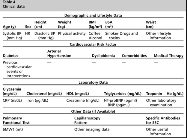
Demographic characteristics, complete medical history, comorbidities, symptoms and signs, labo- ratory values, electrocardiogram (ECG) parame- ters, coronary angiography and other imaging results, medications, and in-hospital and long- term outcomes will be systematically collected for all patients via standardized forms to obtain as much information as possible (Table 4). Spe- cific attention will be paid to excluding the pres- ence of any pulmonary condition (apart from patients with PH due to chronic lung diseases and/or hypoxia) that may lead to a pathologic pul- monary hemodynamic response during exercise.
Follow-up would be performed at 6 months and then once a year for at least 5 years during outpa- tient clinical visits or by telephone call. Events
recorded will be death, major cardiovascular events (myocardial infarction, stroke, coronary revascularization, acute heart failure), hospitaliza- tion, and diagnosis of PH by invasive recording of an mPAP greater than 25 mm Hg at rest.
Resting echocardiographic Doppler examination
TTE examinations will be performed at rest with commercially available equipment on all subjects, according to standardized protocols.75A detailed case report form (CRF) will be used by all labora- tories. All the measurements included in the CRF will be assessed by the operators and performed according to the recommendations for echocar- diographic assessment of the left and right heart from the American Society of Echocardiography or the European Association of Cardiovascular Im- aging76–78(Table 5).
Exercise echocardiographic Doppler examination
Exercise TTE will be performed according to the current recommendations on a semirecumbent cycle ergometer with an incremental workload
Table 4 Clinical data
Demographic and Lifestyle Data
Age (y) Sex Height (cm)
Weight (kg)
BMI (kg/m2)
BSA (m2)
Waist (cm) Systolic BP
(mm Hg)
HR Diastolic BP (mm Hg)
Physical activity Coffee Alcohol
Smoker Drugs and toxins
Other lifestyle information Cardiovascular Risk Factor
Diabetes
Arterial
Hypertension Dyslipidemia Comorbidities Medical Therapy Previous
cardiovascular events or interventions
— — — —
Laboratory Data Glycaemia
(mg/dL) Cholesterol (mg/dL) HDL (mg/dL) Triglycerides (mg/dL) Troponin Hb (g/dL) CRP (m/dL) Iron (mg /dL) Creatinine (mg/dL) NT-proBNP (pg/ml)
BNP (pg/mL)
Other laboratory examination Other Data (if Available)
Pulmonary Functional Test
Capillaroscopy Pattern
Specific Antibodies for SSC
6MWT (mt) Other imaging data Other useful
information
Abbreviations:BMI, body mass index; BNP, B-type natriuretic peptide; BP, blood pressure; BSA, body surface area; CRP, C- reactive protein; Hb, hemoglobin; HDL, high-density lipoprotein; HR, heart rate; NT-proBNP, N-terminal pro b-type natri- uretic peptide; SSC, scleroderma; 6MWT, 6 minutes walking test.
Table 5
Key echocardiographic measurements at rest and during exercise
Echocardiographic View Measurement
Parasternal long axes LV end-diastolic diameter LV end-systolic diameter
Interventricular septum thickness (diastole) Inferolateral wall thickness (diastole) LVOT diameter (zoom)
Wall motion abnormalities Aortic and mitral function Pericardial effusion
Parasternal short axes RVOT diameter
Pulmonary artery diameter RVOT acceleration time RVOT TVI notch
PR early diastolic velocity Peak tricuspid velocity Wall motion abnormalities
Aortic, mitral, tricuspid and pulmonary valve function Pericardial effusion
Apical 4-chamber LV end-diastolic volume
LV end-systolic volume Wall motion abnormalities LA volume
E, A, deceleration time e’ TDI lateral
e’ TDI septal S0systolic TDI
Mitral and tricuspid function Pericardial effusion
Apical 5-chamber Peak aortic velocity
LVOT TVI
Aortic valve function
Apical 2-chamber LV end-diastolic volume
LV end-systolic volume Wall motion abnormalities LA volume
Mitral valve function Pericardial effusion Focused apical on the RV RV dimension 1
RV dimension 2 RV diastolic area RV systolic area RA volume TAPSE
Peak tricuspid velocity E, A
S0systolic TDI e’ TDI, a’ TDI
Tricuspid valve function Pericardial effusion
Subcostal IVC diameter
IVC collapse
RV free wall thickness Pericardial effusion
Abbreviations:A, mitral inflow E velocity as measured by PW Doppler; E, mitral inflow E velocity as measured by PW Doppler;
e’, early diastolic velocity of the mitral annulus as measured by tissue Doppler; IVC, inferior vena cava; LA, left atrial; LV, left ventricular: LVOT, left ventricular outflow tract; PR, pulmonary regurgitation; RA, right atrial; RVOT, RV outflow tract;
S’ pulsed Tissue Doppler velocity of lateral tricuspid annulus; TDI, tissue Doppler imaging; TVI, time-velocity integral.
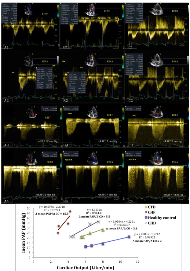
Fig. 1.Relationship between mPAP and CO at rest and during incremental exercise in 3 different diseases compared with normal response to exercise in an age-matched healthy control. (A1–A4) A 53-year-old woman with scleroderma (New York Heart Association [NYHA] class II). (B1–B4) A 64-year-old woman with patent ductus
of 25 every 2 minutes up to the symptom-limited maximal tolerated workload.18,27,79 In subjects with reduced functional capacity, the exercise protocol may consist of lower incremental work- load (10–20 WU every 2 minutes), and will be specified in the records. The exercise table will be tilted laterally by 20 to 30 to the left.
Heart rate (ECG lead) will be continuously moni- tored, and blood pressure will be monitored by sphygmomanometer at baseline and during the last 15 seconds of each workload step. Termina- tion criteria and/or positive test criteria for induc- ible myocardial ischemia will follow the current recommendations.18,79 Key echocardiographic
measurements will be acquired at baseline, at 50 WU, at peak exercise, and after 5-minutes re- covery, including left ventricular and RV function, valvular and subvalvular gradients, regurgitant flows, left and right heart hemodynamics (sPAP, mPAP, PCWP, PVR, right atrial pressure [RAP] and CO) (Figs. 1 and 2, see Table 5;
Table 6).
Oxygen saturation
Transcutaneous arterial oxygen saturation will be measured at a fingertip with a pulse oxymeter at baseline, 50 WU, peak exercise, and after 5-mi- nutes recovery.
=
arteriosus, not corrected (NYHA class II). (C1–C4) A 75-year-old woman with diagnosis of heart failure (NYHA class III) with midrange reduced ejection fraction of 46%. TheDmPAP/DCO of 3.4 mm Hg/L/min in patient with sclero- derma (D), 5.5 mm Hg/L/min in patient with congenital heart disease (), and 15.8 mm Hg/L/min in heart failure patient (>) are indicative of an abnormal pulmonary vascular response to exercise compared with normal response in healthy control (,) (DmPAP/DCO of 2 mm Hg/L/min; normal ranges from 0.5 to 3.0). mPAP (mm Hg)50.6sPAP12; SV5p(LVOT/2)2LVOT VTI; CO (L/min)5SVHR. CHD, congenital heart disease;
CHF, chronic heart failure; CTD, connective tissue disease; LVOT, left ventricular outflow tract; TVI, time- velocity integral. (Courtesy ofEcho Lab Cava de’ Tirreni and Amalfi Coast Division of Cardiology, University Hos- pital of Salerno, Italy.)
Fig. 2. Methodology of exercise stress echocardiography. Agitated saline may be used by mixing saline solution with room air (9:1 ratio) and vigorously agitating between 2 syringes using a 3-way stopcock, in cases of poor TR Doppler signals. A 5-mL bolus will be rapidly administered during exercise while simultaneously obtaining images of the right and left heart in the apical 4-chamber view. For each echocardiographic (echo)-Doppler parameter in case of poor and/or missing images it will be indicated not feasible and/or not assessed, respectively. Key echo-Doppler parameters that will be measured are specified inTables 5and6. 2D, 2-dimensional; E, mitral inflow E velocity as measured by pulsed-wave Doppler; e’, early diastolic velocity of the lateral mitral annulus and septal (average) as measured by TDI; LVOT, left ventricular outflow tract; PASP, pulmonary artery systolic pressure; RVOT, right ventricular outflow tract; TDI, tissue Doppler imaging (pulsed Doppler sample volume is placed at the tricuspid annulus, interventricular septum, and lateral mitral annulus in the apical 4-chamber view); TRV, tricuspid regurgitant velocity; VTI, velocity time integral; * RVOT VTI will be measured only at rest, peak exercise and after 5-minutes recovery.
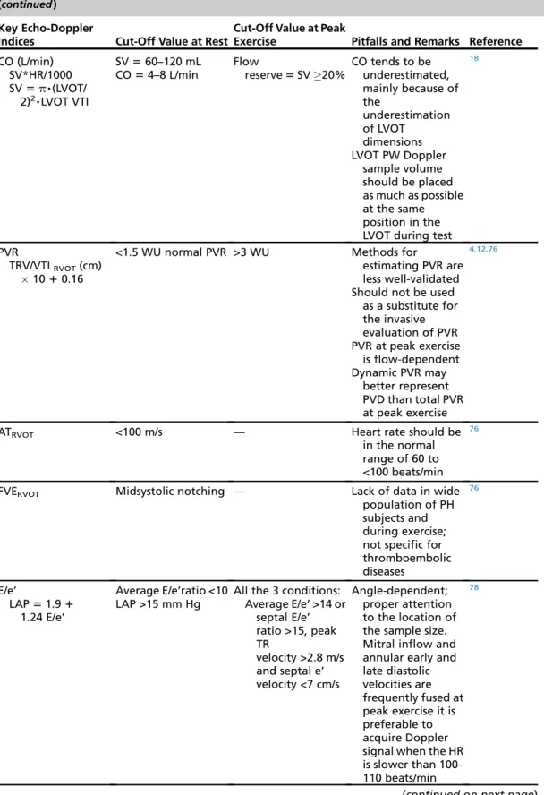
Table 6
Key echocardiographic indices for evaluation of the right heart pulmonary circulation unit at rest and during exercise
Key Echo-Doppler
Indices Cut-Off Value at Rest
Cut-Off Value at Peak
Exercise Pitfalls and Remarks Reference Pulmonary Hemodynamics
sPAP (mm Hg) 4TRV21RAP
TRV >2.8–2.9 m/s or not measurable sPAP >34–36 mm Hg
TRV >3.1 m/s sPAP >40 mm Hg for
healthy patients sPAP >55–60 mm Hg
for athletes
Signal acquisition may be difficult during exercise because of increased respiratory rate and excursion Postexercise is less
reliable because sPAP is known to return to baseline quite quickly Sweep velocity
should be at least 100 mm/s
measuring only the well-defined dense spectral profile If there is a weak TR
jet the intravenous use of agitated saline may provide a more complete TR envelope with attention to avoiding artifacts (fringes) and overestimation
12,18,21,30,68,76
RAP (mm Hg) IVC size and
collapsibility
<2.1 cm,
collapse >50%:
RAP53–5 mm Hg
— The baseline RA
pressure is used for all calculations because of the difficulty of imaging the IVC and estimating RA pressure during exercise
This assumption may result in
underestimation of sPAP
18,76
mPAP (mm Hg) (0.6
PASP12)1RAP
25 mm Hg 34 mm Hg at a CO<10 L/min, 45 mm Hg at a CO<20 L/min and 52 mm Hg at a CO<30 L/min
mPAP–CO relationship is preferable for studying the pulmonary vascular response to exercise
30
(continued on next page)
Table 6 (continued) Key Echo-Doppler
Indices Cut-Off Value at Rest
Cut-Off Value at Peak
Exercise Pitfalls and Remarks Reference CO (L/min)
SV*HR/1000 SV5p,(LVOT/
2)2,LVOT VTI
SV560–120 mL CO54–8 L/min
Flow
reserve5SV20%
CO tends to be underestimated, mainly because of the
underestimation of LVOT
dimensions LVOT PW Doppler
sample volume should be placed as much as possible at the same position in the LVOT during test
18
PVR
TRV/VTIRVOT(cm) 1010.16
<1.5 WU normal PVR >3 WU Methods for
estimating PVR are less well-validated Should not be used
as a substitute for the invasive evaluation of PVR PVR at peak exercise
is flow-dependent Dynamic PVR may
better represent PVD than total PVR at peak exercise
4,12,76
ATRVOT <100 m/s — Heart rate should be
in the normal range of 60 to
<100 beats/min
76
FVERVOT Midsystolic notching — Lack of data in wide
population of PH subjects and during exercise;
not specific for thromboembolic diseases
76
E/e’
LAP51.91 1.24 E/e’
Average E/e’ratio <10 LAP >15 mm Hg
All the 3 conditions:
Average E/e’ >14 or septal E/e’
ratio >15, peak TR
velocity >2.8 m/s and septal e’
velocity <7 cm/s
Angle-dependent;
proper attention to the location of the sample size.
Mitral inflow and annular early and late diastolic velocities are frequently fused at peak exercise it is preferable to acquire Doppler signal when the HR is slower than 100–
110 beats/min
78
(continued on next page)
Table 6 (continued) Key Echo-Doppler
Indices Cut-Off Value at Rest
Cut-Off Value at Peak
Exercise Pitfalls and Remarks Reference RV function
TAPSE <16 mm MeanSD (range)
342 (30–38) D572 (3–11)
<19 mm in primary MR
Angle- and load- dependent; not fully
representative of RV global function There are no well-
defined reference values to assess RV contractile reserve with exercise
10,18,69,76
RV FAC
100(EDA ESA)/EDA
<35% MeanSD (range)
53.76.4 (40.9–
66.5)a
Poorly reproducible in case of
suboptimal image quality especially during exercise There are no well-
defined reference values to assess RV contractile reserve with exercise
76
S0TDI <10 cm/s MeanSD (range)
254 (18–29) D5102 (6–14)
Angle-dependent;
not fully
representative of RV global function There are no well-
defined reference values to assess RV contractile reserve with exercise
10,76
RV-PV coupling
TAPSE/PASP ratio <0.5 for healthy MeanSD (range) 0.70.2 (0.3–1.1) D50.60.3 (1.2–
0)
Conceptually, the lower the ratio, the worse the
association of PH and RV
dysfunction
10,60,69
DmPAP/DCO mm Hg/L1/min1
— >3 abnormal
pulmonary vascular reserve
An increase in mPAP of 1–2 mm Hg/l/
min of CO represents a normal pulmonary vascular response Does not clarify
whether the abnormal pressure is attributable to PVD or upstream transmission of a high LAP
4,30
(continued on next page)
Image analysis and quality control
All participating centers will be chosen according to recommended standard operational proced- ures in terms of data imaging acquisition (opera- tional modes, machine settings), data storage (data format, transfer procedure), and data pro- cessing (software used and measurement proced- ures). All participating centers will fulfill established advanced standard criteria for echocardiographic laboratories.80 All echocardiographic recordings, both at rest and exercise, will be reviewed and analyzed off line by certified operator experts in TTE. A quality control procedure will be set to reduce variability among laboratories and opera- tors and to maintain and improve the quality of subsequent collections of data.81The Echo Core Lab will be established with 3 certified cardiolo- gists, experts in TTE, and with specific docu- mented experience in patients with PH. The Echo Core Lab will issue a user manual with a detailed description on how to measure each single param- eter, according to the most recent American and European recommendations and guidelines.76–81 The user manual will be sent to all participating centers and will be the reference for TTE assess- ment. All operators participating in the study who will be in charge of taking measurements on the
echocardiographic examinations will undergo quality control consisting of 2 steps. For step 1 (Fig. 3), each year the Echo Core Lab will prepare 20 multiple-choice questions on a dedicated on- line questionnaire. All participating centers will be invited by email to access the questionnaire (pass- word protected). Questions will address echocar- diographic issues, especially about the right heart and noninvasive hemodynamics. At least 18 out of 20 correct answers are needed to pro- ceed to step 2. The Echo Core Lab will provide dedicated personal feedback to the participating centers, when needed. For step 2 (Fig. 4), each year the Echo Core Lab will send a compact disc (CD) with 10 complete echocardiographic exami- nations in DICOM format, including resting, 50 W, peak stress, and recovery acquisition in the whole spectrum of enrolled subjects (healthy sub- jects, elite athletes and patients with overt and at risk of PH). All images and videos will be completely anonymized to protect subjects’ pri- vacy. All operators will be requested to measure a prespecified set of data, including all parameters listed in the CRF (seeTables 4and5). The opera- tors will directly measure the requested parame- ters by uploading the 10 cases from the CD to their echocardiographic machine. The DICOM Table 6
(continued) Key Echo-Doppler
Indices Cut-Off Value at Rest
Cut-Off Value at Peak
Exercise Pitfalls and Remarks Reference RVESPAR
sPAP/RV end-systolic area
— — The ratio of peak
exercise to resting RVESPAR may be a promising
noninvasive index of RV contractile reserve. A ratio of 1.64 had respective sensitivity and specificity of 82%
and 96% (AUC 0.94 [95% CI: 0.87–
1.02]) for
identifying CTEPH patients
RVESPAR correlate strongly with data obtained by ExCMR
11
Abbreviations:AT, acceleration time (by PW Doppler); CTEPH, chronic thromboembolic PH; CO, SV*HR/1000; E, mitral inflow E velocity as measured by PW Doppler; e’, early diastolic velocity of the mitral annulus as measured by TDI;
EDA, end-diastolic area; ExCMR, exercise cardiac magnetic resonance; FAC, fractional area change; HR, heart rate; LAP, LA pressure; PASP, pulmonary artery systolic pressure; PV, pulmonary vascular; PW, pulsed-wave; RAP, RA pressure; RVES- PAR, RV end-systolic pressure-area ratio; SV, stroke volume; TRV, TR peak velocity; VTI, velocity-time integral.
aExercise stress test was performed to maximum exercise tolerance on a graded treadmill.
format will easily allow the measurement proced- ure. All operators will be then asked to enter their measurements in a dedicated Excel file, which will be then sent to the coordinating center for analysis. The gold standard value for each mea- surement will be established according to the reading of the Echo Core Lab (by unanimous approval). The Echo Core Lab will also establish acceptable ranges for correct measurement of continuous parameters, when appropriate. Intra- observer and interobserver variability will then be estimated. Intraclass correlation coefficient (ICC) will be calculated, and for those operators with an ICC less than 0.75, a second slot of
measurements will be requested, until a proper agreement is reached. These operators will also be contacted by the Echo Core Lab for personal feedback aimed at understanding the reasons for discrepancy. If needed, operators from the Echo Core Lab will travel to the participating center site for retraining to guarantee robustness in data acquisition and analysis.
DATA MANAGEMENT AND STATISTICAL ANALYSIS
Patient data will be uploaded on a dedicated Web- based platform with secured access credentials Fig. 3.Quality control step 1.
and protocols transporter layer security (TLS)/Hy- perText Transfer Protocol over Secure Socket Layer (HTTPS) and will be completely anonymized before the processing phase by using a hashing function (a noninvertible algorithm capable of mapping the identification data in a unique alphanumeric string).
Dedicated technical staff with specific expertise will ensure the correct operative functioning of the plat- form and the safety of data will be guaranteed. Data management and statistical analyses will be carried out at the Institute of Clinical Physiology–consiglio nazionale delle ricerche (CNR) in Pisa, Italy. Descrip- tive statistics will comprise of the usual scale and frequency statistics. Data will be presented as mean plus or minus standard deviation, or median and interquartile ranges, as appropriate. Different clinical conditions will be compared by 2-sided stu- dent’s t-tests or Wilcoxon rank sum tests, as appro- priate. Correlation analysis will be performed with Pearson or Spearman correlation analysis.
Cox regression analysis and Kaplan-Meier survival estimation will be used to analyze mortality and time-to-clinical-worsening or time-to-event data.
P-values less than 0.05 will be considered as statis- tically significant.
All analyses will be performed by the SPSS/PC software package (SPSS, Chicago, IL, USA) and GraphPad Prism (GraphPad Software Inc, San Diego, CA, USA).
LEGAL AND ETHICAL ASPECTS
The study will be performed in accordance with the Declaration of Helsinki in its current version (2013).
Subjects of the prospective cohort will be informed of the nature and scope of the proposed study, in particular about the possible benefits for their health and potential risks. They will be informed verbally and with a special written document before inclusion to the study. Their consent will be docu- mented by signing the consent form. Participation of subjects in the study is voluntary; the consent to participate may be withdrawn at any time. In case of withdrawal from the study, the subject can require his or her already obtained data to be deleted. The protocol will be submitted to the ethics Fig. 4. Quality control step 2. CD, compact disc; ICC, intraclass correlation coefficient QC, quality control.
committee of each participating center. Other cooperating centers will apply for approval at their local ethics committees. Subjects will be included after the committee has stated no objections against the proposed study. The name of the sub- ject and other confidential information that are sub- ject to medical confidentiality are subject to the provisions of the Federal Data Protection Act and the Data Protection Law of Baden-Wu¨rttemberg, according to the new European Union directive of protection of personal data (GDPR). Transfer for analysis will be performed with pseudonym-coded data. Personal data that may lead to identification of the subject will not be transferred.
SIDE EFFECTS AND RISKS
The risks of this study are limited to the risks associ- ated with a stress echocardiography, which are very rare. Studies unanimously show the excellent safety profile of stress echocardiography, especially with exercise.79 Leg muscle aching might occur the day after the diagnostic test. In some rare cases, a transient ischemia with consequent chest pain or ECG abnormalities could occur. Drug administra- tion is rarely medically indicated to resolve this side effect. In some cases, hypotension may occur that could lead to dizziness and sweating. In the case of premature interruption of the test, the sub- jects will be asked to lie with legs elevated to restore arterial pressure rapidly. In rare cases, life- threatening arrhythmias might occur.
SAFETY MEASURES
During the study procedure, an ECG will be recorded and the blood pressure on the right arm will be measured every 2 to 3 minutes. These mea- sures will be performed until the subject is exhausted or if symptoms such as chest pain or shortness of breath occur, or if the doctor perform- ing the examination considers it necessary to halt the procedure due to changes in the ECG or blood pressure. Subjects will be told to inform the doctor immediately if any symptoms or chest pain, dys- pnea, pain in the legs, or other discomfort occurs during the examination. All necessary equipment needed to perform cardiopulmonary resuscitation, as well as medications required to manage major medical events (eg, myocardial ischemia, syncope, arrhythmias, lung insufficiency) will be pre- sent in the room where the stress echocardiography will be performed. All the medical staff performing the study will be able to treat possible complications and to behave properly in the very rare occurrence of life-threatening arrhythmias. Subjects will be su- pervised at least 30 minutes after finishing the test
and written informed consent will be obtained for each subject.
CLINICAL IMPLICATIONS
As recently reviewed,8 exercise-induced PH defined by an abnormally high mPAP alone or in combination with elevated PAWP and CO, measured invasively or noninvasively, has been re- ported in subjects susceptible to high altitude pul- monary edema, healthy family members of patients with idiopathic PAH, systemic sclerosis, chronic obstructive pulmonary disease or intersti- tial lung diseases, heart failure with decreased or preserved ejection fraction, mitral valve dis- ease, aortic stenosis, late closure of atrial septal defects, and chronic thromboembolism (see Ta- bles 1–3).19–64 Exercise-induced PH has been typically diagnosed in patients referred for short- ness of breath and exercise intolerance without obvious pulmonary or cardiac cause.82 In these patients, there is an inverse relationship between the slope of mPAP–CO and VO2max, suggesting RV afterload-related limitation of maximum CO, such as that observed in patients with manifest PH83and, in fact, in healthy subjects in normoxia or in hypoxia.32,42Thus, modulation of aerobic ex- ercise capacity by the afterload-sensitive RV seems to be a universal phenomenon; however, of course, it is exacerbated in exercise-induced PH or manifest PH compared with healthy con- trols. Exercise-induced PH has been shown to be a major risk factor for the development of resting PH in patients with systemic scle- rosis53,54,84,85and in healthy carriers of a BMPR2 mutation.86Exercise-induced PH has been shown by limited size studies to be of prognostic rele- vance in systemic sclerosis84,87 and in valvular heart diseases, such as mitral regurgitation61,62 or aortic stenosis.63Thus, at this stage, it remains to be defined whether noninvasively diagnosed exercise-induced PH with updated rigorous meth- odology as prespecified in the RIGHT-NET pre- dicts later development of manifest PH, clinical deterioration, or decreased survival. It is unclear whether exercise-induced changes in RV function increase the prognostic relevance of exercise TTE in evaluating the pulmonary circulation. The RIGHT-NET protocol is expected to answer to these questions.
REFERENCES
1. Wood P. Pulmonary hypertension with special refer- ence to the vasoconstrictive factor. Br Heart J 1958;20(4):557–70.
2. Badesch DB, Champion HC, Sanchez MA, et al.
Diagnosis and assessment of pulmonary arterial hy- pertension. J Am Coll Cardiol 2009;54:S55–66.
3. Hoeper MM, Bogaard HJ, Condliffe R, et al. Defini- tions and diagnosis of pulmonary hypertension.
J Am Coll Cardiol 2013;62(25suppl):D45–50.
4. Lewis GD, Bossone E, Naeije R, et al. Pulmonary vascular hemodynamic response to exercise in car- diopulmonary diseases. Circulation 2013;128:
1470–9.
5. Naeije R, Vanderpool R, Dhakal BP, et al. Exercise- induced pulmonary hypertension: physiological ba- sis and methodological concerns. Am J Respir Crit Care Med 2013;187:576–83.
6. Herve P, Lau EM, Sitbon O, et al. Criteria for diag- nosis of exercise pulmonary hypertension. Eur Re- spir J 2015;46:728–37.
7. Naeije R, Vonk Noordegraaf A, Kovacs G. Exercise- induced pulmonary hypertension: at last! Eur Respir J 2015;46:583–6.
8. Naeije R, Saggar R, Badesch D, et al. Exercise- induced pulmonary hypertension. Translating patho- physiological concepts into clinical practice. Chest 2018. [Epub ahead of print].
9. Kovacs G, Herve P, Barbera JA, et al. An official Eu- ropean Respiratory Society statement: pulmonary haemodynamics during exercise. Eur Respir J 2017;50(5) [pii:1700578].
10.D’Alto M, Pavelescu A, Argiento P, et al. Echocardio- graphic assessment of right ventricular contractile reserve in healthy subjects. Echocardiography 2017;34:61–8.
11.Claessen G, La Gerche A, Voigt JU, et al. Accuracy of echocardiography to evaluate pulmonary vascular and RV function during exercise. JACC Cardiovasc Imaging 2016;9(5):532–43.
12.Galie` N, Humbert M, Vachiery JL, et al. 2015 ESC/
ERS guidelines for the diagnosis and treatment of pulmonary hypertension: the joint task force for the diagnosis and treatment of pulmonary hypertension of the European Society of Cardiology (ESC) and the European Respiratory Society (ERS): endorsed by:
Association for European Paediatric and Congenital Cardiology (AEPC), International Society for Heart and Lung Transplantation (ISHLT). Eur Heart J 2016;37(1):67–119.
13.Ferrara F, Gargani L, Ostenfeld E, et al. Imaging the right heart pulmonary circulation unit: Insights from advanced ultrasound techniques. Echocardiogra- phy 2017;34(8):1216–31.
14.Bossone E, Ferrara F, Gru¨nig E. Echocardiography in pulmonary hypertension. Curr Opin Cardiol 2015;30(6):574–86.
15.Bossone E, D’Andrea A, D’Alto M, et al. Echocardi- ography in pulmonary arterial hypertension: from diagnosis to prognosis. J Am Soc Echocardiogr 2013;26(1):1–14.
16.Rudski LG, Gargani L, Armstrong WF, et al. Stress- ing the cardiopulmonary vascular system: the role of echocardiography. J Am Soc Echocardiogr 2018. [Epub ahead of print].
17.Picano E, Pellikka PA. Stress echo applications beyond coronary artery disease. Eur Heart J 2014;
35(16):1033–40.
18.Lancellotti P, Pellikka PA, Budts W, et al. The clin- ical use of stress echocardiography in non- ischaemic heart disease: recommendations from the European Association of Cardiovascular Im- aging and the American Society of Echocardiog- raphy. J Am Soc Echocardiogr 2017;30(2):
101–38.
19.Himelman RB, Stulbarg M, Kircher B, et al. Noninva- sive evaluation of pulmonary artery pressure during exercise bysaline-enhanced Doppler echocardiog- raphy in chronic pulmonary disease. Circulation 1989;79:863–71.
20.Oelberg DA, Mascotte F, Kreisman H, et al. Evalua- tion of right ventricular systolic atrial pressure during incremental exercise by Doppler echocardiography in adults with septal defect. Chest 1998;113:
1459–65.
21.Bossone E, Rubenfire M, Bach DS, et al. Range of tricuspid regurgitation velocity at rest and during ex- ercise in normal adult men: Implications for the diag- nosis of pulmonary hypertension. J Am Coll Cardiol 1999;33:1662–6.
22.Gru¨nig E, Mereles D, Hildebrandt W, et al. Stress Doppler echocardiography for identification of sus- ceptibility to high altitude pulmonary edema. J Am Coll Cardiol 2000;35:980–7.
23.Kiencke S, Bernheim A, Maggiorini M, et al. Exer- cise-induced pulmonary artery hypertension: a rare finding? J Am Coll Cardiol 2008;51:513–4.
24.Gru¨nig E, Weissmann S, Ehlken N, et al. Stress doppler echocardiography in relatives of patients with idiopathic and familial pulmonary arterial hy- pertension: results of a multicenter European anal- ysis of pulmonary artery pressure response to exercise and hypoxia. Circulation 2009;119:
1747–57.
25.Mahjoub H, Levy F, Cassol M, et al. Effects of age on pulmonary artery systolic pressure at rest and dur- ing exercise in normal adults. Eur J Echocardiogr 2009;10(5):635–40.
26.Mo¨ller T, Brun H, Fredriksen PM, et al. Right ventric- ular systolic pressure response during exercise in adolescents born with atrial or ventricular septal defect. Am J Cardiol 2010;105:1610–6.
27.Argiento P, Chesler N, Mule` M, et al. Exercise stress echocardiography for the study of the pulmonary cir- culation. Eur Respir J 2010;35:1273–8.
28.La Gerche A, MacIsaac AI, Burns AT, et al. Pulmo- nary transit of agitated contrast is associated with enhanced pulmonary vascular reserve and right