The effects of lower body compression on left ventricular rotational mechanics in lymphoedema (from the MAGYAR-Path Study)
Attila Nemes1, Árpád Kormányos1, Péter Domsik1, Anita Kalapos1, Lajos Kemény2and GyőzőSzolnoky2*
1Department of Medicine, Albert Szent-Györgyi Clinical Center, University of Szeged, Szeged, Hungary; and2Department of Dermatology and Allergology, Albert Szent-Györgyi Clinical Center, University of Szeged, Szeged, Hungary
Abstract
Aims Lower body half compression of bilateral secondary leg lymphoedema (LE) without relevant cardiac insufficiency gives rise to whether external leg compression may influence left ventricular (LV) function. Patients with LE were subjected to base- line two-dimensional transthoracic echocardiography (2DTTE) for general assessment then three-dimensional speckle-tracking echocardiography (3DSTE) before and 1 h after lower body half external compression for LV torsion analysis.
Methods and results Baseline 2DTTE was performed in the cohort of 25 LE patients, and the results were compared with those of age- and gender-matched 52 healthy controls (mean age: 47.8 ± 12.8 vs. 40.7 ± 14.0 years, 24 women/1 man vs.
49 women/3 men, respectively). 3DSTE was conducted for the assessment of LV rotational mechanics where apical (AR), and basal rotations (BR) were measured before and 1 h after the use of compression class 2 (ccl 2)flat-knitted medical com- pression pantyhoses (pressure range: 23–32 mmHg). 2DTTE showed significantly larger LV end-diastolic volume and ejection fraction among LE patients compared with control subjects (108.3 ± 20.1 vs. 98.5 ± 21.7 mL, 69.8 ± 4.8 vs. 65.5 ± 4.3%, respec- tively) and notably smaller LV end-systolic diameter and posterior wall thickness (28.9 ± 3.5 vs. 31.2 ± 3.4 mm, 8.1 ± 1.0 vs.
9.0 ± 1.7 mm, respectively). The results of 20 patients with LE were considered in 3DSTE examinations due to the drop-out offive probands with technical failures. The data of four LE patients showing significant LV rotational abnormalities were man- aged separately, and the rotational parameters of the remaining sixteen patients did not differ significantly from those of matched controls except significant reduction of LV BR following the application of medical compression stockings (MCS) ( 2.70 ± 1.26 degrees after 1 h use of pantyhose in patient group vs. 4.28 ± 2.18 degrees of the control group;P<0.05).
Conclusions The application of compression pantyhoses moderately but significantly decreased LV BR without a remarkable impact on twisting mechanism in LE patients in the absence of LV rotational abnormalities.
Keywords Left venticular; Rotation; Speckle-tracking; Three-dimensional; Echocardiography; Lymphoedema
Received:10 February 2021; Revised: 1 May 2021; Accepted: 7 June 2021
*Correspondence to: Győző Szolnoky, Department of Dermatology and Allergology, Albert Szent-Györgyi Clinical Center, University of Szeged, Szeged, Hungary.
Email: szolnokygyozo@gmail.com
Introduction
Lymphoedema (LE) is a debilitating condition with the chronic accumulation of interstitial fluid.1 The permanent pitting stage 2 oedema can evolve into stage 3 non-pitting oedema withfibrosis and adipose tissue enlargement if remains un- treated. Proper control of LE results in ameliorated immune surveillance, less propensity for microbial infections, better function and quality of life.1 Subsequent to intensive
decongestion the application of medical compression stock- ing (MCS) maintains long-term volume reduction.1
Lower limb compression-associated systemic effects are fairly undisputed, and research is mostly related to different degrees of heart failure (HF).2 However, a recent report on compression induced leg changes and their haemodynamic effects in healthy individuals comprises the measurement of heart rate, cardiac output, and stroke volume.3 One of the common features of these studies is the absence of three- ESC Heart Failure(2021)
Published online in Wiley Online Library (wileyonlinelibrary.com)DOI:10.1002/ehf2.13487
dimensional (3D) speckle-tracking echocardiography (3DSTE) as an advanced clinical diagnostic procedure with an accurate assessment of global and regional left ventricular (LV) function and mechanics as well as subclinical cardiac dysfunction.4,5 Maintenance treatment of peripheral secondary lymphoedema without relevant cardiac insufficiency using lower body half compression gives rise to study whether local changes of the treated limbs may influence heart function.
Aims
Analysis of LV rotational mechanics with 3DSTE was conducted where apical and basal rotations were assessed before and 1 h after the use of MCS among LE patients, and their results were also compared with that of age- and gender-matched healthy controls.
Methods
Patient population
The study comprised 25 patients; however, 20 subjects (two patients with recurrent erysipelas-related bilateral secondary leg lymphoedema, four patients with malignant melanoma undergoing bilateral inguinal lymphadenectomy, 13 patients with gynaecological cancer treatment-related lymphoedema and one patient with idiopathic bilateral lymphoedema) with- out significant venous haemodynamic or morphological changes and known cardiovascular diseases were compared to 52 age- and gender-matched healthy controls. Five patients had to be excluded from 3DSTE measurements due to inferior image quality. The present study was a part of theMAGYAR-Path Study(MotionAnalysis of the heart and Great vessels bY three-dimensionAl speckle-tRacking echo- cardiography inPathological cases), which aims to investigate the impact of various disorders on LV myocardial mechanics among others (‘magyar’ means ‘Hungarian’ in Hungarian language). The university ethical committee approved the study which complied with the Declaration of Helsinki, and a written informed consent was given by each patient.
Echocardiography
The two-dimensional transthoracic echocardiography (2DTTE) and 3DSTE were performed in control persons and lymphoedema patients before and 1 h after the use of MCS by experienced operators (PD, AK) using Toshiba Artida™ echocardiography system (Toshiba Medical Systems, Tokyo, Japan) equipped with a 1–5 MHz PST-30SBP phased-array transducer. Left atrial and LV internal dimensions and LV ejection fraction were assessed in accordance with recent
guidelines.6 Valvular regurgitations were visually graded by colour Doppler echocardiography. ECG-gated full volume 3D dataset was acquired from six cardiac cycles during a brief expiratory breath hold and constant RR intervals using a PST-25SX matrix-array probe with 3DSTE capability.7,8 Offline LV analysis was assisted by 3D Wall Motion Tracking software (version 2.7, Toshiba Medical Systems, Tokyo, Japan). Apical 4-chamber (AP4CH) and 2-chamber views (AP2CH) and LV apical, mid-ventricular and basal short-axis views were automatically selected at end-diastole. The oper- ator traced LV endocardial border by setting several refer- ence points at the edges of the mitral valve and at the apex on AP2CH and AP4CH views. Following tracking of these points, LV endocardial surface was reconstructed through the heart cycle, and a virtual 3D LV cast was then created.
LV BR (degree of clockwise rotation of LV basal myocardial segments), LV apical rotation (AR) (degree of counter-clockwise rotation of LV apical myocardial seg- ments), and LV twist (net difference between LV BR and AR) were the measured variables (Figure1).
Experimental protocol
After each 3DSTE measurement patients began to wear their MCSs. Bauerfeind black VenoTrain CuraFlow flat-kitted compression class (ccl) 2 (23–32 mmHg) (Bauerfeind, Zeulenroda, Germany) panty-hoses made of 73% polyamid and 27% elasthane were used. Subgarment measure using Picopress device (Microlab Elettronica, Nicolò, Italy) at B1 point9 in standing position revealed a mean pressure of 27.5 ± 4.16 mmHg. The second echocardiographic procedure was appointed to the end of compression therapy. During the 60-min stocking application period patients could stand or sit with straight legs and prior to the echocardiographic procedure stayed relaxed in supine position for 5 min so as to minimize the effects of previous and immediate postural changes and the activity of muscle pump. Room temperature and relative humidity were stable at 21–22°C and 45–50%, respectively.
Statistical analysis
Categorical data were reported in frequencies and percentage, while continuous data were presented as mean ± standard deviation. A value ofP<0.05 was consid- ered statistically significant. All tests were two-sided. Kolmo- gorov–Smirnov test was used for normality of datasets.
Normally distributed datasets were analysed by Student’s t-test, while Mann–Whitney–Wilcoxon test was used for non-normally distributed datasets. Fisher’s exact test was used for analysis of categorical variables. RStudio was used for statistical analysis (RStudio Team (2015) RStudio:
Integrated Development for R. RStudio, Inc., Boston, MA).
Data analysis was performed by MATLAB version 8.6 software package was used for (The MathWorks Inc., Natick, MA, 2015).
Results
Demographic and 2D echocardiographic data
Clinical and 2D echocardiographic data of lymphoedema pa- tients and control subjects are presented Table 1. None of them have cardiovascular symptoms and diseases.
3DSTE-derived left ventricular rotational mechanics
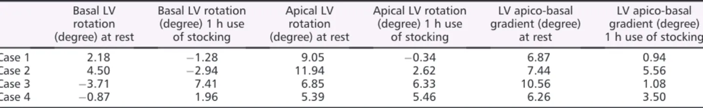
The data of four LE patients showing significant LV rotational abnormalities were managed separately (Table2). In the re- maining group, none of LV rotational parameters differed sig- nificantly from those of matched controls except significant reduction of LV BR following the use of MCS (Table3).
Discussion
We sought to determine the cardiovascular effects of medical compression panty-hose measured by 3DSTE among patients
Figure 1 Three-dimensional (3D) speckle-tracking echocardiographic image of a patient with lymphedema. Apical four chamber (A) and two-chamber views (B), short-axis views at different levels of the left ventricle (LV) (C3, C5, C7), 3D cast of the LV, LV volumetric data and LV apical (white arrow) and basal (dashed arrow) rotations are presented. EDV, end-diastolic volume; EF, ejection fraction; ESV, end-systolic volume; LV, left ventricle.
with bilateral secondary leg LE. Earlierfindings proved that LE is associated with increased arterial stiffness.10 Our recent data disclosed only a few differences in heart LV morphology and rotational mechanics of patients with lower limb lymphoedema compared to those of age-matched control persons.11 2DTTE showed larger LA diameter, LV end-diastolic volume and ejections fraction, while smaller LV end-systolic diameter, interventricular septum, and LV poste- rior wall thickness were also detectable in the lymphoedema cohort.113DSTE could not discriminate between LE and con- trol group in terms of LV rotational mechanics.11
Conversely, none of the previous clinical trials used 3DSTE in the same study population for the assessment of lower limb external compression-related LV mechanics; however, we ear- lier measured the alterations of LV rotations with and without
MCSs among women with lipoedema, the LE masquerading, symmetrical, disproportional fatty enlargement.9
Left ventricular rotation and twist are pivotal components of LV myocardial mechanics referring to the myocardial mo- tion around the LV longitudinal axis. Under standard clinical circumstances the LV base rotates clockwise (negative value), while the LV apex oppositely (positive value). The net differ- ence in this towel-wringing movement is called LV twist. Twist plays an essential role in the mechanical efficiency of the heart, allowing that only a moderate fibre shortening leads to a robust reduction of the LV volume.8,12Our research group preferred the use of 3DSTE over 2D TTE because 3DSTE shows more accurate information from the whole LV thus giving a comprehensive assessment of the entire LV rotational mechanics. 3DSTE has a better correlation with cardiac
Table 2 Changes in LV rotational mechanics in lymphoedema patients with severe LV rotational abnormalities Basal LV
rotation (degree) at rest
Basal LV rotation (degree) 1 h use
of stocking
Apical LV rotation (degree) at rest
Apical LV rotation (degree) 1 h use
of stocking
LV apico-basal gradient (degree)
at rest
LV apico-basal gradient (degree) 1 h use of stocking
Case 1 2.18 1.28 9.05 0.34 6.87 0.94
Case 2 4.50 2.94 11.94 2.62 7.44 5.56
Case 3 3.71 7.41 6.85 6.33 10.56 1.08
Case 4 0.87 1.96 5.39 5.46 6.26 3.50
LV, left ventricular.
Table 3 Changes of LV rotational parameters 1 h after the use of stocking in patients with lymphoedema with normally directed LV rotations
Controls (n= 52) Lymphoedema patients at rest (n= 16)
Lymphoedema patients 1 h after the use of MCS (n= 16)
Basal LV rotation (degree) 4.28 ± 2.18 3.36 ± 1.47 2.70 ± 1.26*
Apical LV rotation (degree) 9.67 ± 4.32 10.86 ± 4.21 10.46 ± 5.95
LV twist (degree) 13.95 ± 4.95 14.22 ± 4.65 13.16 ± 5.92
*P<0.05 vs. controls. LV, left ventricular; MCS, medical compression stocking.
Table 1 Baseline demographic and two-dimensional echocardiographic data on patients and controls
Controls (n= 52) Lymphoedema patients (n= 25)
Age (years) 40.7 ± 14.0 47.8 ± 12.8
Male gender (%) 3 (6) 1 (4)
Hypertension (%) 0 (0) 0 (0)
Diabetes mellitus (%) 0 (0) 0 (0)
Hyperlipidaemia (%) 0 (0) 0 (0)
LA diameter (mm) 35.3 ± 4.1 37.8 ± 4.3*
LV end-diastolic diameter (mm) 46.9 ± 3.7 47.6 ± 3.9
LV end-diastolic volume (mL) 98.5 ± 21.7 108.3 ± 20.1*
LV end-systolic diameter (mm) 31.2 ± 3.4 28.9 ± 3.5*
LV end-systolic volume (mL) 33.9 ± 8.2 33.2 ± 9.4
Interventricular septum (mm) 8.8 ± 1.5 8.0 ± 1.0*
LV posterior wall (mm) 9.0 ± 1.7 8.1 ± 1.0*
LV ejection fraction (%) 65.5 ± 4.3 69.8 ± 4.8*
E (cm/s) 77.6 ± 17.6 76.5 ± 15.6
A (cm/s) 65.5 ± 17.8 67.0 ± 14.5
E/A 1.3 ± 0.4 1.2 ± 0.4
*P<0.05 vs. controls. E and A, early and late transmitralflow velocity, LA, left atrial, LV, left ventricular.
magnetic resonance imaging (MRI) considered as a credible non-invasive assessment of myocardial mechanics.4
Flat-knitted panty-hoses are ideal compression garments for leg LE because they entirely wrap the affected regions, and unlike round-knitted stockings, this stiff material streamlines irregular limbs.13
The aforementioned recent echocardiographic findings of LE patients11gave rise to the measurement of LV rotational characteristics with compression using pantyhose. We in- cluded only patients with stable disease in the maintenance therapeutic phase. After baseline 3DSTE procedure, lower body half was wrapped withflat-knitted panty hose, and after 60 min, the second 3DSTE resulted in a moderate but signifi- cant decrease of LV BR whereas AR as well as twist remained unchanged. These observations suggest that the use of MCS at least does not have detrimental effects on LV rotational mechanics in LE patients without resting LV rotational abnormalities. Increased cardiac afterload usually alters LV rotations and LV twist. The correction of the higher afterload, in turn, usually comes to a decrease of LV rotational parame- ters, including LV BR.12,14
Besides, simultaneous external compression associated haemodynamic changes and their effect on LV mechanisms are also noteworthy. Lattimeret al. proved that ccl 2 below- knee stockings had minimal effects on working venous volume, filling index, and outflow fraction.15 As far as the arterial system is concerned, we found that below-knee ccl 1 MCSs significantly (P = 0.04), and ccl 2 MCSs strongly (P= 0.067) decreased pulse wave velocity (PWV), a measure of aortic stiffness and its amelioration improves cardiovascu- larfitness.16A recent insightful report gives a precise demon- stration of entire limb morphological changes measured by MRI along with the alterations of cardiac output, stroke volume, heart rate detected by echocardiography due to the application of compression tights in supine and upright positions.3Of particular interest, deep arteries and veins of the calf were significantly dilated, whereas superficial veins were considerably slimmer when these limbs were subjected to medium and high pressures3that are closely related to the
pressure range we applied. There was no significant interac- tion effect for these parameters between various pressures and gait. These results suggest that MCSs decrease peripheral arterial resistance which may ameliorate cardiac afterload.
Lower afterload may explain significantly reduced LV BR detected after 60-min use of compression pantyhose.
In summary, our results may foreshadow that 3DSTE can be a sensitive technique to detect limb compression-related LV alterations as well in HF; however, larger clinical studies are needed to validate its applicability.
Limitation
In this study, 3DSTE-derived LV rotational mechanics aimed to be assessed before and 1 h after the use of MCS. Strains and volumes of LV17or other heart chambers18,19were not aimed to be obtained in this study.
Conclusions
The wear of compression pantyhoses moderately, but signifi- cantly reduced LV BR without a remarkable impact on twisting mechanism in LE patients in the absence of LV rotational abnormalities.
Acknowledgement
This study was supported by the funding of the International Phlebology Union (UIP) - Bauerfeind Award 2015-17.
Con fl ict of interest
Authors report no conflict of interest and have nofinancial disclosures.
References
1. Szolnoky G, Dobozy A, Kemény L. To- wards an effective management of chronic lymphedema. Clin Dermatol 2014;32: 685–691.
2. Urbanek T, Juśko M, Kuczmik WB.
Compression therapy for leg oedema in patients with heart failure. ESC Heart Fail2020;7: 2012–2020.
3. Lee DCW, Law HKW, Ali A, Sheridan SE, Wong SHS, Lee SWY. Compression garment-induced leg changes increase hemodynamic responses in healthy indi- viduals.Int J Sports Med2020;41: 3–11.
4. Vachalcova M, Valočik G, Kurečko M, Grapsa J, Taha VA, Michalek P, Jankajová M, Sabol F, Kubikova L, Orban M, Uher T, Böhm A. The three-dimensional speckle tracking echocardiography in distinguishing be- tween ischaemic and non-ischaemic aetiology of heart failure.ESC Heart Fail 2020;7: 2297–2304.
5. Ajmone Marsan N, Michalski B, Cameli M, Podlesnikar T, Manka R, Sitges M, Dweck MR, Haugaa KH. EACVI survey on standardization of cardiac chambers
quantification by transthoracic echocar- diography.Eur Heart J Cardiovasc Imag- ing2020;21: 119–123.
6. Lang RM, Badano LP, Mor-Avi V, Afilalo J, Armstrong A, Ernande L, Flachskampf FA, Foster E, Goldstein SA, Kuznetsova T, Lancellotti P, Muraru D, Picard MH, Rietzschel ER, Rudski L, Spencer KT, Tsang W, Voigt JU. Recommendations for cardiac chamber quantification by echocardiography in adults: an update from the American Society of Echocardi- ography and the European Association
of Cardiovascular Imaging.Eur Heart J Cardiovasc Imaging2015;16: 233–271.
7. Nemes A, Kalapos A, Domsik P, Forster T.
Three-dimensional speckle-tracking echocardiography—a further step in non-invasive three-dimensional cardiac imaging. Orv Hetil 2012; 153: 1570–1577.
8. Nemes A, Kalapos A, Domsik P, Forster T. Left ventricular rotation and twist of the heart. Clarification of some con- cepts.Orv Hetil2012;153: 1547–1551.
9. Nemes A, Kormányos Á, Domsik P, Kalapos A, Kemény L, Szolnoky G.
The impact of lower body compression garment on left ventricular rotational mechanics in patients with lipedema— insights from the three-dimensional speckle tracking echocardiographic MAGYAR-Path Study. Clin Obes 2020;
10: e12380.
10. Yamamoto T, Yamamoto N, Yamashita M, Furuya M, Hayashi A, Koshima I.
Relationship between lymphedema and arteriosclerosis: higher cardio-ankle vas- cular index (CAVI) in lymphedematous limbs. Ann Plast Surg 2016; 76: 336–339.
11. Nemes A, Kormanyos A, Domsik P, Kalapos A, Kemeny L, Forster T, Szolnoky G. Left ventricular rotational mechanics differ between lipedema and
lymphedema: Insights from the three- dimensional speckle tracking echocar- diographic MAGYAR-Path Study.
Lymphology2018;51: 102–108.
12. Stöhr EJ, Shave RE, Baggish AL, Weiner RB. Left ventricular twist me- chanics in the context of normal physiol- ogy and cardiovascular disease: a review of studies using speckle tracking echocardiography.Am J Physiol Heart Circ Physiol 2016; 311: H633–H644.
13. Reich-Schupke S, Stücker M. Round-knit or flat-knit compression garments for maintenance therapy of lymphedema of the leg? - Review of the literature and technical data. J Dtsch Dermatol Ges 2019;17: 775–784.
14. Naeim HA, Abuelatta R, Alatawi FO, Khedr L. Assessment of left ventricular mechanics in patients with severe aortic stenosis after transcatheter aortic valve implantation: 2-D speckle tracking imag- ing study.J Saudi Heart Assoc2020;32: 248–255.
15. Lattimer CR, Kalodiki E, Azzam M, Geroulakos G. Haemodynamic performance of low strength below knee graduated elastic compression stockings in health, venous disease, and lymphoedema.Eur J Vasc Endovasc Surg 2016;52: 105–112.
16. Szolnoky G, Gavallér H, Gönczy A, Bi- hari I, Kemény L, Forster T, Nemes A.
The effects of below-knee medical com- pression stockings on pulse wave veloc- ity of young healthy volunteers. J Strength Cond Res2021;35: 275–279.
17. Nemes A, Kormányos Á, Kalapos A, Domsik P, Gyenes N, Ambrus N, Lengyel C. Normal reference values of left ventricular strain parameters in healthy adults: Real-life experience from the single-center three-dimensional speckle-tracking echocardiographic MAGYAR-Healthy Study. J Clin Ultra- sound2021;49: 368–372.
18. Nemes A, Kormányos Á, Domsik P, Kalapos A, Ambrus N, Lengyel C, Forster T. Normal reference values of right atrial strain parameters using three-dimen- sional speckle-tracking echocardiogra- phy (results from the MAGYAR-Healthy Study).Int J Cardiovasc Imaging2019;
35: 2009–2018.
19. Nemes A, Kormányos Á, Domsik P, Kalapos A, Ambrus N, Lengyel C.
Normal reference values of three-dimen- sional speckle-tracking echocardiogra- phy-derived right atrial volumes and volume-based functional properties in healthy adults (Insights from the MAGYAR-Healthy Study). J Clin Ultra- sound2020;48: 263–268.