Rev. Cardiovasc. Med.2022; 23(1): 005 http://doi.org/10.31083/j.rcm2301005
Copyright: © 2022 The Author(s). Published by IMR Press.
This is an open access article under theCC BY 4.0 license.
Review
Prevalence of left ventricular ‘rigid body rotation’, the near absence of left ventricular twist (insights from the MAGYAR studies)
Attila Nemes*, Árpád Kormányos
Department of Medicine, Medical Faculty, Albert Szent-Györgyi Clinical Center, University of Szeged, 6725 Szeged, Hungary
*Correspondence:nemes@in2nd.szote.u-szeged.hu(Attila Nemes) Academic Editor: Tasneem Z. Naqvi
Submitted: 15 September 2021 Revised: 15 November 2021 Accepted: 24 November 2021 Published: 8 January 2022
Abstract
Left ventricular (LV) twist is defined as the wringing motion of the LV around its long-axis during systole generated by rotation of the LV apex in a counterclockwise direction, as viewed from the apex, while the LV base moves in a clockwise direction. In several cases, the LV apex and base move in the same direction during ejection demonstrating a special condition called as LV ‘rigid body rotation’. The present review aimed to summarize our knowledge about this rare but not fully understood entity demonstrating its theoretic pathophysiologic background, clinical significance, associated diseases, and reversibility based on available literature.
Keywords:Echocardiography; Left ventricular; Rotation; Twist; Speckle-tracking; Two-dimensional; Three-dimensional
1. The normal twisting motion of the heart
Left ventricular (LV) twist represents the wringing motion of the heart along its long-axis during systole gen- erated by rotation of the LV apex in a counterclockwise (CCw) direction, as viewed from the apex, while the LV base moves in a clockwise (Cw) direction [1]. One of the main components of the mechanical efficacy of the heart is the peak systolic LV twist, resulting in 60% LV ejec- tion fraction (EF) with only 15% fiber shortening [2]. This wringing motion is generated by the helical arrangement of myocardial fibers, subendocardially, the spiral holds a left-handed helix, while there is a right-handed helix in the subepicardium. The myocyte helix angle changes continu- ously from the subendocardium to the subepicardium, typi- cally ranging from +60 degree at the subendocardium to –60 degree at the subepicardium [3]. The mid-myocardial cir- cumferential fibers shorten simultaneously with the oblique fibers in the right- and left-handed helices during ejec- tion providing a horizontal counterforce throughout ejec- tion. The subepicardial fibers control the direction of the peak systolic LV twist, mainly due to their longer arm of movement. Therefore, the absence of the endocardial helix would result in increased Cw basal and CCw apical rota- tion, while the absence of the epicardial helix would cause CCw basal and Cw apical LV rotation [4].
2. Estimation of LV rotational mechanics
In earlier years, LV rotational mechanics could be examined only by invasive (microsonometry) or expen- sive (magnetic resonance imaging) methodologies [1].
Two-dimensional (2D) speckle-tracking echocardiography (STE) is a non-invasive opportunity to assess LV rotational mechanics. However, the LV is a three-dimensional (3D)
chamber, therefore its examination in 2D theoretically is associated with loss of information. This fact leads to guidelines not supporting 2DSTE in the quantification of LV rotational mechanics [5]. Moreover, significant dif- ferences between 2DSTE- and 3DSTE-derived LV rota- tional parameters could also be demonstrated [6]. 3DSTE is a novel method for 3D assessment of LV. Shortly, full volume ‘echocloud’ (3D echocardiographic dataset) is ac- quired using 3D capable matrix transducer at the begin- ning of 3DSTE examination in which analysis could be per- formed online or offline using a special software by creating a virtual 3D model of the LV. Volumetric changes in LV re- specting the cardiac cycle, LV contractility represented by LV strains and LV rotational parameters could be calculated at the same time using this cast [7] (Fig.1).
3. MAGYAR studies
The aim of our study conducted in our tertiary car- diovascular center was to assess the diagnostic and poten- tially prognostic values of 3DSTE-derived parameters in- cluding LV rotational parameters. LV apical and basal ro- tations and LV twist were measured in healthy subjects (MAGYAR-Healthy Study) and in several pathological states (MAGYAR-Path Study). LV rotational parameters were measured in elite athletes (MAGYAR-Sport Study), in twins (MAGYAR-Twin Study), in foetuses (MAGYAR- Fetus Study) and during stress protocols (MAGYAR-Stress Study) [7]. (‘Magyar’ means ‘Hungarian’ in Hungarian lan- guage).
4. Near absence of LV twist—LV ‘rigid body rotation’
In special circumstances, LV ‘rigid body rotation’
(RBR), the near absence of LV twist may develop due to
Fig. 1. Left ventricular (LV) rotational mechanics assessed by three-dimensional (3D) speckle-tracking echocardiography.Apical four-chamber (A) and two-chamber views (B) and basal (C3), midventricular (C5) and apical (C7) short-axis views are presented extracted from the acquired 3D volumetric dataset. LV cast (D), curves representing apical (white arrow) and basal (yellow arrow) LV rotations (E) and LV volumetric parameters (F) generated by the software are presented. LA, left atrium; LV, left ventricular; RA, right atrium; RV, right ventricle; EDV, end-diastolic volume; ESV, end-systolic volume; EF, ejection fraction; GLS, LV global longitudinal strain; GCS, LV global circumferential strain; GAC, LV global area strain (change).
changes in the direction of the movement of the myocar- dial fibers, the LV apex and LV base move in the same clockwise (Cw) or counterclockwise (CCw) direction [8]
(Figs.2,3). This phenomenon is considered to be normal in healthy neonates with an immature heart, LV-RBR has occurred due to the counterclockwise rotation of the basal and apical fibers [9]. However, later the normal LV rota- tional mechanics develops. In some special clinical circum- stances, LV-RBR persists in adults, its real clinical signif- icance and prevalence are not known. The present review aimed to summarize related information from known liter- ature mainly based on the results of the MAGYAR Studies.
5. Cardiac diseases associated with LV-RBR
5.1 Noncompaction cardiomyopathy
LV noncompaction cardiomyopathy (NCCM) or spongiform cardiomyopathy is a rare congenital cardiomy- opathy due to an intra-uterine arrest of compaction of the myocardial fibres during embryogenesis [10]. NCCM is characterized by a thin, compacted epicardium and a thick
non-compacted endocardium, with prominent trabeculation and deep intertrabecular recesses [10,11]. Characteristic complications of NCCM include chronic heart failure, life- threatening ventricular arrhythmias and systemic embolic events [10,11].
According to the literature, NCCM is the most exten- sively examined LV-RBR-related disease, where its preva- lence ranged from 26% to 100% (Table1) [11–21]. van Dalenet al. [12] assessed patients with dilated cardiomy- opathy (DCM), healthy subjects and cases with prominent trabeculations, of whom expert opinion in NCCM defined some of them having NCCM, and found that LV-RBR was an objective, quantitative and reproducible functional crite- rion, and had a good predictive value in the diagnosis of NCCM. Specificity and sensitivity of LV-RBR was 88%
and 78% in differentiating NCCM from LV “hypertrabec- ulation”, respectively [12]. Cw LV-RBR was detected in most NCCM cases [11,12,15,18–20], which was found to be related to LV-EF [17]. Almost one-third of the relatives of NCCM patients showed LV-RBR, the ratio of Cw and
Fig. 2. Abnormal clockwise apical (white arrow) and nor- mal clockwise basal (yellow arrow) left ventricular rotations (clockwise left ventricular ‘rigid body rotation’).LV, left ven- tricular; EDV, end-diastolic volume; ESV, end-systolic volume;
EF, ejection fraction; LV-GLS, LV global longitudinal strain; LV- GCS, LV global circumferential strain; LV-GAC, LV global area strain (change).
CCw direction was the same [18]. Ratio of LV-RBR was significantly different between NCCM children and sub- jects with hypertrabecularization [19]. In another study, re- verse LV apical rotation (Cw LV-RBR) was present in 39%
of NCCM children [20].
5.2 Dilated or nonischaemic cardiomyopathy
LV rotational mechanics was examined only in a lim- ited number of studies in series of DCM or nonischaemic (NICM) cardiomyopathy patients. 88–100% of patients showed Cw LV-RBR [22,23]. Reversed apical rotation (Cw LV-RBR) and loss of LV torsion was found to be associ- ated with significant LV remodelling, increased electrical dyssynchrony, reduced systolic function, and increased fill- ing pressures in patients with DCM, indicating a more ad- vanced stage of the disease [22]. The normal torsion pat- tern was observed more frequently in NICM patients with- out mid-wall fibrosis (MWF), and LV-RBR was more fre- quently observed in patients with MWF [24].
5.3 Hypertensive and other special cardiomyopathies Hypertensive heart disease includes a number of com- plications due tohigh blood pressureincluding LV hyper- trophy, heart failure, etc. In hypertensive cardiomyopathy patients with reduced LV-EF, 32% of subjects showed pre- dominantly Cw LV-RBR suggesting compromised LV api- cal rotation similarly to NCCM/DCM/NICM patients [25].
Theoretically hypertension-related abnormalities, fibrosis, etc. could explain these findings. CCw LV-RBR was found in cases with peripartum and chloroquine cardiomyopathies [26,27]. In a small study with hypertrophic cardiomyopathy (HCM), only 1 case out of 18 showed Cw LV-RBR [21].
5.4 Infiltrative cardiac disorders
Regarding the literature, only positron emission tomography-proven cardiac lymphoma [28] and biopsy- proven cardiac amyloidosis (CA) [29] were found to be re- lated to LV-RBR at this moment. Interestingly, CCw LV- RBR could be demonstrated in all cases suggesting com- promised LV basal rotation [28,29]. Infiltration of the my- ocardial tissue by amyloid or lymphomatous tissue could lead to changes in LV twisting deformation [28,29].
5.5 Congenital heart diseases
LV-RBR was reported only in a few case reports in cer- tain congenital heart diseases (CHDs) including hypoplastic right heart syndrome [30], univentricular heart [31] and Eb- stein’s anomaly [32]. Interestingly, hypoplastic right heart syndrome was associated with Cw LV-RBR, while univen- tricular heart and Ebstein’s anomaly were associated with CCw LV-RBR in some individual patients. Abnormalities in the development of myocardial architecture during mor- phogenesis could be theorised to be responsible for LV- RBR in these diseases [30–32].
Tetralogy of Fallot (TOF) is a cardiac anomaly with combination of ventricular septal defect, overriding aorta, pulmonary stenosis and right ventricular hypertrophy [33–
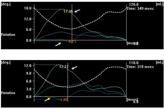
35]. In patients with corrected TOF from the MAGYAR- Path Study, 27% of subjects showed Cw LV-RBR, while 11% of patients had CCw LV-RBR [33]. In case of cor- rected TOF patients with early total reconstruction, the same values proved to be beneficial compared to those who had early palliation and late correction. In another earlier study, the values of the same parameters were 15% and 18%, respectively, with patients with almost zero LV ro- tations [34] (Fig.4). In the study of Dragulescuet al. [35], 38% of corrected TOF patients showed CCw LV-RBR.
6. Disorders with LV-RBR without obvious cardiac involvement
6.1 Acromegaly
Acromegaly is a rare and chronic hormonal disorder that develops due to the hypersecretion of the human growth hormone (GH) and insulin-like growth factor-1 (IGF-1) in adults [36–38]. LV-RBR could be detected in 20% of the cases with normal LV-EF (without obvious signs of clas- sic acromegalic cardiomyopathy), which did not depend on the presence or absence of diabetes mellitus [37,38]. LV- RBR proved to be CCw in 75% of cases, these abnormali- ties could be theorized to long-term effects of GH and IGF- 1.
Fig. 3. Normal counterclockwise apical (white arrow) and abnormal counterclockwise basal (yellow arrow) left ventricular ro- tations (counterclockwise left ventricular ‘rigid body rotation’).LV, left ventricular; EDV, end-diastolic volume; ESV, end-systolic volume; EF, ejection fraction; LV-GLS, LV global longitudinal strain; LV-GCS, LV global circumferential strain; LV-GAC, LV global area strain (change).
6.2 Hypopituitarism
Hypopituitarism is a rare and complex hormonal dis- ease caused by the decreased secretion of one or more hor- mones in the pituitary gland. LV-RBR was present in 13%
of patients, potentially due to abnormal secretion of certain hormones [39].
6.3 Lipedema and lymphedema
While lipedema is a feminine disorder with dispropor- tional, bilateral and symmetrical obesity of unknown path- omechanism, excessive accumulation of lymphatic fluid could be detected due to certain factors in lymphedema [40]. In several cases, lipedema and lymphedema are dif- ficult to be differentiated clinically. However, in both dis- orders, the ratio of LV-RBR was similar (14%) with typical CCw direction in lymphedema patients [40]. The use of compression medical stockings had significant effects on LV rotational mechanics both in lipedema [41] and lym- phedema [42].
6.4 Hidradenitis suppurativa
Hidradenitis suppurativa is a dermatological disorder where chronic inflammation was suggested to be having ef- fects on LV rotational mechanics [43].
6.5 Haemophilia
Haemophilia is an X-linked congenital coagulation factor deficiency. Although significant LV rotational ab- normalities are present in haemophilia with reduced LV api- cal rotation and twist, ratio of patients with LV-RBR is not higher than in the normal population [44].
6.6 Hypereosinophilic syndrome
In hypereosinophilic syndrome (HES), peripheral eosinophilia and eosinophilic tissue/end-organ damage could be demonstrated [45]. In HES patients in the early necrotic stage, LV rotational abnormalities including pres- ence of LV-RBR was found in 13% of cases.
6.7 Following kidney transplantation
In all patients with end-stage renal disease, kidney transplantation is the preferred treatment [46]. In post- transplant patients, ratio of LV-RBR was not significantly higher than in normal controls with CCw direction.
6.8 Twin-to-twin transfusion syndrome
In a recent case report, twin-to-twin transfusion syn- drome was confirmed in the past medical history of an iden- tical pair of twins who showed different patterns of LV- RBR, Cw LV-RBR was described in the recipient twin and CCw LV-RBR was found in the donor twin [47]. Similarly to CHDs, abnormalities in the development of myocardial architecture could lead to LV-RBR in twin-to-twin transfu- sion syndrome theoretically.
7. Healthy subjects and elite athletes
In a current study demonstrating normal reference val- ues of 3DSTE-derived LV rotational parameters and their age- and gender-dependency, 6% of cases proved to have LV-RBR. At the time of 3DSTE, these subjects had no ab- normality, pathologic state, drug use, symptoms, electro- cardiographic or echocardiographic findings, which could explain LV-RBR. Theoretically, these subjects had subclin-
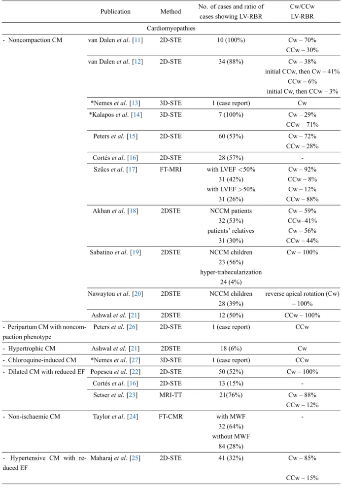
Table 1. Studies confirming left ventricular ‘rigid body rotation’ in different cardiac disorders.
Publication Method No. of cases and ratio of cases showing LV-RBR
Cw/CCw LV-RBR Cardiomyopathies
- Noncompaction CM van Dalenet al. [11] 2D-STE 10 (100%) Cw – 70%
CCw – 30%
van Dalenet al. [12] 2D-STE 34 (88%) Cw – 38%
initial CCw, then Cw – 41%
CCw – 6%
initial Cw, then CCw – 3%
*Nemeset al. [13] 3D-STE 1 (case report) Cw
*Kalaposet al. [14] 3D-STE 7 (100%) Cw – 29%
CCw – 71%
Peterset al. [15] 2D-STE 60 (53%) Cw – 72%
CCw – 28%
Cortéset al. [16] 2D-STE 28 (57%) -
Szűcset al. [17] FT-MRI with LVEF<50% Cw – 92%
31 (42%) CCw – 8%
with LVEF>50% Cw – 12%
31 (26%) CCw – 88%
Akhanet al. [18] 2DSTE NCCM patients Cw – 59%
32 (53%) CCw–41%
patients’ relatives Cw – 56%
31 (30%) CCw – 44%
Sabatinoet al. [19] 2DSTE NCCM children Cw – 100%
23 (56%) hyper-trabecularization
24 (4%)
Nawaytouet al. [20] 2DSTE NCCM children reverse apical rotation (Cw)
28 (39%) – 100%
Ashwalet al. [21] 2DSTE 12 (50%) CCw – 100%
- Peripartum CM with noncom- paction phenotype
Peterset al. [26] 2D-STE 1 (case report) CCw
- Hypertrophic CM Ashwalet al. [21] 2DSTE 18 (6%) Cw
- Chloroquine-induced CM *Nemeset al. [27] 3D-STE 1 (case report) CCw - Dilated CM with reduced EF Popescuet al. [22] 2D-STE 50 (52%) Cw – 100%
Cortéset al. [16] 2D-STE 13 (15%) -
Setseret al. [23] MRI-TT 21(76%) Cw – 88%
CCw – 12%
- Non-ischaemic CM Tayloret al. [24] FT-CMR with MWF -
32 (64%) without MWF
84 (28%) - Hypertensive CM with re-
duced EF
Maharajet al. [25] 2D-STE 41 (32%) Cw – 85%
CCw – 15%
Table 1. Continued.
Publication Method No. of cases and ratio of cases showing LV-RBR
Cw/CCw LV-RBR Infiltrative disorders with cardiac involvement
- Cardiac lymphoma *Nemeset al. [28] 3D-STE 1 (case report) CCw
- Cardiac amyloidosis *Nemeset al. [29] 3D-STE 10 (60%) CCw – 100%
Congenital heart diseases - Hypoplastic right heart syn-
drome
*Nemeset al. [30] 3D-STE 1 (case report) Cw
- Univentricular heart *Nemeset al. [31] 3D-STE 1 (case report) CCw
- Ebstein’ anomaly *Nemeset al. [32] 3D-STE 1 (case report) CCw
- Tetralogy of Fallot *Nemeset al. [33] 3D-STE 26 (38%) Cw – 70%
CCw – 30%
Mentinget al. [34] 2D-STE 82 (34%) Cw – 46%
CCw – 54%
Dragulescuet al. [35] 2D-STE 50 (38%) CCw – 100%
2D, two-dimensional; 3D, three-dimensional; Cw, clockwise; CCw, counterclockwise; LV-RBR, left ventricular ‘rigid body rotation’;
CM, cardiomyopathy; LVEF, left ventricular ejection fraction; MWF, mid-wall fibrosis; NCCM, noncompaction cardiomyopathy;
STE, speckle-tracking echocardiography; FT-CMR, feature-tracking cardiovascular magnetic resonance.
* Results from the MAGYAR-Path Study.
ical abnormalities or undiagnosed disorders which were not known at the time of 3DSTE [48]. In a study compar- ing LV rotational mechanics in NCCM patients and con- trols, 23% of healthy subjects showed CCw LV-RBR which higher rate could be partially explained by selection bias [17]. In contrast, none of the elite athletes doing high dy- namic sports showed 3DSTE-proven LV-RBR in a recent study [49]. The above mentioned results suggest further examinations mainly focusing any diagnostic or prognostic impact of LV-RBR in subjects without symptoms or appar- ent abnormalities.
8. Special considerations regarding LV-RBR
8.1 Clockwise vs. counterclockwise LV-RBR
Normally, the LV base rotates clockwise, while the LV apex rotates in counterclockwise direction. In Cw LV-RBR, apical rotation of the LV has a clockwise direction (negative value on 3DSTE images, Fig.2), while in CCw LV-RBR, basal rotation of the LV has a counterclockwise direction (positive value on 3DSTE images, Fig.3). Direction of LV-RBR and their ratio in certain cardiac and non-cardiac disorders are presented in Tables1 and2 (Ref. [17,36–
40,43–49]). Interestingly, while most cardiomyopathy pa- tients showed Cw LV-RBR [11,12,15,18,19,22,23,25], pa- tients with non-cardiac disorders without obvious cardiac involvement (lymphedema, acromegaly, following kidney transplantation) most likely had CCw LV-RBR [37,40,46].
8.2 Heart failure treatment, reversibility and LV-RBR A recent report described a patient with evidence of re- versible LV-RBR who met the clinical criteria for the diag- nosis of both peripartum cardiomyopathy and LV noncom- paction with reduced LV-EF. Following 6-months heart fail- ure treatment, improvement in LV-EF could be observed to- gether with normalization of LV rotational mechanics [26].
8.3 Vasodilation, stress test and LV-RBR
Vasodilation and stress-related LV-RBR is weakly as- sessed. In a patient with previous coronary stenting pre- sented with stable angina and positive exercise treadmill test, LV-RBR could be demonstrated, its characteristics changed dramatically during dipyridamole-induced vasodi- latation [50]. A case of abnormal CCw rotation of the LV base was reported in a patient with angina pectoris with- out significant coronary artery disease (CAD) at rest, the direction of the rotation of the LV base changed to Cw the- oretically due to dipyridamole-induced vasodilation via im- proved blood supply at maximum hyperaemia [51]. At the recovery phase, LV-RBR could be detected again in this case. On the contrary, a CAD patient showed LV-RBR dur- ing dipyridamole stress, which normalized by the end of stress [52].
8.4 ‘Clear’ LV-RBR vs. LV-RBR with apico-basal gradient LV-RBR is the near absence of LV twist, when rotation at both apical and basal levels of the LV occur in the same Cw or CCw direction (Figs.2,3,4). Although all segments move in the same direction, the amplitude of their motion
Fig. 4. Increased counterclockwise left ventricular apical rotation (white arrow) with almost zero left ventricular basal rotation (yellow arrow).
Fig. 5. Differences between apical and basal left ventricular rotations in the same counterclockwise direction demonstrating high (left panel) or low (right panel) left ventricular apico-basal rotational gradient.LV, left ventricular; EDV, end-diastolic volume; ESV, end-systolic volume; EF, ejection fraction; LV-GLS, LV global longitudinal strain; LV-GCS, LV global circumferential strain; LV-GAS, LV global area strain.
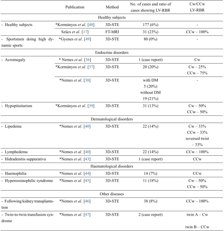
Table 2. Studies confirming left ventricular ‘rigid body rotation’ in different non-cardiac diseases without known cardiac involvement.
Publication Method No. of cases and ratio of cases showing LV-RBR
Cw/CCw LV-RBR Healthy subjects
- Healthy subjects *Kormányoset al. [48] 3D-STE 177 (6%) -
Szűcset al. [17] FT-MRI 31 (23%) CCw – 100%
- Sportsmen doing high dy- namic sports
*Gyeneset al. [49] 3D-STE 80 (0%) -
Endocrine disorders
- Acromegaly * Nemeset al. [36] 3D-STE 1 (case report) Cw
*Kormányoset al. [37] 3D-STE 20 (20%) Cw – 25%
CCw – 75%
*Nemeset al. [38] 3D-STE with DM -
5 (20%) without DM
19 (21%)
- Hypopituitarism *Kormányoset al. [39] 3D-STE 31 (13%) Cw – 50%
CCw – 50%
Dermatological disorders
- Lipedema *Nemeset al. [40] 3D-STE 22 (14%) Cw – 33%
CCw – 33%
reversed twist – 33%
- Lymphedema *Nemeset al.[40] 3D-STE 22 (14%) CCw – 100%
- Hidradenitis suppurativa *Nemeset al. [43] 3D-STE 1 (case report) CCw Haematological disorders
- Haemophilia *Nemeset al. [44] 3D-STE 14 (7%) CCw
- Hypereosinophilic syndrome *Nemeset al. [45] 3D-STE 11 (18%) Cw – 50%
CCw – 50%
Other diseases - Following kidney transplanta-
tion
*Nemeset al. [46] 3D-STE 38 (8%) CCw – 100%
- Twin-to-twin transfusion syn- drome
*Nemeset al.[47] 3D-STE 2 (case report) twin A – Cw
twin B – CCw 3D, three-dimensional; Cw, clockwise; CCw, counterclockwise; LV-RBR, left ventricular ‘rigid body rotation’; STE, speckle-tracking echocardiography; FT-CMR, feature-tracking cardiovascular magnetic resonance.
* Results from the MAGYAR-Path, MAGYAR-Healthy and MAGYAR Sport Studies.
could be different leading to regional differences between apical and basal LV rotations called as LV apico-basal gra- dient (Fig.5). In some disorders, LV apico-basal gradient could be elevated like in case of CA [29]. Its clinical sig- nificance is not known but could be considered as an over- compensation due to absence of normally directed LV rota- tional mechanics. Similar changes could be detected during stress as well [50–52].
9. Prognostic value of LV-RBR
Limited information is available regarding prognostic impact of LV-RBR. In a recent study, reverse LV apical ro- tation (Cw LV-RBR) was not a sensitive but a specific indi- cator of complications in children with NCCM suggesting its prognostic rather than diagnostic value [20].
10. Possible pathophysiological explanations of LV-RBR
Theoretically, changes in LV rotational mechanics may occur due to the above mentioned special factors, as well as due to classic risk factors, such as hypertension, hy- percholesterolaemia, diabetes mellitus, etc. or subclinical diseases (focal/local oedema, inflammation, fibrosis, etc.).
The effect of CAD and related haemodynamic changes could not be excluded either.
11. The role of three-dimensional
speckle-tracking (strain) echocardiography in current cardiology practice
Although 3DSTE is not a guideline-recommended technique at this moment, it has several advantages as com- pared to other echocardiographic methods. It sees the heart as it is: a 3D organ and all volumetric, functional (strain) and rotational parameters of heart chambers respecting the cardiac cycle can be calculated at the same time using the same virtual 3D models based on digitally acquired echocardiographic datasets. Moreover, data acquisition and analysis do not require significantly more time in practi- cal hands than a routine echocardiographic examination.
These facts could highlight its importance in clinical prac- tice in more complicated cases, which require more detailed analysis (for instance in heart failure or following myocar- dial infarction). As demonstrated above, 3DSTE-derived parameters could help in differentiating NCCM from LV
‘hypertrabecularization’ or HCM from cardiac amyloidosis [12,53]. Moreover, prognostic impact of 3DSTE-derived ejection fraction and 3D strain parameters has also been demonstrated [54,55]. 3DSTE allows detailed assessment of the right ventricle and both atria, as well [53,55]. The role of 3DSTE in the assessment of atrioventricular valvu- lar annuli has also been demonstrated [56].
Most of echocardiographic laboratories have access to 2D strain software and LV rotational parameters can be cal- culated with them, 3DSTE-derived ones are recommended according to the guidelines [5]. Although 2DSTE-derived global longitudinal strain is well known and a widely used parameter with a strong progmostic impact [57], limitations of 2DSTE should always be considered, when LV rotations are calculated.
12. Conclusions
LV-RBR is defined as the near absence of LV twist when LV apical and basal segments move in the same clockwise or counterclockwise direction. LV-RBR could be demonstrated in a number of diseases with larger preva- lence in certain disorders with cardiac involvement. How- ever, some non-cardiac diseases without overt cardiovascu- lar involvement show limited prevalence of LV-RBR with- out obvious significance, which would require further in- vestigations.
Author contributions
AN and ÁK designed the research study, performed the research and analyzed the data. All authors contributed to editorial changes in the manuscript. All authors read and approved the final manuscript.
Ethics approval and consent to participate
All subjects gave their informed consent for inclu- sion before they participated in the MAGYAR studies.
The study was conducted in accordance with the Decla- ration of Helsinki, and the protocol was approved by the Ethics Committee of University of Szeged (approval num- ber: 71/2011).
Acknowledgment
We would like to express our gratitude to all those who helped us during the writing of this manuscript. Thanks to all the peer reviewers for their opinions and suggestions.
Funding
This research received no external funding.
Conflict of interest
The authors declare no conflict of interest. Attila Nemes is serving as one of the Editorial Board members of this journal. We declare that Attila Nemes had no involve- ment in the peer review of this article and has no access to information regarding its peer review. Full responsibil- ity for the editorial process for this article was delegated to Tasneem Z. Naqvi.
References
[1] Nakatani S. Left ventricular rotation and twist: why should we learn? Journal of Cardiovascular Ultrasound. 2011; 19: 1–6.
[2] Sallin EA. Fiber orientation and ejection fraction in the human left ventricle. Biophysical Journal. 1969; 9: 954–964.
[3] Geerts L, Bovendeerd P, Nicolay K, Arts T. Characterization of the normal cardiac myofiber field in goat measured with MR- diffusion tensor imaging. American Journal of Physiology-Heart and Circulatory Physiology. 2002; 283: H139–H145.
[4] Sedmera D, Pexieder T, Vuillemin M, Thompsin RP, Ander- son RH. Developmental patterning of the myocardium. The Anatomical Record. 2000; 258: 319–337.
[5] Voigt JU, Pedrizzetti G, Lysyansky P, Marwick TH, Houle H, Baumann R,et al. Definitions for a common standard for 2D speckle tracking echocardiography: consensus document of the EACVI/ASE/Industry Task Force to standardize deforma- tion imaging. European Heart Journal Cardiovascular Imaging.
2015; 16: 1–11.
[6] Andrade J, Cortez LD, Campos O, Arruda AL, Pinheiro J, Vulca- nis L,et al. Left ventricular twist: comparison between two- and three-dimensional speckle-tracking echocardiography in healthy volunteers. European Journal of Echocardiography. 2011; 12:
76–79.
[7] Nemes A, Kalapos A, Domsik P, Forster T. Three-dimensional speckle-tracking echocardiography – a further step in non- invasive three-dimensional cardiac imaging. Orvosi Hetilap.
2012; 153: 1570–1577. (In Hu)
[8] Nemes A, Kalapos A, Domsik P, Forster T. Left ventricular rota- tion and twist of the heart. Clarification of some concepts. Orvosi Hetilap. 2012; 153: 1547–1551. (In Hu)
[9] Notomi Y, Srinath G, Shiota T, Martin-Miklovic MG, Beachler L, Howell K,et al. Maturational and Adaptive Modulation of Left Ventricular Torsional Biomechanics: Doppler tissue imag- ing observation from infancy to adulthood. Circulation. 2006;
113: 2534–2541.
[10] Jenni R, Oechslin E, Schneider J, Attenhofer Jost C, Kaufmann PA. Echocardiographic and pathoanatomical characteristics of isolated left ventricular non-compaction: a step towards classi- fication as a distinct cardiomyopathy. Heart. 2001; 86: 666–671.
[11] van Dalen BM, Caliskan K, Soliman OI, Nemes A, Vletter WB, Ten Cate FJ,et al. Left ventricular solid body rotation in non-compaction cardiomyopathy: a potential new objective and quantitative functional diagnostic criterion? European Journal of Heart Failure. 2008; 10: 1088–1093.
[12] van Dalen BM, Caliskan K, Soliman OII, Kauer F, van der Zwaan HB, Vletter WB,et al. Diagnostic Value of Rigid Body Rotation in Noncompaction Cardiomyopathy. Journal of the American Society of Echocardiography. 2011; 24: 548–555.
[13] Nemes A, Kalapos A, Domsik P, Forster T. Identification of Left Ventricular “Rigid Body Rotation” by Three-Dimensional Speckle-Tracking Echocardiography in a Patient with Noncom- paction of the Left Ventricle: a Case from the MAGYAR-Path Study. Echocardiography. 2012; 29: E237–E240.
[14] Kalapos A, Domsik P, Forster T, Nemes A. Comparative eval- uation of left ventricular function by two-dimensional echocar- diography and three-dimensional speckle-tracking echocardio- graphy in noncompaction cardiomyopathy. Results from the MAGYAR-Path Study. Orvosi Hetilap. 2013; 154: 1352–1359.
(In Hu)
[15] Peters F, Khandheria BK, Libhaber E, Maharaj N, dos Santos C, Matioda H,et al. Left ventricular twist in left ventricular non- compaction. European Heart Journal - Cardiovascular Imaging.
2014; 15: 48–55.
[16] Cortés M, Oliva MR, Orejas M, Navas MA, Rábago RM, Martínez ME, et al. Usefulness of speckle myocardial imag- ing modalities for differential diagnosis of left ventricular non- compaction of the myocardium. International Journal of Cardi- ology. 2016; 223: 813–818.
[17] Szűcs A, Kiss AR, Gregor Z, Horváth M, Tóth A, Dohy Z,et al.
Changes in strain parameters at different deterioration levels of left ventricular function: a cardiac magnetic resonance feature- tracking study of patients with left ventricular noncompaction.
International Journal of Cardiology. 2021; 331: 124–130.
[18] Akhan O, Demir E, Dogdus M, Cakan FO, Nalbantgil S. Speckle tracking echocardiography and left ventricular twist mechanics:
predictive capabilities for noncompaction cardiomyopathy in the first degree relatives. The International Journal of Cardiovascu- lar Imaging. 2021; 37: 429–438.
[19] Sabatino J, Di Salvo G, Krupickova S, Fraisse A, Prota C, Buc- ciarelli V,et al. Left Ventricular Twist Mechanics to Identify Left Ventricular Noncompaction in Childhood. Circulation. Car- diovascular imaging. 2019; 12: e007805.
[20] Nawaytou HM, Montero AE, Yubbu P, Calderón-Anyosa RJC, Sato T, O’Connor MJ,et al. A Preliminary Study of Left Ven- tricular Rotational Mechanics in Children with Noncompaction Cardiomyopathy: Do They Influence Ventricular Function?
Journal of the American Society of Echocardiography. 2018; 31:
951–961.
[21] Ashwal AJ, Mugula SR, Samanth J, Paramasivam G, Nayak K, Padmakumar R. Role of deformation imaging in left ventricular non-compaction and hypertrophic cardiomyopathy: an Indian perspective. The Egyptian Heart Journal. 2020; 72: 6.
[22] Popescu BA, Beladan CC, Călin A, Muraru D, Deleanu D, Roşca
M,et al. Left ventricular remodelling and torsional dynamics in dilated cardiomyopathy: reversed apical rotation as a marker of disease severity. European Journal of Heart Failure. 2009; 11:
945–951.
[23] Setser RM, Kasper JM, Lieber ML, Starling RC, McCarthy PM, White RD. Persistent abnormal left ventricular systolic torsion in dilated cardiomyopathy after partial left ventriculectomy. The Journal of Thoracic and Cardiovascular Surgery. 2003; 126: 48–
55.
[24] Taylor RJ, Umar F, Lin ELS, Ahmed A, Moody WE, Mazur W,et al. Mechanical effects of left ventricular midwall fibrosis in non- ischemic cardiomyopathy. Journal of Cardiovascular Magnetic Resonance. 2016; 18: 1.
[25] Maharaj N, Khandheria BK, Peters F, Libhaber E, Essop MR.
Time to twist: marker of systolic dysfunction in Africans with hypertension. European Heart Journal - Cardiovascular Imaging.
2013; 14: 358–365.
[26] Peters F, Khandheria BK, dos Santos C, Govender S, Botha F, Essop MR. Peripartum Cardiomyopathy Associated with Left Ventricular Noncompaction Phenotype and Reversible Rigid Body Rotation. Circulation: Heart Failure. 2013; 6: e62–e63.
[27] Nemes A, Nagy V, Domsik P, Kalapos A, Kormányos Á, Havasi K, Forster T. Left ventricular rotational abnormalities in chloroquine-induced cardiomyopathy (from the MAGYAR-Path Study). Minerva Cardioangiol. 2018; 66: 786–787.
[28] Földeák D, Kalapos A, Domsik P, Sinkó M, Szeleczki N, Bagdi E,et al. Left ventricular rigid body rotation in a dif- fuse large B-cell lymphoma patient with cardiac involvement:
A case from the three-dimensional speckle-tracking echocardio- graphic MAGYAR-Path Study. Revista Portuguesa de Cardiolo- gia. 2017; 36: 145.e1–145.e5.
[29] Nemes A, Földeák D, Domsik P, Kalapos A, Sepp R, Borbényi Z, et al. Different patterns of left ventricular rotational mechanics in cardiac amyloidosis-results from the three-dimensional speckle- tracking echocardiographic MAGYAR-Path Study. Quantitative Imaging in Medicine and Surgery. 2015; 5: 853–857.
[30] Nemes A, Havasi K, Forster T. “Rigid body rotation” of the left ventricle in hypoplastic right-heart syndrome: a case from the three-dimensional speckle-tracking echocardiographic MAGYAR-Path Study. Cardiology in the Young. 2015; 25:
768–772.
[31] Nemes A, Havasi K, Domsik P, Kalapos A, Forster T. Can uni- ventricular heart be associated with “rigid body rotation”? A case from the three-dimensional speckle-tracking echocardio- graphic MAGYAR-Path Study. Hellenic Journal of Cardiology.
2015; 56: 186–188.
[32] Nemes A, Havasi K, Domsik P, Kalapos A, Forster T. Left Ventricular Rigid Body Rotation in Ebstein’s Anomaly from the MAGYAR-Path Study. Arquivos Brasileiros de Cardiologia.
2016; 160: 544–545.
[33] Nemes A, Rácz G, Kormányos Á, Domsik P, Kalapos A, Gyenes N,et al. Left ventricular rotational abnormalities in adult pa- tients with corrected tetralogy of Fallot following different sur- gical procedures (Results from the CSONGRAD Registry and MAGYAR-Path Study). Cardiovascular Diagnosis and Therapy.
2021; 11: 623–630.
[34] Menting ME, Eindhoven JA, van den Bosch AE, Cuypers JAAE, Ruys TPE, van Dalen BM,et al. Abnormal left ventricular ro- tation and twist in adult patients with corrected tetralogy of Fal- lot. European Heart Journal - Cardiovascular Imaging. 2014; 15:
566–574.
[35] Dragulescu A, Friedberg MK, Grosse-Wortmann L, Redington A, Mertens L. Effect of Chronic Right Ventricular Volume Over- load on Ventricular Interaction in Patients after Tetralogy of Fal- lot Repair. Journal of the American Society of Echocardiogra- phy. 2014; 27: 896–902.
[36] Nemes A, Kormányos Á, Domsik P, Kalapos A, Lengyel C, Valkusz Z,et al. Left ventricular ‘rigid body rotation’ in a patient with acromegaly (from the MAGYAR-Path Study). Quantitative Imaging in Medicine and Surgery. 2017; 7: 378–379.
[37] Kormányos Á, Domsik P, Kalapos A, Orosz A, Lengyel C, Valkusz Z,et al. Left ventricular twist is impaired in acromegaly:
Insights from the three-dimensional speckle tracking echocar- diographic MAGYAR-Path Study. Journal of Clinical Ultra- sound. 2018; 46: 122–128.
[38] Nemes A, Kormányos Á, Domsik P, Kalapos A, Gyenes N, Lengyel C, et al. Diabetes mellitus deteriorates left ven- tricular deformation in acromegaly—analysis from the three- dimensional speckle-tracking echocardiographic MAGYAR- Path Study. Quantitative Imaging in Medicine and Surgery.
2021; 11: 410–414.
[39] Kormányos Á, Gyenes N, Horváth Á, Ambrus N, Lengyel C, Valkusz Z, et al. Left Ventricular Rotational Abnormal- ities in Treated Hypopituitarism: Insights From the Three- Dimensional Speckle-Tracking Echocardiographic MAGYAR- Path Study. Frontiers in Cardiovascular Medicine. 2021; 8:
703146.
[40] Nemes A, Kormányos A, Domsik P, Kalapos A, Keméeny L, Forster T, et al. Left ventricular rotational mechanics differ between lipedema and lymphedema: Insights from the three- dimensional speckle tracking echocardiographic MAGYAR- Path Study. Lymphology. 2018; 51: 102–108.
[41] Nemes A, Kormányos Á, Domsik P, Kalapos A, Kemény L, Szolnoky G. The impact of lower body compression garment on left ventricular rotational mechanics in patients with lipedema- Insights from the three-dimensional speckle tracking echocar- diographic MAGYAR-Path Study. Clinical Obesity. 2020; 10:
e12380.
[42] Nemes A, Kormányos Á, Domsik P, Kalapos A, Kemény L, Szolnoky G. The effects of lower body compression on left ven- tricular rotational mechanics in lymphoedema (from the MAG- YAR‐Path Study). ESC Heart Failure. 2021; 8: 4328–4333.
[43] Nemes A, Kovács R, Kormányos Á, Szolnoky G, Kalapos A, Domsik P, et al. Rigid body rotation of the left ventricle in hidradenitis suppurativa (a case from the three-dimensional speckle-tracking echocardiographic MAGYAR-Path Study).
Quantitative Imaging in Medicine and Surgery. 2018; 8: 547–
550.
[44] Nemes A, Kormányos Á, Domsik P, Ambrus N, Gyenes N, Vezendi K, et al. Left ventricular rotational abnormalities in hemophilia—Insights from the three-dimensional speckle- tracking echocardiographic MAGYAR-Path Study. Quantitative Imaging in Medicine and Surgery. 2022. (in press)
[45] Nemes A, Kormányos Á, Domsik P, Kalapos A, Am- brus N, Modok S, et al. Left ventricular rotational me- chanics in hypereosinophilic syndrome—Analysis from the three‐dimensional speckle‐tracking echocardiographic MAG- YAR‐Path Study. Echocardiography. 2019; 36: 2064–2069.
[46] Borda B, Kormányos Á, Domsik P, Kalapos A, Lengyel C, Am- brus N,et al. Left ventricular rotational abnormalities follow- ing successful kidney transplantation—insights from the three- dimensional speckle-tracking echocardiographic MAGYAR- Path Study. Quantitative Imaging in Medicine and Surgery.
2018; 8: 1095–1101.
[47] Nemes A, Katona M, Domsik P, Kalapos A, Forster T. Dif- ferent patterns of left ventricular ‘rigid body rotation’ in 8- year-old twins with anamnaestic twin-to-twin transfusion syn- drome (from the MAGYAR-Twin Study). Quantitative Imaging in Medicine and Surgery. 2017; 7: 140–141.
[48] Kormányos Á, Kalapos A, Domsik P, Lengyel C, Forster T, Nemes A. Normal values of left ventricular rotational pa- rameters in healthy adults-Insights from the three-dimensional speckle tracking echocardiographic MAGYAR-Healthy Study.
Echocardiography. 2019; 36: 714–721.
[49] Gyenes N, Kormányos Á, Vágvölgyi A, Domsik P, Kalapos A, Ambrus N,et al. Left ventricular rotational mechanics in elite athletes doing high dynamic sports. Insights from the 3D speckle-tracking echocardiographic MAGYAR-Sport Study.
The Journal of Sports Medicine and Physical Fitness. 2021; 61:
1007–1012.
[50] Nemes A, Szántó G, Domsik P, Kormányos Á, Kalapos A, Am- brus N,et al. Change of left ventricular “rigid body rotation” dur- ing dipyridamole-induced vasodilation: a case from the three- dimensional speckle tracking echocardiographic MAGYAR- Stress Study. Journal of Clinical Ultrasound. 2018; 46: 152–156.
[51] Nemes A, Szántó G, Kalapos A, Domsik P, Forster T. Rever- sal of left ventricular “rigid body rotation” during dipyridamole- induced stress in a patient with stable angina: a case from the three-dimensional speckle tracking echocardiographic MAGYAR-Stress Study. Quantitative Imaging in Medicine and Surgery. 2016; 6: 308–311.
[52] Nemes A, Szántó G, Kalapos A, Domsik P, Kormányos Á, Ambrus N,et al. Dipyridamole-induced Left ventricular “Rigid Body Rotation” (a case from the three-dimensional speckle tracking echocardiographic MAGYAR-stress study). Journal of Cardiovascular Echography. 2019; 29: 39–42.
[53] Földeák D, Kormányos Á, Domsik P, Kalapos A, Piros GÁ, Ambrus N,et al. Left atrial dysfunction in light-chain cardiac amyloidosis and hypertrophic cardiomyopathy – a comparative three-dimensional speckle-tracking echocardiographic analysis from the MAGYAR-Path Study. Revista Portuguesa de Cardi- ologia. 2017; 36: 905–913.
[54] Coutinho Cruz M, Moura Branco L, Portugal G, Galrinho A, Timóteo AT, Rio P,et al. Three-dimensional speckle-tracking echocardiography for the global and regional assessments of left ventricle myocardial deformation in breast cancer patients treated with anthracyclines. Clinical Research in Cardiology.
2020; 109: 673–684.
[55] Meng Y, Zhu S, Xie Y, Zhang Y, Qian M, Gao L,et al. Prog- nostic value of right ventricular 3D speckle-tracking strain and ejection fraction in patients with HFpEF. Frontiers in Cardiovas- cular Medicine. 2021; 8: 694365.
[56] Nemes A, Kovács Z, Kormányos Á, Domsik P, Kalapos A, Piros GÁ, et al. The mitral annulus in lipedema: Insights from the three-dimensional speckle-tracking echocardiographic MAGYAR-Path Study. Echocardiography. 2019; 36: 1482–
1491.
[57] Abou R, Goedemans L, Montero-Cabezas JM, Prihadi EA, El Mahdiui M, Schalij MJ,et al. Prognostic Value of Multilayer Left Ventricular Global Longitudinal Strain in Patients with ST- segment Elevation Myocardial Infarction with Mildly Reduced Left Ventricular Ejection Fractions. American Journal of Cardi- ology. 2021; 152: 11–18.