https://doi.org/10.1007/s10554-019-01559-z ORIGINAL PAPER
Normal reference values of three-dimensional speckle-tracking echocardiography-derived left atrial strain parameters (results from the MAGYAR-Healthy Study)
Attila Nemes1 · Árpád Kormányos1 · Péter Domsik1 · Anita Kalapos1 · Csaba Lengyel2 · Tamás Forster1
Received: 7 August 2018 / Accepted: 11 February 2019 / Published online: 19 March 2019
© The Author(s) 2019
Abstract
Left atrial (LA) size and function have been demonstrated to be important imaging biomarkers with powerful potential in predicting clinical outcome in several disorders. The angle-independent three-dimensional (3D) speckle-tracking echocar- diography (3DSTE) has a capability for quantitative assessment of LA volumes and strains in 3D space at the same time from the same 3D acquired datasets. Therefore, the objective of the present study was to define normal values of 3DSTE- derived LA strains in healthy subjects. It was also examined whether there is any age- and gender-dependency of these parameters. The present study comprised 309 healthy volunteers, from which 87 were excluded due to inadequate image quality. The remaining group consisted of 222 subjects (mean age: 36.3 ± 13.7 years, 112 males). Complete two-dimensional echocardiography and 3DSTE have been performed in all cases. Peak circumferential strain (CS) increased with age with a decline > 50 years in females, in males CS remained almost unchanged. While peak longitudinal strain (LS) increased with age with unchanged parameters > 50 years, parallel increase in peak area strain (AS) with age could be demonstrated in both genders with a decline in females > 50 years. While CS and AS at atrial contraction increased with age in females, parallel decrease could be demonstrated in males. LS at atrial contraction increased with age especially in females. Normal values of 3DSTE-derived LA peak strains and strains at atrial contraction are demonstrated together with their age- and gender-dependency.
Keywords Three-dimensional · Speckle-tracking · Echocardiography · Left atrium · Strain · Healthy
Introduction
Left atrial (LA) size and function have been demonstrated to be important imaging biomarkers with powerful poten- tial in predicting clinical outcome in several disorders [1].
Tissue Doppler imaging (TDI) and two-dimensional (2D) speckle-tracking echocardiography (STE) were found to be feasible and reproducible to evaluate LA mechanics by strain and strain rate analysis [2]. However, both techniques
have technical limitations. The most important for TDI is its angle-dependency. Although LA wall has a 3D architecture and a complex motion during the cardiac cycle, echocardio- graphic speckles move out from the 2D plane during their tracking leading to difficulties in the detection of the real myocardial motion during 2DSTE [3]. The angle-independ- ent three-dimensional (3D) STE solves these limitations due to its capability for quantitative assessment of LA volumes and strains in 3D space at the same time from the same 3D acquired datasets respecting LA phasic function [4].
Significant deterioration of LA strains has been demon- strated in several disorders [5–8]. Normal values of 3DSTE- derived LA strains, however, have never been assessed.
Therefore, the objective of the present study was to define normal reference values of global and regional LA strains using 3DSTE in healthy subjects. It was also examined whether there is any age- and gender-dependency of these parameters.
* Attila Nemes
nemes.attila@med.u-szeged.hu
1 2nd Department of Medicine and Cardiology Center, Medical Faculty, Albert Szent-Györgyi Clinical Center, University of Szeged, Semmelweis Street 8, P.O. Box 427, 6725 Szeged, Hungary
2 1st Department of Medicine, Medical Faculty, Albert Szent-Györgyi Clinical Center, University of Szeged, Szeged, Hungary
Patients and methods
Patient populationThe present study comprised 309 healthy volunteers, from which 87 were excluded due to inadequate image quality.
The remaining group consisted of 222 subjects (mean age:
36.3 ± 13.7 years, 112 males). Normal subjects were medical students, hospital employees, and their relatives who partici- pated on a voluntary basis. The eligibility criteria for healthy subjects included (1) no history of hypertension and normal blood pressure at the time of the echocardiographic exami- nation; (2) no history of diabetes mellitus, hyperlipidemia, or cardiovascular disease; (3) no history of any medication use; (4) normal 2D echocardiographic results without val- vular stenosis or grade > 1 regurgitation. All subjects were taken from the MAGYAR-Healthy Study (Motion Analysis of the heart and Great vessels bY three-dimensional speckle- tRacking echocardiography in Healthy subjects), which was created to assess normal values of 3DSTE-derived param- eters among others (‘magyar’ means ‘Hungarian’ in Hungar- ian language). All subjects gave informed consent, the study complied with the Declaration of Helsinki and was approved by the institutional human research committee.
Two‑dimensional Doppler echocardiography
All healthy subjects underwent a complete transthoracic two-dimensional (2D) Doppler echocardiography using a Toshiba Artida™ echocardiography equipment (Toshiba Medical Systems, Tokyo, Japan) with a PST-30SBP (1–5 MHz) phased-array transducer. LA and left ventricular dimensions and ejection fraction were assessed according to recent guidelines [9]. Pulsed Doppler echocardiography was used for measurement of early (E) and late (A) diastolic transmitral flow velocities, while colour Doppler echocar- diography was applied to exclude significant stenoses and regurgitations.
Three‑dimensional speckle‑tracking echocardiography
Toshiba Artida™ echocardiography equipment was used for 3DSTE-derived quantifications (Toshiba Medical Sys- tems, Tokyo, Japan) with a PST-25SX matrix-array trans- ducer with 3DSTE capability. Full-volume 3D datasets were created following acquisition of R-wave triggered wedge- shaped subvolumes from apical window during 6 cardiac cycles with constant RR interval. To improve spatial reso- lution, sector widths of the subvolumes were chosen to be as narrow as possible. During image acquisitions separate
datasets for each chamber including the LA were created for later evaluations. Off-line analyses were performed for the LA using these LA-focused datasets. Acquired 3D datasets were analysed offline using 3D Wall Motion Tracking soft- ware version 2.7 (Toshiba Medical Systems, Tokyo, Japan).
Apical four-chamber (AP4CH), two-chamber (AP2CH) and 3 short-axis LA views were chosen automatically by the software at end-diastole. Following optimisations, mark- ers were set by the reader to the edges of the mitral annulus (MA) and the endocardial side of the apex (Fig. 1). Then the software automatically reconstructed the endocardial surface of the LA and a 3D cast of the LA was created for volumetric and strain analyses [5–8].
End-systolic maximum LA volume (Vmax, before mitral valve opening), early diastolic LA volume before atrial contraction (VPreA, at the time of the P-wave on the ECG) and end-diastolic minimum LA volume (Vmin, before mitral valve closure) were calculated for each subject. Using the same 3D datasets, several global, mean segmental and regional unidirectional [radial (RS), longitudinal (LS) and circumferential (CS)] and two complex/multidirectional [3D (3DS) and area (AS)] LA strains were obtained at the same time. Peak strains were measured during the LA reservoir phase in end-systole, while strains at atrial contraction were obtained at end-diastole (LA systole) during LA booster pump function (Fig. 1) [5–8].
Statistical analysis
All data are reported as mean ± standard deviation or num- ber and percentages. All values were considered significantly different at p < 0.05. Student’s t test was used for compari- sons. Statistical analysis was performed by using RStudio (RStudio Team, RStudio: Integrated Development for R.
RStudio, Inc., Boston, MA, 2015). For offline data analysis and graph creation, a commercial software package was used (MATLAB 8.6, The MathWorks Inc., Natick, MA, 2015).
Results
Demographic and two‑dimensional echocardiographic data
A total of 222 healthy volunteers were included in the study in the following groups of subjects: 18–29 years (n = 99;
mean age: 24.5 ± 2.6, 48 males), 30–39 years (n = 46; mean age: 34.0 ± 2.7 years, 16 males), 40–49 years (n = 26; mean age: 43.7 ± 4.4 years, 12 males) and > 50 years (n = 51, mean age: 57.3 ± 6.3 years, 18 males). Demographic and 2D echo- cardiographic data were in normal ranges as demonstrated in Table 1.
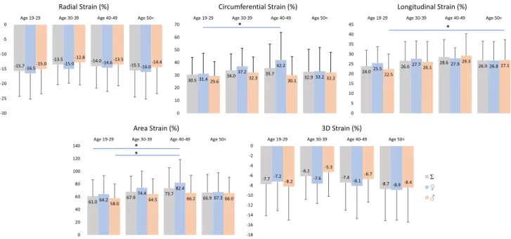
3DSTE‑derived peak LA strains (reservoir function) Peak global LA strains and their gender dependency over decades are demonstrated in Figs. 2 and 4. No significant differences could be demonstrated in strains between the genders, only tendencies could be seen. RS declined with age in females, while an increase in RS could be demon- strated in males > 40 years. CS increased with age with a decline > 50 years in females, in males CS remained almost unchanged. While LS increased with age with unchanged parameters > 50 years, parallel increase in AS with age could be demonstrated in both genders with a decline in females > 50 years. 3DS did not change over the years in females, but an early reduction in males with a later increase could be seen. Peak mean segmental LA strains and differ- ences in regional peak strains over decades are demonstrated in Tables 2 and 3.
3DSTE‑derived LA strains at atrial contraction
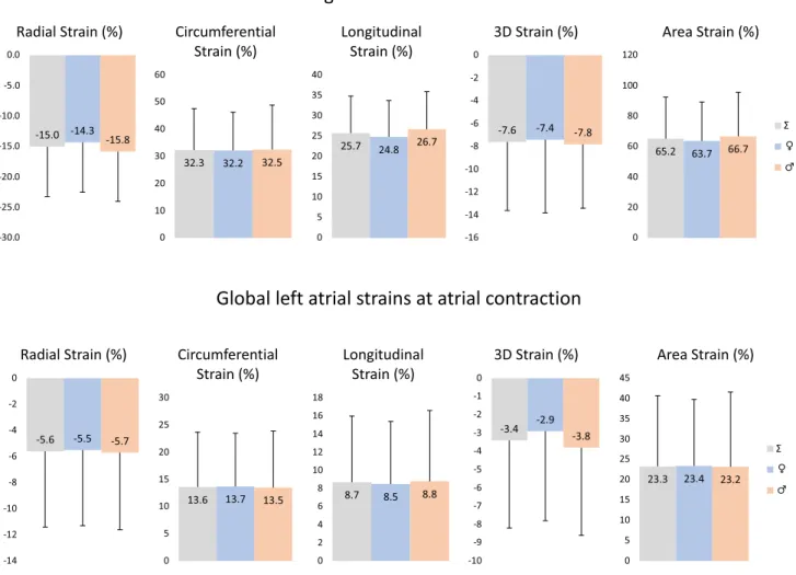
Global LA strains at atrial contraction and their gender dependency over decades are demonstrated in Figs. 3 and 4. No significant changes in any of strains could be dem- onstrated between different age groups, only tendentious alterations could be confirmed. Higher RS at atrial contrac- tion could be seen in older ages. While CS and AS at atrial contraction increased with age in females, parallel decrease could be demonstrated in males. LS at atrial contraction
Fig. 1 Images from three-dimensional (3D) full-volume dataset showing the left atrium (LA) in a healthy subject are demonstrated (a, b): (A) apical four-chamber view, (B) apical two-chamber view, (C3) short-axis view at basal, (C5) mid- and (C7) superior left atrial level. On a, dashed lines represent different planes optimalized on the long-axis and cross-section of the LA. On b, the semi-automated LA border definition, 3D “wire” reconstruction of the LA based on 3D
speckle tracking echocardiographic analysis (red D) and LA volumet- ric data (red E) are presented. Coloured lines represent segmental LA strains while dashed white line represents LA volume changes over the cardiac cycle (red F). White arrow represents peak LA strain, while dashed arrow represents LA strain at atrial contraction. LA left atrium, LV left ventricle, RA right atrium, RV right ventricle
Table 1 Clinical, two-dimensional and volumetric three-dimensional speckle-tracking echocardiographic data of healthy subjects
Data
n 222
Age (years) 36.3 ± 13.7
Male gender (%) 112 (50)
Weight (kg) 72.9 ± 17.5
Height (m) 172.7 ± 10.9
Body surface area (kg/m2) 1.86 ± 0.23
Two-dimensional echocardiography
Left atrium (mm) 37.9 ± 3.8
Left ventricular end-diastolic diameter (mm) 48.3 ± 3.7 Left ventricular end-diastolic volume (ml) 107.1 ± 26.5 Left ventricular end-systolic diameter (mm) 32.5 ± 3.6 Left ventricular end-systolic volume (ml) 38.3 ± 10.5
Interventricular septum (mm) 9.2 ± 1.2
Left ventricular posterior wall (mm) 9.4 ± 1.6
E (cm/s) 77.9 ± 16.4
A (cm/s) 62.2 ± 15.3
Left ventricular ejection fraction (%) 64.9 ± 3.9 Three-dimensional speckle-tracking echocardiography
Maximum left atrial volume (ml) 41.4 ± 13.4 Preatrial contraction left atrial volume (ml) 28.5 ± 12.3 Minimum left atrial volume (ml) 19.9 ± 8.6
increased with age especially in females. 3DS at atrial contraction proved to be highest in females in age group 30–39 years. Mean segmental LA strains at atrial contraction
and differences in regional peak strains over decades are demonstrated in Tables 3 and 4.
Fig. 2 Global peak left atrial strains in males and females over decades. Lines with a star represent significant difference between strains
Fig. 3 Global left atrial strains at atrial contraction in males and females over decades. Lines with a star represent significant difference between strains
Feasibility of 3DSTE‑derived LA measurements 3DSTE measurements were carried out between 2011 and 2016, during this period feasibility improved as the operators
gained experience. During the whole 5-year period 222 meas- urements were successfully performed out of 309 (72%), from which 56 measurements proved to be successful in 66 subjects (85%) in the last year.
Table 2 Age-dependency of three-dimensional speckle- tracking echocardiography- derived peak mean segmental left atrial strain parameters and mean segmental left atrial strain parameters at atrial contraction
† p < 0.05 vs. peak CS Aged 19–29 years
‡ p < 0.05 vs. peak AS Aged 19–29 years
All Aged 19–29 years Aged 30–39 years Aged 40–49 years Aged > 50 years Peak mean segmental strains
RS (%) − 19.5 ± 7.0 − 20.4 ± 7.4 − 18.4 ± 5.9 − 19.0 ± 6.2 − 18.9 ± 7.1 CS (%) 36.7 ± 14.3 35.6 ± 12.9 37.8 ± 12.2 40.0 ± 17.2† 36.4 ± 16.7
LS (%) 29.2 ± 8.3 28.1 ± 7.3 30.0 ± 9.1 31.3 ± 7.9 29.3 ± 9.2
3DS (%) − 12.8 ± 5.2 − 13.3 ± 5.4 − 11.8 ± 4.9 − 12.4 ± 5.0 − 12.8 ± 5.4 AS (%) 71.7 ± 26.3 68.9 ± 25.1 73.9 ± 24.4 79.1 ± 30.2‡ 71.5 ± 27.7 Mean segmental strains at atrial contraction
RS (%) − 8.2 ± 4.9 − 7.6 ± 4.7 − 7.8 ± 4.2 − 8.5 ± 5.7 − 9.3 ± 5.3
CS (%) 15.0 ± 8.4 14.0 ± 7.2 16.0 ± 8.3 15.0 ± 11.7 16.0 ± 8.7
LS (%) 9.9 ± 5.2 9.2 ± 5.1 9.9 ± 5.3 10.6 ± 4.1 10.9 ± 5.4
3DS (%) − 5.4 ± 4.3 − 5.0 ± 4.1 − 5.1 ± 4.1 − 5.6 ± 4.3 − 6.2 ± 4.8 AS (%) 25.8 ± 14.4 24.0 ± 13.2 26.5 ± 13.6 27.2 ± 16.4 28.0 ± 16.0
Table 3 Age-dependency of three-dimensional speckle-tracking echocardiography-derived peak regional left atrial strain parameters
*p < 0.05 vs. RSbasal All; ** p < 0.05 vs. RSsuperior Aged 40–49 years
† p < 0.05 vs. CSmidatrial Aged 19–29 years; ††p < 0.05 vs. CSmidatrial All; †††p < 0.05 vs. CSmidatrial Aged 30–39 years; †††† p < 0.05 vs. CSmidatrial Aged 50 < years
‡ p < 0.05 vs. LSbasal Aged 19–29 years; ‡‡ p < 0.05 vs. LS midatrial All;‡‡‡p < 0.05 vs. LS midatrial Aged 19–29 years; ‡‡‡‡ p < 0.05 vs. LS midatrial Aged 30–39 years; ‡‡‡‡‡p < 0.05 vs. LSmidatrial Aged 40–49 years; ‡‡‡‡‡‡p < 0.05 vs. LSsuperior Aged 40–49 years; ‡‡‡‡‡‡‡p < 0.05 vs. LSmidatrial Aged 50 < years
§ p < 0.05 vs. 3DSsuperior Aged 40–49 years
$ p < 0.05 vs. ASbasal All; $$ p < 0.05 vs. ASbasal Aged 20–29; $$$p < 0.05 vs. AS midatrial Aged 20–29; $$$$p < 0.05 vs. AS basal Aged 30–39
All Aged 19–29 years Aged 30–39 years Aged 40–49 years Aged > 50 years
RSbasal (%) − 18.2 ± 8.9 − 19.3 ± 9.5 − 16.5 ± 7.5 − 16.2 ± 7.2** − 18.5 ± 9.6
RSmidatrial (%) − 19.3 ± 7.7* − 20.3 ± 8.5 − 17.8 ± 6.2 − 18.2 ± 7.1** − 19.3 ± 7.4
RSsuperior (%) − 21.2 ± 12.0* − 22.2 ± 12.4 − 19.8 ± 12.2 − 24.3 ± 12.6 − 19.3 ± 10.6
CSbasal (%) 40.5 ± 14.8†† 39.2 ± 12.1† 41.7 ± 14.9††† 42.1 ± 15.9 41.2 ± 18.5††††
CSmidatrial (%) 31.5 ± 12.8 29.7 ± 11.7 32.7 ± 11.2 34.9 ± 13.3† 32.2 ± 15.3
CSsuperior (%) 38.8 ± 26.1†† 39.2 ± 25.6† 38.3 ± 22.1 44.4 ± 37.1 35.8 ± 23.6
LSbasal (%) 22.1 ± 11.2‡‡ 20.3 ± 10.3 22.8 ± 11.6‡‡‡‡ 21.6 ± 8.5‡‡‡‡‡/‡‡‡‡‡‡ 25.0 ± 12.9‡/‡‡‡‡‡‡‡
LSmidatrial (%) 36.9 ± 13.5 35.5 ± 11.6‡ 38.4 ± 14.1 41.0 ± 17.8 36.4 ± 13.7
LSsuperior (%) 27.0 ± 15.1‡‡ 26.7 ± 14.2‡‡‡ 26.2 ± 14.9‡‡‡‡ 31.3 ± 17.2‡‡‡‡‡ 26.3 ± 15.8‡‡‡‡‡‡‡
3DSbasal (%) − 12.8 ± 7.2 − 13.6 ± 7.7 − 10.9 ± 5.7 − 10.9 ± 5.0§ − 13.6 ± 10.2
3DSmidatrial (%) − 11.8 ± 5.8 − 12.3 ± 6.3 − 10.4 ± 14.9 − 11.3 ± 5.8§ − 12.3 ± 5.4
3DS superior (%) − 13.8 ± 9.0 − 14.0 ± 8.9 − 12.8 ± 9.8 − 15.8 ± 9.4 − 13.2 ± 8.4
ASbasal (%) 61.6 ± 24.1 57.7 ± 19.8 63.3 ± 24.8 61.8 ± 23.2 67.5 ± 29.6$$
ASmidatrial (%) 74.0 ± 27.0 69.9 ± 25.1$$ 78.3 ± 30.4$$$$ 82.1 ± 25.4$$$ 74.5 ± 26.9
ASsuperior (%) 82.2 ± 62.6$ 82.4 ± 59.2$$/$$$ 79.5 ± 54.9 100.7 ± 94.8 75.4 ± 54.1
Discussion
3DSTE is the most recent echocardiographic technique using ‘block-matching’ algorhythm for imaging [4, 10, 11]. The method permits of merging both benefits of 3D and STE imaging: enables to see heart chambers in 3D space by creating a virtual model of a particular chamber and by using this 3D cast, several uni- and multidirectional (dimensional) strains could be calculated over volumetric assessments at the same time [4, 10, 11]. Recently, nor- mal reference values of global and regional LV strains using 3DSTE have already been demonstrated [12]. Due to absence of widely used LA echocardiographic segmen- tation, the same methodology applied for LV was used for strain assessment in the present study. The presented 3DSTE-derived LA strain analysis has already been vali- dated against 2D echocardiography [13], 2DSTE [14] and volumetric real-time 3D echocardiography [15]. Recently, inter- and intraobserver agreements with correlation coeffi- cients have already been demonstrated for 3DSTE-derived LA strain parameters [7].
LA has a distinct phasic function during the cardiac cycle:
it works as a reservoir in systole, it acts as a conduit in early diastole and it works as a booster pump in late diastole helping LV filling through active contraction [16]. During 3DSTE, peak LA strain and LA strain at atrial contraction representing systolic reservoir and late-diastolic booster pump LA functions were calculated automatically from the two-peak curve created by the software using the 3D LA cast in each subject, respectively [16].
In recent studies, significant alterations in 3DSTE- derived LA strain parameters could be demonstrated in several disorders suggesting disease-specific pattern of LA dysfunction [5]. However, LA strains of healthy controls showed differences in these studies suggesting that selec- tion of healthy volunteers and their clinical parameters could have an effect on comparisons [6–8]. Therefore, the current study aimed to establish normal reference ranges of different LA strains using 3DSTE. Over different global and mean segmental LA strains, basal, midatrial and supe- rior (‘apical’) regional strains were defined in males and females among different age groups. In contrast with
Fig. 4 Gender dependency of global peak left atrial strains and left atrial strains at atrial contraction over decades
normal values of 3DSTE-derived LV strains, wide range for LA strains could be demonstrated (large standard devi- ation) [12]. It is likely due to the thin LA wall in combina- tion with limited spatial resolution. Differences were found in LA strains at different LA levels suggesting functional non-uniformity similarly to LV as well [12].
In a recent meta-analysis, the following normal refer- ence ranges for different STE-derived LA strains were revealed: for reservoir (peak) strain of 39% (95% CI 38–41%, from 40 articles), for conduit strain of 23% (95%
CI 21–25%, from 14 articles), and for contractile strain of 17% (95% CI 16–19%, from 18 articles) [17]. LA strain was retrospectively measured by 3D echocardiography for measuring LA strains in healthy children as well using a commercial speckle-tracking package applied to the LA to compute global 3D principal (3DS), longitudinal (GLS), and circumferential (GCS) strains in a recent study [18]. In healthy children, all components of LA strain were found to decline modestly with age. To the best of the authors’
knowledge, this is the first time to demonstrate normal ref- erence values of differed 3DSTE-derived LA strains dem- onstrating their age-dependency. It has also been demon- strated that LA strains have no obvious gender-dependency (except in older ages), but tendencies could be detected together with regional differences.
Limitations
The present study did not aim to assess normal values of LA volumetric data and strain rate parameters. Moreover, only peak strains and strains at atrial contraction were compared. As men- tioned before, inter- and intraobserver agreements with correla- tion coefficients for LA strains have already been confirmed.
Spatial resolution of 3DSTE is relatively poor compared to that of 2D echocardiography. Although healthy subjects were exam- ined relatively high number of them was excluded. Exclusion was mainly based on image quality. Narrow echocardiographic window (as the 3DSTE transducer is more sizeable than its 2D counterpart) and the operators’ inexperience (at the beginning of the study) could be considered as the most important reasons behind exclusion. It is also worthy to note in lesser part that body habitus was also a reason in some cases for exclusion, however extreme obesity was considered pathologic and these patients were not enrolled in the present study.
Conclusion
Normal reference values of 3DSTE-derived LA peak strains and strains at atrial contraction are demonstrated together with their age- and gender-dependency.
Table 4 Age-dependency of three-dimensional speckle-tracking echocardiography-derived regional left atrial strain parameters at atrial contrac- tion
† p < 0.05 vs. CS basal Aged 19–29 years; ††p < 0.05 vs. CS midatrial All; †††p < 0.05 vs. CS midatrial Aged 19–29; ††††p < 0.05 vs. CS midatrial Aged 30–39
‡ p < 0.05 vs. LSbasal Aged 19–29 years; ‡‡p < 0.05 vs. LSbasal All; ‡‡‡p < 0.05 vs. LSbasal Aged 30–39 years; ‡‡‡‡p < 0.05 vs. LSbasal Aged 40–49 years; ‡‡‡‡‡p < 0.05 vs. LSbasal Aged 50 < years
§ p < 0.05 vs. 3DSsuperior Aged 40–49 years
$ p < 0.05 vs. ASbasal Aged 20–29; $$p < 0.05 vs. ASmidatrial Aged 20–29; $$$p < 0.05 vs. ASbasal All; $$$$p < 0.05 vs. ASmidatrial All; $$$$$p < 0.05 vs.
ASsuperior Aged 19–29
All Aged 19–29 years Aged 30–39 years Aged 40–49 years Aged > 50 years
RSbasal (%) − 7.9 ± 5.8 − 7.4 ± 5.6 − 7.4 ± 5.5 − 6.9 ± 5.0 − 9.7 ± 6.6
RSmidatrial (%) − 7.9 ± 5.4 − 7.5 ± 5.1 − 7.5 ± 5.0 − 8.5 ± 6.9 − 8.8 ± 5.4
RSsuperior (%) − 9.1 ± 8.4 − 8.4 ± 8.6 − 8.9 ± 8.4 − 10.8 ± 9.2 − 9.6 ± 7.6
CSbasal (%) 16.5 ± 9.0†† 15.1 ± 7.9††† 18.7 ± 9.9†/†††† 17.0 ± 11.0 16.7 ± 8.8
CSmidatrial (%) 13.0 ± 8.4 11.9 ± 7.5 12.8 ± 7.5 13.2 ± 10.3 14.8 ± 9.5
CSsuperior (%) 15.9 ± 14.6†† 15.7 ± 13.1††† 15.5 ± 15.3 14.8 ± 20.3 16.9 ± 13.6
LSbasal (%) 7.5 ± 5.2 6.8 ± 5.1 7.9 ± 5.6 7.9 ± 4.6 8.5 ± 5.0‡
LSmidatrial (%) 11.4 ± 7.5‡‡ 10.2 ± 7.1‡ 11.9 ± 7.9‡‡‡ 12.6 ± 6.8‡‡‡‡ 12.6 ± 8.0‡‡‡‡‡
LSsuperior (%) 11.0 ± 8.7‡‡ 10.7 ± 8.4‡ 10.0 ± 9.0 11.5 ± 9.5 12.1 ± 8.5‡‡‡‡‡
3DSbasal (%) − 5.4 ± 5.4 − 5.0 ± 5.2 − 4.8 ± 5.1 − 4.5 ± 4.6§ − 6.8 ± 6.4
3DSmidatrial (%) − 5.0 ± 4.7 − 4.7 ± 4.8 − 4.8 ± 4.4 − 4.9 ± 5.5§ − 5.6 ± 4.3
3DSsuperior (%) − 6.0 ± 7.3 − 5.4 ± 6.8 − 5.9 ± 8.0 − 8.5 ± 7.5 − 6.2 ± 7.4
ASbasal (%) 22.7 ± 13.0 20.3 ± 10.9$$$$$ 24.6 ± 14.5 27.1 ± 14.1$ 23.9 ± 14.1
ASmidatrial (%) 25.8 ± 15.3$$$ 23.3 ± 15.3$$$$$ 26.5 ± 13.3 27.0 ± 14.0 29.2 ± 16.9$$
ASsuperior (%) 30.6 ± 29.1$$$/$$$$ 30.4 ± 26.8 29.6 ± 31.6 27.8 ± 35.8 32.5 ± 27.8
Acknowledgements Open access funding provided by University of Szeged (SZTE).
Compliance with ethical standards
Conflict of interest The authors declared that they have no conflict of interest.
Ethical approval All procedures performed in studies involving human participants were in accordance with the ethical standards of the insti- tutional and/or national research committee and with the 1964 Helsinki declaration and its later amendments or comparable ethical standards.
Informed consent Informed consent was obtained from all individual participants included in the study.
Open Access This article is distributed under the terms of the Crea- tive Commons Attribution 4.0 International License (http://creat iveco mmons .org/licen ses/by/4.0/), which permits unrestricted use, distribu- tion, and reproduction in any medium, provided you give appropriate credit to the original author(s) and the source, provide a link to the Creative Commons license, and indicate if changes were made.
References
1. Blume GG, Mcleod CJ, Barnes ME, Seward JB, Pellikka PA, Bastiansen PM, Tsang TS (2011) Left atrial function: physiol- ogy, assessment, and clinical implications. Eur J Echocardiogr 12:421–430
2. Vieira MJ, Teixeira R, Gonçalves L, Gersh BJ (2014) Left atrial mechanics: echocardiographic assessment and clinical implica- tions. J Am Soc Echocardiogr 27:463–478
3. Enzensberger C, Achterberg F, Degenhardt J, Wolter A, Graupner O, Herrmann J, Axt-Fliedner R (2017) Feasibility and reproduc- ibility of two-dimensional wall motion tracking (WMT) in fetal echocardiography. Ultrasound Int Open 3:E26–E33
4. Nemes A, Kalapos A, Domsik P, Forster T (2012) Three- dimensional speckle-tracking echocardiography—a further step in non-invasive three-dimensional cardiac imaging. Orv Hetil 153:1570–1577
5. Nemes A, Domsik P, Kalapos A, Forster T (2016) Is three-dimen- sional speckle-tracking echocardiography able to identify differ- ent patterns of left atrial dysfunction in selected disorders? Short summary of the MAGYAR-Path Study. Int J Cardiol 220:535–537 6. Nemes A, Piros G, Domsik P, Kalapos A, Forster T (2016)
Left atrial volumetric and strain analysis by three-dimensional speckle-tracking echocardiography in noncompaction cardiomyo- pathy: results from the MAGYAR-Path Study. Hellenic J Cardiol 57:23–29
7. Nemes A, Piros GÁ, Lengyel C, Domsik P, Kalapos A, Várkonyi TT, Orosz A, Forster T (2016) Complex evaluation of left atrial dysfunction in patients with type 1 diabetes mellitus by three- dimensional speckle tracking echocardiography: results from the MAGYAR-Path Study. Anatol J Cardiol 16:587–593
8. Havasi K, Domsik P, Kalapos A, McGhie JS, Roos-Hesselink JW, Forster T, Nemes A (2017) Left atrial deformation analysis in patients with corrected tetralogy of fallot by 3D speckle-tracking echocardiography (from the MAGYAR-Path Study). Arq Bras Cardiol 108:129–134
9. Lang RM, Badano LP, Mor-Avi V, Afilalo J, Armstrong A, Ernande L, Flachskampf FA, Foster E, Goldstein SA, Kuznetsova T, Lancellotti P, Muraru D, Picard MH, Rietzschel ER, Rudski L, Spencer KT, Tsang W, Voigt JU (2015) Recommendations for cardiac chamber quantification by echocardiography in adults: an update from the American Society of Echocardiography and the European Association of Cardiovascular Imaging. Eur Heart J Cardiovasc Imaging 16:233–270
10. Ammar KA, Paterick TE, Khandheria BK, Jan MF, Kramer C, Umland MM, Tercius AJ, Baratta L, Tajik AJ (2012) Myocardial mechanics:
understanding and applying three-dimensional speckle tracking echo- cardiography in clinical practice. Echocardiography 29:861–872 11. Urbano-Moral JA, Patel AR, Maron MS, Arias-Godinez JA, Pan-
dian NG (2012) Three-dimensional speckle-tracking echocardi- ography: methodological aspects and clinical potential. Echocar- diography 29:997–1010
12. Kleijn SA, Pandian NG, Thomas JD, Perez de Isla L, Kamp O, Zuber M, Nihoyannopoulos P, Forster T, Nesser HJ, Geibel A, Gorissen W, Zamorano JL (2015) Normal reference values of left ventricular strain using three-dimensional speckle tracking echocardiography: results from a multicentre study. Eur Heart J Cardiovasc Imaging 16:410–416
13. Nemes A, Domsik P, Kalapos A, Lengyel C, Orosz A, Forster T (2014) Comparison of three-dimensional speckle tracking echocar- diography and two-dimensional echocardiography for evaluation of left atrial size and function in healthy volunteers (results from the MAGYAR-Healthy study). Echocardiography 31:865–871 14. Mochizuki A, Yuda S, Oi Y, Kawamukai M, Nishida J, Kouzu H,
Muranaka A, Kokubu N, Shimoshige S, Hashimoto A, Tsuchihashi K, Watanabe N, Miura T (2013) Assessment of left atrial deforma- tion and synchrony by three-dimensional speckle-tracking echo- cardiography: comparative studies in healthy subjects and patients with atrial fibrillation. J Am Soc Echocardiogr 26:165–174 15. Kleijn SA, Aly MF, Terwee CB, van Rossum AC, Kamp O (2011)
Comparison between direct volumetric and speckle tracking meth- odologies for left ventricular and left atrial chamber quantification by three-dimensional echocardiography. Am J Cardiol 108:1038–1044 16. Nemes A, Forster T (2014) Assessment of left atrial size and func- tion—from M-mode to 3D speckle-tracking echocardiography.
Orv Hetil 155:1624–1631
17. Pathan F, D’Elia N, Nolan MT, Marwick TH, Negishi K (2017) Normal ranges of left atrial strain by speckle-tracking echocar- diography: a systematic review and meta-analysis. J Am Soc Echocardiogr 30:59–70
18. Ghelani SJ, Brown DW, Kuebler JD, Perrin D, Shakti D, Wil- liams DN, Marx GR, Colan SD, Geva T, Harrild DM (2018) Left atrial volumes and strain in healthy children measured by three- dimensional echocardiography: normal values and maturational changes. J Am Soc Echocardiogr 31:187–193
Publisher’s Note Springer Nature remains neutral with regard to jurisdictional claims in published maps and institutional affiliations.