573
Adenosine-5
,-diphosphate and Adenosine-5
f-monophosphate
Hans Adam Principle
Adenosine-5'-diphosphate ( A D P ) is phosphorylated with phosphoenolpyruvate and pyruvate kinase (PK). The pyruvate formed is reduced with D P N H and lactic dehydrogenase ( L D H ) . Adenosine-5'-monophosphate ( A M P ) can also be phosphorylated with A T P and myokinase ( M K ) and the resulting two equivalents of A D P can be determined as described. Consequently it is possible to measure individually pyruvate, A D P and A M P in a single operation
1
).
MK, Mg2+
(1) A M P + A T P
v
2 A D PPK, Mg2+, K+
(2) 2 A D P -f- 2 phosphoenolpyruvate ^ 2 A T P + 2 pyruvate Indicator reaction:
L D H
(3) 2 Pyruvate + 2 D P N H + 2 H + ^ = ± 2 lactate + 2 D P N + The equilibrium of reaction (3) is sufficiently far to the r i g h t
2)
(K ^ 10
4
[l./mole]
2
)) so as to reduce all the pyruvate. The equilibrium of reaction (2) (K = 2 x 10
3
at 30°C
3
>
4
)) is sufficiently far to the right to overcome the equilibrium of the A D P dismutation (ca. 6 6 % to the left
5
.
6
)) and allow all the A M P to react.
Reagents *>
1. Perchloric acid, A. R.; sp. gr. 1.67; ca. 70% (w/w) 2. Potassium carbonate,
K 2 C O 3 ,A. R.
3. Potassium hydroxide, A. R., 2 N
4. Triethanolamine, freshly distilled b. p. 277—279° C/150 mm.
5. Magnesium sulphate, MgS04-7H20, A. R.
6. Potassium chloride, KC1, A. R.
7. Ethylene-diamine-tetra-acetic acid, EDTA
disodium salt, E D T A - N a
2
H2
- 2 H2
0 , e.g. Titriplex HI**)8. Hydrochloric acid, 2 N, A. R.
9. Sodium hydroxide, 2 N, A. R.
10. Phosphoenolpyruvate, PEP
crystalline tricyclohexylammonium salt; commercial preparation, see p. 1024.
11. Reduced diphosphopyridine nucleotide, DPNH
disodium salt, D P N H - N a
2
; commercial preparation, see p. 1011.12. Adenosine-5'-triphosphate, ATP
crystalline disodium salt, A T P - N a
2
H2
- 3 H2
0 ; commercial preparation, see p. 1006.13. Adenosines-monophosphate, AMP
disodium salt, A M P - N a
2
; commercial preparation, see p. 1005.*) Complete reagent kits are available commercially, see p. 1035.
**) from E. Merck, Darmstadt (Germany).
J
) H. Adam, Ph. D.-Thesis, Universitat Marburg/L., 1955.
2
) F. Kubowitz and P. Ott, Biochem. Z. 314, 94 [1943].
3) E. Negelein, unpublished; cf. F. Kubowitz and P. Ott, Biochem. Z. 317, 193 [1944].
4
) O. Meyerhof and P. Oesper, J. biol. Chemistry 179, 1371 [1949].
5) H. M. Kalckar, J. biol. Chemistry 148, 127 [1943].
6) L. V. Eggleston and R. Hems, Biochem. J. 52, 156 [1952].
14. Lactic dehydrogenase, LDH
crystalline, from skeletal muscle, suspended in 2.1 M ammonium sulphate solution (pH 5.5);
10 mg. protein/ml. Commercial preparation, see p. 986.
15. Pyruvate kinase, PK
crystalline, from skeletal muscle, suspended in 1.95 M ammonium sulphate solution (pH 5.5);
5 mg. protein/ml. Commercial preparation, see p. 997.
16. Myokinase, MK
from skeletal muscle, suspended in 3.3 M a m m o n i u m sulphate solution (pH ^ 6 ) ; 5 mg. protein/
ml. Commercial preparation, see p. 989.
Purity of the e n z y m e preparations
The L D H should have a specific activity of at least 2 0 0 0 0 units *Vmg. and be free from M K and PK (i.e. less than 0.01 % with respect to the L D H activity). The specific activity of the P K should be at least 2 500 units *tymg. and that of the M K at least 2 0 0 0 units *)/mg. Within the limits given above PK should be free from M K , and M K should be free from ATPase.
Preparation of Solutions
I. Triethanolamine buffer (5 x 10⁻² M; pH 7.55):
Dissolve 7.46 g. triethanolamine in ca. 700 ml. doubly distilled water, adjust to pH 7.55 with ca. 15 ml. 2 N HC1 and dilute with doubly distilled water to 1000 ml.
II. Magnesium sulphate (0.5 M):
Dissolve 6.02 g. MgS04 -7HiO in doubly distilled water and make up to 100 ml.
III. Potassium chloride (2 M):
Dissolve 14.91 g. KC1 in doubly distilled water and make up to 100 ml.
IV. Ethylene-diamine-tetra-acetate (100 mg./ml.):
Dissolve 10 g. EDTA-Na2H2 • 2 H2O in doubly distilled water, neutralize with 2 N NaOH and dilute with doubly distilled water to 100 ml.
V. Phosphoenolpyruvate (ca. 4 x 10~
2
M PEP):
Dissolve 100 mg. PEP (crystalline tricyclohexylammonium salt) in doubly distilled water and make up to 5 ml. Determine the PEP content of the solution enzymatically (see p. 224).
VI. Reduced diphosphopyridine nucleotide (ca. 1 0 -2
M (3-DPNH):
Dissolve 20 mg. DPNH-Na2 in 2 ml. doubly distilled water. Determine the DPNH content of the solution enzymatically (see p. 531).
VII. Adenosine triphosphate (ca. 10~
2
M ATP):
Dissolve 10 mg. ATP-Na2H2 • 3 H2O in 2 ml. doubly distilled water.
VIII. Adenosine monophosphate (ca. 1 0 -2
M AMP):
Dissolve 10 mg. AMP-Na2 in 2 ml. doubly distilled water.
IX. Lactic dehydrogenase, LDH (0.1 mg. protein/ml.):
Dilute 0.01 ml. of a crystalline suspension (10 mg. protein/ml.) to 1 ml. with 2.25 M ammonium sulphate solution (pH 6.5).
*) According to Th. Biicher
1
) one unit is the amount of enzyme dissolved in 1 ml. which decreases the optical density of D P N H by 0.100 in 100 seconds at 366 VC\\L with a 1 cm light path.
7) G. Beisenherz, H. J. Boltze, Th. Biicher, R. Czok, K. H. Garbade, E. Meyer-Arendt and G. Pflei- derer, Z. Naturforsch. 8b, 555 [1953].
V.2.h Adenosine-5'-diphosphate and Adenosine-5'-monophosphate 575
X. Pyruvate kinase, PK (0.5 mg. protein/ml.):
Dilute 0.1 ml. of a crystalline suspension (5 mg. protein/ml.) to 1 ml. with 2.1 M ammonium sulphate solution (pH 5.5).
XI. Myokinase, MK (0.25 mg. protein/ml.):
Dilute 0.05 ml. of a crystalline suspension (5 mg. protein/ml.) to 1 ml. with 3.3 M ammonium sulphate solution (pH 6).
For deproteinization:
2 N KOH; 3.75 M K 2 C 0 3 ; perchloric acid solutions Xa and Xb on p. 541 Procedure
D e p r o t e i n i z a t i o n
Since hydrolysis of ATP must be avoided during extraction of ADP and AMP from tissues, refer to the determination of adenosine-5'-triphosphate with phosphoglycerate kinase (p. 539) for all details connected with the preparation of the extract.
Spectrophotometric m e a s u r e m e n t s
Wavelength: 340 or 366 mu.; light path: 1 cm.; final volume: 2.0 ml.; room temperature.
Prepare the following reaction mixture*) and neutralize:
0.036 ml. magnesium sulphate solution (II) 0.076 ml. potassium chloride solution (III) 0.004 ml. EDTA solution (IV)
0.040 ml. PEP solution (V) 0.030 ml. DPNH solution (VI) 0.007 ml. ATP solution (VII) or a multiple of the individual volumes.
Pipette successively into the cuvettes:
deproteinized sample (extract) and buffer (I) to 2.00 ml.
0.140 ml. reaction mixture.
Mix in (with a small glass or plastic rod flattened and bent at one end) 0.020 ml. LDH suspension (IX).
Follow the optical density until no further change occurs (3—5 min.), read optical density E i . Mix in
0.020 ml. PK suspension (X).
Take readings until reaction stops (6—9 min.) and then measure optical density E2. If the reaction does not come to a complete stop, extrapolate from the point of PK addition:
the change in optical density/min. (from the linear part of the curve) multiplied by the number of minutes since addition of PK, gives the optical density to be subtracted from the final optical density E2.
*) As D P N H preparations occasionally contain some A M P (just as A T P preparations contain some A D P and phosphoenolpyruvate solutions some pyruvate), it is necessary to carry out a blank determination for all three substrates on the salt-substrate buffer mixture. This composite mixture should not be kept for longer than 6 hours without carrying out a new blank determination.
Immediately mix in
0.018 ml. MK suspension (XI).
Take readings until the reaction stops (10 — 13 min.) and then measure optical density E 3 . If the reaction does not stop, extrapolate as described above.
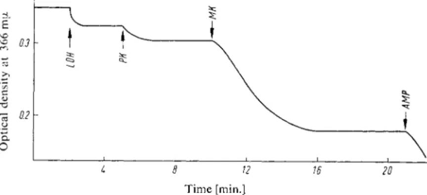
To test the correct functioning of the system mix in ca. 0.01 ml. AMP solution (VIII).
A renewed reaction should occur immediately (Fig. 1). The optical density differences Ei —E2 = A E A D P and E 2 — E3 = A E A M P are used for the calculations.
Time [min.]
Fig. 1. Determination of pyruvate, A D P and A M P in the same mixture; decrease in optical density at 366 m[x.
L D H = lactic dehydrogenase, P K = pyruvate kinase, M K = myokinase, A M P = adenosine- 5'-monophosphate as control.
Calculations
Extinction coefficient for D P N H (25° C):
£340 = 6.22 [CM.
2
/PIMOLE]
£
3 6 6 = 3.30 [CM.
2
/FI.MOLE]
A E
A D P
x VA
x VE
— = LIMOLES A D P in EXTRACT
£ X d x V
P
and
A E
A M
p x VA
x V_= E
amoles A M P in extract 2x £ x
dx
VP
where A E
A
D P = Ei— E2
A E
A
M P = E2
— E3V
A
= volume of test mixture in the cuvette (2.0 ml.) V g = total volume of extract [ml.]Vp = volume of extract added to the cuvette [ml.]
£ = extinction coefficient [cm.
2
/u.mole]
d = light path of the cuvette [1 cm.]
V.2.h Adenosine-5'-diphosphate and Adenosine-5'-monophosphate 577
for measurements at 366 mu under the stated conditions it follows that:
A E
A D P
x 2 X VE
3.30 X 1 X V
P
= A EA D P
X 0.606 X VEV
P
(xmoles A D P in extract andA E
A M P
x 0.303 x VEV
P
[xmoles A M P in extract.These values divided by the fresh weight of tissue taken give the u.moles A D P or A M P per g. tissue.
The results are reproducible to ± 1 . 5 % and agree with U V absorption, phosphate and ribose deter
minations. A s little as 1 0
-8
moles of nucleotide can be measured with this accuracy. Micro-cuvette- allow the determination of 10~
9
moles of nucleotide.
Specificity
I D P , G D P , U D P or C D P are quantitatively converted, but at different rates (refer t o Fig. 2). The time course o f the reactions with I D P , G D P or U D P cannot be differentiated from that with A D P ; only with C D P is a correction by extrapolation possible.
A OP* (JDP
5 10
Time [min.]
15
Fig. 2. Determination of A D P in the presence of about equimolar concentration of I D P , G D P , U D P or C D P ; decrease in optical density at 366 mu, after addition of pyruvate kinase.