Article
Neuronal Modulators from the Coral-Associated Fungi Aspergillus candidus
Gao-Yang Peng1,2,3, Tibor Kurtán4 , Attila Mándi4 , Jing He5, Zheng-Yu Cao5 , Hua Tang6, Shui-Chun Mao1,* and Wen Zhang2,3,*
Citation: Peng, G.-Y.; Kurtán, T.;
Mándi, A.; He, J.; Cao, Z.-Y.; Tang, H.;
Mao, S.-C.; Zhang, W. Neuronal Modulators from the
Coral-Associated FungiAspergillus candidus.Mar. Drugs2021,19, 281.
https://doi.org/10.3390/md 19050281
Academic Editor: Rob Keyzers
Received: 21 April 2021 Accepted: 13 May 2021 Published: 19 May 2021
Publisher’s Note:MDPI stays neutral with regard to jurisdictional claims in published maps and institutional affil- iations.
Copyright: © 2021 by the authors.
Licensee MDPI, Basel, Switzerland.
This article is an open access article distributed under the terms and conditions of the Creative Commons Attribution (CC BY) license (https://
creativecommons.org/licenses/by/
4.0/).
1 School of Pharmacy, Nanchang University, 461 Bayi Road, Nanchang 330006, China;
penggaoyang42@163.com
2 School of Medicine, Tongji University, 1239 Si-Ping Road, Shanghai 200092, China
3 School of Pharmacy, Navy Medical University, 325 Guo-He Rd., Shanghai 200433, China
4 Department of Organic Chemistry, University of Debrecen, POB 400, H-4002 Debrecen, Hungary;
kurtan.tibor@science.unideb.hu (T.K.); mandi.attila@science.unideb.hu (A.M.)
5 State Key Laboratory of Natural Medicines, Department of TCM Pharmacology, School of Traditional Chinese Pharmacy, China Pharmaceutical University, 639 Long-Mian Ave., Nanjing 211198, China;
18796219566@163.com (J.H.); zycao1999@hotmail.com (Z.-Y.C.)
6 Institute of Translational Medicine, Shanghai University, 99 Shang-Da Road, Shanghai 200444, China;
tanghua0309@126.com
* Correspondence: maoshuichun@ncu.edu.cn (S.-C.M.); wenzhang1968@163.com (W.Z.)
Abstract: Three newp-terphenyl derivatives, named 400-O-methyl-prenylterphenyllin B (1) and phenylcandilide A and B (17and18), and three new indole-diterpene alkaloids, asperindoles E–G (22-24), were isolated together with eighteen known analogues from the fungiAspergillus candidus associated with the South China Sea gorgonianJunceela fragillis. The structures and absolute configu- rations of the new compounds were elucidated on the basis of spectroscopic analysis, and DFT/NMR and TDDFT/ECD calculations. In a primary cultured cortical neuronal network, the compounds 6,9,14,17,18and24modulated spontaneous Ca2+oscillations and 4-aminopyridine hyperexcited neuronal activity. A preliminary structure–activity relationship was discussed.
Keywords:coral-associated fungi; secondary metabolites;p-terphenyl; indole-diterpene alkaloids;
spontaneous Ca2+oscillations
1. Introduction
Fungi ofAspergillus candidusis found to be wide-spread in soil [1] and marine en- vironments [2–4], and also co-existing with various animals and plants [5–8]. The fungi are reported to produce prolific secondary metabolites, including p-terphenyls [9–11], terpenes [3], flavonoids [7,12,13], cyclopeptides [6], alkaloids [5], polyketones, and fatty acids [3]. These metabolites display a wide spectrum of biological activities, including cytotoxic [5,8], antibacterial [5,6], antifungal [3,8], antioxidant [6], and immunosuppressive activities [14].
As part of our continuing search for bioactive molecules from marine invertebrates and the associated fungi [15–17], a strain ofA. candiduswas isolated from the internal tissues of the gorgonian coralJunceela fragillis, collected from the Xisha area of the South China Sea. Chemical investigation of the fermentation extract of this fungus resulted in the isolation of three newp-terphenyl derivatives and three new indole-diterpene alkaloids, together with eighteen known analogues. Further,p-Terphenyls are regarded as the main metabolites ofA. candidus. More than 230 analogues have been reported up to date, with the chemical diversity being attributed to the substituents on rings A and C [8,18,19].
The indole-diterpene alkaloids are a cluster of characteristic metabolites from this genus and were firstly reported from the titled fungi [20]. These metabolites are structurally constructed with an indole molecule and a saculatane diterpenoid. The biosynthetic process
Mar. Drugs2021,19, 281. https://doi.org/10.3390/md19050281 https://www.mdpi.com/journal/marinedrugs
Mar. Drugs2021,19, 281 2 of 14
of both types of metabolites were conducted by investigating 60-hydroxy-4,20,30,400-tetra- methoxy-p-terphenyl [18,19,21] forp-terphenyls, and penitrem [22] and lolitrems [23,24]
for the indole-diterpene alkaloids. The bioactivities and the intriguing ring-systems of these molecules also attracted attention for total synthesis [19,25,26]. Herein, we report the isolation, structure elucidation, and neuronal modulatory activity of these compounds.
2. Results
TheA. candidus was cultivated on biomalt agar medium and then extracted with EtOAc. The EtOAc extract was subjected to the usual workup for isolation [15,16,27]
to yield compounds1–24(Figure1), including eighteenp-terphenyl derivatives and six indole-diterpene alkaloids. Based on the spectroscopic analysis and comparison with the reported data, the known compounds were determined as 300-hydroxyl-prenylterphenyllin (2), 3-methoxy-400-deoxyterprenin (3), 3-hydroxyterphenyllin (4) [28], 4-O-methylprenyl- terphenyllin (5) [9], terphenyllin (6) [12,28], 3-methoxyterprenin (7) [14], prenylterphenyllin J (8) [8], 3,300-dihydroxy-60-desmethyl terphenyllin (9) [29], deoxyterhenyllin (10) [30], prenylterphenyllin (11) [4], prenylterphenyllin B (12) [11], candidusins A (13) and B (14) [31], prenylcandidusin A (15) [11], 4-methyl-candidusin A (16) [32], and asperindoles A (19), C (20) and D (21) [20]. A complete NMR assignment is presented for the com- pounds2and3since there is no report for the NMR data of both the compounds. The assignments are fully supported by those of co-occurring prenylterphenyllin (11) [4] and 3-methoxyterprenin (7) [14], two analogues once reported from the soil-derived strains of the same species.
Mar. Drugs 2021, 19, x 2 of 14
and were firstly reported from the titled fungi [20]. These metabolites are structurally con- structed with an indole molecule and a saculatane diterpenoid. The biosynthetic process of both types of metabolites were conducted by investigating 6-hydroxy-4,2,3,4-tetra- methoxy-p-terphenyl [18,19,21] for p-terphenyls, and penitrem [22] and lolitrems [23,24]
for the indole-diterpene alkaloids. The bioactivities and the intriguing ring-systems of these molecules also attracted attention for total synthesis [19,25,26]. Herein, we report the isolation, structure elucidation, and neuronal modulatory activity of these com- pounds.
2. Results
The A. candidus was cultivated on biomalt agar medium and then extracted with EtOAc. The EtOAc extract was subjected to the usual workup for isolation [15,16,27] to yield compounds 1–24 (Figure 1), including eighteen p-terphenyl derivatives and six in- dole-diterpene alkaloids. Based on the spectroscopic analysis and comparison with the reported data, the known compounds were determined as 3-hydroxyl-prenylterphenyl- lin (2), 3-methoxy-4-deoxyterprenin (3), 3-hydroxyterphenyllin (4) [28], 4-O- methylprenyl-terphenyllin (5) [9], terphenyllin (6) [12,28], 3-methoxyterprenin (7) [14], prenylterphenyllin J (8) [8], 3,3-dihydroxy-6-desmethyl terphenyllin (9) [29], deoxyter- henyllin (10) [30], prenylterphenyllin (11) [4], prenylterphenyllin B (12) [11], candidusins A (13) and B (14) [31], prenylcandidusin A (15) [11], 4-methyl-candidusin A (16) [32], and asperindoles A (19), C (20) and D (21) [20]. A complete NMR assignment is presented for the compounds 2 and 3 since there is no report for the NMR data of both the compounds.
The assignments are fully supported by those of co-occurring prenylterphenyllin (11) [4]
and 3-methoxyterprenin (7) [14], two analogues once reported from the soil-derived strains of the same species.
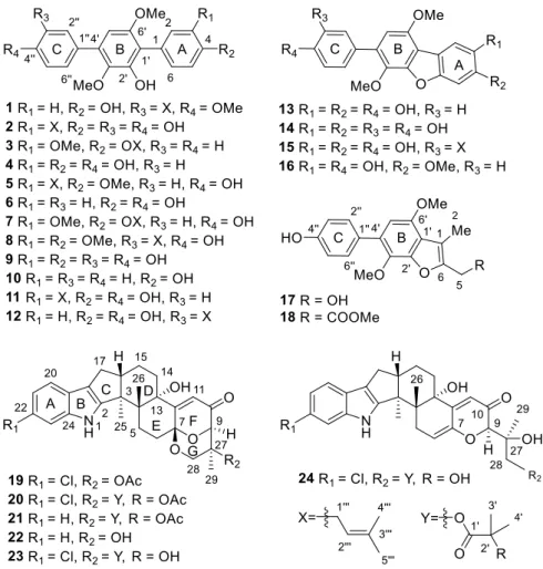
Figure 1. Structures of compounds 1–24.
Figure 1.Structures of compounds1–24.
Mar. Drugs2021,19, 281 3 of 14
Further, 400-O-methyl-prenylterphenyllin B (1) was obtained as a colorless amorphous solid. Its molecular formula was determined as C26H28O5by HRESIMS, requiring 13 degrees of unsaturation. The IR spectrum displayed absorptions for hydroxy (3357 cm−1) and substituted benzol (1609, 1462, 834, and 815 cm−1) functionalities. The presence of benzol rings was supported by the strong UV absorptions at 276, 248, and 209 nm. The NMR spectra of1 displayed resonances for twentysp2carbons and sixsp3carbons, taking into account the ten degrees of unsaturation. The remaining three degrees of unsaturation assigned to the ring system of this molecule were in agreement with that of the terphenyl framework. Its NMR data were almost identical to those of the co-isolated prenylterphenyllin B (12) [11], except for the appearance of an additional methoxy group (δH3.89, 3H, s;δC55.6, CH3). The methoxy group was assigned as 400-OMeviaits HMBC correlations to C-400, and further confirmed by its NOE correlation with H-500(Figure S1). The structure of compound1was therefore determined as 400-O-methyl-prenylterphenyllin B.
Phenylcandilide A (17), obtained as a yellow amorphous solid, had a molecular for- mula of C18H18O5as deduced from the HRESIMS, indicating 10 degrees of unsaturation.
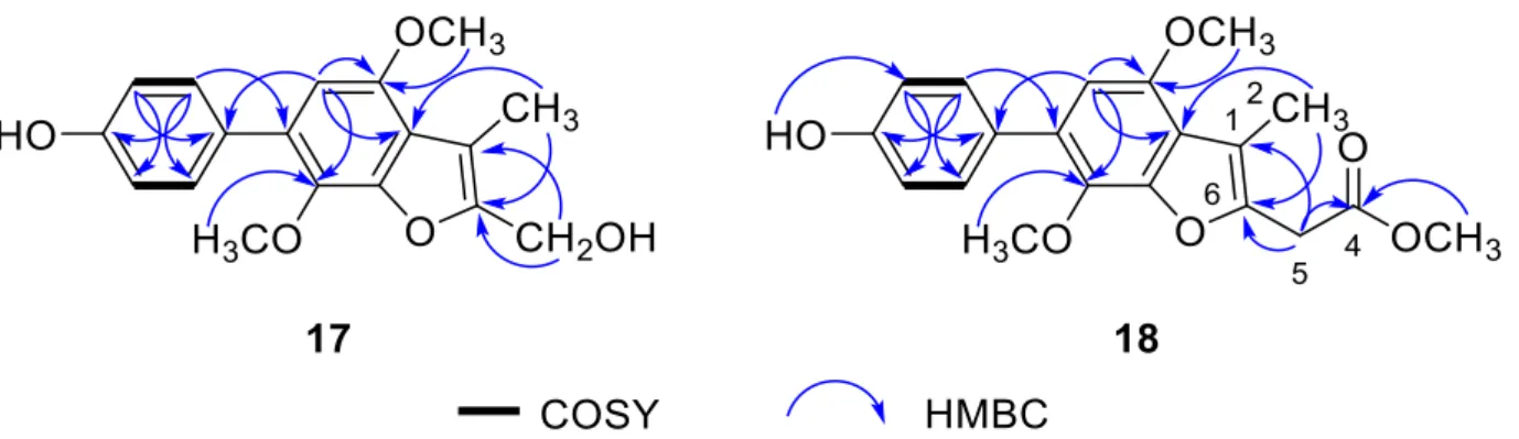
The NMR data of 17 showed similarity to those of 4-methyl-candidusin A (16) [32], regard- ing the signals for the phenol ring C and benzofuran ring B. The C-2 to C-5 diene fragment of the phenol ring A in16, however, was degraded to a methyl and a hydroxymethyl groups in17. The distinct HMBC correlations from H3-2 to C-10and C-6, and H2-5 to C-6 and C-1 confirmed the location of 1-Me and 6-CH2OH (Figure2). The structure of17was determined and nominated as phenylcandilide A.
Further, 4-O-methyl-prenylterphenyllin B (1) was obtained as a colorless amor- phous solid. Its molecular formula was determined as C
26H
28O
5by HRESIMS, requiring 13 degrees of unsaturation. The IR spectrum displayed absorptions for hydroxy (3357 cm
−1) and substituted benzol (1609, 1462, 834, and 815 cm
−1) functionalities. The presence of benzol rings was supported by the strong UV absorptions at 276, 248, and 209 nm. The NMR spectra of 1 displayed resonances for twenty sp
2carbons and six sp
3carbons, taking into account the ten degrees of unsaturation. The remaining three degrees of unsaturation assigned to the ring system of this molecule were in agreement with that of the terphenyl framework. Its NMR data were almost identical to those of the co-isolated prenylter- phenyllin B (12) [11], except for the appearance of an additional methoxy group (δ
H3.89, 3H, s; δ
C55.6, CH
3). The methoxy group was assigned as 4-OMe via its HMBC correlations to C-4, and further confirmed by its NOE correlation with H-5 (Figure S1). The structure of compound 1 was therefore determined as 4-O-methyl-prenylterphenyllin B.
Phenylcandilide A (17), obtained as a yellow amorphous solid, had a molecular for- mula of C
18H
18O
5as deduced from the HRESIMS, indicating 10 degrees of unsaturation.
The NMR data of 17 showed similarity to those of 4-methyl-candidusin A (16) [32], re- garding the signals for the phenol ring C and benzofuran ring B. The C-2 to C-5 diene fragment of the phenol ring A in 16, however, was degraded to a methyl and a hy- droxymethyl groups in 17. The distinct HMBC correlations from H
3-2 to C-1 and C-6, and H
2-5 to C-6 and C-1 confirmed the location of 1-Me and 6-CH
2OH (Figure 2). The structure of 17 was determined and nominated as phenylcandilide A.
Figure 2. Selected COSY and HMBC correlations of compounds 17 and 18.
Phenylcandilide B (18) was obtained as a yellow amorphous solid. Its molecular for- mula was established as C
20H
20O
6on the basis of the HRESIMS. Comparison of its NMR data (Table 1) with those of 17 revealed a similarity in the structures. A difference was recognized for the signals of the hydroxymethyl group in 17 being replaced by a methyl acetate subunit in 18, confirmed by the IR at 1740 cm
–1. The location of the methyl acetate subunit was indicated by the diagnostic HMBC correlations from H
2-5 to C-4, C-1 and C- 6, and 4-OMe to C-4 (Figure 2). The structure of compound 18 was thus determined and nominated as phenylcandilide B.
Table 1.
1H and
13C NMR data for 1, 17 and 18.
Position 1 (in CDCl
3) 17 (in DMSO) 18 (in DMSO)
δ
H a(J in Hz) δ
C b, Type δ
H c(J in Hz) δ
C b, Type δ
H a(J in Hz) δ
C b, Type
1 125.7, C 111.7, C 112.6, C
2 7.36, d (8.5) 132.2, CH 2.30, s 9.5, CH
32.25, s 9.4, CH
33 6.92, d (8.5) 115.2, CH
4 154.8, C 169.4, C
5 6.92, d (8.5) 115.2, CH 4.51, d (5.7) 53.5, CH
23.93, s 31.6, CH
26 7.36, d (8.5) 132.2, CH 152.2, C 145.8, C
1 116.1, C 119.1, C 119.0, C
2 147.3, C 146.9, C 146.9, C
3 138.9, C 135.9, C 135.7, C
4 132.8, C 129.0, C 128.9, C
Figure 2.Selected COSY and HMBC correlations of compounds17and18.
Phenylcandilide B (18) was obtained as a yellow amorphous solid. Its molecular formula was established as C20H20O6on the basis of the HRESIMS. Comparison of its NMR data (Table1) with those of17revealed a similarity in the structures. A difference was recognized for the signals of the hydroxymethyl group in17being replaced by a methyl acetate subunit in18, confirmed by the IR at 1740 cm–1. The location of the methyl acetate subunit was indicated by the diagnostic HMBC correlations from H2-5 to C-4, C-1 and C-6, and 4-OMe to C-4 (Figure2). The structure of compound18was thus determined and nominated as phenylcandilide B.
Mar. Drugs2021,19, 281 4 of 14
Table 1.1H and13C NMR data for1,17and18.
Position 1 (in CDCl3) 17 (in DMSO) 18 (in DMSO)
δHa(Jin Hz) δCb, Type δHc(Jin Hz) δCb, Type δHa(Jin Hz) δCb, Type
1 125.7, C 111.7, C 112.6, C
2 7.36, d (8.5) 132.2, CH 2.30, s 9.5, CH3 2.25, s 9.4, CH3
3 6.92, d (8.5) 115.2, CH
4 154.8, C 169.4, C
5 6.92, d (8.5) 115.2, CH 4.51, d (5.7) 53.5, CH2 3.93, s 31.6, CH2
6 7.36, d (8.5) 132.2, CH 152.2, C 145.8, C
10 116.1, C 119.1, C 119.0, C
20 147.3, C 146.9, C 146.9, C
30 138.9, C 135.9, C 135.7, C
40 132.8, C 129.0, C 128.9, C
50 6.46, s 104.0, CH 6.57, s 105.0, CH 6.58, s 105.2, CH
60 153.5, C 149.6, C 149.3, C
100 130.3, C 128.6, C 128.6, C
200 7.44, d (2.5) 130.0, CH 7.38, d (8.7) 130.3, CH 7.37, d (8.5) 130.3, CH
300 130.2, C 6.83, d (8.7) 115.0, CH 6.83, d (8.5) 115.0, CH
400 157.0, C 156.5, C 156.5, C
500 6.93, d (8.7) 110.3, CH 6.83, d (8.7) 115.0, CH 6.83, d (8.5) 115.0, CH
600 7.45, dd (8.7, 2.5) 127.2, CH 7.38, d (8.7) 130.3, CH 7.37, d (8.5) 130.3, CH 1000 3.38, d (7.5) 28.7, CH2
2000 5.36, t (7.5) 122.5, CH
3000 132.8, C
4000 1.73, s 18.0, CH3
5000 1.75, s 26.0, CH3
4-OMe 3.66, s 52.1, CH3
30-OMe 3.45, s 60.8, CH3 3.71, s 60.4, CH3 3.68, s 60.3, CH3
60-OMe 3.74, s 56.1, CH3 3.85, s 55.7, CH3 3.85, s 55.7, CH3
400-OMe 3.89, s 55.6, CH3
5-OH 5.28, t (5.7)
20-OH 5.94, s
400-OH 9.51, s 9.48, s
a500 MHz.b125 MHz.c600 MHz.
Asperindole E (22) was obtained as an optically active, white powder. Its molecular formula was established as C27H31NO5by HRESIMS, implying 13 degrees of unsaturation.
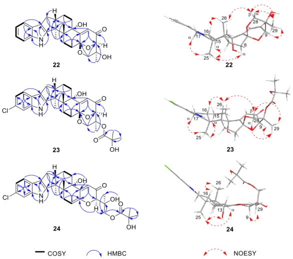
The IR spectrum of 22displayed absorptions for hydroxy (3360 cm–1) and substituted benzol (1632, 1468, 800, and 742 cm−1) functionalities. The presence of anα,β-unsaturated ketone moiety was suggested by the characteristic IR absorption at 1658 cm−1. The strong UV absorptions at 279, 268, 230 and 210 nm were in agreement with the presence of a benzol ring or anα,β-unsaturated ketone moiety in the structure. The NMR spectra (Table2) demonstrated a great similarity to the known metabolites of asperindoles A–D, previously obtained from an ascidian-derived fungusAspergillussp. [20], suggesting the same indole-diterpene framework for these molecules. Signals for the acetyl group in asperindole B were not observed for22. The structure of22was suggested to be the deacetyl analogues of asperindole B, which was fully confirmed by 2D NMR experiments, particularly HMBC and NOESY (Figure3). The absolute configurations of22were the same as that of asperindoles A–D on the basis of the similar ECD spectra (Figure4). The structure of compound22was thus determined as asperindole E.
Table 2.1H and13C NMR data for22–24.
Position 22 (in CDCl3) 23 (in CDCl3) 24 (in DMSO)
δHa(Jin Hz) δCb, Type δHc(Jin Hz) δCb, Type δHa(Jin Hz) δCb, Type
1-NH 7.70, s 7.72, s 10.96, s
2 152.0, C 152.8, C 153.9, C
3 52.0, C 52.1, C 50.4, C
4 39.4, C 39.4, C 42.7, C
5α 1.87, dd
(11.5, 9.2) 27.3, CH2 1.84, ovd 27.2, CH2 2.37, dd
(17.0, 6.0) 30.6, CH2
5β 2.65, dd (11.5, 9.5) 2.60, ovd 3.02, d (17.0)
6α 2.23, dd (13.0, 9.2) 31.1, CH2 2.12, dd (13.2, 9.0) 30.7, CH2
5.69, m 111.5, CH
6β 2.66, dd (13.0, 9.5) 2.61, ovd
7 94.5, C 94.0, C 145.0, C
9 4.05, d (2.0) 84.0, CH 4.81, d (2.4) 79.2, CH 4.25, d (1.5) 82.1, CH
10 196.5, C 196.0, C 194.7, C
11 6.24, s 120.7, CH 6.22, s 120.4, CH 5.84, s 116.1, CH
12 158.9, C 159.8, C 154.5, C
13 78.9, C 78.8, C 73.8, C
14α 2.00, ovd 33.9, CH2 2.00, ovd 33.8, CH2 1.93, ovd 31.7, CH2
14β 1.97, ovd 1.96, dd (13.2, 4.8) 1.83, ovd
15α 2.08, dd (13.0, 4.0) 21.3, CH2 2.09, ovd 21.3, CH2 1.92, ovd 21.1, CH2
15β 1.82, m 1.82, ovd 1.66, m
16 2.83, m 48.6, CH 2.82, m 48.7, CH 2.72, m 49.3, CH
17α 2.45, dd
(13.2, 11.0) 27.7, CH2 2.43, dd
(13.2, 10.2) 27.7, CH2 2.33, dd
(13.0, 10.5) 26.8, CH2
17β 2.74, dd
(13.2, 6.5)
2.71, dd (13.2, 6.0)
2.61, dd (13.0, 7.0)
18 117.4, C 117.5, C 115.3, C
19 125.3, C 123.9, C 123.3, C
20 7.43, dd (6.7, 2.0) 118.7, CH 7.31, d (8.4) 119.3, CH 7.28, d (8.5) 118.9, CH 21 7.08, td (6.7, 2.0) 119.9, CH 7.04, dd (8.4, 1.8) 120.4, CH 6.92, dd (8.5, 2.0) 118.8, CH
22 7.10, td (6.7, 2.0) 120.7, CH 126.4, C 123.9, C
23 7.31, dd (6.7, 2.0) 111.7, CH 7.28, d (1.8) 111.6, CH 7.26, d (2.0) 111.3, CH
24 139.9, C 140.2, C 140.3, C
25 1.38, s 16.4, CH3 1.37, s 16.4, CH3 1.29, s 16.4, CH3
26 1.16, s 24.5, CH3 1.14, s 24.5, CH3 0.99, s 19.7, CH3
27 66.8, C 76.9, C 74.3, C
28α 3.64, dd (12.0, 2.0) 68.2, CH2 4.21, dd (13.2, 2.4) 65.0, CH2
3.88, s 67.9, CH2
28β 3.75, d (12.0) 3.69, d (13.2)
29 1.07, s 18.9, CH3 1.37, s 17.3, CH3 1.30, s 21.9, CH3
10 176.9, C 175.5, C
20 72.5, C 71.3, C
30 1.52, s 27.4, CH3 1.29, s 27.3, CH3
40 1.53, s 27.3, CH3 1.29, s 27.3, CH3
13-OH 4.98, s
27-OH 5.06, d (1.5)
20-OH 5.26, s
a500 MHz.b125 MHz.c600 MHz.doverlapped signals.
Asperindole F (23), obtained as an optically active, white powder, has a molecular formula of C31H36ClNO7as determined by HRESIMS. The presence of a chlorine atom in the molecule was indicated by the isotopic peaks atm/z568/570 [M−H]− with a ratio of 3:1. The NMR spectra of23(Table2) resembled those of asperindole C (20) [20]
(Table S1), except for the acetyl group which is absent, showing the same difference pattern as that between asperindole E (22) and B. Compound23is the deacetylated derivative of asperindole C, and was named as asperindole F. This assignment was further confirmed by NMR and ECD experiments (Figures3and4).
Mar. Drugs2021,19, 281 6 of 14
Mar. Drugs 2021, 19, x 6 of 14
Figure 3. Selected COSY, HMBC, and NOESY correlations of compounds 22–24.
Figure 4. Experimental ECD data of compounds 20 and 22–24 in MeCN.
Asperindole G (24) was obtained as an optically active, white powder. Its HRESIMS gave the same molecular formula as that of asperindole F (23). As expected, the NMR data of 24 (Table 2) showed similarity to those of 23. However, two sp3 carbon atoms (δ 94.0, C;
δ 30.7, CH2) in 23 were replaced by two sp2 carbon atoms (δ 145.0, C; δ 111.5, CH) in 24, suggesting a cleavage of the ether bridge of ring G in 23 to form a C-6 to C-7 double bond,
Figure 3.Selected COSY, HMBC, and NOESY correlations of compounds22–24.
Mar. Drugs 2021, 19, x 6 of 14
Figure 3. Selected COSY, HMBC, and NOESY correlations of compounds 22–24.
Figure 4. Experimental ECD data of compounds 20 and 22–24 in MeCN.
Asperindole G (24) was obtained as an optically active, white powder. Its HRESIMS gave the same molecular formula as that of asperindole F (23). As expected, the NMR data of 24 (Table 2) showed similarity to those of 23. However, two sp3 carbon atoms (δ 94.0, C;
δ 30.7, CH2) in 23 were replaced by two sp2 carbon atoms (δ 145.0, C; δ 111.5, CH) in 24, suggesting a cleavage of the ether bridge of ring G in 23 to form a C-6 to C-7 double bond,
Figure 4.Experimental ECD data of compounds20and22–24in MeCN.
Asperindole G (24) was obtained as an optically active, white powder. Its HRESIMS gave the same molecular formula as that of asperindole F (23). As expected, the NMR data of24(Table2) showed similarity to those of23. However, twosp3carbon atoms (δ94.0, C;δ30.7, CH2) in23were replaced by twosp2carbon atoms (δ145.0, C;δ111.5, CH) in 24, suggesting a cleavage of the ether bridge of ring G in23to form a C-6 to C-7 double bond, and a 28-hydroxymethyl group in24. The assignment for the C-6 to C-7 double bond was confirmed by the HMBC correlations of H-6 with C-4 and C-12, and H-9 with C-7.
In addition, theα-hydroxyisobutyrate moiety was attached to the C-28 methylene group of the side-chain instead of the C-27 of the bridged 1,3-dioxane ring (as those in19–23),
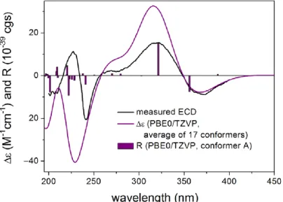
as confirmed by the diagnostic HMBC correlations between H2-28 and the ester carbonyl carbon (δ175.5, C). Compound24was expected to have the same stereochemistry as that of 19–23due to their obvious correlations in biogenetic origin, even though the ECD spectrum of24was significantly different from those of20–23. However, a very weak NOE correlation was observed between H-9 and H3-26 in the NOESY spectrum of24. This suggested that an epimerization might occur at C-9, which is adjacent to the C-10 ketone group. In order to determine the absolute configuration of C-9, TDDFT-ECD [33] and DFT-NMR calculations [34] were performed on the (3S,4R,9R,13S,16S,27S) and (3S,4R,9S,13S,16S,27S) epimers. Ring F, containing anα,β-unsaturated carbonyl chromophore, was expected to have a major impact on the high-wavelength ECD transitions, and thus a large difference was expected between the ECD spectra of the two epimers. The initial Merck molecular force field (MMFF) conformational search resulted in 353 and 156 conformers in a 21 kJ/mol energy window for (3S,4R,9R,13S,16S,27S)-24and (3S,4R,9S,13S,16S,27S)-24, respectively.
The re-optimization of these conformers yielded 15 and 17 low-energy conformers over the 1% Boltzmann distribution at theωB97X/TZVP [35] PCM/MeCN level (Figures S83 and S84). Unexpectedly, the Boltzmann-weighted ECD spectra of both epimers computed at various levels reproduced well the major transitions of the experimental ECD spectrum, indicating that the contribution of ring E is dominant. However, the (9R) epimer could reproduce the 225 nm positive Cotton effect (CE) with a blue shift (Figure5), while the (9S) epimer had only a negative computed CE below 250 nm (Figure6). This small difference suggested that24had (9R) absolute configuration, and the difference in the experimental ECD spectra of20–23and24derives from the different chromophore systems and different planar structures. It is well-documented that even small structural changes can result in markedly different or mirror-image ECD spectra for homochiral derivatives by changing the preferred conformation or electronic properties of the molecule [36]. The (9R) and (9S) epimers were further distinguished by13C NMR DFT calculations, which has been proven an efficient method to distinguish diastereomeric natural products [17,34]. For the NMR calculations, the above MMFF conformers were re-optimized at the B3LYP/6-31 + G(d,p) level yielding 16 and 14 low-energy structures above the 1% Boltzmann distribution, respectively. Despite the DMSO solvent and the presence of the halogen, both causing larger deviations in the computed data, the calculated13C NMR shifts of the (9R) epimer had a slightly smaller MAE average value than those of the (9S) epimer, and the DP4+
statistical analysis [35,37] resulted in an 83.75% confidence for the (9R) epimer (Table S2).
Since both the computed ECD and NMR data suggested (9R) configuration, the H-9 and H3-26 NOE cross-peak must be an artefact, and the absolute configuration of 24was determined to be (3S,4R,9R,13S,16S,27S). The interatomic distance of H-9 and H3-26 is 3.7 Å in the (9S) epimer and it is above 5.0 Å in the (9R) epimer.
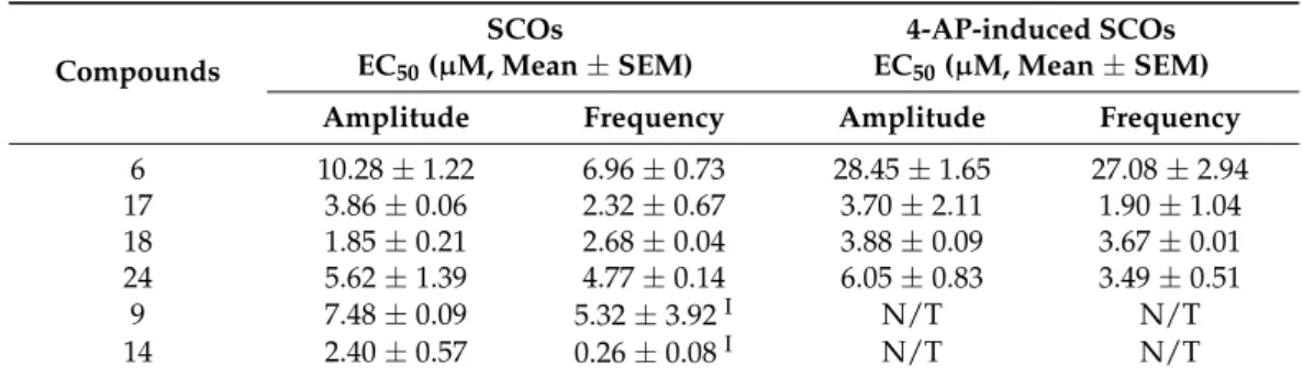
All the isolated compounds were evaluated for their neuronal modulatory activities by testing their effect on spontaneous Ca2+oscillations (SCOs), and the seizurogenic agent 4-aminopyridine (4-AP) induced hyperexcitation in primary cultured neocortical neurons (Table3). SCOs play a crucial role in mediating neuron development, and are closely associated with neuronal excitable and inhibitory neuronal transmission [38–40]. The compounds with modulatory activity on SCOs may have potential in drug candidates for treating neurological diseases such as epilepsy, pain and depression [41].
Mar. Drugs2021,19, 281 8 of 14
Mar. Drugs 2021, 19, x 8 of 14
Figure 5. Experimental ECD spectrum of 24 in MeCN compared with the Boltzmann-weighted CAM-B3LYP/TZVP PCM/MeCN ECD spectrum of (3S,4R,9R,13S,16S,27S)-24. Level of optimiza- tion: ωB97X/TZVP PCM/MeCN. Bars represent the rotatory strength values of the lowest-energy conformer.
Figure 6. Experimental ECD spectrum of 24 in MeCN compared with the Boltzmann-weighted CAM-B3LYP/TZVP PCM/MeCN ECD spectrum of (3S,4R,9S,13S,16S,27S)-24. Level of optimiza- tion: ωB97X/TZVP PCM/MeCN. Bars represent the rotatory strength values of the lowest-energy conformer.
All the isolated compounds were evaluated for their neuronal modulatory activities by testing their effect on spontaneous Ca2+ oscillations (SCOs), and the seizurogenic agent 4-aminopyridine (4-AP) induced hyperexcitation in primary cultured neocortical neurons (Table 3). SCOs play a crucial role in mediating neuron development, and are closely as- sociated with neuronal excitable and inhibitory neuronal transmission [38–40]. The com- pounds with modulatory activity on SCOs may have potential in drug candidates for treating neurological diseases such as epilepsy, pain and depression [41].
Figure 5. Experimental ECD spectrum of 24in MeCN compared with the Boltzmann-weighted CAM-B3LYP/TZVP PCM/MeCN ECD spectrum of (3S,4R,9R,13S,16S,27S)-24. Level of optimization:
ωB97X/TZVP PCM/MeCN. Bars represent the rotatory strength values of the lowest-energy conformer.
Mar. Drugs 2021, 19, x 8 of 14
Figure 5. Experimental ECD spectrum of 24 in MeCN compared with the Boltzmann-weighted CAM-B3LYP/TZVP PCM/MeCN ECD spectrum of (3S,4R,9R,13S,16S,27S)-24. Level of optimiza- tion: ωB97X/TZVP PCM/MeCN. Bars represent the rotatory strength values of the lowest-energy conformer.
Figure 6. Experimental ECD spectrum of 24 in MeCN compared with the Boltzmann-weighted CAM-B3LYP/TZVP PCM/MeCN ECD spectrum of (3S,4R,9S,13S,16S,27S)-24. Level of optimiza- tion: ωB97X/TZVP PCM/MeCN. Bars represent the rotatory strength values of the lowest-energy conformer.
All the isolated compounds were evaluated for their neuronal modulatory activities by testing their effect on spontaneous Ca2+ oscillations (SCOs), and the seizurogenic agent 4-aminopyridine (4-AP) induced hyperexcitation in primary cultured neocortical neurons (Table 3). SCOs play a crucial role in mediating neuron development, and are closely as- sociated with neuronal excitable and inhibitory neuronal transmission [38–40]. The com- pounds with modulatory activity on SCOs may have potential in drug candidates for treating neurological diseases such as epilepsy, pain and depression [41].
Figure 6. Experimental ECD spectrum of 24in MeCN compared with the Boltzmann-weighted CAM-B3LYP/TZVP PCM/MeCN ECD spectrum of (3S,4R,9S,13S,16S,27S)-24. Level of optimization:
ωB97X/TZVP PCM/MeCN. Bars represent the rotatory strength values of the lowest-energy conformer.
In the present study, we found that four compounds6,17,18and24inhibited SCO activity, and 4-AP induced hyperexcitability by decreasing the SCO amplitude and fre- quency in the primary cultured cortical neuronal network. However, compounds9and14 produced a more complicated Ca2+response. Their concentration dependently increased the SCO frequency with the concurrent suppression of the SCO amplitude at concentrations below 10µM and 3µM, respectively, and transiently increased the intracellular Ca2+con- centration which recovered to basal level within 5 min at concentrations of 30µM and 10 µM, respectively (Table3, and Figures S85–S90). For the cluster ofp-terphenyl derivatives, all the active compounds have hydroxyls for both R2and R4and those with hydrogens for both R1and R3displayed the strongest activity. Substitution for one of the R2/R4pair of hydroxyls or one of the R1/R3pair of hydrogens will decrease the activity. Interestingly, the degradation of ring A to a hydroxymethyl group may lead to an increase in activity.
For the cluster of indole-diterpene alkaloids, ring cleavage on the ether bridge of ring G seems critical for the activity since all those compounds that have ring G are not active.
Table 3.Compounds influence SCOs and 4-AP-induced SCOs.
Compounds
SCOs
EC50(µM, Mean±SEM)
4-AP-induced SCOs EC50(µM, Mean±SEM)
Amplitude Frequency Amplitude Frequency
6 10.28±1.22 6.96±0.73 28.45±1.65 27.08±2.94
17 3.86±0.06 2.32±0.67 3.70±2.11 1.90±1.04
18 1.85±0.21 2.68±0.04 3.88±0.09 3.67±0.01
24 5.62±1.39 4.77±0.14 6.05±0.83 3.49±0.51
9 7.48±0.09 5.32±3.92I N/T N/T
14 2.40±0.57 0.26±0.08I N/T N/T
Data represent mean values of five independent experiments; “N/T” means not tested. ”I” indicates increase in the SCO frequency.
3. Experimental Section
3.1. General Experimental Procedures
Optical rotations were determined with a Rudolph Autopol VI polarimeter. UV spectra were recorded on a Shimadzu UV-2700 spectrophotometer. IR spectra were recorded on a Bruker TENSOR II spectrophotometer. The ECD spectra were measured with a Jasco- 715 spectropolarimeter. NMR spectra were recorded on Bruker DRX-600 and DRX-500 spectrometers, and the signals of residual CHCl3(δH7.26 ppm;δC77.0 ppm), DMSO (δH
2.50 ppm;δC39.6 ppm) and CH3OH (δH3.31 ppm;δC49.0 ppm) were used as references for chemical shifts. The HR-ESI-MS data were recorded on an Agilent 1290-6545 UHPLC-Q- TOF-MS spectrometer. Semipreparative HPLC was performed using an Agilent Technology 1100 system with an ODS column (YMC Pack ODS-A, 10× 250 mm, 5µM). Column chromatography (CC) was performed with Sephadex LH-20 gel (SE-751 84 Uppsala, GE Healthcare Bio-Sciences AB) and silica gel (200–300 mesh, 400–600 mesh; Yantai, China), respectively. The thin-layer chromatography (TLC) experiments were conducted with silica gel plates (HSGF-254, Yantai, China), and detected by heating after spraying with anisaldehyde sulfuric acid reagent.
3.2. Fungal Material
The fungal strain SG-8-5 was isolated from the internal tissues of the gorgonian coral J. fragillis, which was collected from the Xisha area of the South China Sea, and identified asA. candidusby 18sRNA sequence (GenBank accession number AB008396.1). The fungus was deposited in Tongji University, Shanghai, China.
3.3. Culture, Extraction and Isolation
The fungal strainA. candidus was cultivated on 20.0 L of 5% w/v biomalt (Villa Natura, Germany) solid agar medium (20.0 g/L biomalt, 15.0 g/L agar, 800 mL/L artificial seawater) at room temperature for 28 days. The fungal mycelia with the medium were extracted with EtOAc (4.0 L×5) under ultrasonic conditions. The EtOAc extract was concentrated under vacuum (13.6 g) and was separated into thirteen fractions (Fr.1–13) by silica gel CC, eluting with a gradient CH2Cl2/MeOH (v/v100:0 to 4:1). Fraction 2 (52.7 mg) was subjected to a Sephadex LH-20 CC (CH2Cl2/MeOH 2:1) and then purified by HPLC (MeOH/H2O 89:11, 1.5 mL/min) to give3(1.3 mg,tR18 min). Fraction 5 (29.9 mg) was subjected to a Sephadex LH-20 CC (CH2Cl2/MeOH 2:1) and HPLC (MeOH/H2O 80:20, 2.0 mL/min) to afford5(3.1 mg, tR24 min). Fraction 6 (644.9 mg) was subjected to a Sephadex LH-20 CC (CH2Cl2/MeOH 2:1) to give eight subfractions (Fr.6a to Fr.6h). The Fr.6e (215.8 mg) was separated by silica gel CC using a gradient petroleum (PE) in acetone (39:1 to 6:4), then split by HPLC to yield7(1.2 mg, MeOH/H2O 79:21, 1.5 mL/min,tR18 min),8(0.9 mg, MeOH/H2O 79:21, 1.5 mL/min,tR21 min),1(1.2 mg, MeOH/H2O 80:20,
Mar. Drugs2021,19, 281 10 of 14
2.0 mL/min,tR33 min),19(1.8 mg, MeOH/H2O 80:20, 2.0 mL/min,tR50 min),20(2.3 mg, MeOH/H2O 80:20, 2.0 mL/min,tR63 min),21(1.1 mg, MeOH/H2O 80:20, 2.0 mL/min,tR 36 min). Fr.6f (54.7 mg) was separated by silica gel CC (PE-EtOAc 9:1 to 1:1) and purified by HPLC (MeOH/H2O 80:20, 2.0 mL/min) to afford23(1.2 mg,tR44 min) and24(0.7 mg,tR
25 min). Fr.6g (18.7 mg) was purified by HPLC (MeOH/H2O 70:30, 2.0 mL/min) to afford 10(0.8 mg,tR23 min) and22(0.9 mg,tR64 min). Fr.6h (52.4 mg) was purified by HPLC (MeOH/H2O 65:35, 2.0 mL/min) to yield16(1.6 mg,tR30 min),11(1.5 mg,tR48 min), and12(1.0 mg,tR51 min). Fraction 8 (164.7 mg) was subjected to a Sephadex LH-20 CC (CH2Cl2/MeOH 2:1) to yield thirteen subfractions (Fr.8a to Fr.8m). Fr.8h (19.8 mg) was further purified by HPLC (MeOH/H2O 70:30, 1.5 mL/min) to afford6(3.6 mg,tR13 min), 2(0.5 mg,tR20 min), and15(3.1 mg,tR41 min). Fr.8j (33.4 mg) was purified by HPLC (MeOH/H2O 65:35, 2.0 mL/min) to afford13(13.9 mg,tR18 min). Fraction 9 (364.2 mg) was subjected to a Sephadex LH-20 CC (CH2Cl2/MeOH 2:1) to yield4(50.0 mg). Fraction 10 (649.6 mg) was subjected to a Sephadex LH-20 CC (CH2Cl2/MeOH 2:1) to furnish fourteen subfractions (Fr.10a to Fr.10n) and pure compound14(11.6 mg). Fr.10g (29.3 mg) was further purified by HPLC (MeOH/H2O 70:30, 2.0 mL/min) to afford18(2.0 mg,tR27.5 min). Fr.10i (84.5 mg) was separated by silica gel CC with the eluent of gradient petroleum ether-ethyl acetate from 9:1 to 1:1, followed by the purification of HPLC (MeOH/H2O 58:42, 2.0 mL/min) to yield17(2.5 mg,tR42 min). Fraction 11 (201.0 mg) was applied on a Sephadex LH-20 CC (CH2Cl2/MeOH 2:1) and purified by HPLC (MeOH/H2O 55:45, 2.0 mL/min) to afford9(13.0 mg,tR11 min).
The 400-O-methyl-prenylterphenyllin B (1): colorless, amorphous solid; UV (MeCN) λmax(logε) 276 (3.39), 248 (3.12), 209 (3.64) nm; IR (film)νmax3357, 2922, 2852, 1659, 1609, 1462, 1246, 1073, 1029, 834, 815 cm−1;1H and13C NMR data see Table1; HRESIMSm/z 419.1870 [M−H]−(calcd for C26H27O5, 419.1864).
The 300-hydroxyl-prenylterphenyllin (2): colorless, amorphous solid; UV (MeCN)λmax
(logε) 277 (3.28), 250 (3.13), 210 (3.65) nm; IR (film)νmax3346, 2922, 2853, 1665, 1607, 1461, 1260, 1093, 799, 722 cm−1;1H NMR (CD3OD, 500 MHz):δH7.11 (1H, d,J= 2.2 Hz, H-200), 7.05 (1H, d,J= 2.2 Hz, H-2), 7.01 (1H, dd,J= 8.2, 2.2 Hz, H-6), 6.96 (1H, dd,J= 8.2, 2.2 Hz, H-600), 6.83 (1H, d,J= 8.2 Hz, H-500), 6.77 (1H, d,J= 8.2 Hz, H-5), 6.42 (1H, s, H-50), 5.37 (1H, m, H-2000), 3.67 (3H, s, 60-OMe), 3.41 (3H, s, 30-OMe), 3.32 (2H, m, H-1000), 1.73 (3H, s, H-5000), 1.72 (3H, s, H-4000);13C NMR (CD3OD, 125 MHz):δC154.8 (C, C-4), 149.1 (C, C-20), 146.1 (C, C-300), 145.9 (C, C-400), 140.8 (C, C-30), 134.2 (C, C-40), 133.2 (CH, C-2), 132.7 (C, C-3000), 131.7 (C, C-100), 130.3 (CH, C-6), 128.2 (C, C-3), 126.3 (C, C-1), 124.2 (CH, C-2000), 121.5 (CH, C-600), 119.0 (C, C-10), 117.2 (CH, C-200), 116.2 (CH, C-500), 115.1 (CH, C-5), 105.0 (CH, C-50), 60.8 (CH3, 30-OMe), 56.5 (CH3, 60-OMe), 29.3 (CH2, C-1000), 26.0 (CH3, C-5000), 17.9 (CH3, C-4000);
HRESIMSm/z423.1787 [M + H]+(calcd for C25H27O6, 423.1802).
The 3-methoxy-400-deoxyterprenin (3): colorless, amorphous solid; UV (MeCN)λmax
(logε) 275 (3.27), 250 (3.14), 202 (3.68); IR (film)νmax3356, 2924, 2853, 1664, 1601, 1461, 1266, 1075, 829, 771 cm−1;1H NMR (CDCl3, 500 MHz): δH7.64 (2H, dd,J= 7.5, 1.5 Hz, H-600), 7.46 (2H, td,J= 7.5, 1.5 Hz, H-500), 7.39 (1H, tt,J= 7.5, 1.5 Hz, H-400), 7.02 (1H, dd,J= 8.7, 2.0 Hz, H-6), 7.02 (1H, d,J= 2 Hz, H-2), 6.98 (1H, d,J= 8.7 Hz, H-5), 6.50 (1H, s, H-50), 5.93 (1H, s, 20-OH), 5.57 (1H, m, H-2000), 4.64 (2H, d,J= 7 Hz, H-1000), 3.88 (3H, s, 3-OMe), 3.76 (3H, s, 60-OMe), 3.46 (3H, s, 30-OMe), 1.79 (3H, s, H-5000), 1.75 (3H, s, H-4000);13C NMR (CDCl3, 125 MHz):δC153.6 (C, C-60), 149.1 (C, C-3), 147.8 (C, C-4), 147.4 (C, C-20), 139.0 (C, C-30), 138.2 (C, C-100), 137.6 (C, C-3000), 133.0 (C, C-40), 128.9 (CH, C-200, 600), 128.6 (CH, C-300, 500), 127.7 (CH, C-400), 125.4 (C, C-1), 123.0 (CH, C-6), 120.3 (CH, C-2000), 117.0 (C, C-10), 114.4 (CH, C-2), 112.6 (CH, C-5), 104.2 (CH, C-50), 65.8 (CH2, C-1000), 61.1 (CH3, 30-OMe), 56.2 (CH3, 60-OMe), 56.1 (CH3, 3-OMe), 26.0 (CH3, C-5000), 18.4 (CH3, C-4000); HRESIMSm/z 421.2027 [M + H]+(calcd for C26H29O5, 421.2010).
Phenylcandilide A (17): yellow, amorphous solid; UV (MeCN)λmax(logε) 276 (3.41), 256 (3.16), 242 (3.45), 227 (3.33), 209 (3.50), 203 (3.49) nm; IR (film)νmax3359, 2922, 2852, 1658, 1469, 1266, 1206, 1091, 831, 738 cm−1;1H and13C NMR data see Table1; HRESIMS m/z337.1055 [M + Na]+(calcd for C18H18NaO5, 337.1046).
Phenylcandilide B (18): yellow, amorphous solid; UV (MeCN)λmax(logε) 275 (3.58), 255 (3.37), 240 (3.61), 227 (3.54), 212 (3.64) nm; IR (film)νmax3354, 2924, 2853, 1740, 1660, 1497, 1267, 1213, 1172, 1104, 830, 671 cm−1;1H and13C NMR data see Table1; HRESIMS m/z357.1349 [M + H]+(calcd for C20H21O6, 357.1333).
Asperindole E (22): white powder; [α]20D +50.00 (c0.03, CHCl3); UV (MeCN) λmax (logε) 279 (2.78), 268 (2.75), 230 (3.45), 210 (3.24) nm; ECD (0.045 mM, MeCN)λmax(∆ε) 238 (−20.91) nm; IR (film)νmax3360, 2920, 2851, 1720, 1658, 1632, 1468, 1260, 1016, 800, 742 cm−1;1H and13C NMR data see Table3; HRESIMSm/z472.2086 [M + Na]+(calcd for C27H31NO5Na, 472.2094).
Asperindole F (23): white powder; [α]20D +40.56 (c 0.03, CHCl3); UV (MeCN) λmax (logε) 285 (2.89), 267 (2.76), 236 (3.63), 209 (3.23) nm; ECD (0.035 mM, MeCN)λmax(∆ε) 242 (−39.92) nm; IR (film)νmax3358, 2920, 2851, 1730, 1659, 1633, 1467, 1260, 1013, 799, 704 cm−1;1H and13C NMR data see Table3; HRESIMSm/z568.2120 [M−H]−(calcd for C31H35ClNO7, 568.2108).
Asperindole G (24): white powder;[α]20D +31.11 (c0.03, CHCl3); UV (MeCN)λmax(log ε) 303 (2.92), 293 (2.90), 286 (2.91), 270 (2.85), 236 (3.56), 211 (3.33) nm; ECD (0.035 mM, MeCN),λmax(∆ε) 373 (−8.91), 362 (−7.78), 322 (+15.46), 269 (+2.49), 241 (−20.56), 227 (+11.34) nm; IR (film)νmax3349, 2920, 2851, 1730, 1659, 1462, 1260, 1018, 798 cm−1;1H and
13C NMR data see Table3; HRESIMSm/z592.2068 [M + Na]+(calcd for C31H36ClNO7Na, 592.2073).
3.4. Neuronal Modulatory Activity Assay In Vitro
The neuronal modulatory activities of1–24were evaluated by testing the effect on spontaneous Ca2+oscillations (SCOs) and seizurogenic agent 4-aminopyridine (4-AP)- induced hyperactive SCOs frequency and amplitude in primary cultured neocortical neu- rons as described previously [16,38]. Neocortical neurons at 9 days in vitro (DIV) were used to investigate the influence of tested compounds on intracellular Ca2+concentration ([Ca2+]i). Briefly, the neurons were loaded with Fluo-4 for 1 h at 37◦C in Locke’s buffer.
After recording the baseline spontaneous Ca2+oscillations for 5 min, different concentra- tions of compounds were added to the corresponding well, and the [Ca2+]iwas monitored for 15 min using FLIPRtetra®. To test anti-epileptic potential of the meroterpenoids, 4-AP (10µM) was added and the monitoring for [Ca2+]iwas continued for an additional 10 min.
The presented data were values of F/F0, where F is the fluorescence intensity at any time point whereas F0is the basal fluorescence. An event with∆F/F0over 0.1 unit was consid- ered to be an SCO. The frequency and amplitude of SCOs were quantified using Origin software (V7.0) from a time period of 5 min after first or second addition of compound or vehicle (0.1% DMSO).
3.5. Computational Section
Mixed torsional/low-frequency mode conformational searches were carried out by means of the Macromodel 10.8.011 software by using the Merck molecular force field (MMFF) with an implicit solvent model for CHCl3[42]. Geometry re-optimizations were carried out at the B3LYP/6-31 + G(d,p) level in vacuo and theωB97X/TZVP level with the PCM solvent model for MeCN. TDDFT-ECD calculations were run with various functionals (B3LYP, BH&HLYP, CAMB3LYP, and PBE0) and the TZVP basis was set as implemented in the Gaussian 09 package, with the same or no solvent model as in the preceding DFT optimization step [43]. ECD spectra were generated as sums of Gaussians with 3000 and 2700 cm−1widths at half-height, using dipole-velocity-computed rotational strength val- ues [44]. NMR calculations were performed at the mPW1PW91/6-311 + G(2d,p) level [45].
Computed NMR shift data were corrected with I = 185.2853 and S =−1.0306 [46]. Boltz- mann distributions were estimated from the B3LYP andωB97X energies. The MOLEKEL software package was used for visualization of the results [47].
Mar. Drugs2021,19, 281 12 of 14
4. Conclusions
From the fungiA. candidus, associated with the South China Sea gorgonianJunceela fragillis, twenty-four metabolites havingp-terphenyl and indole-diterpene frameworks were obtained with their structures and absolute configurations being elucidated on the basis of spectroscopic analysis and computational calculations. It was found that small structural changes can result in a markedly different ECD spectra, and13C NMR DFT calculations are an efficient method to distinguish diastereomeric natural products. Six compounds could modulate SCOs and 4-aminopyridine hyperexcited neuronal activity in thein vitrobiotest. A preliminary structure–
activity relationship was discussed, which may give a reference for further investigation or chemical optimization of SCO modulators.
Supplementary Materials:The following are available online athttps://www.mdpi.com/article/
10.3390/md19050281/s1, tables for the experimental NMR data of20and computed NMR data of 24, figures for the 2D correlations of1–3, MS, IR, UV, NMR spectra of1–3,17–18and22–24, the low energy conformers of the computed structures of24, and the in vitro effects of6,9,14,17,18and24 on SCOs.
Author Contributions:G.-Y.P. performed the experiments. T.K. and A.M. performed the quantum mechanical calculation. J.H. and Z.-Y.C. contributed to the bioassay. H.T. was responsible for the fungal isolation and fermentation. S.-C.M. and W.Z. conceived and designed the experiments. All authors have read and agreed to the published version of the manuscript.
Funding: This research was funded by the National Nature Science Foundation of China, grant numbers 81820108030 and 41806088. T.K and A.M. were supported by the National Research, Devel- opment and Innovation Office (K120181 and FK134653) and the János Bolyai Research Scholarship of the Hungarian Academy of Sciences.
Institutional Review Board Statement:The study was conducted according to the guidelines of the Declaration of Helsinki, and approved by the Institutional Review Board of China Pharmaceutical University (protocol code 2016-0011, approved at 2 March 2016).
Data Availability Statement:Data are contained within the article or Supplementary Materials.
Acknowledgments: The Governmental Information-Technology Development Agency (KIFÜ) is acknowledged for CPU time.
Conflicts of Interest:The authors declare no competing financial interest.
References
1. Klich, M.A. Biogeography ofAspergillusspecies in soil and litter.Mycologia2002,94, 21–27. [CrossRef]
2. Lin, Y.-K.; Xie, C.-L.; Xing, C.-P.; Wang, B.-Q.; Tian, X.-X.; Xia, J.-M.; Jia, L.-Y.; Pan, Y.-N.; Yang, X.-W. Cytotoxicp-terphenyls from the deep-sea-derivedAspergillus candidus.Nat. Prod. Res.2019,24, 1–5. [CrossRef]
3. Han, J.; Lu, F.; Bao, L.; Wang, H.; Chen, B.; Li, E.; Wang, Z.; Xie, L.; Guo, C.; Xue, Y.; et al. Terphenyl derivatives and terpenoids from a wheat-born moldAspergillus candidus.J. Antibiot.2020,73, 189–193. [CrossRef]
4. Wei, H.; Inada, H.; Hayashi, A.; Higashimoto, K.; Pruksakorn, P.; Kamada, S.; Arai, M.; Ishida, S.; Kobayashi, M. Prenylter- phenyllin and its dehydroxyl analogs, new cytotoxic substances from a marine-derived fungusAspergillus candidusIF10. J.
Antibiot.2007,60, 586–590. [CrossRef] [PubMed]
5. Buttachon, S.; Ramos, A.A.; Inácio,Â.; Dethoup, T.; Gales, L.; Lee, M.; Costa, P.M.; Silva, A.M.S.; Sekeroglu, N.; Rocha, E.; et al.
Bis-indolyl benzenoids, hydroxypyrrolidine derivatives and other constituents from cultures of the marine sponge-associated fungusAspergillus candidusKUFA0062.Mar. Drugs2018,16, 119. [CrossRef]
6. Shan, T.J.; Wang, Y.Y.; Wang, S.; Xie, Y.Y.; Cui, Z.H.; Wu, C.Y.; Sun, J.; Wang, J.; Mao, Z.L. A newp-terphenyl derivative from the insect-derived fungusAspergillus candidusBdf-2 and the synergistic effects of terphenyllin.PeerJ2020,8, 15. [CrossRef] [PubMed]
7. Ma, J.; Zhang, X.L.; Wang, Y.; Zheng, J.Y.; Wang, C.Y.; Shao, C.L. Aspergivones A and B, two new flavones isolated from a gorgonian-derivedAspergillus candidusfungus.Nat. Prod. Res.2016,31, 32–36. [CrossRef] [PubMed]
8. Zhou, G.; Chen, X.; Zhang, X.; Che, Q.; Zhang, G.; Zhu, T.; Gu, Q.; Li, D. Prenylatedp-terphenyls from a mangrove endophytic fungus,Aspergillus candidusLDJ-5.J. Nat. Prod.2020,83, 8–13. [CrossRef]
9. Yan, W.; Li, S.J.; Guo, Z.K.; Zhang, W.J.; Wei, W.; Tan, R.X.; Jiao, R.H. Newp-terphenyls from the endophytic fungusAspergillussp.
YXf3.Med. Chem. Lett.2017,27, 51–54. [CrossRef]
10. Liu, S.S.; Zhao, B.B.; Lu, C.H.; Huang, J.J.; Shen, Y.M. Two newp-terphenyl derivatives from the marine fungal strainAspergillus sp. AF119.Nat. Prod. Commun.2012,7, 1057–1062. [CrossRef] [PubMed]
11. Cai, S.; Sun, S.; Zhou, H.; Kong, X.; Gu, Q. Prenylated polyhydroxy-p-terphenyls fromAspergillus taichungensisZHN-7-07.J. Nat.
Prod.2011,74, 1106–1110. [CrossRef] [PubMed]
12. Marchelli, R.; Vining, L.C. Terphenyllin, a novelp-terphenyl metabolite fromAspergillus Candidus.J. Antibiot.1975,28, 328–331.
[CrossRef] [PubMed]
13. Munden, J.E.; Butterworth, D.; Hanscomb, G.; Verrall, M.S. Production of chlorflavonin, an antifungal metabolite ofAspergillus candidus.Appl. Microbiol.1970,19, 718–720. [CrossRef] [PubMed]
14. Kamigauchi, T.; Sakazaki, R.; Nagashima, K.; Kawamura, Y.; Yasuda, Y.; Matsushima, K.; Tani, H.; Takahashi, Y.; Ishii, K.;
Suzuki, R.; et al. Terprenins, novel immunosuppressants produced byAspergillus candidus. J. Antibiot. 1998, 51, 445–450.
[CrossRef] [PubMed]
15. Liu, D.-H.; Sun, Y.-Z.; Kurtán, T.; Mándi, A.; Tang, H.; Li, J.; Su, L.; Zhuang, C.-L.; Liu, Z.-Y.; Zhang, W. Osteoclastogenesis regulation metabolites from the coral-associated fungusPseudallescheria boydiiTW-1024-3. J. Nat. Prod. 2019,82, 1274–1282.
[CrossRef]
16. Wang, H.-L.; Li, R.; Li, J.; He, J.; Cao, Z.-Y.; Kurtán, T.; Mándi, A.; Zheng, G.-L.; Zhang, W. Alternarin A, a drimane meroterpenoid, suppresses neuronal excitability from the coral-associated fungiAlternariasp. ZH-15.Org. Lett.2020,22, 2995–2998. [CrossRef]
17. Sun, P.; Cai, F.Y.; Lauro, G.; Tang, H.; Su, L.; Wang, H.L.; Li, H.H.; Mándi, A.; Kurtán, T.; Riccio, R. Immunomodulatory biscembranoids and assignment of their relative and absolute configurations: Data set modulation in the density functional theory/nuclear magnetic resonance approach.J. Nat. Prod.2019,82, 1264–1273. [CrossRef]
18. Li, W.; Li, X.B.; Lou, H.X. Structural and biological diversity of naturalp-terphenyls. J. Asian. Nat. Prod. Res. 2017,20, 1–13.
[CrossRef]
19. Liu, J.-K. Natural terphenyls: developments since 1877.Chem. Rev.2006,106, 2209–2223. [CrossRef]
20. Ivanets, E.V.; Yurchenko, A.N.; Smetanina, O.F.; Rasin, A.B.; Zhuravleva, O.I.; Pivkin, M.V.; Popov, R.S.; Von Amsberg, G.;
Afiyatullov, S.S.; Dyshlovoy, S.A. Asperindoles A–D and ap-terphenyl derivative from the ascidian-derived fungusAspergillussp.
KMM 4676.Mar. Drugs2018,16, 232. [CrossRef]
21. Tian, S.-Z.; Pu, X.; Luo, G.; Zhao, L.-X.; Xu, L.-H.; Li, W.-J.; Luo, Y. Isolation and characterization of newp-terphenyls with antifungal, antibacterial, and antioxidant activities from halophilic actinomyceteNocardiopsis gilvaYIM 90087. J. Agric. Food.
Chem.2013,61, 3006–3012. [CrossRef] [PubMed]
22. Liu, C.; Tagami, K.; Minami, A.; Matsumoto, T.; Oikawa, H. Reconstitution of biosynthetic machinery for the synthesis of the highly elaborated indole diterpene penitrem.Angew. Chem. Int. Ed.2015,54, 5529. [CrossRef]
23. Munday-Finch, S.C.; Wilkins, A.L.; Miles, C.O.; Tomoda, H.; ¯Omura, S. Isolation and structure elucidation of lolilline, a possible biosynthetic precursor of the lolitrem family of tremorgenic mycotoxins.J. Agric. Food. Chem.1997,45, 199–204. [CrossRef]
24. Jiang, Y.; Ozaki, T.; Harada, M.; Miyasaka, T.; Sato, H.; Miyamoto, K.; Kanazawa, J.; Liu, C.; Maruyama, J.-I.; Adachi, M.; et al.
Biosynthesis of indole diterpene lolitrems: Radical-induced cyclization of an epoxyalcohol affording a characteristic lolitremane skeleton.Angew. Chem. Int. Ed.2020,59, 17996–18002. [CrossRef] [PubMed]
25. Kawada, K.; Arimura, A.; Tsuri, T.; Fuji, M.; Komurasaki, T.; Yonezawa, S.; Kugimiya, A.; Haga, N.; Mitsumori, S.; Inagaki, M.;
et al. Total synthesis of terprenin, a highly potent and novel immunoglobulin E antibody suppressant.Angew. Chem. Int. Ed.
1998,37, 973–975. [CrossRef]
26. Enomoto, M.; Morita, A.; Kuwahara, S. Total synthesis of the tremorgenic indole diterpene paspalinine.Angew. Chem. Int. Ed.
Engl.2012,51, 12833–12836. [CrossRef]
27. Li, J.; Tang, H.; Kurtán, T.; Mándi, A.; Zhuang, C.-L.; Su, L.; Zheng, G.-L.; Zhang, W. Swinhoeisterols from the south china sea spongeTheonella swinhoei.J. Nat. Prod.2018,81, 1645–1650. [CrossRef]
28. Kurobane, I.; Vining, L.C.; McInnes, A.G.; Smith, D.G. 3-Hydroxyterphenyllin, a new metabolite ofAspergillus candidus.Structure elucidation by H and C nuclear magnetic resonance spectroscopy.J. Antibiot.1979,32, 559–564. [CrossRef]
29. Belofsky, G.N.; Gloer, K.B.; Gloer, J.B.; Wicklow, D.T.; Dowd, P.F. Newp-terphenyl and polyketide metabolites from the sclerotia ofPenicillium raistrickii.J. Nat. Prod.1998,61, 1115–1119. [CrossRef]
30. Takahashi, C.; Yoshihira, K.; Natori, S.; Umeda, M. The structures of toxic metabolites ofAspergillus candidus. I. The compounds A and E, cytotoxicp-terphenyls.Chem. Pharm. Bull.1976,24, 613–620. [CrossRef]
31. Kobayashi, A.; Takemura, A.; Koshimizu, K.; Nagano, H.; Kawazu, K. Candidusin A and B: Newp-terphenyls with cytotoxic effects on sea urchin embryos.Agric. Biol. Chem.1982,46, 585–589. [CrossRef]
32. Wang, W.; Liao, Y.; Tang, C.; Huang, X.; Luo, Z.; Chen, J.; Cai, P. Cytotoxic and antibacterial compounds from the coral-derived fungusAspergillus triticiSP2-8-1.Mar. Drugs2017,15, 348. [CrossRef] [PubMed]
33. Mándi, A.; Kurtán, T. Applications of OR/ECD/VCD to the structure elucidation of natural products.Nat. Prod. Rep.2019,36, 889–918. [CrossRef] [PubMed]
34. Li, W.S.; Yan, R.J.; Yu, Y.; Shi, Z.; Mándi, A.; Shen, L.; Kurtán, T.; Wu, J. Determination of the absolute configuration of super- carbon-chain compounds by a combined chemical, spectroscopic, and computational approach: Gibbosols A and B.Angew. Chem.
Int. Ed.2020,59, 13028–13036. [CrossRef] [PubMed]
35. Smith, S.G.; Goodman, J.M. Assigning stereochemistry to single diastereoisomers by GIAO NMR calculation: The DP4 probability.
J. Am. Chem. Soc.2010,132, 12946–12959. [CrossRef]
Mar. Drugs2021,19, 281 14 of 14
36. El-Kashef, D.H.; Daletos, G.; Plenker, M.; Hartmann, R.; Mándi, A.; Kurtán, T.; Weber, H.; Lin, W.; Ancheeva, E.; Proksch, P.
Polyketides and a dihydroquinolone alkaloid from a marine-derived strain of the fungusMetarhizium marquandii.J. Nat. Prod.
2019,82, 2460–2469. [CrossRef]
37. Grimblat, N.; Zanardi, M.M.; Sarotti, A.M. Beyond dp4: An improved probability for the stereochemical assignment of isomeric compounds using quantum chemical calculations of nmr shifts.J. Org. Chem.2015,80, 12526–12534. [CrossRef]
38. Cao, Z.; Cui, Y.; Nguyen, H.M.; Jenkins, D.P.; Wulff, H.; Pessah, I.N. Nanomolar bifenthrin alters synchronous Ca2+oscillations and cortical neuron development independent of sodium channel activity.Mol. Pharmacol.2014,85, 630–639. [CrossRef]
39. Zheng, J.; Chen, J.; Zou, X.; Zhao, F.; Guo, M.; Wang, H.; Zhang, T.; Zhang, C.; Feng, W.; Pessah, I.N.; et al. Saikosaponin d causes apoptotic death of cultured neocortical neurons by increasing membrane permeability and elevating intracellular Ca2+
concentration.Neurotoxicology2019,70, 112–121. [CrossRef]
40. Sinner, B.; Friedrich, O.; Zink, W.; Martin, E.; Fink, R.H.; Graf, B.M. Ketamine stereoselectively inhibits spontaneous Ca2+- oscillations in cultured hippocampal neurons.Anesth. Analg.2005,100, 1660–1666. [CrossRef]
41. Nathalie, P.; Ana, M.L.M.; Shapiro, M.S. New invitrophenotypic assay for epilepsy: Fluorescent measurement of synchronized neuronal calcium oscillations.PLoS ONE2014,9, e84755.
42. MacroModel. Schrödinger LLC, 2015. Available online:http://www.schrodinger.com/macromodel(accessed on 17 March 2021).
43. Frisch, M.J.; Trucks, G.W.; Schlegel, H.B.; Scuseria, G.E.; Robb, M.A.; Cheeseman, J.R.; Scalmani, G.; Barone, V.; Mennucci, B.;
Petersson, G.A.; et al.Gaussian 09; Revision E.01; Gaussian, Inc.: Wallingford, CT, USA, 2013.
44. Stephens, P.J.; Harada, N. ECD cotton effect approximated by the Gaussian curve and other methods.Chirality2010,22, 229–233.
[CrossRef] [PubMed]
45. Adamo, C.; Barone, V. Exchange functionals with improved long-range behavior and adiabatic connection methods without adjustable parameters: ThemPW andmPW1PW models.J. Chem. Phys.1998,108, 664–675. [CrossRef]
46. Pierens, G.K.1H and13C NMR scaling factors for the calculation of chemical shifts in commonly used solvents using density functional theory.J. Comput. Chem.2014,35, 1388–1394. [CrossRef]
47. Varetto, U.Molekel 5.4; Swiss National Supercomputing Centre: Manno, Switzerland, 2009.