Communication
Bioactive Compounds of Nigella Sativa Essential Oil as Antibacterial Agents against Chlamydia Trachomatis D
Tímea Mosolygó1,*, Ahmad Mouwakeh2, Munira Hussein Ali1, Annamária Kincses1 , Csilla Mohácsi-Farkas2 , Gabriella Kiskó2 and Gabriella Spengler1
1 Department of Medical Microbiology and Immunobiology, Faculty of Medicine, University of Szeged, 6720 Szeged, Hungary; munirahusseinali@gmail.com (M.H.A.);
kincses.annamaria@med.u-szeged.hu (A.K.); spengler.gabriella@med.u-szeged.hu (G.S.)
2 Department of Microbiology and Biotechnology, Faculty of Food Science, Szent István University, 1118 Budapest, Hungary; ahmad.mouwakeh@hotmail.com (A.M.); farkas.csilla@etk.szie.hu (C.M.-F.);
kisko.gabriella@etk.szie.hu (G.K.)
* Correspondence: mosolygo.timea@med.u-szeged.hu; Tel.:+36-62-546-112
Received: 26 July 2019; Accepted: 18 September 2019; Published: 19 September 2019
Abstract:Urogenital tract infection caused by obligate intracellular bacteriumChlamydia trachomatis D (CtrD) is a leading cause of sexually transmitted diseases. Essential oil (EO) ofNigella sativahas a broad antimicrobial spectrum. The aim of this study was to evaluate the antimicrobial activity of the bioactive compounds (p-cymene, thymoquinone, carvacrol, and thymol) of N. sativaEO againstCtrD. The cytotoxic effects of the compounds were determined by MTT assay. In order to quantify the anti-chlamydial activity of the compounds, HeLa cells were infected withCtrD orCtrD treated previously with the compounds. The titer of the infectiousCtrD was determined by indirect immunofluorescence assay. The minimum inhibitory concentrations of the compounds were evaluated by direct quantitative PCR. None of the compounds showed a cytotoxic effect on HeLa cells in the concentrations tested. According to the immunofluorescence assay, all of the compounds significantly inhibited the growth ofCtrD. The quantitative PCR revealed that the minimum concentration that exerted anti-chlamydial activity was 3.12µM in the case of thymoquinone and p-cymene, while that of carvacrol and thymol was 6.25µM. Therefore, it can be concluded that bioactive compounds of N. sativaEO could be used as effective antimicrobial agents againstCtrD.
Keywords: Chlamydia trachomatis; antibacterial activity;Nigella sativa
1. Introduction
Although antibiotic therapy eliminates bacterial infections, there is emerging evidence of bacteria developing antimicrobial resistance (AMR). AMR together with a lack of development of new antimicrobial agents has become a global public health concern [1].
Chlamydia trachomatisis an obligate intracellular bacterium that causes a wide spectrum of human diseases, such as genitourinary, pulmonary, and ocular infections. The most common genitourinary infections caused by C. trachomatis serovars D to K are mucopurulent cervicitis in females and non-gonococcal urethritis in males. Additionally, cases of untreated infections can lead to various complications, such as pelvic inflammatory disease (PID), ectopic pregnancy, chronic prostatitis, and infertility [2].Chlamydiaspp. are characterized by typical lifecycles. First, the elementary body (EB), which is the infectious form, infects the host cell. After the EB enters the host cell, the formation of inclusion occurs and the EB transforms into the reticulate body (RB). The RB is characterized by its high metabolic activity and further division by binary fission. This process subsequently results in the
Microorganisms2019,7, 370; doi:10.3390/microorganisms7090370 www.mdpi.com/journal/microorganisms
filling of the entire cytoplasm and dislocation of the nucleus. Approximately 24 to 72 h later, there is a final transition of RBs into EBs that ends with host cell lysis [3].
Chlamydial infections can be managed by azithromycin, tetracyclines, and fluoroquinolones.
However, rates of clinical treatment failures range from 5% to 23%, which might be attributed to AMR [4]. Azithromycin resistance ofC. trachomatisserovar L2 is caused by a mutation in therplD gene that codes for ribosomal protein L4. This alteration results in a declining activity of antibiotics by interfering with protein synthesis [5]. C. trachomatisresistance to fluoroquinolone is attributed to a point mutation of thegyrA[6]. Although chlamydiae are replicating in a membrane bound vacuole, horizontal gene transfer could be involved in the occurrence of AMR. A recent study reported that tetracycline resistance inChlamydiaspp. is associated with the horizontal gene transfer of antibiotic resistance genes (tetC,tetR), which encode efflux pumps [7]. AMR of chlamydiae could be the result of selective pressure of continuous exposure to antimicrobial drugs at subinhibitory concentrations [4].
Furthermore, chlamydiae can transform to persistent forms, which further enhances their resistance to antimicrobial drugs [8].
Phythochemicals have garnered attention over the past decade because of their therapeutic potential against a wide range of pathogenic microorganisms. The antimicrobial activity of essential oils (EOs) extracted from medicinal plants is well demonstrated [9,10]. EO obtained fromNigella sativa (black cumin), which is rich in phenolic compounds, has a broad antimicrobial spectrum including both Gram-negative and Gram-positive bacteria, viruses, parasites, and fungi [11]. In addition,N. sativa EO effectively reduced the development of bacterial biofilm ofStaphylococcus aureusin an in vitro study [12]. Among the phenolic constituents, p-cymene (p-cy) and thymoquinone (Thq) are the major components ofN. sativaEO [13]. Carvacrol (Car) and thymol (Thy) can also be found in the EO extracted fromN. sativa[14,15].
To the best of our knowledge, only one study has been published in association with the anti-chlamydial activity of EOs or other formulations of phythochemicals. Specifically, the anti-chlamydial effect of EO obtained fromMentha suaveolenswas investigated on the lymphogranuloma venereum strain ofC. trachomatis[16]. The aim of our study was to evaluate the antimicrobial activity ofN. sativaEO and its bioactive compounds (p-cy, Thq, Car, and Thy) againstC. trachomatisserovar D.
2. Materials and Methods
2.1. Bacterial Strain and Cell Line
Chlamydia trachomatis(serovar D, UW-3/Cx) was propagated on HeLa 229 cells (ATCC, CCL-2.1).
The infected cells were purified by density gradient centrifugation, as previously described [17].
The titer of infectious elementary bodies (EBs) was determined by indirect immunofluorescence assay and was expressed in inclusion forming unit/mL (IFU/mL) [18]. HeLa cells were maintained in minimal essential medium (MEM) comprising 10% fetal bovine serum, 2 mM L-glutamine, 1×nonessential amino acids, 1×MEM vitamins, 25µg/mL gentamicin, and 1µg/mL fungizone [19].
2.2. Essential Oil and Active Compounds
N. sativaEO extraction was performed as reported earlier [14]. Thymoquinone (Thq), thymol (Thy), and carvacrol (Car) were purchased from MilliporeSigma ( St. Louis, MO, USA) and p-cymene (p-cy) was purchased from Alfa Aesar (Haverhill, MA, USA). EO, Thy, and Thq were diluted using dimethyl sulfoxide (DMSO, MilliporeSigma), while ethanol was used as diluent for Car and p-cy to prepare stock solutions, and further dilutions were performed with medium used for the maintenance of HeLa cells.
2.3. Cytotoxicity Assay
The effects of increasing concentrations of the compounds on HeLa cell growth were tested as described by ˙Zesławska et al. [20]. Briefly, 2×10⁴HeLa cells in 100µL of medium were added to
each well, with the exception of the medium control wells. After an overnight incubation period, the compounds were diluted and added to the cells. Initial concentrations of the bioactive compounds were 100µM, while in the case of the EO it was 0.04% (v/v). After 48 h, 20µL of MTT (thiazolyl blue tetrazolium bromide, MilliporeSigma) solution (from a 5 mg/mL stock) were added to each well.
After 4 h, 100µL of sodium dodecyl sulfate (SDS, MilliporeSigma) was added to each well and the plates were further incubated at 37◦C overnight. The cell growth was determined by measuring the optical density. Inhibitory concentration 50 (IC50) was evaluated, where the compounds reduced the growth of the treated HeLa cells by 50%.
2.4. Anti-Chlamydial Assay
EBs ofC. trachomatisD (4×10⁴IFU/mL) were incubated withN. sativaEO (0.0025%v/v) and its bioactive compounds at various concentrations (25, 50µM) in a sucrose–phosphate–glutamic acid buffer (SPG) for 2 h at 37◦C. As a control,C. trachomatisD was also incubated in SPG alone. To quantify the anti-chlamydial effects of compounds, confluent HeLa cells were infected with compounds-treated C. trachomatisD or the non-treated controls. After 48 h, the cells were fixed with acetone at−20◦C for 10 min, and the number ofC. trachomatisD inclusions was determined by immunofluorescence assay [18].
2.5. Determination of Minimal Inhibitory Concentrations
Minimal inhibitory concentrations (MICs) of the effective compounds were evaluated by a previously described method [21]. Briefly, HeLa cells were infected withC. trachomatisD (1 multiplicity of infection) and treated with the compounds in two-fold dilutions for 1 h at 37◦C. The initial concentrations of compounds were 100µM. HeLa cells infected withC. trachomatisD alone were used as controls. After 48 h, the cells were washed and resuspended in water. The number of infectious EBs was determined by direct quantitative PCR using the following primers:pykFforward 5’-GTT GCC AAC GCC ATT TAC GAT GG-3’;pykFreverse 5’-TGC ATG TAC AGG ATG GGC TCC TA-3’.
2.6. Statistical Analysis
All values are expressed as a mean±standard deviation of three replicates from three independent experiments. Statistical analysis of the data was carried out with SigmaPlot for Windows Version 12.0 software (Systat Software, San Jose, CA USA), using the two-tailedt-test for independent samples.
Differences were considered statistically significant atp<0.05.
3. Results
3.1. Cytotoxicity Assay
Before the assessment of the anti-chlamydial activity of the compounds, HeLa cells were incubated with increasing concentrations ofN. sativaEO and its bioactive components for 48 h. The cell viability was measured by MTT assay, and IC50values were evaluated (Table1). No significant cytotoxicity was observed following the exposure of HeLa cells to p-cy, Thq, Car, and Thy up to 100µM. By contrast, N. sativaEO exerted cytotoxic properties towards HeLa cells and its IC50value was defined at 0.009%
(v/v). A four-fold lower concentration than its IC50was used in the anti-chlamydial assay, in order to avoid the direct toxic effects of EO.
3.2. Anti-Chlamydial Assay
In order to determine the anti-chlamydial activity ofN. sativaEO and its compounds, 0.0025%
(v/v) of EO were incubated with the EB suspension for 2 h. The active components of EO were tested at concentrations of 25 or 50µM. As shown in Figure1, all of the compounds tested significantly reduced the infectivity yield after 2 h of treatment. Treatment of EBs withN. sativaEO completely inhibited the replication ofC. trachomatisD. The same results were observed when the chlamydial EB suspension
was treated with Thq, Car, or Thy at concentrations of 50µM (Figure2). Moreover, exposure to 25µM of Thq was able to reduce the formation of inclusions by 100%. Among the components ofN. sativa, p-cy proved to be the least effective, although it inhibited the growth ofC. trachomatisD by more than 50% even at the lowest concentration examined.
Table 1.Cytotoxic effects ofNigella sativaessential oil (EO) and its bioactive compounds on HeLa cells.
Compounds IC50
p-cymene >100µM thymoquinone >100µM carvacrol >100µM
thymol >100µM
N. sativaessential oil 0.009% (v/v)
Figure 1.Anti-chlamydial effects of compounds at 25 and 50µM. TheN. sativaessential oil (EO) was tested at a concentration of 0.0025% (v/v). p-cy: p-cymene; Thq: thymoquinone; Car: carvacrol; Thy:
thymol; *p<0.05.
Figure 2. Immunofluorescence-stained inclusions ofC. trachomatisD in HeLa cells. The cells were infected with (A)C. trachomatisD alone or withC. trachomatisD pre-incubated with (B) thymoquinone;
(C) carvacrol; (D) thymol at a concentration of 50µM. Pictures were acquired by a digital camera attached to a fluorescence microscope.
3.3. Determination of Minimal Inhibitory Concentrations
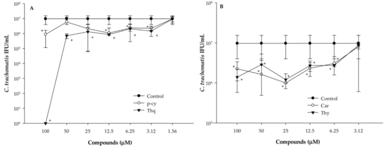
As all of the bioactive compounds tested showed antimicrobial activity in the anti-chlamydial assay, their MICs were evaluated by direct quantitative PCR (Figure3). HeLa cells were infected withC. trachomatisD and at the same time treated with two-fold serial dilutions of p-cy, Thq, Car, or Thy. After 1 h, the cells were washed and the medium was replaced. Direct PCR was performed from the cells 48 h later in order to determine the number of infectiousC. trachomatisD. Untreated but infected cells were used as controls. Treatment of the EBs with 100µM of Thq for 1 h completely inactivated the EBs ofC. trachomatisD. We did not observe complete inhibition for the other bioactive compounds—even at the highest concentrations tested. The MICs of p-cy and Thq were defined at 3.12µM, while the lowest concentration that significantly inhibited the replication ofC. trachomatisD was 6.25µM in the cases of Car and Thy.
Figure 3.Inhibitory effects of the bioactive compounds ofN. sativaEO onC. trachomatisD at different concentrations evaluated by direct quantitative PCR. HeLa cells infected withC. trachomatisD alone were used as controls. (A) p-cy: p-cymene; Thq: thymoquinone; (B) Car: carvacrol; Thy: thymol;
*p<0.05.
4. Discussion
The emergence of AMR is considered as a major public health problem due to the appearance of reduced or missing response of microorganisms to the applied antimicrobial agents. C. trachomatis infection is the most commonly reported sexually transmitted, bacterial infection, with an estimated 131 million new cases [22]. In addition, it has been found that Chlamydia spp. possess several different mechanisms associated with AMR development, despite their unique lifecycle characteristics.
Under exposure to certain conditions, such as the presence of interferon-γ, β-lactam antibiotics, or deprivation of nutrients,C. trachomatiscan transform to a persistent state, which can be defined by reduced replication and the occurrence of aberrant bodies [8]. Moreover, a recently published study demonstrated that azithromycin, which is the first choice drug in the therapy of chlamydial infections, could induce persistent infection in vitro [23]. Subinhibitory concentrations of the antimicrobial drugs were also able to induce AMR of certain chlamydial strains [5–7]. The ideal anti-chlamydial agents would be able to inhibit the growth of chlamydiae without exerting selective pressure for the development of AMR. The main advantages of natural-based products are that they apply less selective pressure against pathogens and exert remarkable effects on the inhibition of efflux pumps and AMR reversal [24,25]. The most common natural bioactive agents are volatile phenolic compounds, such as p-cy, Thq, Car, Thy, cinnamaldehyde, eugenol, limonene, and menthol, which are secondary metabolites of medicinal plants [15].
Our previous study revealed that EO extracted fromN. sativainhibited the growth ofS. aureus, including methicillin resistantS. aureus, and exerted antibiofilm activity. Regarding the bioactive compounds ofN. sativaEO, both staphylococcus strains were sensitive to Thq and Car [12]. In this
present study, we demonstrated that theN. sativaEO was able to completely inactivate the EBs of C. trachomatis D after 2 h exposure time at a concentration that was four-fold lower than its IC50 evaluated on HeLa cells. Moreover, all of the bioactive constituents (p-cy, Thq, Car, Thy) showed a direct antibacterial effect againstC. trachomatisD. As only one study related to anti-chlamydial activity of EOs has been published, further studies are needed to clarify the exact mechanisms of their effects. Car and Thq were able to damage the cell membrane ofS. aureusandListeria monocytogenes[14,26]. Thy, which is the most common constituent of EOs obtained fromThymusspp. and p-cy, exerted antimicrobial activity against a broad spectrum of pathogens, including Gram-positive and Gram-negative bacteria and fungi. Similar to other monoterpenes, Thy and p-cy were able to damage bacterial lipid membranes;
therefore, the possible mechanisms related to anti-chlamydial activity of Thq, Car, Thy, and p-cy might be associated with the disruption of the lipid bilayers [15,27].
MICs of the compounds were evaluated by direct quantitative PCR and defined at 6.25 (Car, Thy) and 3.12µM (p-cy, Thq), respectively. We were not able to detect complete inhibition ofC. trachomatis D, except in the case of Thq, which could be the result of the shorter exposure time (1 h). This finding supports the fact that the efficacy of their antimicrobial activity is time-dependent [16].
We are planning further experiments to evaluate the antimicrobial effects ofN. sativaEO and its bioactive compounds on intracellularly replicatingC. trachomatisRBs and their synergistic effects with clinically used antibiotics.
5. Conclusions
It can be concluded that bioactive compounds of N. sativa EO inhibited the replication of C. trachomatisD in vitro. These findings suggest thatN. sativaEO or its bioactive constituents could be used as effective antimicrobial agents againstC. trachomatisD. As numerous EOs possess antimicrobial activity and in turn can enhance the effect of antibiotics, further studies could support the use of bioactive components ofN. sativaEO as potential phytotherapeutics in anti-chlamydial therapy.
Author Contributions:T.M., A.M., M.H.A., and A.K. performed the experiments; T.M. wrote the original draft preparation; A.M., M.H.A., A.K., C.M.-F., and G.K. revised and edited the original draft; G.S. designed and supervised the study, revised the final manuscript.
Funding:This study was supported by the GINOP-2.3.2-15-2016-00012 project (University of Szeged, Hungary).
G.S. was supported by the János Bolyai Research Scholarship of the Hungarian Academy of Sciences. A.K. was supported by theÚNKP-18-3 New National Excellence Program of the Ministry of Human Capacities of Hungary.
A.M., C.M.F and G.K. were supported by the European Union and co-financed by the European Social Fund (grant agreement no. EFOP-3.6.3-VEKOP-16-2017-00005).
Conflicts of Interest:The authors declare no conflict of interest.
References
1. Allcock, S.; Young, E.H.; Holmes, M.; Gurdasani, D.; Dougan, G.; Sandhu, M.S.; Solomon, L.; Török, M.E.
Antimicrobial resistance in human populations: Challenges and opportunities. Glob. Health Epidemiol.
Genom.2017,2, e4. [CrossRef] [PubMed]
2. Murray, P.R.; Rosenthal, K.S.; Pfaller, M.A.Medical Microbiology, 8th ed.; Elsevier: Philadelphia, PA, USA, 2016; ISBN 978-0-323-29956-5.
3. Elwell, C.; Mirrashidi, K.; Engel, J. Chlamydia cell biology and pathogenesis.Nat. Rev. Microbiol.2016,14, 385–400. [CrossRef] [PubMed]
4. Mestrovic, T.; Ljubin-Sternak, S. Molecular mechanisms ofChlamydia trachomatisresistance to antimicrobial drugs.Front. Biosci.2018,23, 656–670. [CrossRef]
5. Binet, R.; Maurelli, A.T. Frequency of Development and Associated Physiological Cost of Azithromycin Resistance inChlamydia psittaci6BC andC. trachomatisL2.Antimicrob. Agents Chemother.2007,51, 4267–4275.
[CrossRef] [PubMed]
6. Dessus-Babus, S.; Bébéar, C.M.; Charron, A.; Bébéar, C.; de Barbeyrac, B. Sequencing of gyrase and topoisomerase IV quinolone-resistance-determining regions ofChlamydia trachomatisand characterization of quinolone-resistant mutants obtained In vitro.Antimicrob. Agents Chemother.1998,42, 2474–2481. [CrossRef]
[PubMed]
7. Marti, H.; Kim, H.; Joseph, S.J.; Dojiri, S.; Read, T.D.; Dean, D. Tet(C) Gene Transfer betweenChlamydia suis Strains Occurs by Homologous Recombination after Co-infection: Implications for Spread of Tetracycline-Resistance amongChlamydiaceae.Front. Microbiol.2017,8, 156. [CrossRef]
8. Schoborg, R.V. Chlamydia persistence—A tool to dissect chlamydia–host interactions.Microbes Infect.2011, 13, 649–662. [CrossRef]
9. Dorman, H.J.D.; Deans, S.G. Antimicrobial agents from plants: Antibacterial activity of plant volatile oils.
J. Appl. Microbiol.2000,88, 308–316. [CrossRef]
10. Warnke, P.H.; Becker, S.T.; Podschun, R.; Sivananthan, S.; Springer, I.N.; Russo, P.A.J.; Wiltfang, J.;
Fickenscher, H.; Sherry, E. The battle against multi-resistant strains: Renaissance of antimicrobial essential oils as a promising force to fight hospital-acquired infections.J. Craniomaxillofac. Surg.2009,37, 392–397.
[CrossRef]
11. Forouzanfar, F.; Bazzaz, B.S.F.; Hosseinzadeh, H. Black cumin (Nigella sativa) and its constituent (thymoquinone): A review on antimicrobial effects.Iran. J. Basic Med. Sci.2014,17, 929–938.
12. Mouwakeh, A.; Kincses, A.; Nové, M.; Mosolygó, T.; Mohácsi-Farkas, C.; Kiskó, G.; Spengler, G.Nigella sativa essential oil and its bioactive compounds as resistance modifiers againstStaphylococcus aureus.Phytother. Res.
2019,33, 1010–1018. [CrossRef] [PubMed]
13. Shaaban, H.A.; Sadek, Z.; Edris, A.E.; Saad-Hussein, A. Analysis and antibacterial activity ofNigella sativa essential oil formulated in microemulsion system.J. Oleo Sci.2015,64, 223–232. [CrossRef] [PubMed]
14. Mouwakeh, A.; Telbisz,Á.; Spengler, G.; Mohácsi-Farkas, C.; Kiskó, G. Antibacterial and Resistance Modifying Activities ofNigella sativaEssential Oil and its Active Compounds AgainstListeria monocytogenes.
In Vivo2018,32, 737–743. [CrossRef] [PubMed]
15. Marchese, A.; Orhan, I.E.; Daglia, M.; Barbieri, R.; Di Lorenzo, A.; Nabavi, S.F.; Gortzi, O.; Izadi, M.;
Nabavi, S.M. Antibacterial and antifungal activities of thymol: A brief review of the literature.Food Chem.
2016,210, 402–414. [CrossRef] [PubMed]
16. Sessa, R.; Di Pietro, M.; De Santis, F.; Filardo, S.; Ragno, R.; Angiolella, L. Effects ofMentha suaveolensessential oil onChlamydia trachomatis.BioMed Res. Int.2015,2015, 508071. [CrossRef] [PubMed]
17. Burián, K.; Hegyesi, H.; Buzás, E.; Endrész, V.; Kis, Z.; Falus, A.; Gönczöl, E.Chlamydophila (Chlamydia) pneumoniaeinduces histidine decarboxylase production in the mouse lung.Immunol. Lett.2003,89, 229–236.
[CrossRef]
18. Balogh, E.P.; Faludi, I.; Virók, D.P.; Endrész, V.; Burián, K.Chlamydophila pneumoniaeinduces production of the defensin-like MIG/CXCL9, which has in vitro antichlamydial activity.Int. J. Med. Microbiol.2011,301, 252–259. [CrossRef]
19. Raffai, T.; Burián, K.; Janovák, L.; Bogdanov, A.; Hegemann, J.H.; Endrész, V.; Virok, D.P. Vaginal Gel Component Hydroxyethyl Cellulose Significantly Enhances the Infectivity ofChlamydia trachomatisSerovars D and E.Antimicrob. Agents Chemother.2019,63, e02034–e020318. [CrossRef] [PubMed]
20. ˙Zesławska, E.; Kincses, A.; Unger, V.; Tóth, V.; Spengler, G.; Nitek, W.; Tejchman, W. Exocyclic Sulfur and Selenoorganic Compounds Towards Their Anticancer Effects: Crystallographic and Biological Studies.
Anticancer Res.2018,38, 4577–4584. [CrossRef] [PubMed]
21. Eszik, I.; Lantos, I.; Önder, K.; Somogyvári, F.; Burián, K.; Endrész, V.; Virok, D.P. High dynamic range detection ofChlamydia trachomatisgrowth by direct quantitative PCR of the infected cells.J. Microbiol. Methods 2016,120, 15–22. [CrossRef]
22. Newman, L.; Rowley, J.; Vander Hoorn, S.; Wijesooriya, N.S.; Unemo, M.; Low, N.; Stevens, G.; Gottlieb, S.;
Kiarie, J.; Temmerman, M. Global Estimates of the Prevalence and Incidence of Four Curable Sexually Transmitted Infections in 2012 Based on Systematic Review and Global Reporting. PLoS ONE2015, 10, e0143304. [CrossRef] [PubMed]
23. Xue, Y.; Zheng, H.; Mai, Z.; Qin, X.; Chen, W.; Huang, T.; Chen, D.; Zheng, L. An in vitro model of azithromycin-induced persistentChlamydia trachomatisinfection.FEMS Microbiol. Lett.2017,364. [CrossRef]
[PubMed]
24. Kincses, A.; Varga, B.; Csonka,Á.; Sancha, S.; Mulhovo, S.; Madureira, A.M.; Ferreira, M.-J.U.; Spengler, G.
Bioactive compounds from the African medicinal plantCleistochlamys kirkiias resistance modifiers in bacteria.
Phytother. Res.2018,32, 1039–1046. [CrossRef] [PubMed]
25. Ferreira, R.J.; Kincses, A.; Gajdács, M.; Spengler, G.; dos Santos, D.J.V.A.; Molnár, J.; Ferreira, M.-J.U.
Terpenoids fromEuphorbia pedroias Multidrug-Resistance Reversers. J. Nat. Prod. 2018,81, 2032–2040.
[CrossRef] [PubMed]
26. Burt, S.A.; Ojo-Fakunle, V.T.A.; Woertman, J.; Veldhuizen, E.J.A. The Natural Antimicrobial Carvacrol Inhibits Quorum Sensing inChromobacterium violaceumand Reduces Bacterial Biofilm Formation at Sub-Lethal Concentrations.PLoS ONE2014,9, e93414. [CrossRef]
27. Marchese, A.; Arciola, C.R.; Barbieri, R.; Silva, A.S.; Nabavi, S.F.; Tsetegho Sokeng, A.J.; Izadi, M.; Jafari, N.J.;
Suntar, I.; Daglia, M.; et al. Update on Monoterpenes as Antimicrobial Agents: A Particular Focus on p-Cymene.Materials2017,10, 947. [CrossRef] [PubMed]
©2019 by the authors. Licensee MDPI, Basel, Switzerland. This article is an open access article distributed under the terms and conditions of the Creative Commons Attribution (CC BY) license (http://creativecommons.org/licenses/by/4.0/).