Introduction
The discovery of antibiotics and their subsequent intro- duction into clinical medicine has been one of the main prerequisites for our current – modern day – healthcare to develop (Gaynes 2017). Previously lethal infections have become manageable, the life expectancy of people worldwide has changed drastically and novel medical interventions (e.g., cancer chemotherapy, invasive sur- gery, organ transplantation, and neonatology) were made possible (Laxminarayan et al. 2013). Bacterial pathogens have also reacted to the use of these agents and developed various resistance mechanisms to avoid their lethal effects (Nikaido 2009). The development of resistant isolates was to be expected by the laws of Darwinian evolution;
however, the misuse and overuse of these agents have catalyzed this process to become a severe health problem in the span of only a few decades (Chang et al. 2015).
Currently, the emergence of multidrug-resistant (MDR) bacteria is a global public health issue, which severely hinders clinicians in providing patients with adequate antimicrobial treatment regimens (Gajdács and Albericio 2019; Munita and Arias 2016). Several national and global public health authorities have published reports and esti-
mates on the global impact of antibiotic resistance (World Health Organization 2014). The grimmest predictions may be found in the O’Neill report (from the National Health Service of the United Kingdom), projecting 10 million deaths per year by 2050 and 100 billion USD worth of economic burden (O’Neill 2014). Rice et al. have defined the „ESKAPE” bacteria (including E: Enterococcus faecium, S: Staphylococcus aureus, K: Klebsiella pneumoniae, A: Acinetobacter baumannii, P: Pseudomonas aeruginosa, E:
Enterobacter spp., or recently Enterobacteriaceae) as the most concerning from the standpoint of clinical impact, both mortality-wise and economically (Rice 2010).
The issue of antibiotic resistance in the 21st century is a three-sided problem: i) on one hand, the emerge of drug- resistant mutants against newly developed antibiotics is an inevitable evolutionary process (which is common against any kind of noxious agents), ii) while the non-prudent use of antibiotics (e.g., for viral infections or other inappro- priate indications) only exacerbates this process; iii) the costs of clinical research and the development of novel antibiotics – coupled with the relatively modest returns on investment from these drugs – lead to a shift in the interest of pharmaceutical companies to instead develop drugs for chronic (i.e. more „profitable”) illnesses; this has resulted in a „discovery void”, with very limited amount
ARTICLE
Non-antibiotic pharmaceutical agents as antibiotic adjuvants
Márió Gajdács1,2
1 Department of Pharmacodynamics and Biopharmacy, Faculty of Pharmacy, University of Szeged, Szeged, Hungary
2 Institute of Medical Microbiology, Faculty of Medicine, Semmelweis University, Budapest, Hungary The emergence of multidrug-resistant bacteria is a global public health
issue, which severely hinders clinicians in providing patients with adequate antimicrobial treatment regimens. The strategy of drug repurposing is an emerging strategy in antimi- crobial chemotherapy, during which new pharmacological uses are identifi ed for drugs already approved. The aim of our present study was to assess the adjuvant properties of several existing and widely-used pharmacological agents against bacteria in combination with reference antibiotics. Staphylococcus aureus ATCC 25923, S. epidermidis ATCC 12228, Escherichia coli ATCC 25922 and Klebsiella pneumoniae ATCC 700603 were selected for our experiments. The minimum inhibitory concentrations (MICs) of the tested compounds were determined using the broth microdilution method, while a MIC reduction assay was performed to ascertain the eff ect of the tested compounds on the MICs of standard antibiotics (ciprofl oxacin and gentamicin). Eight tested compounds (namely atorvastatin, celecoxib, clotrimazole, diclofenac-epolamine, ivermectin, lidocaine, mebendazole and terbinafi ne) showed antibacterial activity on the tested bacterial strains and several agents presented with various degrees of adjuvant (MIC-reducing) properties. Further experiments involving the screening of additional pharmaceutical compounds for their secondary antibacterial and adjuvant properties are warranted.
Acta Biol Szeged 64(1):XX-XX (2020) ABSTRACT
ciprofl oxacin combination therapy gentamicin
non-antibiotic compounds pharmaceutical compounds KEY WORDS
http://abs.bibl.u-szeged.hu/index.php/abs
Submitted 04 June 2020 Accepted 24 July 2020
*Corresponding author
E-mail: mariopharma92@gmail.com
iologica cta zegediensis
DOI:10.14232/abs.2020.1.xx-xx
ARTICLE INFORMATION
of novel agents receiving marketing authorization since the 2000s (Gajdács 2019; Gajdács et al. 2020). In fact, no new broad-spectrum antibiotics were developed since the introduction of the fluoroquinolones in the 1980s (Darrow and Kesselheim 2014). Without new agents, researchers have investigated alternative strategies to combat bacte- rial pathogens more effectively (Rios et al. 2016). One of the proposed strategies is combination therapy: while the use of two or more existing antibiotics simultane- ously in clinical situations is a controversial topic (with very few verified indications), however, the inclusion of non-antibiotic adjuvants seems to be a promising strat- egy (Ahmed et al. 2014; Tangdén 2014; Szerencsés et al.
2019). These adjuvants include enzyme inhibitors (e.g., clavulanic acid, a β-lactamase inhibitor), efflux pump inhibitors, modulators of bacterial membrane potential, membrane permeabilizers, inhibitors of bacterial cell- cell communication (quorum sensing) and monoclonal antibodies (Wright 2016; Kealey et al. 2017; Drawz and Bonomo 2010). However, it must be noted that most of these molecules did not receive clinical approval due to their toxicity in vivo (Tegos et al. 2011). Recently, the ad- juvant properties of existing pharmaceutical compounds have received substantial attention. This strategy is termed
„drug repurposing” (or drug re-profiling), during which new pharmacological uses are identified for drugs already approved, outside of their original indications (Pushpakom et al. 2019). As the physicochemical, pharmacokinetic and toxicological profile of these compounds have already been established, the initial stages of the drug authoriza- tion process (Phase I–II clinical trials) may be avoided, leading to substantial monetary benefits for the pharma- ceutical companies; if this new indication of the tested compound is appropriate, pharmaceutical companies may once again expect financial returns for their investments (Miró-Canturri et al. 2019; Paul et al. 2010; Pushpakom et al. 2019; Soo et al. 2017). Drug re-profiling is also an emerging strategy in antimicrobial chemotherapy:
some of these compounds have antibacterial properties themselves, while others have secondary mechanisms of action (some of which are unknown as of now). These mechanisms may include: bacteriostatic properties, inhibi- tion of bacterial cell-cell communication, modulation of virulence factor-expression, biofilm-inhibition and so on (Miró-Canturri et al. 2019; Paul et al. 2010; Pushpakom et al. 2019; Soo et al. 2017; Yang et al. 2009) However, there are still significant gaps in the knowledge in the field of drug repurposing for antimicrobial purposes.
The aim of our present study was to assess the ad- juvant properties of several existing and widely-used pharmacological agents against bacteria in combination with reference antibiotics, in an in vitro study.
Materials and methods Culture media
The following culture media were used during our ex- periments: cation-adjusted Mueller-Hinton broth (Bio- Rad, Hercules, CA, USA), Luria–Bertani broth (Bio-Rad, Hercules, CA, USA), 5% sheep blood agar (bioMérieux, Marcy-l’Étoile, France) and eosine-methylene blue agar (bioMérieux, Marcy-l’Étoile, France).
Bacterial strains
Staphylococcus aureus ATCC 25923 and S. epidermidis ATCC 12228 were used as representative Gram-positive strains, while E. coli ATCC 25922 and K. pneumoniae ATCC 700603 were selected as representative Gram-negative strains for our experiments. For shorter time periods (<1 month), the bacterial strains were maintained on blood agar and eosine-methylene blue agar plates (for Gram-negatives) with continuous passage. For longer periods, the strains were kept in a -80 °C freezer, in a 1:4 mixture of 85%
glycerol and liquid Luria-Bertani medium.
Antibiotics and non-antibiotic compounds
Ciprofloxacin and gentamicin (Sigma-Aldrich, St. Louis, MI, USA; will be listed as SA in the subsequent text) were selected as antibiotic controls for our studies. Twenty (n
= 20) pharmacological agents, encompassing drug with different chemical structures and mechanisms of action were tested during our experiments: acetaminophen (SA), amantadine (SA), acyclovir (Teva Pharmaceuticals, Petah Tikva, Israel; will be listed as TPh in the subsequent text), atorvastatin (SA), azelastine (SA), celecoxib (Pfizer Hungary, Budapest, Hungary), cetirizine (SA), clotrima- zole (TPh), diclofenac-epolamine (SA), enalapril-maleate (SA), ivermectin (SA), lidocaine (SA), mebendazole (SA), metformin (SA), metoprolol-succinate (SA), prazozine (SA), sitagliptine (SA), terbinafine (GlaxoSmithKline, Brentford, UK), valsartan (SA) and xylomethazoline (SA). The compounds were chosen on the basis of being available as over-the-counter (OTC) medication or being frequently prescribed for common chronic conditions as hypertension or diabetes mellitus. Pharmaceutical compounds were dissolved in phosphate-buffered saline (PBS), except for atorvastatin, which was dissolved in dimethyl sulfoxide (DMSO). All solutions were prepared on the day of the assay. The concentration of DMSO was below 1 V/V% in all experiments.
Antibacterial activity of non-antibiotic compounds, MIC determination
The minimum inhibitory concentrations (MICs) of the tested compounds were determined using the standard broth microdilution method, based on the recommenda-
tions of the Clinical and Laboratory Standards Institute (CLSI; M07-A10). The experiments were performed in 96- well polystyrene microtiter plates, using cation-adjusted Mueller–Hinton broth. The tested concentrations of the compounds were ranging between 1.95-250 µg/mL, the two-fold serial dilutions of the tested compounds were made starting in the third row of the microtiter plates.
During the experiments with S. aureus ATCC 25922 and S. epidermidis ATCC 12228, the Mueller-Hinton broth was supplemented by 2% NaCl, as based on CLSI protocols.
The plates were incubated at 37 °C in an air thermostat.
The MIC values of the tested compounds were recorded after 16-18 h of incubation; the interpretation of the results was performed visually. All experiments were performed in triplicate.
MIC reduction assay
To ascertain the effect of the tested compounds on the MICs of standard antibiotics (i.e. ciprofloxacin and gen- tamicin), a MIC reduction assay was performed (Sarker et al. 2007). The assay was performed in a 96-well microtiter plate, using cation-adjusted Mueller-Hinton broth. The setup of the plates was the following: in rows A-D of the plate, serial dilutions were made for the reference antibiotic, in rows E-H the same serial dilutions were performed for the reference antibiotic with the addition of
the non-antibiotic compounds in a constant concentration as adjuvants (MIC/4 in cases where MIC was ≤ 250 µg/
mL and 125 µg/mL where MICs were higher than 250 µg/mL) in all the wells, except for medium control and cell control wells (Sarker et al. 2007). The inoculation of the plates and the incubation was performed according to a standard broth microdilution method, described previously. The modified MICs (compared to the MICs of the antibiotics alone) were determined visually, as the concentration, where no visible growth of bacteria could be observed. All experiments were carried out in triplicate.
Results
Antibacterial activity of pharmaceutical compounds
The MICs observed for the non-antibiotic pharmaceu- tical compounds is presented in Table 1. Eight tested compounds (namely atorvastatin, celecoxib, clotrimazole, diclofenac-epolamine, ivermectin, lidocaine, mebendazole and terbinafine) showed antibacterial activities in the tested concentration range, while compounds with MICs
>250 µg/mL were not considered to be active.
MIC reduction assays
The results of the MIC reduction assays for Gram-positive
Minimal inhibitory concentrations (µg/mL)
Compounds S. aureus ATCC 25923 S. epidermidis ATCC 12228 E. coli ATCC 25922 K. pneumoniae ATCC 700603
Acetaminophen >250 >250 >250 >250
Amantadine >250 >250 >250 >250
Acyclovir >250 >250 >250 >250
Atorvastatin 125 125 250 >250
Azelastine >250 >250 >250 >250
Celecoxib 15.6 31.2 >250 >250
Cetirizine >250 >250 >250 >250
Clotrimazole 125 62.5 >250 >250
Diclofenac-epolamine 250 250 >250 >250
Enalapril-maleate >250 >250 >250 >250
Ivermectin 31.2 125 >250 >250
Lidocaine 250 250 250 250
Mebendazole 62.5 125 62.5 250
Metformin >250 >250 >250 >250
Metoprolol-succinate >250 >250 >250 >250
Prazozine >250 >250 >250 >250
Sitagliptine >250 >250 >250 >250
Terbinafine 250 125 >250 >250
Valsartan >250 >250 >250 >250
Xylomethazoline >250 >250 >250 >250
Table 1 Minimum inhibitory concentrations (MICs) of the tested pharmaceutical compounds on reference bacterial strains.
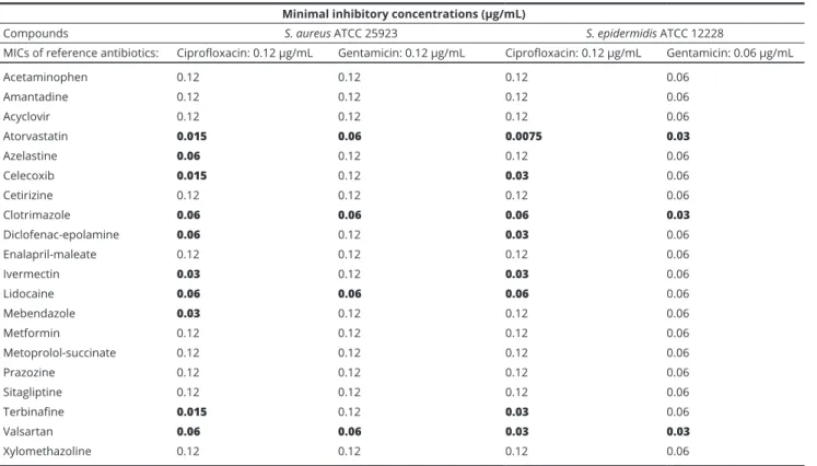
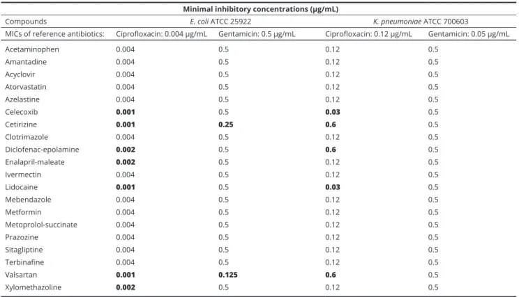
bacteria are presented in Table 2., while results for Gram- negative bacteria are shown in Table 3. Overall, the tested non-antibiotics were the most potent adjuvants against Gram-positive bacteria and they enhanced the antibacte- rial activity (i.e. they reduced the MICs) or ciprofloxacin to the highest extent (reducing the MICs 50-93.25% or 2-5-fold), while having modest effects on Gram-negative bacteria (E. coli and K. pneumoniae) and on the MICs of gentamicin. Interestingly, azelastine and valsartan on Gram-positive bacteria, while cetirizine, enalapril, val- sartan and xylomethazoline on Gram-negative bacteria had MIC-reducing effects, without having any intrinsic antibacterial properties themselves (see Table 1.).
Discussion
Infections caused by MDR bacteria are associated with an increased mortality rate and decreased quality of life in the affected patients worldwide (Falagas et al. 2008).
Since the 2000s, the development of novel antibiotics has been shown to keep up with the development in the levels of bacterial resistance (Falagas et al. 2008). Combination therapy with non-antibiotic compounds may provide a
straightforward, attractive and financially reasonable drug development avenue (Medina and Pieper 2016; Lyddiard et al. 2016; Spellberg 2014). Several reports have been published in the literature on the antibacterial properties of non-antibiotic drugs; however, the systematic screening of drugs for such purposes have not yet been performed (Kruszewska et al. 2002; Lagadinou et al. 2020). Doxo- rubicin and bleomycin are antitumor agents (frequently termed as “anticancer antibiotics”) show antibacterial properties on a variety of bacterial strains: the proposed mechanism of action is related to their intercalation into bacterial DNA (similar to the mechanism of the fluoro- quinolones) and the generation of oxidative free radicals in the presence of Fe2+-ions (which are indispensable for the biochemical pathways of bacteria) (Kruszewska et al. 2002; Lagadinou et al. 2020; Soo et al. 2017). Several antipsychotic drugs (e.g., phenothiazine, thioridazine) have also been described as DNA-intercalators; in ad- dition, their efflux pump-inhibitory properties were also experimentally verified on many bacterial strains (Amaral et al., 2004). The adjuvant properties of non- steroidal anti-inflammatory drugs (NSAIDs) in bacterial infections have been supported in the clinical practice by empirical evidence, while laboratory studies have also
Minimal inhibitory concentrations (µg/mL)
Compounds S. aureus ATCC 25923 S. epidermidis ATCC 12228
MICs of reference antibiotics: Ciprofloxacin: 0.12 µg/mL Gentamicin: 0.12 µg/mL Ciprofloxacin: 0.12 µg/mL Gentamicin: 0.06 µg/mL
Acetaminophen 0.12 0.12 0.12 0.06
Amantadine 0.12 0.12 0.12 0.06
Acyclovir 0.12 0.12 0.12 0.06
Atorvastatin 0.015 0.06 0.0075 0.03
Azelastine 0.06 0.12 0.12 0.06
Celecoxib 0.015 0.12 0.03 0.06
Cetirizine 0.12 0.12 0.12 0.06
Clotrimazole 0.06 0.06 0.06 0.03
Diclofenac-epolamine 0.06 0.12 0.03 0.06
Enalapril-maleate 0.12 0.12 0.12 0.06
Ivermectin 0.03 0.12 0.03 0.06
Lidocaine 0.06 0.06 0.06 0.06
Mebendazole 0.03 0.12 0.12 0.06
Metformin 0.12 0.12 0.12 0.06
Metoprolol-succinate 0.12 0.12 0.12 0.06
Prazozine 0.12 0.12 0.12 0.06
Sitagliptine 0.12 0.12 0.12 0.06
Terbinafine 0.015 0.12 0.03 0.06
Valsartan 0.06 0.06 0.03 0.03
Xylomethazoline 0.12 0.12 0.12 0.06
Results in boldface represent cases when the MICs have decreased due to the effect of the adjuvants.
Table 2 Results of the MIC reduction assays on Gram-positive bacterial strains using ciprofloxacin and gentamicin as reference antibiotics.
provided results that some NSAIDs (e.g., acetamino- phen, acetyl-salicylic acid, ibuprofen, indomethacin, metamizol-sodium, etoricoxib) and local anesthetics (e.g., lidocaine) may have mechanisms enhancing the effects of antibiotics in vivo (Al-Bakri et al. 2009; Chan et al.
2017; D’Angelo et al. 2018; Johnson et al. 2008; Ogundeji et al. 2016; Thangamani et al. 2015). These mechanisms may include inhibition of biofilm-formation, adherence, reduction of motility and the modulating the release of antibiotics by leukocytes (Al-Bakri et al. 2009; Chan et al.
2017; D’Angelo et al. 2018; Johnson et al. 2008; Ogundeji et al. 2016; Thangamani et al. 2015). Allopurinol (a gout medication) increased the potency of anti-tuberculosis medications against Mycobacterium tuberculosis (Naftalin et al. 2017). The antibacterial activity of azole antifungals and ivermectin against Gram-positive bacteria only was previously described (Ghannoum and Rice 1999; Ashraf et al. 2018). The exact mechanism of action is not well- defined, but it is probably associated with affecting the binding to the terminal D-alanyl-D-alanine of the penta- peptide peptidoglycan precursors in the cell wall (Ghan- noum and Rice 1999; Ashraf et al. 2018). The mechanism of statins (including simvastatin and atorvastatin among others) regarding their antibacterial potency also needs
further studies, however, it has been suggested that they interfere with the mevalonate pathway, limiting the syn- thesis of the major lipid constituents of cell membrane microdomains (Ko et al. 2017). Apart from drugs, some publications also reported on the adjuvant properties of vitamins, enhancing the bactericidal activity of antibiot- ics; these publications highlight the role of high-dose of lipid soluble vitamins (ADEK) and Vitamin C (Andrade et al. 2014; Kwiencinska-Piróg et al. 2019).
Conclusions
The aim of our present study was to assess a selection of non-antibiotic pharmaceutical compounds – sourced from diverse clinical indications and molecular char- acteristics – as antibiotic adjuvants. Currently, there are around 6000-9000 drug compounds marketed for human therapeutic purposes; these agents may be con- sidered as potential combination agents with reference antibiotics, to potentiate their antibacterial properties in clinical situations. The pharmacokinetic parameters and in vivo tolerability of these compounds have already been described; thus, these compounds are already one
Minimal inhibitory concentrations (µg/mL)
Compounds E. coli ATCC 25922 K. pneumoniae ATCC 700603
MICs of reference antibiotics: Ciprofloxacin: 0.004 µg/mL Gentamicin: 0.5 µg/mL Ciprofloxacin: 0.12 µg/mL Gentamicin: 0.05 µg/mL
Acetaminophen 0.004 0.5 0.12 0.5
Amantadine 0.004 0.5 0.12 0.5
Acyclovir 0.004 0.5 0.12 0.5
Atorvastatin 0.004 0.5 0.12 0.5
Azelastine 0.004 0.5 0.12 0.5
Celecoxib 0.001 0.5 0.03 0.5
Cetirizine 0.001 0.25 0.6 0.5
Clotrimazole 0.004 0.5 0.12 0.5
Diclofenac-epolamine 0.002 0.5 0.6 0.5
Enalapril-maleate 0.002 0.5 0.12 0.5
Ivermectin 0.004 0.5 0.12 0.5
Lidocaine 0.001 0.5 0.03 0.5
Mebendazole 0.004 0.5 0.12 0.5
Metformin 0.004 0.5 0.12 0.5
Metoprolol-succinate 0.004 0.5 0.12 0.5
Prazozine 0.004 0.5 0.12 0.5
Sitagliptine 0.004 0.5 0.12 0.5
Terbinafine 0.004 0.5 0.12 0.5
Valsartan 0.001 0.125 0.6 0.5
Xylomethazoline 0.002 0.5 0.12 0.5
Table 3 Results of the MIC reduction assays on Gram-negative bacterial strains using ciprofloxacin and gentamicin as reference antibiotics.
Results in boldface represent cases when the MICs have decreased due to the effect of the adjuvants.
step closer into their clinical utilization. The highlights of the study include the study of twenty pharmaceutical compounds that are frequently used by patients. Eight tested compounds showed antibacterial activity on the tested bacterial strains and several agents presented with various degrees of adjuvant (MIC-reducing) properties.
Further experiments involving the screening of additional pharmaceutical compounds for their secondary antibac- terial and adjuvant properties is definitely warranted.
Acknowledgements
M.G. was supported by the János Bolyai Research Scholarship of the Hungarian Academy of Sciences (BO/00144/20/5). M.G. would also like to acknowledge the support of the ESCMID’s “30 under 30” Award.
References
Ahmed A, Azim A, Gurjar M, Baronia AK (2014) Current concepts in combination antibiotic therapy for critically ill patients. Indian J Crit Care Med 18:310-314.
Al-Bakri AG, Othman G, Bustanji Y (2009) The assessment of the antibacterial and antifungal activities of aspi- rin, EDTA and aspirin–EDTA combination and their effectiveness as antibiofilm agents. J Appl Microbiol 107:280-286.
Amaral L, Viveiros M, Molnár J (2004) Antimicrobial activ- ity of phenothiazines. In vivo 18:725-731.
Andrade JC, Morais-Braga, MFB, Guedes GMM, Freitas TMA, Menezes IRA, Coutinho DMH (2014) Enhance- ment of the antibiotic activity of aminoglycosides by alpha-tocopherol and other cholesterol derivates. Biomed Pharmacother 68:1065-1069.
Ashraf S, Chaudhry U, Raza A, Ghosh D, Zhao X (2018) In vitro activity of ivermectin against Staphylococcus aureus clinical isolates. Antimicrob Res Infect Control 7:e27.
Chan EWL, Yee ZY, Raja I, Yap JKY (2017) Synergistic effect of non-steroidal anti-inflammatory drugs (NSAIDs) on antibacterial activity of cefuroxime and chloramphenicol against methicillin-resistant Staphylococcus aureus. J Glob Antimicrob Resist 10:70-74.
Chang HH, Cohen T, Grad YH, Hanage WP, O’Brien TF, Lipsitch M (2015) Origin and proliferation of multiple- drug resistance in bacterial pathogens. Microbiol Mol Biol Rev 79:101-116.
D’Angelo F, Baldelli V, Halliday N, Pantalone P, Polticelli F, Fiscarelli E, Williams P, Visca P, Leoni L, Rampioni (2018) Identification of FDA-approved drugs as antivirulence agents targeting the pqs quorum-sensing system of Pseudomonas aeruginosa. Antimicrob Agents Chemother
24:e01296-18.
Darrow JJ, Kesselheim AS (2014) Drug development and FDA approval, 1938-2013. N Engl J Med 370:e39.
Drawz SM, Bonomo RA (2010) Three decades of β-lactamase inhibitors. Clin Microbiol Rev 23:160-201.
Falagas ME, Rafailidis PI, Matthaiou DK, Virtzili S, Nikita D, Michalopoulos A (2008) Pandrug-resistant Klebsiella pneumoniae, Pseudomonas aeruginosa and Acinetobacter baumannii infections: Characteristics and outcome in a series of 28 patients. Int J Antimicrob Agents 32:450-454.
Gajdács M, Albericio F (2019) Antibiotic resistance: From the bench to patients. Antibiotics 8:e129.
Gajdács M (2019) The concept of an ideal antibiotic: Implica- tions for drug design. Molecules 24:e892.
Gajdács M, Paulik E, Szabó A (2020) Knowledge, attitude and practice of community pharmacists regarding antibiotic use and infectious diseases: A cross-sectional survey in Hungary (KAPPhA-HU). Antibiotics 9:e41.
Gaynes R (2017) The discovery of penicillin—New insights after more than 75 Years of clinical use. Emerg Infect Dis 23:849-853.
Ghannoum MA, Rice LB (1999) Antifungal agents: Mode of action, mechanisms of resistance, and correlation of these mechanisms with bacterial resistance. Clin Microbiol Rev 12:501-517.
Johnson SM, Saint John BE, Dine AP (2008) Local anesthetics as antimicrobial agents: A review. Surg Infect 9:205-213.
Kealey C, Creaven CA, Murphy CD, Brady CB (2017) New approaches to antibiotic discovery. Biotechnol Lett 39:805-817.
Ko HTH, Lareu RR, Dix BR, Hughes JD (2017) Statins: an- timicrobial resistance breakers or makers? PeerJ http://
dx.doi.org/10.7717/peerj.3952
Kwietcinska-Piróg J, Skowron K, Bogiel T, Bialucha A, Przekwas J, Gospodarek-Komkowksa E (2019) Vitamin C in the presence of sub-inhibitory concentration of aminoglycosides and fluoroquinolones alters Proteus mirabilis biofilm inhibitory rate. Antibiotics 8:e116.
Kruszewska H, Zareba T, Tyski S (2002) Search of antimi- crobial activity of selected non-antibiotic drugs. Acta Pol Pharm 59:436-439.
Lagadinou M, Onisor MO, Rigas A, Musetescu DV, Gkentzi D, Assimakopoulos SF, Panos G, Marangos M (2020) Antimicrobial properties on non-antibiotic drugs in the era of increased bacterial resistance. Antibiotics 9:e107.
Laxminarayan R, Duse A, Wattal C, Zaidi AKM, Wertheim HFL, Sumpradit N, Vlieghe E, Hara GL, Gould IM, Goossens H, Greko C, So AD, Bigdeli M, Tomson G, Woodhouse W, Ombaka E, Peralta AQ, Qamar FN, Mir F, Kariuki S, Bhutta ZA, Coates A, Bergstrom R, Wright GD, Brown ED, Cars O (2013) Antibiotic resistance-the need for global solutions. Lancet Infect Dis 13:1057-1098.
Lyddiard D, Jones GL, Greatrex BW (2016) Keeping it simple:
Lessons from the golden era of antibiotic discovery.
FEMS Microbiol Lett 363:fnw084.
Medina E, Pieper DH (2016) Tackling threats and future problems of multidrug-resistant bacteria. Curr Top Microbiol Immunol 398:3-33.
Miró-Canturri A, Ayerbe-Algaba R, Smani Y (2019) Drug repurposing for the treatment of bacterial and fungal infections. Front Microbiol 10:e41.
Munita JM, Arias CA (2016) Mechanisms of antibiotic re- sistance. Microbiol Spectr doi:10.1128/microbiolspec.
VMBF-0016-2015.
Naftalin CM, Verma R, Gurumurthy M, Lu Q, Zimmerman M, Yeo BCM, Tan KH, Lin W, Yu B, Dartois Y, Paton NI (2017) Coadministration of allopurinol to increase antimycobacterial efficacy of pyrazinamide as evaluated in a whole-blood bactericidal activity model. Antimicrob Agents Chemother 61:e00482-17.
Nikaido H (2009) Multidrug resistance in bacteria. Annu Rev Biochem 78:119-146.
Ogundeji AO, Pohl CH, Sebolal OM (2016) Repurposing of aspirin and ibuprofen as candidate anti-cryptococcus drugs. Antimicrob Agents Chemother 60:4799-4808.
O’Neill J (2014) Antimicrobial resistance: Tackling a crisis for the health and wealth of nations. Available online: https://
amr-review.org/sites/default/files/AMRReviewPaper- Tacklingacrisisforthehealthandwealthofnations_1.pdf (accessed on 4th of July, 2020).
Paul SM, Mytelka DS, Dunwiddie CT, Persinger CC, Munos BH, Lindborg SR, Schacht AL (2010) How to improve R&D productivity: The pharmaceutical industry’s grand challenge. Nat Rev Drug Discov 9:203-214.
Pushpakom S, Iorio F, Eyers PA, Escott KJ, Hopper S, Wells A, Doig A, Guilliams T, Latimer J, McNamee C, Norris A, Sanseau P, Cavalla D, Pirmohamed M (2019) Drug repurposing: Progress, challenges and recommendations.
Nat Rev Drug Discov 18:41-58.
Rice LB (2010) Progress and challenges in implementing the research on ESKAPE pathogens. Infect Control Hosp Epidemiol 31:S7-S10.
Rios AC, Moutinho CG, Pinto FC, Del Fiol FS, Jozala A, Chaud MC, Vila MCDM, Teixeria JA, Balcao VM (2016) Alternatives to overcoming bacterial resistances: State- of-the-art. Microbiol Res 191:51-80.
Sarker SD, Nahar L, Kumarasamy Y (2007) Microtitre plate-based antibacterial assay incorporating resazurin as an indicator of cell growth, and its application in the in vitro antibacterial screening of phytochemicals.
Methods 42:321-324.
Shallcross LJ, Howard SJ, Fowler T, Davies SC (2015) Tack- ling the threat of antimicrobial resistance: From policy to sustainable action. Philos Trans R Soc Lond B Biol Sci 370:20140082.
Soo VWC, Kwan BW, Quezada H, Castillo-Juárez I, Pérez- Eretza B, García-Contreras SJ, Martínez-Vázquez M, Wood TK, García-Contreras R (2017) Repurposing of anticancer drugs for the treatment of bacterial infections.
Curr Top Med Chem 17:1157-1176.
Spellberg B (2014) The future of antibiotics. Crit Care 18:e228.
Szerecsés B, Mülbacher A, Vágvölgyi C, Pfeiffer I (2019) In vitro interactions of amphotericin B and non-antifungal compounds against opportunistic human pathogen Cryptococcus neoformans. Acta Biol Szeged 63:181-184.
Tandgén T (2014) Combination antibiotic therapy for mul- tidrug-resistant Gram-negative bacteria. Ups J Med Sci 119:149-153.
Thangamani S, Younis W, Seleem MN (2015) Repurpos- ing celecoxib as a topical antimicrobial agent. Front Microbiol 28:e750.
Tegos GP, Haynes M, Strouse JJ, Khan MM, Bologa CG, Oprea TI, Sklar LA (2011) Microbial efflux pump inhi- bition: Tactics and strategies. Curr Pharm Des 17:1291- 1302.
World Health Organisation (2014) Antimicrobial resis- tance: Global report on surveillance. 2014, pp. 1–256.
Available online: http://apps.who.int/iris/bitstream /10665/112642/1/9789241564748_eng.pdf?ua=1 (ac- cessed on 4th of July, 2020).
Wright GD (2016) Antibiotic adjuvants: Rescuing antibiotics from resistance. Trends Microbiol 24:862-871.
Yang L, Rybtke MT, Jakobsen TH, Hentzer M, Bjarnsholt T, Givskov M, Tolker-Nielsen T (2009) Computer-aided identification of recognized drugs as Pseudomonas aeru- ginosa quorum-sensing inhibitors. Antimicrob Agents Chemother 53:2432-2443.