Novel Antibiotics as Metabolic Inhibitors
Franklin M. Harold
No one considers the lion which leaps on its prey to be a parasite nor the snake which injects venom into the wound of its victim before eating it.
Here there is nothing equivocal; one creature destroys the life of another to preserve its own . . . . The conception is so simple that no one has ever thought of giving it a name. This condition, instead of being examined in isolation, can appear as a factor in more complex phenomena. In order to simplify words we will call it antibiosis.
P. Vuillemin, 1899, translated by H. W. Florey CO.
I. Introduction 306 II. Inhibitors of Cell Wall Synthesis 307
A. Penicillin 307 B. New Inhibitors of Peptidoglycan Biosynthesis 309
C. Inhibitors of Chitin Synthesis 311 III. Interaction of Antibiotics with Lipid Membranes 311
A. Disorganization of Lipid Membranes 311
B. Ion-Conducting Antibiotics 313 IV. Inhibitors of the Respiratory Chain and Energy Transfer 319
A. Respiratory Inhibitors 320 B. Antibiotics and ATPase 322 V. Inhibitors of D N A Replication and Transcription 323
A. Antibiotics That Bind to D N A 323 B. Inhibitors of Replication 327 C. Inhibitors of RNA Polymerase and Transcription 329
VI. Antibiotic Inhibitors of Protein Synthesis 331 A. Inhibitors of Amino Acid Activation 333 B. Novel Inhibitors of Ribosome Function 333
VII. A Potpourri of Antibiotics 338 A. Purine Nucleosides 339 B. Mycophenolic Acid 340 C. Sideromycins 340 D. Showdomycin 341 VIII. Chemical Structure and Biological Activity of Antibiotics 342
A. Ion Conduction by Antibiotics 342 B. Association of Antibiotics with Membrane Lipids 343
305
306 FRANKLIN Μ. HAROLD
IX
C. Interaction with D N A
D. Binding of Antibiotics to Proteins..
E. The Limits of Specificity
The Origin and Function of Antibiotics References
3 4 4 3 4 4 3 4 6 3 4 6 3 4 9
I. INTRODUCTION
Most of the antibiotics familiar to the general public, including penicil
lin, tetracycline, chloramphenicol, and streptomycin, were already in use by 1950. The systematic search for new antibiotics during the past two decades has added very greatly to the roster. Recent treatises by Umezawa (2) and Korzybski et al. (3) each record chemical and other data for close to a thousand distinct antibiotics. Their structures range in complexity from analogs of small metabolites such as cycloserine to the proteinaceous bacteriocins. Only about one antibiotic in twenty has the degree of selective toxicity for parasites required for use in medical practice (kanamycin, lincomycin, and the rifamycins are among the recent additions to the physicians' arsenal), but students of cellular physiology and biochemistry are less demanding. The sites and mecha
nisms of action of many antibiotics have been determined quite precisely, and these substances find increasing application as specific inhibitors of complex processes such as protein synthesis or ion transport. The use of antibiotics as specific metabolic inhibitors rests on the assumption, which must be validated in each instance, that a given antibiotic has only a single site or mechanism of action. It is quite remarkable how often this assumption is found to be legitimate, at least for low concen
trations of the drug.
It is not easy to define the term antibiotic in a manner both rigorous and practical. Waksman's original definition (4) read "substances pro
duced by microorganisms which inhibit the growth of other microorga
nisms." With the discovery of antimicrobial agents produced by plants and animals, there is now a tendency to use the term in a wider sense to include any "substance produced by a living organism, which inhibits the growth or activity of another living organism" (5). As a guide to the composition of a literature review the latter conception is too broad, since it embraces the pharmacology of all natural products. I have there
fore adhered to the original definition of antibiotics, arbitrary as it is, excluding with relief the alkaloids and toxins and with some regret the synthetic antimicrobial drugs such as isoniazid and nalidixic acid.
The present review will focus on the mode of action of antibiotics at the molecular level, that is, on the interaction of antibiotics with their receptor molecules and the chain of biochemical events initiated by this interaction. We shall be concerned primarily with antibiotics that have but recently attracted the attention of biochemists and cell physiologists. Studies on the classical antibiotics fall within the purview of this chapter only insofar as they provide the background against which we can assess research on more novel and exotic substances.
A number of symposia and general review articles summarize the cur- rent state of knowledge concerning the mode of action of antibiotics old and new (6-10), but no investigator in this field can do without the treatise on antibiotics edited by Gottlieb and Shaw (11). This pro- vides comprehensive coverage of the literature through 1966 and serves as the point of departure for this chapter. In order to keep the number of literature citations within reasonable bounds, this review makes refer- ence almost exclusively to papers published from 1966 through 1970 in the major western languages.
II. INHIBITORS OF CELL WALL SYNTHESIS
A. Penicillin
Any discussion of antibiotics that inhibit the synthesis of microbial cell walls must begin with penicillin since it is the classical representative of the genre and continues to receive intense attention. Various aspects of this subject have been reviewed by Flynn and Godzeski (12), Strominger and his associates (13, 14), Jones (15), and Perkins (16).
B y 1957 it was generally recognized that penicillin interferes with forma- tion of normal cell walls by bacteria (but not by fungi). Bacteria grow- ing in the presence of the antibiotic give rise to forms that are osmoti- cally fragile and that undergo lysis unless stabilized. At the same time complex nucleotides accumulate both in the cells and in the medium.
A typical one was shown to have the structure uridine diphosphate-iV- acetylmuramic acid-L-Ala-D-Glu-L-Lys-D-Ala-D-Ala. Nucleotides of this general type were subsequently found to be derived from intermediates in the biosynthesis of the peptidoglycan component of the wall (Fig. 1).
Peptidoglycan (sometimes called murein or mucopeptide) is a polymer unique to bacteria which confers rigidity and mechanical strength upon the wall. The site of action of penicillin has been unequivocally localized
MurNAc-P-P-uridine L- A l a - D- G l u - L- L y s - D- A l a - D- A l a
UMP
MurNAc-P-P-lipid
D- · G l u - L- L y s - D- A l a - D- A l a
P-Lipid a-COOH
Bacitracin
Vancomycin
• Ristocetin
e-NH. DP-GlcNAc UDP GlcNAc-MurNAc-P-P-lipid
L- A l a - · D - - G l u - L- L y s - D- A l a - D- A l a α-COOH I \ Nr„ +
€-NH2 N H<
/ (ATP)
GlcNAc-MurNAc-P-P-lipid
L- A l a i G l u - L- L y s - D- A l a - D- A l a α-CONH, I
e-NH2
GlcNAc-MurNAc-P-P-lipid
L- A l a - D- G l u - L- L y s - D- A l a - D- A l a
Glycyl-tRNA
Acceptor
GlcNAc-MurNAc-acceptor
L- A l a - D- G l u - L- L y s - D- A l a - D- A l a a-CONH2 I
6-NH
a-CONH2 I e-NH (Gly)5
NH,
Penicillin Cephalosporin
(Gly).
NH2
-GlcNAc-MurNAc- I
GlcNAc-MurNAc-acceptor
L- A l a - D- G l u - L- L y s - D- A l a - D- A l a a-CONH2 I
c-NH (Gly).
NH L- A l a - D- G l u - L- L y s - D- A l a
I ι ι a-CONH2 I
I e-NH
L- A l a - D- G l u - L- L y s - D- A l a - D- A l a a-CONH2 I
e-NH (Gly).
NH2
- Glc Ν Ac - Mur Ν Ac - (Gly).
NH2
FIG. 1. Sites of inhibition of peptidoglycan synthesis by antibiotics. [Redrawn from Perkins (16), with kind permission of the author and Academic Press.]
308
at the final step in peptidoglycan synthesis; low concentrations of peni- cillin, and also of the closely related cephalosporins, block a cross-linking reaction catalyzed by a specific transpeptidase (Fig. 1). It would appear that penicillin is a conformational analog of the D-alanyl-D-alanine terminus of the precursor nucleotide. The antibiotic thus occupies the active site of the enzyme and acylates it by formation of a thiol ester between the protein and penicilloic acid; the block is irreversible. A second enzyme, D-alanine carboxypeptidase, is inhibited competitively by penicillin (16-19). The chain of events that ensues makes penicillin a particularly effective bacteriocidal agent. The protoplast continues to grow and to synthesize cell wall precursors which can be incorporated into the growing peptidoglycan polymer but which are not cross-linked.
At the same time specific hydrolases, whose normal function is to prepare sites for the insertion of new units into the growing wall, break down and weaken the existing structure. The weakened wall bulges under osmotic stress, generally at the site of septum formation, leading eventu- ally to splitting of the old wall and release of the protoplast or sphero- plast. Indeed, in some strains of E. coli low concentrations of penicillin inhibit normal septation even though no effect on cross-linking is detect- able. The sites of septum formation appear to be the most sensitive targets of penicillin action (20, 21).
B. New Inhibitors of Peptidoglycan Biosynthesis
Production of osmotically sensitive cells and the accumulation of peptidoglycan precursors are two features diagnostic of antibiotics that block peptidoglycan synthesis. The sites of action of a number of such antibiotics are shown in Fig. 1, which is based on the classic work of Strominger, Park, and their associates (for reviews, see references 13, 14, 16). Bacitracin, which has complex effects on both biosynthesis of the wall and the integrity of the cytoplasmic membrane, is now known to block a particular step in peptidoglycan synthesis: the dephosphoryla- tion of the lipid carrier which transports wall precursors from the cyto- plasm to the site of assembly, external to the membrane (22, 23). The mode of action of vancomycin and ristocetin, related antibiotics of un- certain structure, is not as well defined. Both inhibit peptidoglycan syn- thesis by preventing the transfer of new precursor units to an acceptor element of the preexisting cell wall (13, 14, 16, 18). Perkins (24) recently reported that both antibiotics form very tight, equimolar complexes with the terminal D-alanyl-D-alanine moiety of peptidoglycan precursor, and
310 FRANKLIN Μ. HAROLD
indeed with any acyl-D-alanyl-D-alanine, and plausibly suggested that formation of such a complex lies at the root of the inhibition of pepti- doglycan synthesis. Binding of vancomycin to membrane fragments may reflect interaction of the antibiotic with membrane-bound precursors (24, 25). On the other hand, completed cell wall peptidoglycan also binds vancomycin with very high affinity, and it has been argued that bound antibiotic may sterically block extensions of the peptidoglycan chain (26, 27). Vancomycin, in vivo binds at both sites (28), and the receptor responsible for lethal binding remains uncertain. Vancomycin is a mix
ture of several components (29), which certainly complicates the in
terpretation of the experiments.
Several novel antibiotics exert their effects in this region of the meta
bolic map. Phosphonomycin (Fig. 2) is bacteriocidal to both gram-posi
tive and gram-negative bacteria; it gives rise to spheroplasts but does not induce the accumulation of cell wall precursors. The antibiotic in
hibits a very early step in peptidoglycan synthesis, binding irreversibly to the enzyme pyruvate-uridine diphospho-iV-acetylglucosamine trans
ferase. This enzyme is intracellular, and the antibiotic gains access to it via a transport system whose normal substrate is glycerol phosphate A second group of antibiotics containing phosphorus was reported under the names prasinomycin (82, 88) and moenomycin (84). These also inhibit peptidoglycan synthesis but at a relatively late stage since both elicit the accumulation of precursor nucleotides of the kind found in cells inhibited by penicillin. The precise mode of action of these anti
biotics remains to be determined. The structures of both moenomycin and prasinomycin are partially known (85, 86). Enduracidin, a basic polypeptide active against gram-positive bacteria, also causes accumula
tion of these precursor nucleotides (87).
(80, 81).
ο
HO OH HOCH
CH2OCNH5
Η Η
*""C—c*'"
H3C ^ \Q/ ^ P Q3H2 Polyoxin D Phosphonomycin
F I G. 2. Novel antibiotic inhibitors of cell wall synthesis in bacteria and fungi.
C. Inhibitors of Chitin Synthesis
Fungal cell walls lack peptidoglycan but contain another polysac- charide, chitin. The biosynthesis of this polymer is inhibited by members of the polyoxin group of antibiotics (38, 39), one of which is shown in Fig. 2. The polyoxins are structural analogs of UDP-iV-acetyl- glucosamine (40, 41), the natural substrate of chitin synthetase, and inhibit the enzyme competitively. The affinity of chitin synthetase for polyoxin D is a thousandfold higher than for the natural substrate.
Exposure of Neurospora to the antibiotic induces formation of sphero- plasts (39).
III. INTERACTION OF ANTIBIOTICS WITH LIPID MEMBRANES
Membranes participate in a bewildering variety of physiological processes, but their most general and elementary role is to serve as barriers to the passage of ions and other polar compounds. The existence of such barriers makes possible a milieu interieur whose composition differs from that of the environment; but beyond this, it is increasingly evident that differences of ion concentration, and the gradients of pH and electrical potential that this implies, lie at the heart of oxidative phosphorylation, photosynthesis, and active transport. In the last analy- sis, the barrier function of membranes depends upon their lipid constitu- ents, since lipids make up the nonpolar regions that exclude both water and polar solutes. The integrity of this hydrophobic phase is breached by antibiotics in various ways, ranging from gross disorientation to con- duction of specific ions.
A. Disorganization of Lipid Membranes
Antibiotics that disrupt lipid membranes and render them leaky to small molecules have been familiar for some three decades. These include the tyrocidines, polymyxins, and colistins, all of which are in effect ionic detergents of biological origin (for reviews, see references 4^-44)- A second class of antibiotics that bind to and disorganize lipid membranes are the polyenes. The molecular mechanism is quite well understood:
312 F R A N K L I N Μ. HAROLD
Polyenes complex specifically with steroids; the association distorts the membrane, resulting in the formation of pores that are visible in electron micrographs (reviews, 44~46).
In considering recent additions to this collection, it should be kept in mind that membrane disorganization is in the first instance detected as general leakiness to small molecules. Many antibiotics that bind to a specific membrane component may induce leakiness—either secondarily or at high doses (bacitracin, novobiocin, vancomycin, even strepto
mycin) . B y the same token, it may turn out that some of the compounds listed below have more specific primary sites of action.
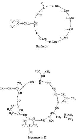
1. Surfactin is a proteolipid of known structure (Fig. 3 ) , first described under the name "protoplast-bursting factor." Produced by Bacillus subtilis it is in effect a cationic detergent. Disruption of cell membranes
CH2
ov
\ \ ^ — L-G1U
H3CvH t v
> - ( C H2)9— C H H,C
CH
/
L-Leu D-Leu
\
L-Val
V-Leu *>-A sP
D - L e u "
Surfactin
/C H3 CH
/
CH3 / "C° H ^ N H C H / / Ο
CH CO
HC— CH, CH— CH,
/ 3
Ν—CH2
HN—N /
H2C -C H 2\CO HC
\N Η N C H2- < 3
H N ' <f £c CH3
H2C^H/CH2 C I OH Monamycin D
FIG. 3. Recent antibiotics that disorganize lipid membranes.
and leakiness apparently result from interaction with membrane phos- pholipids such as phosphatidyl choline and phosphatidyl ethanolamine
(47,48).
2. Streptomycetes produce a number of antibiotics that render microbial membranes leaky. One of these is monamycin, a cyclodepsi- peptide (Fig. 3) which forms complexes with alkali metal cations. Its antibacterial action, however, appears to be due to cell lysis; cations that associate with the antibiotic antagonize its biological effects (49).
Desertomycin induces leakiness in both bacterial and fungal membranes (50). The same is true of azalomycin (51), which is structurally related to the polyenes; judging by the available reports, its primary mode of action is likely to be more specific but is presently unknown.
B. Ion-Conducting Antibiotics
The realization that certain antibiotics exert their effects by facili- tating the diffusion of ions across lipid membranes is a comparatively recent development; it is barely mentioned in the treatise of Gottlieb and Shaw (11). I propose to review this subject in some detail, not only because of its novelty but because I believe it to be among the most significant developments in molecular biology. Ion-conducting agents have revealed novel pharmacological principles and have become potent tools for the dissection of energy-linked membrane functions.
Several recent reviews cover aspects of this field (9, 44, 46, 52-58).
To my knowledge, the concept of ion conduction was first formulated by Mitchell as part of the chemiosmotic hypothesis of oxidative phos- phorylation (59, 60). Briefly, this hypothesis states that the respiratory chain of mitochondria is so arranged as to generate H+ and O H- on opposite sides of the cristae membrane. The membrane is essentially impermeable to these ions, so that respiration creates a differential of pH and of electrical potential across the membrane. The gradient of proton activity is thought to reverse the direction of the ATPase asso-
ciated with the cristae membrane and to thus drive the net synthesis of ATP. This is not the place to assess the merits of the chemiosmotic and rival theories of oxidative phosphorylation, a task performed with admirable detachment by Greville (61) and Racker (62). Suffice it to note here Mitchell's insight that any substance which facilitates the diffusion of protons across the membrane would collapse the differential of pH and electrical potential and thus uncouple ATP synthesis from respiration. It seems quite clear that this is, in fact, the molecular basis
3 1 4 FRANKLIN Μ. HAROLD of uncoupling by dinitrophenol and many other well-known synthetic substances (44, $4, 60, 63-66). However, I am not aware of any com
pound of biological origin which acts as a proton conductor in Mitchell's original sense and shall refrain from pursuing the matter further.
The full significance of the concept of ion conduction became apparent with the discovery by Pressman, Chappell, and their associates that certain antibiotics facilitate diffusion of alkali metal cations across bio
logical membranes. These antibiotics may belong to any of a number of structural classes and range in selectivity from the promiscuous gramicidins to the highly fastidious valinomycin. The essence of their mode of action is formation of a complex such that the cation is encaged within the antibiotic molecule; the latter presents a hydrophobic surface to the outside and can thus "dissolve" in and pass across lipid membranes.
The physiological consequences include loss of K+, uncoupling of oxi
dative or photosynthetic phosphorylation, inhibition of active transport, and others—depending on the specificity of the antibiotic and on the magnitude and direction of ion gradients. Nature has devised several variations on this basic theme.
1. VALINOMYCIN AND ENNIATINS
Valinomycin, a cyclodepsipeptide antibiotic (Fig. 4 ) , first attracted the attention of biologists following the discovery that it is a potent uncoupler of oxidative phosphorylation. Examination of some peculiar features of this uncoupling led to the discovery that valinomycin induces K+ accumulation by mitochondria (56, 67, 68). It is now clear that the basic effect of valinomycin is to facilitate movement of K+ across mem-
FIQ. 4. Structure of the antibiotic valinomycin and its K+ complex. [Redrawn from Pinkerton and Steinrauf (79) with kind permission of the authors and Aca
demic Press.]
branes of all kinds—mitochondrial, bacterial, mammalian plasma mem- branes, and artificial lipid bilayers (55, 56, 66, 67, 69-76). Shemyakin and his associates (57, 58) have prepared numerous analogs and deriva- tives of valinomycin and have shown excellent correlation between anti- biotic activity and ion conduction. Valinomycin is highly selective; in artificial lipid bilayers it prefers K+ to N a+ by a factor of 400, and its specificity in mitochondrial membrane is still higher (53, 66, 67, 72, 74).
The remarkable properties of valinomycin are fully explained by the three-dimensional configuration of the antibiotic and of its K+ complex, which have been established by several laboratories. Upon interaction with K+, the essentially planar molecule puckers up such that the ester carbonyl groups point inward into the ring, hexagonally arranged about the K+ ion. The antibiotic molecule shields the K+ ion from the solvent, presenting a hydrophobic and almost spherical surface (Fig. 4 ) . Thus complexed, K+ can pass across a nonpolar lipid phase, equilibrating with K+ in the aqueous phases on either side (77-80). The proposition that valinomycin serves as a carrier for K+ is now supported by a large body of experimental evidence (55, 56, 66, 69, 80). Of great importance is the fact that the valinomycin-K+ complex bears a net positive charge;
movement of the complex is thus strongly influenced by an electrical potential (54, 60, 69).
The same basic mechanisms are involved in the mode of action of enniatins and of beauvericin. These are structurally related to valino- mycin and also form positively charged, lipid-soluble complexes with K+ but are less selective (81-83).
It is increasingly accepted that these structural features provide an essentially complete explanation of the biological effects of valinomycin, summarized by the statement that this antibiotic mediates passive trans- location of K+ across membranes in the direction of the electrochemical potential [but see Pressman (55, 56) for an alternative view]. Full documentation of this assertion would lead too far afield, but some ex- amples are appropriate. The case is simplest for erythrocytes (84) and resting cells of Streptococcus faecalis (73). Valinomycin causes such cells to lose K+ to the medium, provided electroneutrality can be main- tained by the movement of other ions. Where an electrical potential exists across the membrane, the effect of valinomycin depends on the total ion fluxes. Thus, the antibiotic has little effect on chloroplasts, which pump protons inward (85), or on K+ accumulation by bacteria (86), but induces K+ accumulation by mitochondria, which do not do so ordinarily (55, 56, 67, 87).
316 FRANKLIN Μ. HAROLD
Valinomycin, and some of the other antibiotics discussed below, can thus serve as biochemical probes to detect a membrane potential, to estimate its magnitude, and to assess its role in biological processes (54, 87, 88). Beyond that, valinomycin may offer a model of the kind of reactions that occur at natural transport sites for K+ and other ions
(66).
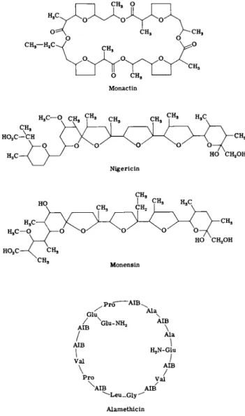
2 . MACROTETROLIDES
Though totally different in structure (Fig. 5 ) , the macrotetrolide anti
biotics closely resemble valinomycin in their mode of action. Monactin and its homologs form a specific complex with K+ in which the molecule enfolds the cation in a hydrophobic shell (89-91). The complex again bears a net positive charge and passes across membranes in response to the electrochemical gradient. Like valinomycin, monactin induces elec
trical conductance in lipid membranes and facilitates K+ movement across biological membranes (70, 72, 76, 92-98). It should be remem
bered, however, that monactin is somewhat less K+ selective than is valinomycin.
3 . NlGERICIN, M O N E N S I N , AND OTHER CARBOXYLIC POLYETHERS
Valinomycin and monactin were initially described as inducers of K+ uptake in mitochondria. Another class of ion conductors was first recog
nized by their capacity to antagonize the action of valinomycin and discharge K+ from mitochondria (55, 56, 69, 99, 100). Nigericin, monensin, dianemycin, and several less studied compounds fall into this group.
Once again, physical experiments together with the determination of molecular structures (101-103) have provided an essentially complete explanation of the mode of action of these antibiotics. The structures of nigericin and monensin are shown in Fig. 5 . It will be noted that both are monocarboxylic acids and can thus exist both in the protonated form and as neutral salts. However, salt formation is accompanied by a major change in conformation such that the cation is once again en
folded within a hydrophobic shell. The basis of the mode of action of these antibiotics is thus that they can shuttle both protons and cations across membranes; nigericin prefers K+, whereas monensin prefers N a+
(55, 56, 69, 70, 85, 102, 103). The complexes are electrically neutral, and thus the antibiotics tend to carry out exchange of cations for protons, or "antiport" in Mitchell's apt terminology (54, 60). Monensin appar-
-AIB,.
Glu Ala
AIB
\
Val
ΑΪΒ - ^G A1 U ' \
/ Ala
\
H2N-Glu
ν AIB
\ / Pro y a l
XA I B AIB
^ L e u _ G l y ^ Alamethicin
FIG. 5. Some ionophorous antibiotics ( A I B , α-aminoisobutyric acid).
ently catalyzes strictly equimolar antiport of N a+ and H+ and therefore does not increase the electrical conductance of the membrane; nigericin, however, can carry protons alone to some degree (53).
The biological effects of these antibiotics are explicable in terms of the ion translocations which they induce. Consideration of some of these illustrates the use of "ionophorous" antibiotics (69) in exploring the
318 FRANKLIN Μ. HAROLD
relationship of pH and electrical gradients to active transport, oxidative phosphorylation, and photosynthesis.
In mitochondria, for instance, nigericin causes loss of K+ by exchange for protons from the medium (69). It is not, however, an uncoupler of oxidative phosphorylation. This presumably reflects the fact that the greater part of the proton-motive force of mitochondria, which supports A T P synthesis according to the chemiosmotic theory, is made up of the membrane potential; electrically neutral exchange of one cation for another should not affect the membrane potential (54, 60, 87, 88). How
ever, nigericin does block uptake of phosphate by the mitochondria (100), suggesting that phosphate uptake depends on the pH gradi
ent—which is obliterated by the antibiotic (104,105).
In the glycolytic bacterium Streptococcus faecalis, monensin and nigericin also abolish the pH gradient and stop transport processes that presumably depend on this gradient. However, monensin does not inhibit net accumulation of K+, which is believed to be directed by the membrane potential (86, 106). Readers are referred to a monograph by Mitchell
(54) for a detailed consideration of the use of antibiotics as probes in cellular electrophysiology.
A note of caution is appropriate at this point. The ionophorous anti
biotics, like all other drugs, are not free of misleading side effects. Thus, valinomycin does complex H+, although at physiological pH the H+ con
centration is sufficiently low that proton conduction is usually negligible (75). Nigericin facilitates K+- H+ antiport but will accept N a+ as well, albeit with lower affinity (53, 70, 72); it also acts to some extent as a proton conductor (53). Indeed, it may not always be legitimate to regard the antibiotics as carriers whose effect on membrane structure can be neglected. Finer et al. (107) have reported distortion of certain lipid membranes following addition of the antibiotics; some degree of nonspecific leakiness may be expected in biological systems and has been encountered in the experiments of at least the present author.
4. GRAMICIDINS
The gramicidins are hardly recently discovered antibiotics, having been known since about 1939, but only during the past five years has their mode of action been clarified. Gramicidins are a family of closely related linear peptides in which the terminal amino group is formylated and the terminal carboxyl group is masked by ethanolamine (for review, see reference 108). Gramicidin A, for example, has the sequence H C O - L - Val-Gly- L - Ala- D -Leu- L-Ala-D-Val-L-Val- D-Val- L-Try- D-Leu- L-Try- D-
Leu-L-Try-D-Leu-L-Try-NHCH2CH2OH. Like the other antibiotics con- sidered in this section, gramicidins induce ion transport across a variety of membranes, including lipid bilayers and bacterial, mitochondrial, and erythrocyte membranes. However, the gramicidins do not appreciably discriminate between cations, translocating K+, N a+, and even H+ (67, 68, 70, 72, 73, 109). The molecular basis of ion movements induced by gramicidin is uncertain. It has been suggested that gramicidins form cation complexes which shuttle across the membrane. On the other hand, there is evidence that gramicidin may interact with the matrix of the membrane, distorting its structure to form a pore through which the cations can pass (107, 110, 111). Be this as it may, the fact that grami- cidins increase the conductance of lipid bilayer membranes (72) is evidence that they permit net movement of electrical charges.
The biological effects of gramicidins combine those of cation con- ductors and of proton conductors as well, limiting the utility of these antibiotics in physiological research.
5. ALAMETHICIN
Alamethicin is a cyclic depsipeptide whose structure (Fig. 5) has re- cently been determined (112). It has been reported to induce ion uptake in mitochondria. However, it is strongly surface active and may inhibit the growth of some gram-positive bacteria by inducing lysis.
This compound is of great interest, not as a metabolic inhibitor in the usual sense, but as an inducer of action potentials and related phenomena in artificial lipid membranes (53, 113). This involves associa- tion with, and reorganization of, the phospholipid structure (114). Some- what similar electrokinetic phenomena are induced by monazomycin
(53), a compound of unknown structure which induces cation uptake in mitochondria but has otherwise been little studied.
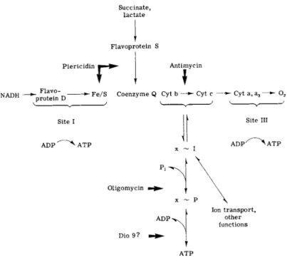
IV. INHIBITORS OF THE RESPIRATORY CHAIN AND ENERGY TRANSFER
In aerobic organisms, energy from catabolism is made available by the passage of protons and electrons over the cascade of carriers called the respiratory chain. Its components are generally similar in animals, plants, and microorganisms although there are substantial differences in detail. The loci at which free energy is tapped for the synthesis of
320
Succinate, lactate
FRANKLIN M. HAROLD
Flavoprotein S Piericidin
r
AntimycinNADH —+~ F I?V° " t. ^ Fe/S Coenzyme Q Cyt b - - • Cyt c — » - Cyt a, ag —- 02 protein ϋ
ν J ^ J V J
Site I ADP ^ A T P
Site III ADP > A T P
Oligomycin
Dio 9 ?
Ion transport, other functions
FIG. 6. Sites of inhibition of the respiratory chain and of energy transfer by antibiotics (cyt., cytochrome).
ATP are called coupling sites. In animal mitochondria, coupling site I lies between N A D H dehydrogenase and coenzyme Q, site II between cytochromes b and c, and site III in the region of cytochrome oxidase
(Fig. 6).
A. Respiratory Inhibitors
Most of the antibiotics that block the respiratory chain were initially recognized by virtue of their fungicidal activity. The bacterial respira
tory chain is in many cases inaccessible to inhibitors except in membrane fragments. Whether this reflects peculiarities of wall or membrane struc
ture is unclear.
The respiratory inhibitor par excellence is antimycin A, which inhibits the respiratory and photosynthetic chains in the region of coupling site II. The antimycins have been thoroughly and lucidly reviewed by Rieske (115). The work of the past few years has been mostly concerned with elucidation of the structure of the coupling site and of the antimycin
receptor. Site II apparently includes two molecules of cytochrome b, one of cytochrome c, and a site for a single molecule of antimycin.
The antibiotic is now thought to bind to one of the cytochrome b mole
cules in a noncovalent manner, since it can be removed by solvent extrac
tion. The binding is cooperative (allosteric), and it is assumed that attachment of antimycin changes the conformation of one or more pro
teins so as to bring electron flow to a halt [116,117).
Coupling site I has for years been studied with the use of barbiturates and rotenone, but at present the inhibitor of choice is the new antibiotic piericidin. First recognized as an insecticide (118), piericidin proved to be a potent inhibitor of respiration in mitochondria and in bacterial membranes. The structure of piericidin (119) resembles that of coenzyme Q (Fig. 7), suggesting that the mode of action may involve competition with a quinone. This appears to be correct. Piericidin inhibits N A D H oxidation and reduction of coenzyme Q in mitochondria and at higher concentrations blocks electron flow between succinate and coenzyme Q as well (120-126). In some instances (127, 128) inhibition can be re
versed by ubiquinones, but this is not always the case.
Piericidin is now generally thought to block a step on the oxygen side of N A D H dehydrogenase. Inhibition of electron flow by piericidin, like that due to antimycin, can be reversed by gentle extraction with organic solvents, suggesting that lipids are involved in the binding of the antibiotic (122, 124). Recent work, however, has also implicated the N A D H dehydrogenase itself. Each molecule of the enzyme appears to bind two molecules of the antibiotic. The two binding sites are indis
tinguishable kinetically but make unequal contributions to the respira
tory chain (129-131).
A number of other antibiotics have been shown to block the respiratory chain, but their primary sites of action remain ill defined. The antifungal agent pyrrolnitrin (Fig. 7) inhibits respiration of both mitochondria and Saccharomyces cerevisiae, and this is likely to be its primary site of action. In yeast, the block is located between succinate or N A D H and coenzyme Q (132, 133). However, in Candida, pyrrolnitrin induces
CH,0 CH3O.
Η OH
Piericidin A Pyrrolnitrin
FIG. 7. Novel inhibitors of the respiratory chain.
322 FRANKLIN Μ. HAROLD
leakage of cell constituents and disruption of the cytoplasmic membrane (134). It remains to be determined whether this is a secondary, non
specific effect or a clue to the primary receptor. Other antibiotics that inhibit the respiratory chain include flavensomycin (135), U19718 (136), and U24544 (137). Their modes of action need further definition; the same is true of such relatively ancient antibiotics as usnic acid and pyocyanine. Napthomycin, an analog of vitamin Κ (138), may well prove useful in the dissection of the respiratory chain.
B. Antibiotics and ATPase
Although the distinction is somewhat arbitrary, the term "inhibitors of energy transfer" designates those compounds that act on the pathway of energy coupling beyond the respiratory carriers proper (Fig. 6). The classic example is oligomycin, whose discovery by Lardy and his asso
ciates was the first fruit of their systematic effort to apply antibiotics to the dissection of oxidative phosphorylation.
Readers are referred to the review by Shaw (139) for a detailed con
sideration of the mode of action of oligomycin. Suffice it to point out here that oligomycin inhibits respiration only so long as it is coupled to phosphorylation; dinitrophenol and other uncouplers relieve the in
hibition of respiration. Oligomycin, and also the related antibiotic ruta- mycin, clearly affect a step beyond the respiratory chain. This is now known to be the ATPase of the mitochondrial membrane, which catalyzes the final step of oxidative phosphorylation, the synthesis of A T P itself (62). The ATPase is subject to inhibition when associated with the mitochondrial membrane, but it is resistant once solubilized. This led to the finding that oligomycin binds not to the ATPase itself but to a protein that forms part of the stalk to which the ATPase is attached (140-143). Inhibition of the ATPase is thought to involve a transmitted effect on the conformation of the enzyme protein. The structures of oligomycin and rutamycin are still unknown, but this does not detract from the usefulness of these inhibitors in studies of mitochondrial metab
olism. To cite but one example: Ion transport by mitochondria is energy linked and can be driven either by respiration or by exogenous ATP.
Oligomycin blocks ion transport energized by ATP but not that driven by respiration, which points to the existence of a high-energy state or intermediate preceding ATP.
Ossamycin, peliomycin, and venturicidin have been shown to act much like oligomycin (144). The mode of action of Dio 9 is somewhat differ
ent; unfortunately it is a mixture of several components of unknown
structure. Dio 9 inhibits the ATPase of mitochondrial, chloroplast, and bacterial membranes but, unlike oligomycin, it inhibits the solubilized as well as the bound enzyme (145-148)- Its metabolic effects are com- plex, including both inhibition and uncoupling of oxidative phosphoryla- tion, the induction of mitochondrial swelling, and the inhibition of active transport in Streptococcus faecalis [145, 148, 149)- To what extent the physiological effects of Dio 9 can be attributed to inhibition of the ATPase remains to be determined.
V. INHIBITORS OF DNA REPLICATION AND TRANSCRIPTION
Genetic information is normally encoded in immensely long, double- stranded D N A molecules which serve as templates for two related processes: replication and transcription. The multitude of antibiotics that affect these processes have traditionally been subdivided into one group that primarily blocks D N A synthesis and another that inhibits R N A synthesis. The growth of knowledge has blurred this distinction.
We now recognize that antibiotics which bind to the D N A template generally inhibit both replication and transcription, although not neces- sarily to the same degree. It therefore seems appropriate to classify the antibiotics in terms of their target molecule rather than their target process.
Another recent development that must be kept in mind is the demon- stration (150) that the classical Kornberg D N A polymerase (polymerase I) is not, after all, the enzyme responsible for D N A replication in E.
coli. This will require the revaluation of conclusions drawn from experi- ments on the effects of antibiotics on the Kornberg polymerase. We can anticipate many reports on the action of drugs on the novel D N A - polymerizing enzymes associated with the cytoplasmic membrane of bacteria (151-153), and these may well resolve some of the uncertainties that becloud the discussion below.
A. Antibiotics That Bind to DNA
1. ACTINOMYCIN
The prototype of all antibodies that associate with D N A is actino- mycin (Fig. 8 ) ; the extensive work on this antibiotic has been reviewed by Reich et al (154) and Balis (155). Suffice it to recall here that, in a variety of organisms ranging from bacteria to mammalian cells,
Actinomycin D Anthramycin Daunomycin
FIG. 8. Antibiotics that complex with DNA.
actinomycin inhibits the synthesis of R N A far more strongly than that of D N A . The same result is obtained in extracts; the antibiotic has little effect on Romberg's D N A polymerase I but blocks R N A poly
merase completely at low concentrations. Actinomycin blocks elongation of the R N A chain rather than the initiation of transcription {156). The antibiotic forms a tight complex with double-stranded D N A , but not with R N A or single-stranded D N A ; binding requires G-C pairs. These observations, together with the results of X-ray diffraction studies, were rationalized by Reich and his associates (154) m a detailed model.
Actinomycin is thought to be bound to the outer surface of the D N A helix, held in the minor groove by hydrogen bonds between the antibiotic and deoxyguanosine residues. The presence of the antibiotic molecules in the groove blocks movement of the polymerase and arrests transcription.
324 F R A N K L I N Μ . H A R O L D
Despite its widespread acceptance, Reich's model has been challenged on the basis of detailed physicochemical studies of the interaction of actinomycin with D N A . Miiller and Crothers (157) have proposed that complex formation involves intercalation of the planar chromophore moiety of the antibiotic between D N A bases, usually close to G-C pairs.
At the same time, the peptide rings undergo conformational changes which allow each ring to associate with one phosphodiester backbone.
The slowness of this conformational change accounts for the stability of the actinomycin-DNA complex, once it has formed. Recently, Waring (158) has provided strong support for the intercalation of the actino- mycin chromophore from studies on uncoiling of closed circular D N A molecules when they bind the antibiotic. Even the role of G—C pairs in binding actinomycin may not be universal, since Wells and Larson (159) have reported that synthetic polynucleotides lacking deoxyguanylic acid still bind the antibiotic.
Intercalation was originally proposed by Lerman (160) to account for the interaction of D N A with acridines. It has now become apparent that intercalation is a major pharmacological mechanism, which is in- creasingly being invoked for antibiotics as well.
2. ANTHRACYCLINE ANTIBIOTICS
The anthracycline group includes nogalamycin, daunomycin, and others (155, 161, 162). All share the basic structure of a tetrahydrotetra- cenquinone chromophore linked to a sugar moiety; aglycones have little antibiotic activity. The structure of daunomycin (163) is shown in Fig.
8. The biological effects of anthracyclines are in general similar to those of actinomycin. In both bacterial and mammalian cells they inhibit R N A synthesis more severely than D N A synthesis. Ribosomal R N A synthesis is particularly sensitive to inhibition by nogalamycin (164).
The activity of R N A polymerase is strongly inhibited by members of this group; they block elongation of the R N A chain but do not prevent initiation of transcription (156). The activity of D N A polymerase is also inhibited by anthracyclines to various degrees (165).
The anthracyclines bind to D N A but apparently not in a very specific manner. It has been reported that nogalamycin preferentially associates with A-T pairs (162) but not all investigators find this to be true (165).
Daunomycin, indeed, forms complexes not only with D N A but with R N A and with oligonucleotides as well (165, 166). Complex formation with D N A is accompanied by striking changes in sedimentation proper- ties. Linear molecules exhibit increased viscosity and reduced density,
3 2 6 FRANKLIN Μ. HAROLD
while circular D N A molecules undergo the characteristic uncoiling process. These observations provide convincing evidence that the planar chromophores intercalate between the D N A bases. The sugar residues, essential both for biological activity and for complex formation, are thought to interact with the backbone (158,166-168).
A novel antibiotic that may be related to nogalamycin is steffimycin.
It inhibits the synthesis of all classes of R N A by Bacillus subtilis and binds to D N A . However, like nogalamycin, it can also complex with t R N A and inhibit protein synthesis in extracts in this way (169, 170).
3 . CHROMOMYCIN, MITHRAMYCIN, AND OLIVOMYCIN
Chromomycin, whose structure is shown in Fig. 8, is one of a group of related antibiotics which, like the anthracyclines, feature a chromo- phore substituted by various sugars. Chromomycin is a potent inhibitor of R N A synthesis in vivo and of R N A polymerase in vitro, with double- stranded D N A as template. It probably blocks elongation of the R N A chain (165, 171, 172). Although chromomycin is generally thought of as an inhibitor of transcription, in Bacillus subtilis it inhibits D N A synthesis equally well (173).
Again, formation of a complex between the antibiotic and D N A is thought to lie at the heart of its mode of action. Binding, and the conse
quent interference with the template function of D N A , are favored by G-C pairs and also by divalent cations. Many of the results are consistent with intercalation of the chromophore portion of the antibiotic (165, 174). However, chromomycin induced little or no uncoiling of a circular D N A duplex, which argues against intercalation (158).
Mithramycin and olivomycin are other members of this group, similar to, but not identical with, chromomycin both in structure and in biologi
cal activity. Mithramycin preferentially inhibits R N A synthesis in B.
subtilis, unlike chromomycin (173). Olivomycin has been shown to block the growth of R N A chains during transcription, but it has little effect on initiation (175). According to Waring (158), there is no evidence for intercalation of mithramycin.
4 . OTHER ANTIBIOTICS
Intercalation is likely to prove an important mode of interaction be
tween antibiotics and D N A , but evidently not the only one. The novel antibiotic anthramycin (Fig. 8) lacks the structural features required for intercalation, yet it binds to D N A very tightly and inhibits R N A
polymerase about as strongly as does actinomycin {176). It binds only to double-stranded native D N A , perhaps by formation of a labile co- valent bond {177).
Numerous other antibiotics form complexes with D N A and inhibit R N A synthesis (and sometimes D N A synthesis as well). Examples in- clude distamycin {178, 179); kanchanomycin {180, 181), which is thought to interfere with D N A synthesis by blocking the template but with R N A synthesis by inhibiting the polymerase; pluramycin {155, 182, 183); streptonigrin {184); quinomycin {165, 185-187); and grana- ticin {188). The molecular mechanisms by which these compounds bind to D N A are as yet unknown. Intercalation, binding to functional groups, and associations that induce single-strand scissions have all been considered.
Antibiotics that bind to D N A tend, as a general rule, to inhibit transcription both in vivo and in vitro. From time to time, however, antibiotics have been claimed to bind to D N A yet preferentially inhibit replication. An example is mitomycin, which is now known to affect various aspects of R N A metabolism as well. It is doubtful that anti- biotics exist which bind to D N A yet do not block transcription.
Phleomyan is an antibiotic of unknown structure which has been used as an inhibitor of D N A synthesis in E. coli. The earlier literature has been summarized by Bhuyan {162). Phleomycin interacts with D N A by a mechanism involving thymine {189, 190), but it now appears that it forms complexes with R N A as well. Indeed, Watanabe and August {191) suggest that phleomycin primarily inhibits transcription. Bleo- mycins are thought to resemble phleomycins in structure as well as in name and perhaps also in mode of action {183,192,193).
Hedamycin and rubiflavin, whose structures are unfortunately not known, also no longer seem to be very promising candidates. In intact E. coli, these antibiotics inhibit D N A synthesis far more rapidly than that of R N A or protein. They form various complexes with D N A , all of unknown structure, and it is possible that binding prevents separation of the strands during D N A replication {194). Hedamycin, in vitro, in- hibits D N A polymerase I but also blocks transcription by R N A poly- merase {195). In sum, at present no antibiotic is known which binds to D N A and specifically blocks replication.
B. Inhibitors of Replication
The mechanisms that ensure the accurate replication of D N A and the equal distribution of the products have proven far more complex
328 F R A N K L I N Μ. HAROLD
than originally envisaged. Antibiotics that selectively interfere with replication without reacting with the template itself may well exist, but none can be placed in this category with any assurance.
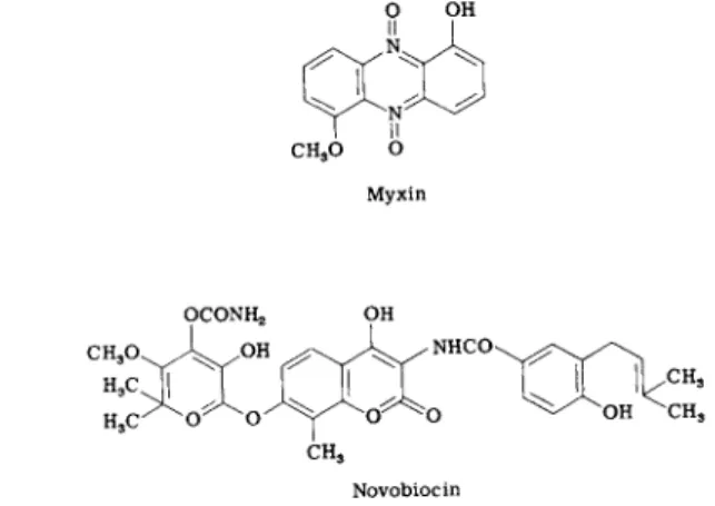
A good prospect is novobiocin (Fig. 9 ) , which was first found to affect wall synthesis and membrane permeability. Smith and Davis [196, 197) then showed that it inhibits D N A synthesis before any other effect is manifest and totally blocks cell division. The antibiotic inhibits R o m berg's D N A polymerase I in extracts but apparently does not bind to the template. The physiological results do point to replication as the target of novobiocin, and the association of D N A replication with the plasma membrane may ultimately provide an explanation for the effects of the antibiotic on permeability (198, 199). N o studies on the effect of novobiocin on the novel D N A replicase II (151-158) have yet been reported.
A more recent addition to the short list is myxin (Fig. 9 ) , an antibiotic produced by a myxobacterium. In Escherichia coli, myxin caused rapid cessation of D N A synthesis, followed by its degradation to acid-soluble compounds. After some time, R N A synthesis was inhibited as well (200, 201). Apparently, myxin is not bound to D N A itself. Its primary site of binding may, in fact, be the cytoplasmic membrane. Myxin-treated cells exhibit various cytological changes in membrane structure, including vacuolation and filamentous growth. The antibiotic does not block mem
brane synthesis per se, even at high concentrations. On the other hand, concentrations so low as to have no effect on D N A synthesis still block cell division (201, 202). In view of the close association, at least in bacteria, between D N A and plasma membrane, these results are very
ο OH
CH3O ο
Myxin
Novobiocin
FIG. 9. Possible inhibitors of D N A replication.
suggestive. Myxin also causes bleaching of Euglena, presumably by in
hibiting the replication of chloroplast D N A (203).
C. Inhibitors of RNA Polymerase and Transcription
1. RIFAMYCINS AND STREPTOVARICINS
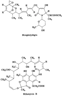
The rifamycins, and their semisynthetic derivatives such as rifampicin, are among the relatively few recent antibiotics to have found extensive clinical use. They are potent inhibitors of gram-positive bacteria and of mycobacteria, inhibit the replication of some viruses, but have little toxicity for animals. The structure of a rifamycin is shown in Fig. 1 0 .
The resemblance of rifamycins to the macrolides led to the expectation that they would prove to be inhibitors of protein synthesis, but this is not the case. Rifamycins inhibit almost instantaneously the synthesis of all classes of R N A by gram-positive bacteria; synthesis of D N A and protein continues for some time. In 1 9 6 8 , no fewer than six labora-
OH Streptolydigin
Rifamycin Β
FIG. 10. Antibiotic inhibitors of RNA polymerase.
330 FRANKLIN Μ. HAROLD
tories discovered independently that rifamycins strongly inhibit R N A polymerase of bacteria (both gram-positive and gram-negative), whereas mammalian R N A polymerases are resistant {204-210). By a variety of criteria it was demonstrated that inhibition of R N A polymerase is indeed the process responsible for the inhibition of growth. Most com
pelling are the characteristics of mutants resistant to rifamycin. These contain a resistant polymerase, and the locus that specifies rifamycin resistance defines the structural gene for the enzyme (204, 205, 208, 210, 211).
The reaction catalyzed by R N A polymerase is complex. The enzyme is thought to associate with double-stranded D N A and cause local de- naturation or "melting" of the secondary structure. This partial unwind
ing is required to enable one of the strands to be transcribed. An "initia
tion complex" is next formed which includes D N A , enzyme, and the first nucleotide; addition of subsequent ribonucleotides results in elonga
tion with formation of the phosphodiester links. Rifamycin does not block the binding of the enzyme to the D N A template but inhibits the initiation of transcription. It may block formation of an active binary complex between D N A and the polymerase (212) or the stabiliza
tion of the initiation complex by addition of the first nucleotide (213) or the formation of the first phosphodiester bond (214). All investigators seem to agree that, once the process of transcription has begun, rifamycin no longer inhibits it (212, 213, 215-217). Even in intact cells, it has been possible to distinguish initiation from elongation. Rifamycin blocks initiation of the transcription of the tryptophan operon in E. coli but not transcription already in progress (218, 219).
The enzyme R N A polymerase consists of several subunits which make up the "core" responsible for the polymerization step, together with fac
tors such as those that determine whether a particular D N A strand is transcribable. All the evidence indicates that rifamycin binds to the enzyme core. The R N A polymerase from resistant mutants does not bind the antibiotic (210, 213, 220). The macrocyclic ring is involved in the binding reaction, since alterations in its structure profoundly affect antibiotic activity (221). A very stable complex is formed in which each enzyme molecule binds one antibiotic molecule at a site close to, but distinct from, the DNA-binding site (210, 213, 216, 220).
The specific affinity of rifamycins for R N A polymerase has already proved to be of great utility in analyzing the mechanism of transcription.
Readers are referred to the 1969 Lepetit symposium (222) but a few later examples should be cited briefly. Among the rifamycin-resistant mutants of Bacillus sub tilts, some proved to be deficient in sporulation
as well. This led to the discovery (223-225) of a factor that controls the specificity of R N A polymerase during sporulation. Doolittle and Pace (226) employed rifamycin to block the initiation of transcription in E. coli; from the residual transcription of transfer R N A they deduced the existence of precursor species of high molecular weight. A third appli
cation has been to the evolution of mitochondria: Is mitochondrial R N A polymerase homologous with the bacterial enzyme, as are mitochondrial ribosomes? Two laboratories reported that mitochondrial R N A poly
merase is resistant to rifamycin and is thus presumably synthesized under nuclear control (227, 228). Two others have found it to be sensitive
(229, 280). Chloroplast R N A polymerase also appears to be sensitive to rifamycin (281).
Rifamycin is thus well established as a selective inhibitor of R N A polymerase, but it would appear that one of its most beneficial uses may have a different basis: Rifamycins inhibit the multiplication of vaccinia (pox) virus and may find clinical application as antiviral agents (282). Vaccinia virus does contain an R N A polymerase, but the antiviral effect is not due to the inhibition of transcription (288, 284) · Replication of the R N A phage Q-β is also blocked (285), and again there is no reason to invoke DNA-dependent polymerases. The molecular basis of the antiviral effects of rifamycin remains to be established.
The streptovaricins are a group of antibiotics structurally related to rifamycin. Like rifamycins, they inhibit bacterial R N A polymerase at the initiation step. Indeed, mutants resistant to streptovaricins are often resistant to rifamycins as well (286-289).
2 . STREPTOLYDIGIN
Streptolydigin, whose structure is shown in Fig. 10, promises to be of great utility in cell biology. Like rifamycin, it is an inhibitor of R N A polymerase, and mutants resistant to streptolydigin boast an R N A polymerase whose core has undergone alteration (289). However, strep
tolydigin is an inhibitor of chain elongation rather than of initiation and should be a valuable complement to rifamycin (289, 240).
VI. ANTIBIOTIC INHIBITORS OF PROTEIN SYNTHESIS
Perhaps no aspect of molecular biology has attracted as many investi
gators as has the mechanism of protein synthesis. The exacting task
![FIG. 1. Sites of inhibition of peptidoglycan synthesis by antibiotics. [Redrawn from Perkins (16), with kind permission of the author and Academic Press.]](https://thumb-eu.123doks.com/thumbv2/9dokorg/1152286.82849/4.648.85.582.68.826/inhibition-peptidoglycan-synthesis-antibiotics-redrawn-perkins-permission-academic.webp)