I N T R O D U C T O R Y L E C T U R E
My main object in this paper is to discuss receptor mechanisms in human vision; but I shall first briefly review certain ultrastructural and chemical arrangements found generally in vertebrates [i].
The vertebrate photoreceptors—both rods and cones—are derived embryonically from such ciliated ependymal cells as typically line the ventricles of the brain. In the rods and cones it is the expanded and otherwise modified shafts of cilia that form the outer segments [2].
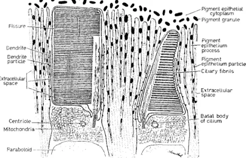
The latter have a layered structure. In rods the lamellae take the form of a pile of flattened sacs (Sjöstrand's double-membrane discs), each bordered by a differentiated rim and all enclosed within the plasma membrane. In cones the layers are ordinarily produced by the repeated infolding of the plasma membrane on the side away from the ciliary process [3]. These features are shown diagrammatically in Fig. 1. The lamellar membranes in both rods and cones are about 5 nm thick, about as thick therefore as the diameter of a molecule of cattle rhodopsin, if it is spherical in shape.
The outer segments stand in the closest anatomical and functional relationship, on the one hand with the inner segments, on the other with the pigment epithelium. A system of delicate cytoplasmic pro- cesses extends downward from the pigment epithelial cells among the outer segments, overlapping there with a similar system of processes ('dendrites') springing from the apices of the inner segments and surrounding the outer segment like a palisade (Fig. 1) [4]. These probably facilitate exchanges of metabolites between the outer segment and the adjoining structures. It is noteworthy that the region of the inner segment from which the dendrites arise contains a dense clump of mitochondria—the ellipsoid—and is apparently a site of intense metabolic activity, as is also the pigment epithelium. Such dendrites are widespread though not universal structures ; they are present in a number of amphibia and mammals, including the rhesus monkey and man, though they seem to be absent in rats and mice [4].
133
GE O R G E WA L D
Biological Laboratories of Harvard University, Cambridge, Mass., U.S.A.
134 M O L E C U L A R A N D F I N E S T R U C T U R E O F R E C E P T O R S
A system of deeply staining micelles has been observed in the lamellae of the rod outer segments of the mudpuppy, Necturus [4].
They may contain the visual pigment, in this case porphyropsin. The micelles are so regular in size and spacing as to resemble a crystalline
FISSURE -
Centriole- Mitochondria-
Paraboloid
Pigment epitheLial cytoplasm Pigment granule
Pigment -epithelium
process Pigment
-epithelium particle
FIG. Ι. Diagram showing ultrastructural arrangements of the outer segments of a rod and cone, in this case as found in the retina of the salamander Necturus, the mudpuppy. The outer segment is derived from a primitive cilium and retains a ciliary process as stalk and back- bone structure. The transverse membranes have a different form in rods and cones: in rods, flattened sacs with a differentiated rim; in cones, repeated infoldings of the plasma membrane on the side across from the cilium. In such large amphibian rods the outer segment is typically carved into lobules by deep longitudinal fissures. Also in Necturus rods the transverse membranes display a system of highly regular, deeply staining micelles which may contain the visual pigment.
The outer segments are inserted between two metabolically active tissues : the pigment epithelium and the inner segments. Long, slender processes extend from both so as to surround the outer segments, so facilitating mutual exchanges of metabolites. At the region of the inner segment from which such processes, the dendrites, arise, a dense clump of mitochondria (the ellipsoid) and a heavy store of glycogen (the paraboloid) imply intense metabolic activity, perhaps associated with the function of the outer segment (from BROWN, GIBBONS and
WALD [4]).
array. If they do contain the visual pigment, each micelle contains on the average about 50 molecules of porphyropsin.
The outer segments of the rods and cones are primarily solid state structures, quasi-crystalline in the sense that many of their component molecules are fixed in position and highly oriented. The lamellar structure and the presence of systems of lamellar micelles are evidences of this condition. Furthermore, W . J . SC H M I D T showed years ago that frog rod outer segments are dichroic, owing to the fact that their rhodopsin is oriented so that its chromophores lie transversely, in the planes of the lamellae. Direct microspectrophotometry of single rod outer segments has recently shown this orientation to be almost perfect [1,5]. Owing to this condition the absorption of light passing down the axis of the outer segment, its normal direction in the eye, is increased about 1-4 times as compared with the same concentration of rhodopsin randomly oriented as in free solution.
The quasi-crystalline structure of outer segments has suggested that such typical solid state phenomena as exciton migration (inductive resonance) and photoconductivity may play a large role in visual excitation. These are by no means to be taken for granted, however ; indeed, their a priori prospects are poor [1, 4]. The efficiency of resonance transfer of molecular excitation falls off rapidly with distance, and is believed not to occur at all beyond about 10 nm. This makes difficulties for such a conjugated protein as rhodopsin, the diameter of which if spherical is about 4 nm, so that its chromophores must on the average lie that far apart even if the molecules were closely packed. There is some possibility of such close packing in lamellar micelles, with, in addition, some clustering of the chromophores.
Such devices would make exciton migration easier within a micelle, but correspondingly more difficult from one micelle to another. Even in frog rods, in which rhodopsin is unusually concentrated and highly oriented, no experimental evidence could be found that molecular excitation migrates over distances comparable with the dimensions of the outer segment [6]. It looks at present as though rod or cone excita- tion must occur at or quite close to the site of absorption of light.
The visual pigments of vertebrates share a common structure. Each consists of a specific type of protein, an opsin, bearing as chromophore a particular configuration of retinaldehyde (vitamin A aldehyde, formerly retinene), the 1 i-cis isomer. The only action of light in vision is to isomerize this chromophore from the 11-as to the all-trans
136 M O L E C U L A R A N D F I N E S T R U C T U R E O F R E C E P T O R S
C H3 H C H3 H C H3
I I I I I OH
H H H H H, C S 32 I £ — C H3
CXL I C H3
1U
1 II
All-trans RETINOL (VITAMIN Λ)
RCTINALDCHVDC
C H , H C H , Η I I I I
Τ
11-EIS RETINOLI
Η PI
H2C \ / Ç - C P I3 C . II I C H3 3 Ι
H2 C H2O H
FIG. 2. Structures of dXX-trans and 11-CM retinol (vitamin A ) and retinaldehyde (retinene).
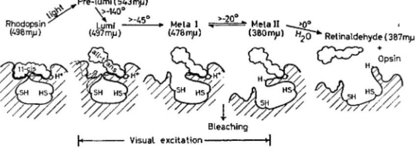
mixture of opsin and all-trans retinaldehyde (Fig. 3). This last reaction is too slow to have a part in visual excitation. The intermediates represent progressive stages in the opening-up of the opsin structure, during which new chemical groups are exposed. One of these stages is responsible for triggering visual excitation, perhaps owing to the exposure of a catalytic site on opsin, converting it to an active enzyme.
We have realized for many years that a dark adapted rod can be excited through one of its rhodopsin molecules absorbing a photon. An efficient catalysis might amplify the effects of such a one-molecule event the requisite amount—perhaps one million times—needed to transform it into a nervous excitation [1].
configuration (Fig. 2) [7]. All other changes—chemical, physiological or psychological—are 'dark' consequences of this one light reaction.
This first product, in the case of rhodopsin called pre-lumirhodop- sin [8], is highly unstable at ordinary temperatures. In the dark it is transformed rapidly over a series of intermediates—lumirhodopsin, metarhodopsin I and metarhodopsin II [9]—hydrolysing finally to a
Bleaching
Visual excitation •]
FIG. 3. Stages in the bleaching of rhodopsin. Rhodopsin has as chromophore 11 -eis retinaldehyde, which fits closely a section of the opsin structure. The only action of light is to isomerize retinaldehyde from the 11 -eis to the all-trans configuration (pre-lumirhodopsin).
Then the structure of opsin opens progressively (lumi- and the metarhodopsins), ending in the hydrolysis of retinaldehyde from opsin. Bleaching occurs in going from metarhodopsin I to I I ; and visual excitation must have occurred by this stage. The opening of opsin exposes new chemical groups, including two —SH groups and one H+-binding group. The absorption maxima shown are for pre- lumirhodopsin at — i90°C, lumirhodopsin at — 65 °C, and the other pigments at room temperature.
either molecule or both must be exchanged for or isomerized to the 11-ay configuration.
T o these general arrangements, which they share with other vertebrates, the primates—monkeys, apes and man—add two special features : the presence of a central yellow patch, or macula lutea ; and colour vision, which though not by any means confined to primates is peculiarly accessible in them, and particularly in man.
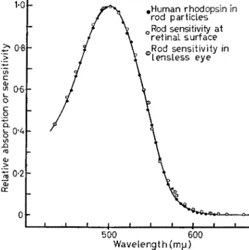
Human rods contain a typical rhodopsin [11]. Its absorption spectrum, measured in suspensions of rod outer segments or by micro- spectrophotometry of human retinas [12], has Am ax about 500 nm, and accounts precisely for the spectral sensitivity of human rod vision, In vertebrate retinas, owing to the presence of the enzyme alcohol dehydrogenase, the retinaldehyde released by the bleaching of visual pigments is reduced to retinol (vitamin A ) . In frogs this enzyme is soluble, and the most prevalent co-factor of biological oxido-reduc- tions, D P N (in England N A D ) acts as coenzyme. FUTTERMAN [10] has recently found that in cattle retinas a particle-bound alcohol dehydro- genase may react preferentially with T P N as coenzyme. T o regenerate a visual pigment, retinol must be re-oxidized to retinaldehyde, and
138 M O L E C U L A R A N D F I N E S T R U C T U R E O F R E C E P T O R S
J 1 J I I I I I L
500 600 Wavelength (mu)
FIG. 4. Absorption spectrum of human rhodopsin, measured in a suspension of rod outer segments, compared with the spectral sensi- tivity of human rod vision, as at the retinal surface. The latter data involve either the average scotopic luminosity corrected for ocular transmission, or uncorrected measurements of the spectral sensitivity of rod vision in the aphakic (lensless) eye [15] (from WALD and BROWN [11]).
rhodopsin exhibits much the same properties [12]. In both man and monkey the rhodopsin molecules are oriented in the rod outer segments much as in frogs and cattle [12].
Recently the difference spectra of the red- and green-sensitive pigments of the cones have been measured by direct microspectro- photometry of human and monkey foveas [12]. The spectrum of the fovea was measured first in the dark, then after exhaustive bleaching whether measured in normal subjects and corrected for ocular transmission, or measured without correction in the lensless eye (Fig.
4). Human rhodopsin, extracted into aqueous solution, bleaches in the usual way to opsin and all-trans retinaldehyde, and can be regenerated in solution from opsin and 1 1- e i s retinaldehyde. Rhesus monkey
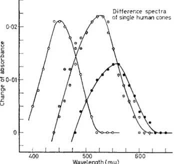
with deep red light, and again after further bleaching with shorter wavelengths. The difference spectrum of the red-sensitive pigment was found to have its maximum at 565-570 nm, that of the green- sensitive pigment at about 535 nm. Both pigments are regenerated with 11-as retinaldehyde, showing that this is their chromophore, joined to different opsins.
Recently, also, it has proved possible to make such measurements in single parafoveal rods and cones in human and monkey retinas [13,14].
Recording spectra in such exceedingly small fields inevitably involves some bleaching of the visual pigments. MARKS et al incorporate a predetermined correction for this factor in their measurements. In our experiments the pigments bleach 20-30 per cent in the course of a single scan. By recording the spectrum in both directions—from red to violet, and in the reverse direction—and averaging the results, one obtains spectra that are not greatly distorted from their proper form and position.
Such measurements of the difference spectra of the visual pigments in single human cones reveal the presence of (at least) three groups of cones, red-, green- and blue-sensitive (Fig. 5). These data provide the first direct evidence of the existence of such groups of cones, reasonably consistent in their properties with the hypothetical components of trichromatic theory. Too few cones have yet been measured in this way to define the average absorption maxima reliably. A central problem awaiting such measurements is whether each type of cone contains a single visual pigment or mixtures of pigments.
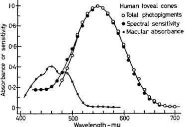
We have also determined, by direct microspectrophotometry, the absorption spectra of human and monkey maculas [12]. Some years ago the human macular pigment was shown to be a carotenoid, apparently lutein or leaf xanthophyll, C ^ H s ^ O H ^ [15]. Monkey maculas appear to possess the same pigment. The entire human or monkey macula yields a typical carotenoid spectrum, with absorption maxima at about 430, 455 and 485 nm. The average absorbance at 455 nm in eight series of measurements of human maculas was 0-49, representing an average absorption at this wavelength of 68 per cent.
When allowance is made for ocular and macular absorptions, the absorption spectrum of the total visual pigments of the human fovea accounts exactly for the foveal spectral sensitivity (the photopic luminosity function) (Fig. 6) [12].
A simple sensory procedure was recently devised for measuring the spectral sensitivities of the human colour vision pigments [16]. The
I40 M O L E C U L A R A N D F I N E S T R U C T U R E O F R E C E P T O R S
Difference spectra
400 500 600 Wavelength (mjj)
FIG. 5. Difference spectra of the visual pigments in single cones of the human parafovea. In each case the absorption spectrum was recorded in the dark from 650 to 380 nm, then again after bleaching with a flash of yellow light. The differences between these spectra are shown. They apparently represent a blue-sensitive cone with about 450 nm, two green-sensitive cones with Am ax about 525 nm, and a red-sensitive cone with Am ax about 555 nm (from BROWN and WALD [14]).
spectrum of the red-sensitive pigment. In effect the blue background illumination selectively bleaches the blue- and green-sensitive pig- ments to such levels that the foveal thresholds are due to the red- sensitive pigment alone. Similarly, exposure to bright yellow light isolates the action spectrum of the blue-sensitive pigment; and exposure to a mixture of red and blue, hence purple, light isolates the spectrum of the green-sensitive pigment.
spectral sensitivity of the dark adapted fovea represents the composite action spectrum of all the foveal photopigments. By performing such measurements on intense coloured backgrounds, one can isolate the action spectra of the individual pigments. So, for example, determining visual thresholds throughout the spectrum, superimposed on a steady background illumination of intense blue light, reveals the action
Measured at the level of the cornea, such action spectra vary from one person to another, owing to differences in ocular and macular transmission. The uncorrected spectral sensitivity curves for the blue-, green- and red-sensitive pigments are maximal at 440-450 nm, 540-550 nm and 575-580 nm (Fig. 7).
FIG. 6. Difference spectrum of the total photopigments of the human fovea (average of 5) compared with the spectral sensitivity of foveal vision, measured as at the level of the cones. T o obtain the latter function, the average photopic luminosity curve was converted to a quantum basis and corrected for ocular and macular transmission.
The spectrum of a human macula is shown at the left. The corrected luminosity curve agrees with the difference spectrum of the foveal photopigments down to about 510 nm. Below this wavelength the difference spectrum falls off owing to the formation of coloured
products of bleaching (from BROWN and WALD [12]).
After correction for ocular and macular transmission, these action spectra become invariant, and have the force of absorption spectra of the colour-vision pigments. Their maxima lie at about 430, 540 and 575 nm (Fig. 7).
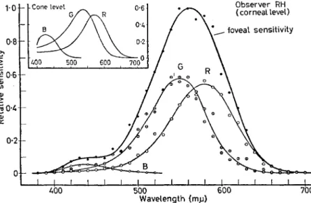
The spectral sensitivities of the three colour-vision pigments, measured at the corneal level, add up to yield the average photopic luminosity curve when their maxima are in the proportions, B : G : R = 0-018:0-58:0-54. Corrected for ocular and macular trans- missions, hence as though at the level of the cones, these ratios become about 0-09:0-59:0-52 (Fig. 7) [16].
Of. J I I I j I ι 1 1 1 1 1 1 1 L_
500 600 700 Wavelength ~mjj
142 M O L E C U L A R A N D F I N E S T R U C T U R E O F R E C E P T O R S
Wavelength (ΗΑΜ)
FIG. 7. Contributions of the individual colour-receptor mechanisms to the total foveal sensitivity of observer R.H. The main graph shows measurements at the corneal level, the inset corresponding curves at the level of the cones. In R.H., who is particularly blue-sensitive, when the total foveal sensitivity is given a maximal height of i-o, the heights of the blue-, green- and red-sensitive curves are as B: G: R = 0-053 : 0*575 10-542 at the corneal level, and 0-2810-59 -0-52 at the level of the cones. In the average observer the blue-component is only about one-third as high (from WALD [16]).
in fact, four major types, the so-called deuteranopes forming two distinct classes [cf. 17].
One mechanism of colour-blindness involves the lack of one of the three colour-vision pigments (hence achromia). Three types of achromia occur, depending upon which of the pigments is missing:
blue-blindness or acyanopia, in which the blue-sensitive pigment is lacking, though this contributes so little to the total spectral sensitivity that the luminosity function remains almost normal ; red-blindness or anerythropia, in which the red-sensitive pigment is absent and the
The spectra of these pigments also account for the photopic lumin- osity curves associated with all forms of human colour-blindness (the dichromias). Though it is usually assumed that human dichromats are of three kinds—protanopes, deuteranopes and tritanopes—there are,
luminosity function depends essentially on the green-sensitive pigment alone; and one type of 'deuteranopia', green-blindness or achloropia, in which the green-sensitive pigment is lacking, and the luminosity function is governed primarily by the red-sensitive pigment alone.
In the other mechanism of colour-blindness (' synchromia ') all three visual pigments are present in more-or-less normal proportions and the photopic luminosity curve is therefore relatively normal, but matters are so arranged that two of the visual pigments excite a single sensation. They might do so through being mixed in one type of cone ; alternatively the neural pathways from two of the three types of cone converge or overlap. This condition is apparent in the second class of ' deuteranopes in whom the photopic luminosity curve is normal, but the red- and green-sensitive pigments stimulate a single sensation, (hence red-green or R - G synchromia). It seems likely, also, that the rare type of colour-blindness described as tetartanopia may involve similar fusions of the red and blue, or the green and blue mechanisms.
It is significant that all types of red-green confusers—in the older terminology protanopes and both classes of deuteranope—apparently see all wavelengths longer than the neutral point, not as green or red, but as yellow. This implies that all such dichromats retain all three central mechanisms of colour response, the simultaneous excitation of the red and green mechanisms yielding a fused sensation of yellow. In that case the essential failure in colour-blindness lies more peri- pherally. In a red-blind the green-sensitive cones, and in a green-blind the red-sensitive cones must excite indiscriminately the central mechanisms for both the red and green sensations. Similarly, in red- green synchromia, through the operation of such mechanisms as suggested above, both pigments stimulate both sensations indis- criminately.
R E F E R E N C E S
ι. More adequate references for this portion of the paper will be found in WALD G . , BROWN P.K. and GIBBONS L R . (1962) In Biological Receptor Mechanisms (J.Beament, ed.), p. 32, Cambridge University Press;
(1963) J. opt. Soc. Amer. 5 3 , 20.
2. DE ROBERTIS Ε . (1960)^. gen. Physiol. 4 3 , Suppl. 2, 1 ; DOWLING J.E. and GIBBONS L R . (1961) In The Structure of the Eye (G.K.Smelser, ed.), p. 85, Academic Press, N.Y.
3. SJÖSTRAND F . S . (1959) Ergebn. Biol. 2 1 , 128; (1959) Revs. mod. Phys.
3 1 , 301. (1961) In The Structure of the Eye (G.K.Smelser, ed.), p. 1, Academic Press, N.Y.
144 M O L E C U L A R A N D F I N E S T R U C T U R E O F R E C E P T O R S
4. BROWN P . K . , GIBBONS I.R. and WALD G . (1963) J. cell Biol. 1 9 , 79.
5. LIEBMAN P.A. (1962) Biophys. J. 2 , 161.
6. HAGINS W.A. and JENNINGS W . H . (i960) Discuss. Faraday Soc. No. 27, 180.
7. HUBBARD R. and KROPF A. (1958) Proc. nat. Acad. Sei. Wash. 4 4 , 130.
8. YOSHIZAWA T . and WALD G . (1963) Nature, Lond. 1 9 7 , 1279 ; (1964) 2 0 1 , 340.
9. MATTHEWS R . G . , HUBBARD R., BROWN P . K . and WALD G . (1963) J . gen.
Physiol. 4 7 , 215.
10. FUTTERMAN S. (1963)^. biol. Chem. 2 3 8 , 1145.
11. WALD G . and BROWN P . K . (1958) Science 1 2 7 , 222.
12. BROWN P . K . and WALD G . (1963) Nature, Lond. 2 0 0 ,3 7 . Also unpublished observations (cf. 16).
13. MARKS W . B . , DOBELLE W . H . and MACNICHOL E.F. Jr. (1964) Science
1 4 3 , 1181.
14. BROWN P . K . and WALD G . (1964) Science 1 4 4 , 45.
15. WALD G . (1945) Science 1 0 1 , 653 ; (1949) Docum. Ophthal. 3, 94.
16. WALD G . (1964) Science 1 4 5 , 1007.
17. WILLMER E.N. (1955) Docum. Ophthal. 9 , 235.