1 23
Strahlentherapie und Onkologie Journal of Radiation Oncology, Biology, Physics
ISSN 0179-7158 Strahlenther Onkol
DOI 10.1007/s00066-019-01513-x
Dosimetric comparison of inverse
optimisation methods versus forward optimisation in HDR brachytherapy of breast, cervical and prostate cancer
Georgina Fröhlich, Gyula Geszti, Júlia Vízkeleti, Péter Ágoston, Csaba Polgár &
Tibor Major
1 23
Commons Attribution license which allows
users to read, copy, distribute and make
derivative works, as long as the author of
the original work is cited. You may self-
archive this article on your own website, an
institutional repository or funder’s repository
and make it publicly available immediately.
ORIGINAL ARTICLE
https://doi.org/10.1007/s00066-019-01513-x Strahlenther Onkol
Dosimetric comparison of inverse optimisation methods versus forward optimisation in HDR brachytherapy of breast, cervical and prostate cancer
Georgina Fröhlich1,2 · Gyula Geszti2· Júlia Vízkeleti1· Péter Ágoston1· Csaba Polgár1,3· Tibor Major1,3
Received: 4 April 2019 / Accepted: 8 August 2019
© The Author(s) 2019
Abstract
Objective Dosimetric comparison of HIPO (hybrid inverse planning optimisation) and IPSA (inverse planning simulated annealing) inverse and forward optimisation (FO) methods in brachytherapy (BT) of breast, cervical and prostate cancer.
Methods At our institute 38 breast, 47 cervical and 50 prostate cancer patients treated with image-guided interstitial high-dose-rate BT were selected. Treatment plans were created using HIPO and IPSA inverse optimisation methods as well as FO. The dose–volume parameters of different treatment plans were compared with Friedman ANOVA and the LSD post-hoc test.
Results IPSA creates less dose coverage to the target volume than HIPO or FO: V100 was 91.7%, 91% and 91.9% for HIPO, IPSA and FO plans (p= 0.1784) in breast BT; 90.4%, 89.2% and 91% (p= 0.0045) in cervical BT; and 97.1%, 96.2% and 97.7% (p= 0.0005) in prostate BT, respectively. HIPO results in more conformal plans: COIN was 0.72, 0.71 and 0.69 (p= 0.0306) in breast BT; 0.6, 0.47 and 0.58 (p< 0.001) in cervical BT; and 0.8, 0.7 and 0.7 (p< 0.001) in prostate BT, respectively. In breast BT, dose to the skin and lung was smaller with HIPO and FO than with IPSA. In cervical BT, dose to the rectum, sigmoid and bowel was larger using IPSA than with HIPO or FO. In prostate BT, dose to the urethra was higher and the rectal dose was smaller using FO than with inverse methods.
Conclusion In interstitial breast and prostate BT, HIPO results in comparable dose–volume parameters to FO, but HIPO plans are more conformal. In cervical BT, HIPO produces dosimetrically acceptable plans only when more needles are used. The dosimetric quality of IPSA plans is suboptimal and results in unnecessary larger active lengths.
Keywords Inverse optimisation algorythms · HIPO hybrid inverse planning optimisation · IPSA inverse planning simulated annealing · Breast brachytherapy · Prostate brachytherapy · Cervical brachytherapy
Dosimetrischer Vergleich von inversen Optimierungsmethoden versus Vorwärtsoptimierung bei Hochdosis-Brachytherapie des Mamma-, Zervix- und Prostatakarzinoms
Zusammenfassung
Ziel Es erfolgte ein dosimetrischer Vergleich von inversen und Vorwärtsoptimierungsmethoden (FO) aus dem Bereich HIPO („hybrid inverse planning optimization“) und IPSA („inverse planning simulated annealing“) in der Brachytherapie (BT) des Mamma-, Zervix- und Prostatakarzinoms.
Methoden In die Studie wurden 38 Mamma-, 47 Zervix- und 50 Prostatakarzinompatienten aufgenommen, die mit bildgesteuerter interstitieller hochdosierter BT behandelt worden waren. Behandlungspläne wurden mittels inversen sowie Vorwärtsoptimierungsmethoden aus dem Bereich HIPO und IPSA erstellt. Die Dosisvolumenparameter der verschiedenen Behandlungspläne wurden durch Friedman-ANOVA („analysis of variance“) und auf die LSD („least significant difference“) bezogene Post-hoc-Tests miteinander verglichen.
Georgina Fröhlich, Ph.D.
frohlich.georgina@gmail.com
1 Centre of Radiotherapy, National Institute of Oncology, Ráth György Street 7–9, 1122 Budapest, Hungary
2 Faculty of Science, Eötvös Loránd University, Pázmány Péter mall 1/A, 1117 Budapest, Hungary
3 Faculty of Medicine, Department of Oncology, Semmelweis University, Ráth György Street 7–9, 1122 Budapest, Hungary
Ergebnisse Bei IPSA wird die Zielvolumenabdeckung durch den Einsatz geringerer Dosis im Vergleich zu HIPO oder FO erreicht; V100 war 91,7%, 91% und 91,9% für HIPO-, IPSA- und FO-Pläne (p= 0,1784) bei Mamma-BT; 90,4%, 89,2%
und 91% (p= 0,0045) bei Zervix-BT sowie 97,1%, 96,2% und 97,7% (p= 0,0005) bei Prostata-BT. Durch HIPO entstehen gleichmäßigere Pläne, der COIN („conformal index“) betrug 0,72; 0,71 und 0,69 (p= 0,0306) bei Mamma-BT; 0,6; 0,47 und 0,58 (p< 0,001) bei Zervix-BT sowie 0,8; 0,7 und 0,7 (p< 0,001) für Prostata-BT. Bei Mamma-BT war die erfasste Dosis in Haut und Lungen mit HIPO und FO niedriger als mit IPSA. Im Rahmen der Zervix-BT war die Dosis in Rektum, Sigma und Darm bei Nutzung von IPSA erhöht im Vergleich zu HIPO oder FO. Bei FO-Anwendungen war die Dosis bei Prostata-BT im Vergleich zu den inversen Methoden an der Urethra erhöht, die Rektaldosis dagegen erniedrigt.
Schlussfolgerung Bei der interstitiellen Mamma- und Prostata-BT erreicht HIPO vergleichbare Dosisvolumen-Parameter- werte wie FO, die HIPO-Pläne sind jedoch deutlich konformaler. Bei Zervix-BT generiert HIPO dosimetrisch akzeptable Pläne nur bei Verwendung von mehreren Nadeln. Die dosimetrische Qualität von IPSA-Plänen ist suboptimal und führt zu unnötiger zusätzlicher Bestrahlung.
Schlüsselwörter Inverse Optimierung algorithmus · HIPO hybrid inverse planning optimisation · IPSA inverse planning simulated annealing · Mamma-Brachytherapie · Prostata-Brachytherapie · Zervix-Brachytherapie
Introduction
In spite of some early publications on inverse optimisa- tion in brachytherapy (BT) [1, 2], inverse dose planning has played a significant role in external beam radiotherapy (EBRT) treatment planning since 2000 [3]. In brachyther- apy these methods have become widespread in the past decade [4]. Beside the reproducibility of the plans, their practical advantage is reduced planning time. However, it needs the accurate setup of the initial preset and it is ben- eficial only if sufficient degrees of freedom are available for the algorithm. Therefore, inverse optimisation works only for interstitial BT treatments, where a large number of needles and source positions are available. Although, the planning time is shorter than with forward optimisation methods, more volumes (i.e. organs at risk) are generally needed. For example, automatic skin contouring is appro- priate for forward optimisation (FO) in interstitial breast BT, but a special 5 mm thick skin shell is needed for inverse methods. Even though inverse methods are currently gener- ally used in several types of interstitial BT, only a few stud- ies with a small number of patients have been conducted on this [5–11]. Comprehensive dosimetric evaluation and comparison with the forward method are still awaited.
Several inverse methods were developed during the past decade, but only two are applied widely. Hybrid inverse planning optimisation (HIPO) is a heuristic, hybrid de- terministic stochastic dose–volume-based inverse optimi- sation method [12]. The stochastic algorithm, called simu- lated annealing, searches the optimal catheter distributions for a given set of dose objectives. The deterministic algo- rithm, called dose–volume histogram-based optimisation, optimizes 3D dose distribution quickly by moving straight downhill once it is in the advantageous region of the search space given by the stochastic algorithm. For optimisation of the dwell times of the radioactive source, the limited-mem- ory Broyden–Fletcher–Goldfarb–Shanno (L-BFGS) quasi-
Newtonian algorithm is used. The given dose–volume con- straints are reached at the same time with the minimalisation of several cost functions.
Inverse planning simulated annealing (IPSA) is a heuris- tic stochastic anatomy-based inverse optimisation method [13]. It is determined by the cost function, which repre- sents the dose prescription and constraints. Since it was implemented for low-dose-rate seed prostate treatments, it optimises the dwell positions of the source, too. Both HIPO and IPSA inverse optimisation algorithms have been imple- mented in Elekta BT treatment planning products (Elekta Brachytherapy, Veendendaal, the Netherlands).
The aim of present study is to analyse the dosimetric effect of HIPO and IPSA inverse optimisation algorithms and compare it to the forward optimisation method in high- dose-rate interstitial BT treatments of breast, cervical and prostate cancer.
Materials and methods
At our institute, 38 breast, 47 cervical and 50 prostate can- cer patients treated with image-guided high-dose-rate inter- stitial BT were selected for this study.
Postoperative multicatheter breast BT implantation was performed using a preimplant CT image set. Based on the postimplant CT, the planning target volume (PTV) and or- gans at risk were created and the treatment plan was nor- malised to the basal dose points using the optimal value of the F-factor and graphical and manual optimisation [11]
were used (Oncentra Brachy v4.5.3, Elekta Brachytherapy, Veendendaal, the Netherlands). The prescribed dose was 30.1 Gy in 7 fractions, twice a day. The detailed descrip- tion of our treatment method can be found in our previous publication [14].
BT boost treatment for cervical cancer was delivered with a combined interstitial intracavitary technique, given
Strahlenther Onkol
Fig. 1 Dose distributions using HIPO (hybrid inverse planning optimisation), IPSA (inverse planning simulated annealing) and forward optimi- sation (FO) in interstitial BT (brachytherapy) of breast (a), cervical (b) and prostate (c) cancer.Red dots:active dwell positions (volumes:red:
PTV [planning target volume];agreen:non-target breast,blue:ipsilateral lung,pink:ribs;byellow:bladder,green:rectum,violet:sigmoid,pink:
vagina;cyellow:urethra,green:rectum)
1 or 2 fractions weekly. Patients were treated with 4 BT fractions of 7 Gy. Initial and post-teletherapy MRI were used to determine the number and position of needles in the ring- or Fletcher-type interstitial applicator. The im- plantation was transrectal US guided. The delineation of PTV, bladder, rectum, sigmoid and bowel was performed on postimplant CT, also using information from post-telether- apy MRI. During treatment planning, graphical and manual optimisation was used to achieve an optimal dose distribu- tion (Oncentra Brachy v4.5.3, Elekta Brachytherapy, Veen- dendaal, the Netherlands). The detailed description of our treatment method can be found in our previous publications [11,15].
Transrectal US-guided transperineal interstitial prostate implantation was performed during the 4 weeks of the EBRT course in a single fraction. After scanning the prostate with US, a virtual preimplant plan was generated (Oncentra Prostate v3.1, Elekta Brachytherapy, Veenden- daal, the Netherlands). The HIPO optimisation method was used, and the prescribed dose was 10 Gy to the whole prostate gland (V100≥95%). Based on this plan, metal needles were inserted into the prostate through a template under live US guidance. The optimisation procedure was used again for the dwell times in the inserted needles to achieve the final dose distribution. The detailed description
of our treatment method can be found in our previous publication [16].
Patients were treated with a high-dose-rate (HDR) re- mote afterloading unit (microSelectron v3, Elekta Brachy- therapy, Veendendaal, the Netherlands) using an Ir-192 stepping source (type v2) with an initial contained activity of 370 GBq (reference air kerma rate of 40.7 mGy * m2/h).
The used source step was 2.5 mm.
Additional treatment plans were created using HIPO and IPSA inverse or forward (graphical and manual) optimisa- tion methods (Fig. 1). To avoid inter-observer variations, only one physicist who is well-experienced in interstitial BT made all the plans. With both inverse methods, our library preset was used first (the used constraints are in Ta- bles4and5in the Appendix), then this initial preset was modified to achieve the optimal dose distribution in each plan. It has to be noted that inverse algorithms can plan the optimal catheter distribution only in the Oncentra prostate planning system. In breast and cervical BT, this feature is not needed, as the dose planning process is not in real-time.
We defined the catheter positions in a simple manual way before implantation and did not use this automatic option in prostate planning neither [11,14–16]. With FO and HIPO, active dwell positions were inside or on the surface of the PTV, while IPSA optimised the source dwell positions and created active dwells outside the PTV, too. Using HIPO, the recommended 0.2 value of the dwell time gradient re- striction was used to modulate the ratio of dwell times of the adjacent dwell positions [17].
The following dose–volume parameters were used for quantitative evaluation of the plans:
V100, V150:the volume of the PTV receiving 100% and 150% of the prescribed dose (%) [18],
V100, V150, VPTV: absolute volume irradiated by 100% and 150% of the prescribed dose (cc) and the volume of the PTV,
D90: the minimum dose delivered to 90% of PTV (Gy) [19],
DNR: dose nonuniformity ratio [20,21]
DHI: dose homogeneity index [22,23],
COIN: conformal index [24],
D2(x): the minimal dose to the most exposed 2 cc ofthe critical organ x(% or Gy) [25],
D1(x): the minimal dose to the most exposed 1 cc ofthe critical organ x(%),
D0.1(x): the minimal dose to the most exposed 0.1 cc of the critical organ x(%),
V50(x): absolute volume tothe critical organ xirradiated by 50% of the prescribed dose (cc), where x isnon-tar- get breast, contralateral breast, skin, lung, heart, blad- der, rectum, sigmoid or bowel.
The dose–volume parameters do not follow the Gaus- sian distribution (F-tests were significant for all parame- ters), so the studied parameters of different treatment plans were compared with non-parametric Friedman ANOVA and the LSD post-hoc test (Statistica 12.3, StatSoft, Tulsa, OK, USA).
Results
Both HIPO and IPSA methods decrease the time of plan- ning process to FO, from 25 to 15 min in breast BT, from 10 to 7 min in cervical BT and from 20 to 10 min on average in prostate BT.
IPSA created less dose coverage to the target volume than HIPO or FO, V100 was 91.7%, 91% and 91.9% for HIPO, IPSA and FO plans (p= 0.1784) in breast BT, 90.4%, 89.2% and 91% (p= 0.0045) in cervical BT and 97.1%, 96.2% and 97.7% (p= 0.0005) in prostate BT, respectively.
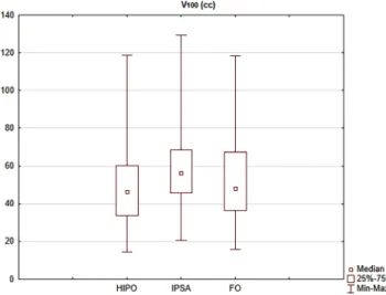
In cervical BT plans, IPSA created larger volumes ir- radiated by the prescribed dose, V100 was 48.5 cc, 59.6 cc and 52.8 cc in HIPO, IPSA and FO plans, respectively (p< 0.001; Fig.2). The volumes irradiated with high doses were also larger in IPSA plans: V150 was 26.7 cc, 30.3 cc and 28.9 cc (p< 0.001), respectively. In the case of breast and prostate BT plans, there were no significant differences in these parameters (p= 0.0806 and 0.1038).
In breast BT, all of the plans were appropriately homo- geneous, DNR was 0.3, 0.3 and 0.29 in HIPO, IPSA and FO plans, respectively (p= 0.1524). In the case of cervical BT, IPSA resulted in the most homogeneous plans, with DNR values of 0.55, 0.50 and 0.54 (p< 0.001), respectively.
HIPO plans were more homogeneous in prostate BT, DHI was 0.7, 0.6 and 0.6 (p< 0.001).
Fig. 2 The absolute volume irradiated by 100% of the prescribed dose (V100) using HIPO (hybrid inverse planning optimisation), IPSA (in- verse planning simulated annealing) and forward optimisation (FO) methods in interstitial cervical BT (brachytherapy) plans
Strahlenther Onkol
Table 1 Dose–volume parameters (mean and range, median) in interstitial breast BT using HIPO (hybrid inverse planning optimisation), IPSA (inverse planning simulated annealing) and forward optimisation (FO)
Dose–volume parameter HIPO IPSA FO p-value
(ANOVA)b
p-value (post hoc)b
Nr. of needles 13.5 (7–28)a – –
VPTV(cc) 60.2 (26.9–173.6) – –
V100 (%) 91.7 (87.6–96.4) 91.0 (88.8–98.3) 91.9 (90.0–96.0) 0.1784 –
V150 (%) 35.5 (25.8–50.9) 33.2 (16.1–60.3) 36.0 (23.0–42.5) 0.0205 HIPO-IPSA: 0.0409 IPSA-FO: 0.0169
DNR 0.30 (0.25–0.45) 0.30 (0.20–0.50) 0.29 (0.25–0.37) 0.1524 –
COIN 0.72 (0.50–0.80) 0.71 (0.50–0.80) 0.69 (0.49–0.82) 0.0306 HIPO-FO: 0.0339
IPSA-FO: 0.0493 V50(non-target breast, cc) 0.8 (0.3–1.2) 1.3 (0.5–1.7) 0.9 (0.3–1.5) 0.0027 HIPO-IPSA: 0.0245
IPSA-FO: 0.0436 D1(contralateral breast,
%)
1.4 (0.1–1.9) 1.6 (0.6–5.8) 1.5 (0.3–2.3) 0.3146 –
D1(skin, %) 19.8 (8.9–26.8) 21.8 (13.2–38.0) 17.1 (4.2–25.7) 0.0425 HIPO-IPSA: 0.0485 IPSA-FO: 0.0210 D0.1(lung, %) 42.7 (8.5–64.0) 57.4 (8.0–68.7) 44.7 (25.0–74.0) 0.0457 HIPO-IPSA: 0.0024
IPSA-FO: 0.0083 D0.1(heart, %) 22.7 (7.5–49.2) 23.7 (6.1–55.6) 22.9 (8.0–41.0) 0.8984 –
V100, V150the volume of the PTV (planning target volume) receiving 100% and 150% of the prescribed dose (%),VPTVthe volume of the PTV, DNRdose nonuniformity ratio,COINconformal index,V50(non-target breast)absolute volume of the non-target breast irradiated by 50% of the prescribed dose (cc),D1(contralateral breast), D1(skin)the minimal dose of the most exposed 1 cc of the contralateral breast and skin (%), D0.1(lung), D0.1(heart)the minimal dose of the most exposed 0.1 cc of lung and heart (%)
amedian dose–volume parameters
bFriedman ANOVA and LSD post-hoc test (italicizedp-values are significant)
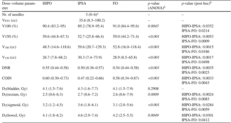
Table 2 Dose–volume parameters (mean and range, median) in interstitial cervical BT (brachytherapy) using HIPO (hybrid inverse planning optimisation), IPSA (inverse planning simulated annealing) and forward optimisation (FO)
Dose–volume param- eter
HIPO IPSA FO p-value
(ANOVA)b
p-value (post hoc)b
Nr. of needles 3 (0–6)a – –
VPTV(cc) 35.6 (8.3–100.2) – –
V100 (%) 90.4 (83.2–95) 89.2 (78.9–95.4) 91.0 (84.4–95.6) 0.0045 HIPO-IPSA: 0.0352 IPSA-FO: 0.0214 V150 (%) 59.6 (44.8–67.3) 52.7 (25.8–66.4) 59.0 (44.2–71.4) <0.001 HIPO-IPSA: 0.0053
IPSA-FO: 0.0009 V100(cc) 48.5 (14.6–118.6) 59.6 (20.7–129.3) 52.8 (16.0–118.4) <0.001 HIPO-IPSA: 0.0015
IPSA-FO: 0.0186 V150(cc) 26.7 (7.8–68.2) 30.3 (7.4–73.9) 28.9 (8.5–65.8) <0.001 HIPO-IPSA: 0.0017
IPSA-FO: 0.0498 DNR 0.55 (0.44–0.58) 0.50 (0.36–0.57) 0.54 (0.44–0.58) <0.001 HIPO-IPSA: 0.0035
IPSA-FO: 0.0023 COIN 0.60 (0.30–0.73) 0.47 (0.22–0.66) 0.58 (0.34–0.87) <0.001 HIPO-IPSA: 0.0033
IPSA-FO: 0.0043
D2(bladder, Gy) 4.1 (1.5–7.6) 4.3 (1.6–7.7) 4.1 (1.5–7.9) 0.2908 –
D2(rectum, Gy) 2.5 (0.6–6.3) 2.7 (0.6–7.2) 2.6 (0.6–7.9) 0.0009 HIPO-IPSA: 0.0024 IPSA-FO: 0.0083 D2(sigmoid, Gy) 3.2 (1.2–4.5) 3.6 (1.8–6.1) 3.1 (2.0–5.6) <0.001 HIPO-IPSA: 0.0284
IPSA-FO: 0.0059 D2(bowel, Gy) 4.1 (1.8–6.2) 4.6 (2.9–7.4) 4.2 (2.5–5.5) 0.0049 HIPO-IPSA: 0.0301
IPSA-FO: 0.0412
V100, V150the volume of the PTV (planning target volume) receiving 100% and 150% of the prescribed dose (%),V100, V150, VPTVabsolute volume irradiated by 100% and 150% of the prescribed dose (cc) and the volume of the PTV,DNRdose nonuniformity ratio,COINconformal index,D2(bladder), D2(rectum), D2(sigmoid), D2(bowel)the minimal dose of the most exposed 2 cc of the bladder, rectum, sigmoid and bowel (Gy)
amedian dose-volume parameters
bFriedman ANOVA and LSD post-hoc test (italicizedp-values are significant)
Table 3 Dose–volume parameters (mean and range, median) in HDR (high-dose-rate) interstitial prostate BT (brachytherapy) using HIPO (hybrid inverse planning optimisation), IPSA (inverse planning simulated annealing) and forward optimisation (FO)
Dose–volume pa- rameter
HIPO IPSA FO p-value
(ANOVA)b
p-value (post hoc)b
Nr. of needles 18 (16–20)a – –
VPTV(cc) 39.5 (20.1–74.0) – –
V100 (%) 97.1 (89.0–99.0) 96.2 (94.8–98.5) 97.7 (97.0–98.5) 0.0005 HIPO-IPSA: 0.0466 IPSA-FO: 0.0237 V150 (%) 30.1 (22.1–37.0) 38.0 (30.5–56.3) 38.7 (22.0–59.9) <0.001 HIPO-IPSA: 0.0011
HIPO-FO: 0.0008
DHI 0.70 (0.61–0.82) 0.60 (0.44–0.73) 0.61 (0.38–0.77) <0.001 HIPO-IPSA: 0.0024
HIPO-FO: 0.0016 COIN 0.82 (0.73–0.91) 0.70 (0.61–0.72) 0.70 (0.42–0.74) <0.001 HIPO-IPSA: 0.0008
HIPO-FO: 0.0003 D0.1(urethra, %) 113.8
(107.0–119.1)
112.6 (107.4–123.9)
124.6 (113.7–140.3)
<0.001 HIPO-FO: 0.0046 IPSA-FO: 0.0022 D2(rectum, %) 57.4 (46.2–91.0) 59.2 (52.4–72.2) 50.5 (34.8–59.4) <0.001 HIPO-FO: 0.0057 IPSA-FO: 0.0011
V100, V150the volume of the PTV (planning target volume) receiving 100% and 150% of the prescribed dose (%),VPTVthe volume of the PTV, DHIdose homogeneity index,COINconformal index,D0.1(urethra)the minimal dose of the most exposed 0.1 cc of the urethra (%),D2(rectum)the minimal dose of the most exposed 2 cc of the rectum (%)
amedian dose–volume parameters
bFriedman ANOVA and LSD post-hoc test (italicizedp-values are significant)
HIPO resulted in more conformal plans, COIN was 0.72, 0.71 and 0.69 (p= 0.0306) in breast BT, 0.6, 0.47 and 0.58 (p< 0.001) in cervical BT and 0.8, 0.7 and 0.7 (p< 0.001) in prostate BT, respectively.
In breast BT, the dose to the skin and lung was smaller with HIPO and FO than with IPSA. D1(skin) was 19.8%, 21.8% and 17.1% (p= 0.0425), D0.1(lung) was 42.7%,
Fig. 3 The active dwell positions in the intracavitary applicator and in the interstitial needles in the case of HIPO (hybrid inverse planning optimisation) and IPSA (inverse planning simulated annealing) optimisation methods.aRed dots:active dwell positions,red dots with yellow background:active dwell positions inside the target volume.bRed dots:active dwell positions,blue:the interstitial intracavitary applicator (volumes:red:PTV (planning target volume),yellow:bladder,green:rectum,violet:sigmoid,pink:vagina)
57.4% and 44.7% (p= 0.0457) in HIPO, IPSA and FO plans, respectively. In cervical BT, the dose to the rectum, sigmoid and bowel was larger using IPSA. D2(rectum) was 2.5 Gy, 2.7 Gy and 2.6 Gy (p= 0.0009), D2(sigmoid) was 3.2 Gy, 3.6 Gy and 3.1 Gy (p< 0.001) and D2(bowel) was 4.1 Gy, 4.6 Gy and 4.2 Gy (p= 0.0049), respectively. In prostate BT, the dose to the urethra was higher in FO plans
Strahlenther Onkol
than in inverse optimised plans, D0.1(urethra) was 113.8%, 112.6% and 124.6% (p< 0.001), respectively. However, the rectal dose was smaller using FO, D2(rectum) was 57.4%, 59.2% and 50.5% (p< 0.001), respectively.
The detailed statistical data are in Table 1 for breast, Table2for cervical and Table3for prostate BT plans.
Discussion
In several types of cancer, image-guided interstitial brachy- therapy has no better alternative at present, including high- tech EBRT as volumetric modulated arc therapy or stereo- tactic radiation therapy [3]. In spite of the fact that inverse dose optimisation is one of the hot topics in EBRT, its role in BT planning has not been evaluated systematically.
Choi et al. compared HIPO and IPSA algorithms for HDR interstitial tongue BT [6]. They found that these meth- ods generate similar dosimetric results; however, the total dwell time with IPSA is 4 s longer than that of HIPO. They used 4–8 needles. Graphical optimisation was needed for the target coverages to satisfy the clinical goal. Then, the total dwell time was increased by approximately 10%.
We found superfluous active lengths in interstitial breast BT cases using the IPSA algorithm. Additionally, despite that the contour of PTV was not a concave shape, there were inactive dwell positions between two active positions.
The coverage of the PTV with the prescribed dose was sig- nificantly lower with IPSA (D90: 101%) than with HIPO (102%) or FO (102.7%), but this difference is not important clinically. The volumes irradiated by a high dose (V150) were larger with HIPO (35.5%) and FO (36.0%) than with IPSA (33.2%), but the conformality was higher with HIPO and IPSA than with FO (COIN: 0.72, 0.71, 0.69, respec- tively). Nevertheless, forward-optimised plans were as ho- mogeneous as inverse plans, DNR was 0.30 with HIPO, 0.30 using the IPSA algorithm and 0.29 with FO. The dose to the skin and lung was significantly lower using HIPO and FO compared to the IPSA method. Overall, FO and HIPO generated dosimetrically acceptable treatment plans in in- terstitial breast BT. If all the necessary volumes of interest are available, HIPO reduces the overall planning time.
Thibault et al. investigated the clinical outcome of in- verse-planned interstitial gynaecological BT [8]. They used a perineal template for implantation of a median of 17 nee- dles and the IPSA algorithm for dose optimisation. They found that the D90 parameter correlates with local tumour control. Matias et al. compared the dosimetric results of FO and HIPO and found that the two methods are dosimet- rically comparable [26]. Trnková et al. compared forward and inverse interstitial cervical BT plans using the obsolete Plato planning system for FO and the special gynaecologic treatment planning system Oncentra Gyn for inverse plan-
ning [9]. They stated that the conformality is the highest using HIPO, and the treatment time is less than in FO and IPSA. IPSA tends to overload the needles and needs addi- tional contours to work. Our present work shows that IPSA generated lower-quality treatment plans than FO and HIPO methods in all the examined dosimetric parameters. IPSA resulted in longer active lengths and inactive dwell positions between active ones, as can be seen in Fig. 3. It resulted in significantly larger volumes irradiated by the prescribed dose (Fig.2) and high-dose volumes (V150). In the optimal case, isodose surface volume correlates with the volume of the PTV [27]. FO and HIPO generated clinically similar dosimetric results, but HIPO needs additional adjustment with FO to reach the dose–volume constraints, especially in the case of a small number of needles. The number of needles correlates with the dosimetric quality of the treat- ment plan, as we showed in our previous study [11].
Pokharel et al. evaluated the HIPO algorithm in HDR prostate BT [28]. They found that HIPO can provide treat- ment plans with comparable target coverage to that of FO with a reduction in dose to the critical structures; how- ever, HIPO resulted lower target coverage compared to FO.
Panettieri et al. compared IPSA and HIPO [10] and stated that IPSA generates large dwell times in particular positions of the catheter, which can be the cause of the resultant lower homogeneity compared to the HIPO method. Poulin et al.
made a comparison of optimisation algorithms in prostate BT and found that dose optimisation engines give similar dosimetric results [29]. Dinkla et al. made a comparison be- tween graphical, IPSA and HIPO optimisation methods in HDR/PDR prostate BT [7]. They found that dose–volume parameters are comparable for all methods, and inverse al- gorithms resulted in shorter planning time than graphical optimisation (6.7 vs. 7.6 min, on average). We also experi- enced reduction of the optimisation time with HIPO and IPSA methods, but the effectiveness of IPSA was sub- optimal: it generated dosimetrically acceptable plans, but the value of all the dose–volume parameters was inferior to using FO or HIPO methods. Additionally, IPSA cre- ated superfluous active lengths outside the prostate besides the underdosed prostate region close to rectum. FO and HIPO resulted in dosimetrically similar plans, but the PTV dose coverage was higher using FO (D90: 112.2% with FO vs. 110.4% with HIPO), the high-dose volumes were smaller with HIPO (V150: 30.1% with HIPO vs. 38.7%
with FO), and HIPO was also more homogeneous (DHI:
0.7 vs. 0.6) and conformal (COIN: 0.8 vs. 0.7, respectively).
The dose to the urethra was lower with HIPO (D0.1: 113.8%
vs. 124.6%), but the rectal dose was higher (D2: 57.4% vs.
50.5%).
Taking every result into account, the IPSA optimisation method resulted in suboptimal treatment plans and used un- necessarily longer active lengths in interstitial breast, cervi-
cal and prostate BT (this superfluous length is usually 1 or 2 dwell positions, so 2.5–5 mm). Using the HIPO algorithm, active dwell positions can be determined before the optimi- sation of the dwell times in postimplant planning (in breast and cervical BT), and there is an option to plan the needles and active dwells inversely during live planning (in prostate BT) based on our predefined rules. Additionally, with the dwell time gradient restriction option, HIPO can produce homogeneous dwell time distribution. In HDR interstitial breast and prostate BT, HIPO can be recommended for dose optimisation; however, FO also results in dosimetrically ac- ceptable plans, but with longer planning time. In the case of HDR interstitial cervical BT, sometimes a small number of needles does not give enough opportunities for inverse optimisation. To achieve the recommended dose coverage of PTV, additional FO is needed. With FO, isodoses can be expanded in the areas where organs at risk are not close to the PTV, while they can be decreased near to these tissues.
Conclusion
In HDR interstitial breast and prostate BT, HIPO results in comparable dose–volume parameters to FO, but HIPO plans are more conformal. FO needed more planning time and more experience of the physicist. In cervical BT, HIPO
produces dosimetrically acceptable plans only if a larger number of needles are used, and in this case the combination of FO and HIPO is recommended. The dosimetric quality of IPSA plans is suboptimal and results in unnecessarily larger active lengths.
Acknowledgements This study would not have been possible without the support of Michael Niekamp (Elekta Brachytherapy, Veendendaal, the Netherlands), who ensured the practice of inverse optimisation al- gorithms for our brachytherapy team.
Funding This paper was supported by the János Bolyai Research Scholarship of the Hungarian Academy of Sciences and the ÚNKP- 18-4 New National Excellence Program of the Ministry of Human Capacities.
Funding Open access funding provided by National Institute of On- cology (OOI).
Conflict of interest G. Fröhlich, G. Geszti, J. Vízkeleti, P. Ágoston, C. Polgár, and T. Major declare that they have no competing interests.
Open Access This article is distributed under the terms of the Creative Commons Attribution 4.0 International License (http://
creativecommons.org/licenses/by/4.0/), which permits unrestricted use, distribution, and reproduction in any medium, provided you give appropriate credit to the original author(s) and the source, provide a link to the Creative Commons license, and indicate if changes were made.
Strahlenther Onkol
Appendix
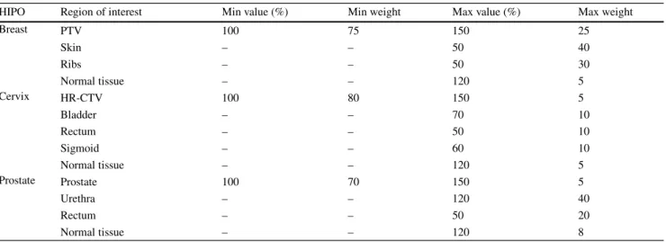
Table 4 Initial dose–volume constraints for the HIPO optimisation algorithms in interstitial breast, cervical and prostate BT
HIPO Region of interest Min value (%) Min weight Max value (%) Max weight
Breast PTV 100 75 150 25
Skin – – 50 40
Ribs – – 50 30
Normal tissue – – 120 5
Cervix HR-CTV 100 80 150 5
Bladder – – 70 10
Rectum – – 50 10
Sigmoid – – 60 10
Normal tissue – – 120 5
Prostate Prostate 100 70 150 5
Urethra – – 120 40
Rectum – – 50 20
Normal tissue – – 120 8
Table 5 Initial dose–volume constraints for the IPSA optimisation algorithms in interstitial breast, cervical and prostate BT
Surface Volume
IPSA Region of interest
Min value (%)
Min weight
Max value (%)
Max weight Min value (%)
Min weight
Max value (%)
Max weight
Breast PTV 100 100 150 1 100 100 150 1
Skin – – 40 40 – – – –
Ribs – – 50 30 – – – –
Non-target breast
– – 120 5 – – 35 100
Cervix HR-CTV 200 100 150 15 200 100 150 10
Bladder – – 70 15 – – 70 15
Rectum – – 50 15 – – 50 15
Sigmoid – – 60 15 – – 60 15
Prostate Prostate 100 5 150 1 100 5 200 0.1
Urethra 100 1 108 3 100 1 108 3
Rectum – – 60 3 – – 60 3
References
1. Pouliot J, Tremblay D, Roy J et al (1996) Optimization of perma- nent 125I prostate implants using fast simulated annealing. Int J Radiat Oncol Biol Phys 36(3):711–720
2. Sloboda RS (1992) Optimization of brachytherapy dose distribu- tions by simulated annealing. Med Phys 19(4):955–964
3. Georg D, Kirisits C, Hillbrand M et al (2008) Image-guided ra- diotherapy for cervix cancer: high-tech external beam therapy versus high-tech brachytherapy. Int J Radiat Oncol Biol Phys 71:1272–1278
4. De Boeck L, Belien J, Egyed W (2011) Dose optimization in HDR brachytherapy: a literature review of quantitative models. HUB Re- search Papers, 2011/32
5. Major T, Polgár CS (2017) Treatment planning for multicatheter interstitial brachytherapy of breast cancer—from Paris system to anatomy-based inverse planning. J Contemp Brachyther 9:89–98 6. Choi CH, Park SY, Park JM et al (2018) Comparison of the
IPSA and HIPO algorithms for interstitial tongue high-dose-rate brachytherapy. PLoS ONE 13(10):205–229
7. Dinkla AM, van der Laarse R, Kaljouw E et al (2015) A com- parison of inverse optimization algorithms for HDR/PDR prostate brachytherapy treatment planning. Brachytherapy 14(2):279–288 8. Thibault I, Lavallée MC, Aubin S et al (2012) Inverse-planned
gynecologic high-dose-rate interstitial brachytherapy: clinical outcomes and dose-volume histogram analysis. Brachytherapy 11:181–191
9. Trnková P, Baltas D, Karabis A et al (2010) A detailed dosi- metric comparison between manual and inverse plans in HDR intracavitary/interstitial cervical cancer brachytherapy. J Contemp Brachyther 2(4):163–170
10. Panettieri V et al (2014) Comparison of IPSA and HIPO inverse planning optimization algorithms for prostate HDR brachytherapy.
J Appl Clin Med Phys 15(6):50–55
11. Fröhlich G, Vízkeleti J, Anhhong NN et al (2018) Dosimetric eval- uation of combined intracavitary-interstitial image-guided adaptive brachytherapy of cervical cancer and comparison with conventional treatment techniques. Magy Onkol 62(4):242–248
12. Lahanas M, Baltas D, Zamboglou N (2003) A hybrid evolutionary multiobjective algorithm for anatomy based dose optimisation al- gorithm in HDR Brachytherapy. Phys Med Biol 48:399–415 13. Lessard E, Pouliot J (2001) Inverse planning anatomy-based dose
optimization for HDR-brachytherapy of the prostate using fast sim- ulated annealing algorithm and dedicated objective function. Med Phys 28(5):773–779
14. Major T, Polgár CS, Lövey K, Fröhlich G (2011) Dosimetric char- acteristics of accelerated partial breast irradiation with CT image- based multicatheter interstitial brachytherapy: A single institution’s experience. Brachytherapy 10:421–426
15. Vízkeleti J, Fröhlich G, Anhhong NN et al (2018) Clinical re- sults of combined intracavitary-interstitial image-guided adaptive brachytherapy in locally advanced cervical cancer. Magy Onkol 62:249–257
16. Fröhlich G, Ágoston P, Lövey J et al (2010) Dosimetric evaluation of high-dose-rate interstitial brachytherapy boost treatments for lo- calized prostate cancer. Strahlenther Onkol 186(7):388–395 17. Mavroidis P, Katsilieri Z, Kefala V et al (2010) Radiobiological
evaluation of the influence of dwell time modulation restriction in HIPO optimized HDR prostate brachytherapy implants. J Contemp Brachyther 2(3):117–128
18. ICRU Report 89 (2013) Prescribing, recording and reporting Brachytherapy for cancer of the cervix. Oxford University Press, Oxford, United Kingdom
19. Dimopoulos JCA, Lang S, Kirisits C et al (2009) Dose-volume his- togram parameters and local control in magnetic resonance image- guided cervical cancer brachytherapy. Int J Radiat Oncol Biol Phys 75:56–63
20. Kovács GY, Hebbinghaus D, Dennert P (1996) Conformal treat- ment planning for interstitial brachytherapy. Strahlenther Onkol 172:469–474
21. Vicini FA, Kestin LL, Edmundson GK, Jaffray DA, Wong JW, Kini VR, Chen PY, Martinez AA (1999) Dose-volume analysis for quality assurance of interstitial brachytherapy for breast cancer. Int J Radiat Oncol Biol Phys 45:803–810
22. Saw CB, Suntharalingam N, Wu A (1993) Concept of dose nonuni- formity in interstitial brachytherapy. Int J Radiat Oncol Biol Phys 26:519–527
23. Wu A, Ulin K, Sternick ES (1988) A dose homogeneity index for evaluating Ir-192 interstitial breast implants. Med Phys 15:104–107 24. Baltas D, Kolotas C, Geramani K, Mould RF, Ioannidis G, Kekchidi M, Zamboglou N (1998) A conformal index (COIN) to evaluate implant quality and dose specification in brachytherapy.
Int J Radiat Oncol Biol Phys 40(2):515–524
25. Georg P, Kirisits C, Goldner G et al (2009) Correlation of dose- volume parameters, endoscopic and clinical rectal side effects in cervix cancer patients treated with definitive radiotherapy including MRI-based brachytherapy. Radiother Oncol 91:173–180
26. Matias LDS, Palmquist T et al (2015) Dosimetric and Radiobio- logical evaluation of hybrid inverse planning and optimization for cervical cancer Brachytherapy. Anticancer Res 35:6091–6096 27. Serban M, Kirisits Ch, Pötter R et al (2018) Isodose surface vol-
umes in cervix cancer brachytherapy: Change of practice from standard (Point A) to individualized image guided adaptive (EM- BRACE I) brachytherapy. Radiother Oncol 129:567–574
28. Pokharel S et al (2013) Evaluation of hybrid inverse planning and optimization (HIPO) algorithm for optimization in real-time, high- dose-rate (HDR) brachytherapy for prostate. J Appl Clin Med Phys 14(4):4198
29. Poulin E et al (2016) Comparison of dose and catheter optimization algorithms in prostate high-dose-rate brachytherapy. Brachytherapy 15:102–111