Comparative analysis of image-guided adaptive interstitial brachytherapy and intensity-modulated arc therapy versus conventional treatment
techniques in cervical cancer using biological dose summation
Georgina Fröhlich, M.Sc., PhD
1, Júlia Vízkeleti, MD, PhD
1, Anhhong Nhung Nguyen, MD
1, Tibor Major, M.Sc., PhD
1, Csaba Polgár, MD, D.Sc.
1,21Centre of Radiotherapy, National Institute of Oncology, Budapest, Hungary, 2Department of Oncology, Faculty of Medicine, Semmelweis University, Budapest, Hungary
Abstract
Purpose: To compare image-guided adaptive interstitial brachytherapy (BT) and intensity-modulated arc therapy (IMAT) with conventional treatment techniques in cervical cancer using an alternative biological dose summation method.
Material and methods: Initially, 21 interstitial BT and IMAT plans of patients with cervical cancer were included and additional plans were created (inverse optimized interstitial, optimized intracavitary, non-optimized intracavitary BT plans, and conformal external beam radiotherapy [EBRT]). The most exposed volume of critical organs in BT were identified manually on EBRT CT images. Biological total doses (EQD2) were calculated and compared between each combination of BT and EBRT plans. This method was compared with uniform dose conception (UDC) in IMAT and conformal EBRT plans.
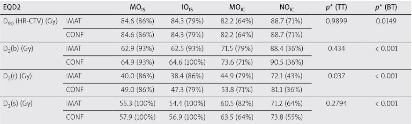
Results: The D90 of high-risk CTV and D2 of bladder and sigmoid were different in BT techniques only: p = 0.0149,
< 0.001, < 0.001, respectively. The most advantageous values were obtained in the interstitial treatment plans and in- verse optimized interstitial plans did not differ dosimetrically from these, while optimized intracavitary plans resulted in worse dose-volume parameters, and the worst of all were intracavitary plans without optimization. The D2 of rec- tum was significantly lower with IMAT than with conformal EBRT plans (p = 0.037) and showed the same trend in BT plans as the other parameters (p < 0.001). The UDC dose summation method overestimated D2 of bladder, rectum, and sigmoid (p < 0.001 for all).
Conclusions: Although optimization improves the quality of conventional BT plans, interstitial plans produce sig- nificantly higher dose coverage of high-risk clinical target volume (HR-CTV) and lower doses to organs at risk (OARs).
IMAT plans decrease the dose to the rectum. UDC overestimates OARs doses.
J Contemp Brachytherapy 2019; 11, 1: 69–75 DOI: https://doi.org/10.5114/jcb.2019.82999 Key words: cervical cancer, dose summation, integrated biological doses, intensity-modulated arc therapy, inter- stitial brachytherapy.
Purpose
The standard of care in the curative treatment of local- ly advanced cervical cancer (stages IB2-IVA) are external beam radiotherapy (EBRT) and intracavitary (ic.) or in- terstitial (is.) brachytherapy (BT) boost with concomitant chemotherapy. Both radiotherapy modalities have devel- oped rapidly, with increasing sophisticated techniques appearing to escalate the dose to the tumor and spare or- gans at risk (OARs). These include intensity-modulated
arc therapy (IMAT) [1] and image-guided adaptive inter- stitial brachytherapy (IGABT) [2,3]. In this situation, an accurate and reliable dose reporting is essential.
The use of BT boost has been linked with pelvic con- trol and overall survival [4]. Adaptive, conformal BT ap- proaches result in further improved clinical outcomes [5], with dose coverage of the target volume (D90, the mini- mum dose delivered to 90% of the high-risk clinical target volume [HR-CTV]) correlating with local tumor control Address for correspondence: Georgina Fröhlich, PhD, Centre of Radiotherapy, National Institute
of Oncology, 7-9 Ráth György St., H-1122 Budapest, Hungary, phone: +36 1 224 8600, fax: +36 1 224 8620,
e-mail: frohlich.georgina@gmail.com
Received: 03.10.2018 Accepted: 06.02.2019 Published: 28.02.2019
[2,6,7], and the minimal dose of the most exposed 2 cc of the OARs with normal tissue toxicity [8,9].
To report these dose-volume parameters properly, overall volumetric doses have to be integrated with EBRT and BT. As simple physical dose summation does not take into consideration the different biological effects, the equivalent dose given in 2 Gy fractions (EQD2) has to be calculated [10,11]. In the GEC-ESTRO recommendations, based on the EMBRACE study [12], the dose distribution of the EBRT is assumed to be completely uniform for the target volume and OARs (uniform dose conception – UDC) [13]. However, the EBRT dose is not always uni- formly distributed. In the intensity-modulated radiother- apy (IMRT) technique, the most exposed 2 cc of the OARs is not a disjunct volume, since its voxels are dispersed in the organ, as we showed earlier [14]. In previous in- vestigations, authors added BT and EBRT dose-volume histogram (DVH) of EQD2 doses [15] or made rigid im- age registration of BT and EBRT CT or MR images [16].
It was also shown before that the most exposed part of the OARs in the integrated plans evolves in the region where the maximum dose is in BT. However, this 2 cc is not in the same location as the most exposed part of EBRT [14]. Therefore, the simple DVH addition method sums the dose of two different 2 cc volumes. The rigid image registration technique does not take into account the de- formation of the regions of interest and from this, doses of different tissues are summarized. Only deformable image registration (DIR) could be an appropriate method to integrate BT and EBRT doses for HR-CTV and OARs, but “currently no DIR program is capable of tracking the location and dose-exposure history of relevant biological structures within the target volumes and OARs” (ICRU report 89) [17]. The main problem is a foreign body (an applicator) in situ, which is not present on EBRT image data sets.
The aim of the present study is to present an alter- native method for adding the biologically effective doses of EBRT and BT in the absence of adequate deformable registration algorithms and compare it to the recent UDC method. Using biological dose summation of the most ex- posed volumes of the critical organs, we compared IMAT and interstitial IGABT versus conventional treatment techniques in cervix cancer.
Material and methods
At our Institute, 21 interstitial IGABT and IMAT plans of patients with cervical cancer were included in this study. Selection criteria was stage IB2-IVA with poor response to EBRT. Patients were examined with pel- vic magnetic resonance imaging (MRI) and staged with computed tomography (CT) or positron emission tomog- raphy-computed tomography (PET-CT) at the beginning, and the therapeutic effect was assessed with an MRI at the end of EBRT. The EBRT was delivered with an ener- gy of 10 MV using 2 full arcs. The prescribed dose was 1.8/50.4 Gy for the whole pelvis. Based on our local IGRT protocol, CBCT verification was made from 1st to 3rd frac- tions; the systematic error was calculated and corrected before the 4th fraction and weekly verification was com-
pleted for patient positioning. EBRT was complemented with BT boost, delivered with combined interstitial-in- tracavitary technique, starting 1 week after EBRT, given 1 or 2 fractions weekly. Because of a given EBRT boost or due to a weak condition, thirteen patients were treated with 4 BT fractions of 7 Gy, five with 3, two with 2, and one with 1 fraction. Initial and post- EBRT MRI were used to determine the number and position of needles in the ring or Fletcher type interstitial applicator [18,19,20]. The implantation was transrectal US-guided. The delineation of HR-CTV of bladder, rectum, sigmoid, and bowel was performed on post-implant CT, using information of post-EBRT MRI. During treatment planning, manual op- timization (MO) was used to achieve an optimal dose dis- tribution. In clinical routine, the EUD method was used to determine the dose constraints for HR-CTV and OARs in remaining BT fractions. The total doses were also cal- culated with this method [21,22].
Besides manual optimized interstitial (MOIS) and IMAT treatment plans, additional BT and EBRT plans were created:
• In inverse optimized interstitial BT plans (IOIS), only the HIPO (hybrid inverse planning optimization) dose-volume-based algorithm was used. The weight of different DVH constraints (cost functions) was tuned to achieve optimal dose distribution;
• Manual optimized intracavitary BT plans (MOIC) were 3D optimized (based on CT), but without us- ing the needles. Dose was prescribed to points A and manual optimization was used;
• Non-optimized intracavitary BT plans (NOIC) dosim- etry was based on points A without optimization;
• Conformal EBRT plans (CONF) used the convention- al 4 field box technique, with 18 MV photon beams.
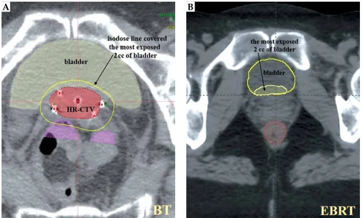
Since the most exposed part of the OARs is in the re- gion where the maximum dose is in BT, the most exposed 2 cc of bladder, rectum, and sigmoid were determined in BT CTs (Oncentra Brachy v. 4.5.3, Elekta Brachythera- py, Veendendaal, The Netherlands). Subsequently, these most exposed 2 cc from BT were manually identified on EBRT CT images for every patient (Eclipse v. 13.7, Varian Medical Systems, Palo Alto, USA) (Figure 1). We inves- tigated BT and EBRT CT image sets on the same moni- tor in axial, sagittal, and coronal views, and delineated 2 cc volume on EBRT CT in the same anatomical place where the isodose surface of the dose of the most exposed 2 cc was found in the BT CT. To reduce subjectivity, one BT expert physicist along with one radiation oncologist (experienced in gynecologic BT) performed this critical part of the investigation.
The total EQD2 doses of these volumes were calcu- lated in each combination of BT and EBRT plans using the linear-quadratic radiobiological model. The α/β of HR-CTV was assumed 10 Gy, while for OARs, 3 Gy was used. The following dose-volume parameters were used for quantitative evaluation of plans:
• D90: the minimum dose delivered to 90% of HR-CTV (Gy);
• D2(x): the minimal dose of the most exposed 2 cc of the critical organ x (Gy),
where x is the bladder (b), rectum (r), or sigmoid (s).
Two-way ANOVA and Fisher-LSD (least significant difference) post-hoc tests were used (Statistica 12.5, Stat- Soft, Tulsa, OK, USA) to compare biological total dose of different treatment combinations:
• IMAT EBRT + MOIS BT, + IOIS BT, + MOIC BT, + NOIC BT;
• CONF EBRT + MOIS BT, + IOIS BT, + MOIC BT, + NOIC BT.
This dose summation method was compared with UDC in combined MOIS BT plans and IMAT, or confor- mal EBRT plans using Wilcoxon-matched pairs test.
Results
The mean volume of the HR-CTV after EBRT (residu- al tumor volume) was 46.1 cc (range, 24.1-100.2 cc). Com- paring different combinations of BT and EBRT plans, we found that D90 of high-risk CTV and D2 of bladder and sigmoid were different in BT techniques only: p = 0.0149,
< 0.001, < 0.001, respectively. The most advantageous val- ues were obtained in the MOIS plans. IOIS plans did not differ dosimetrically from these plans, while MOIC plans resulted in worse dose-volume parameters, and the worst of all were NOIC plans (Table 1). The D2 of rectum was significantly lower with IMAT than with CONF EBRT plans (p = 0.037) and showed the same trend in BT plans as the other parameters (p < 0.001). D2(r) were 40.0 Gy, 38.4 Gy, 44.9 Gy, and 72.1 Gy for IMAT and MOIS, IOIS, MOIC, and NOIC combinations, respectively, and 49.0 Gy,
47.3 Gy, 53.8 Gy, and 81.1 Gy for CONF EBRT and MOIS, IOIS, MOIC, and NOIC plans (Figure 2).
The post-hoc test showed significant differences in all variables between is. (MOIS and IOIS) and ic. (MOIC and NOIC) BT plans, while D2(b) and D2(r) differed between MOIC and NOIC plans.
It was found that the HR-CTV was exposed at least with the recommended EQD2 total dose (85 Gy) in 86% of the patients with the IMAT + MOIS IGABT technique. With the IOIS, MOIC, and NOIC plans, this was only 79%, 64%, and 71%, respectively. The same proportions were derived with CONF EBRT. In 86% of the patients, the D2 rectal dose was below recommended tolerance dose in MOIS BT and IMAT or CONF EBRT plans, while for IOIS BT plans, it was 86% and 79%, for MOIC plans 79% and 71%, and NOIC BT plans only 43% and 36%, respectively (Table 1).
Comparing our dose summation method to UDC in IMAT and CONF EBRT plans, we found that the UDC overestimates D2 of bladder by 12% and 8.5%, rectum by 55% and 26.5% (Figure 3) and sigmoid by 17.2% and 12%, respectively. Detailed EQD2 and p values are presented in Table 2.
Discussion
Brachytherapy boost has a fundamental role in the radiotherapy of locally advanced cervical cancer [23].
Presently, there are no better alternatives [1,24]; however, several high-tech EBRT techniques are possible competi- tors such as conformal [25], image-guided [1] and inten- sity-modulated EBRT [26,27], arc therapy [28], helical to- motherapy [29], and stereotactic radiotherapy with linear accelerators [30,31,32] or with CyberKnife [33].
Fig. 1. The most exposed 2 cc of bladder in BT (A) contoured in EBRT CT (B) in an axial slice
A B
Although MRI-based BT has been considered the
‘gold standard’ by international recommendations [13], in the lack of its broad availability, CT-based contouring and planning can also lead to similar dosimetrical and clinical results with the use of post-EBRT MRI [34,35,36].
The latter case has an advantage over MRI-based BT: im- age registration is easier between post-implant CT and EBRT CT than between post-implant MRI and EBRT CT.
At this moment, DIR is not yet available even between similar imaging modalities. The fundamental reasons are Table 1. Mean EQD2 total doses of different combinations of BT and EBRT plans
EQD2 MOIS IOIS MOIC NOIC p* (TT) p* (BT)
D90 (HR-CTV) (Gy) IMAT 84.6 (86%) 84.3 (79%) 82.2 (64%) 88.7 (71%) 0.9899 0.0149 CONF 84.6 (86%) 84.3 (79%) 82.2 (64%) 88.7 (71%)
D2(b) (Gy) IMAT 62.9 (93%) 62.5 (93%) 71.5 (79%) 88.4 (36%) 0.434 < 0.001 CONF 64.9 (93%) 64.6 (100%) 73.6 (71%) 90.5 (36%)
D2(r) (Gy) IMAT 40.0 (86%) 38.4 (86%) 44.9 (79%) 72.1 (43%) 0.037 < 0.001 CONF 49.0 (86%) 47.3 (79%) 53.8 (71%) 81.1 (36%)
D2(s) (Gy) IMAT 55.3 (100%) 54.4 (100%) 60.5 (82%) 71.2 (64%) 0.2794 < 0.001 CONF 57.9 (100%) 56.9 (100%) 63.5 (64%) 73.8 (55%)
MOIS – manual optimized interstitial, IOIS – inverse optimized interstitial, MOIC – manual optimized intracavitary, NOIC – non-optimized intracavitary BT plans, IMAT – intensity-modulated arc therapy, CONF – conformal EBRT plans, D90 – the minimum dose delivered to 90% of HR-CTV, D2(b), D2(r), D2(s) – the minimal dose of the most exposed 2 cc of bladder, rectum, and sigmoid
In brackets: percentage of plans, which fulfilled the criteria of GEC-ESTRO Recommendation. *2-way ANOVA and Fisher-LSD post-hoc test
MOIS IOIS MOIC NOIC
IMAT EBRT CONF EBRT
Fig. 2. Total EQD2 of the most exposed 2 cc of rectum in combinations of intensity-modulated arc therapy (IMAT) or conformal (CONF) EBRT and manual optimized inter- stitial (MOIS), inverse optimized interstitial (IOIS), manual optimized intracavitary (MOIC), and non-optimized intra- cavitary (NOIC) BT plans
Fig. 3. Total EQD2 of the most exposed 2 cc of rectum in interstitial brachytherapy and intensity-modulated arc therapy (IMAT) or conformal (CONF) EBRT using our dose summation method and using uniform dose concep- tion (UDC)
IMAT Conformal EBRT UDC Median 25-75% Min-Max
Rectum D2 (Gy) Rectum D2 (Gy)
100 90 80 70 60 50 40 30 20 10
100 80 60 40 20 0 EQD2 of the most exposed 2 cc of the rectum
Vertical bars denote 0.95 confidence intervals EQD2 of the most exposed 2 cc of the rectum
Table 2. The EQD2 total doses of interstitial BT plus intensity-modulated arc therapy (IMAT) or conformal (CONF) EBRT plans and the same parameters calculated by the UDC method
D90 (HR-CTV) (Gy) D2(b) (Gy) D2(r) (Gy) D2(s) (Gy)
IMAT 84.6 62.9 40.0 55.3
p* 0.6547 < 0.001 0.0012 0.0033
CONF 84.6 64.9 49.0 57.9
p* 0.6547 < 0.001 < 0.001 0.0081
UDC 84.5 70.4 62.0 64.8
D90 – the minimum dose delivered to 90% of HR-CTV, D2(b), D2(r), D2(s) – the minimal dose of the most exposed 2 cc of bladder, rectum, and sigmoid. *Wilcox- on-matched pairs test
the foreign body (plastic or metal applicator) in situ and the deformation of organs due to application. Other au- thors added BT and EBRT DVHs directly [15] or used rig- id image registration [16] instead of DIR. We mimicked DIR ‘in mind’ by defining the most exposed 2 cc of crit- ical organs in BT CT, and then delineating this volume on EBRT CT. In this way, the addition of biological doses of the same volumes (2 cc) became possible without soft- ware image registration. Obviously, this manual delinea- tion has also limitations such as inter-observer variability.
It is a time-consuming method, and identification of these 2 cc volumes is not trivial with different bladder and rec- tal filling.
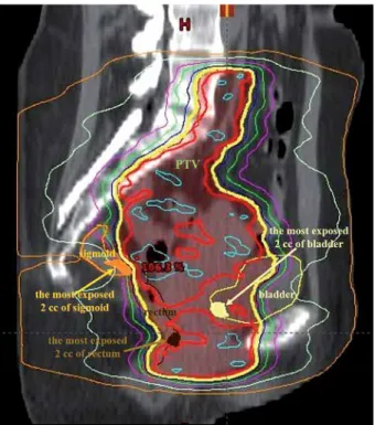
Gelover et al. [15] did not find statistically significant differences between EQD2 doses of OARs in conformal and IMRT EBRT techniques by adding EBRT and BT DVHs; however, they did not add the dose of the same volumes of OARs. In our analysis, D2 (EQD2) of the rectum was significantly lower by 9 Gy (on average) in IMAT than in CONF EBRT plans. This may be due to the fact that the most exposed volumes of OARs are not iden- tical in the IMAT and CONF plans, since the dose of crit- ical organs can be decreased with the IMAT technique, as is shown in Figure 4.
The effect of BT technique on dose-volume param- eters was also investigated in our study. Of note, all examined dosimetric parameters were statistically sig- nificant. The most valuable plans were obtained using MOIS and IOIS, while MOIC resulted in worse dose-vol- ume parameters, and the worst of all were NOIC plans.
Conventional A-point-based (NOIC) plans often resulted in an overdosage of the HR-CTV (D90 88.7 vs. 82.2 Gy) compared to MOIC plans, but with higher doses to OARs. Paul et al. demonstrated that the dosimetric ad- vantages of volume-based intracavitary planning pro- duced more conformal plans than point-A-based plans.
Volume-based plans resulted in a 6-12% reduction in the total dose to 2 cc of the OARs as well as an 8-37%
reduction per BT fraction compared to point-A-based plans [37]. Previous studies have pointed out the strong correlation between local tumor control and D90 of HR- CTV, with the best results above 85 Gy EQD2 [24]. In our case, 86% of the patients received at least this dose during the treatment (IMAT + MOIS BT). Patients who received EBRT boost and fewer fractions of BT are the
‘underdosed’ cases, with a D90 of 79.2 Gy. However, for standard fractionation, D90 is 90.1 Gy (p = 0.0006). As the HR-CT is an integral part of the target volume in EBRT plans and the same constraint was used for IMAT and for CONF EBRT (95% of prescribed dose has to cover 95% of the planning target volume homogeneously), no differences in the coverage of the HR-CTV were noted.
The D2 OARs doses are the predictors of side effects. In 86% of cases, rectal D2 remained below recommended tolerance level during the treatment, though this pro- portion would have been only 43% using NOIC BT, and 36% with NOIC BT and CONF EBRT. Moreover, the ef- fect of the BT technique used is also larger than the EBRT technique. Van de Kamer et al. [16] showed that without EBRT boost, DVH parameter adding is a good approx- imation method, but underestimates D90 by 2.8%. They
used rigid image registration, and only 5 patients were investigated in this study. Our dose summation method showed that UDC overestimates D2 of bladder, rectum, and sigmoid in both EBRT techniques. The maximal de- viation was 22 Gy (D2(r)). There was no difference be- tween plans with or without EBRT boost, but there were only 8 cases of the former.
Overall treatment time (OTT) is another influential factor of tumor control; 85 Gy should be delivered to HR- CTV within 50 days [23]. At our Institute, it takes 61 days (on average), and we are planning to introduce OTT fac- tor into our dose summation technique.
This study is the starting point of the development of an algorithm for the summation of EBRT and BT bi- ologically effective doses, which uses an artificial intel- ligence-based DIR algorithm to match the critical ana- tomical structures in the two radiotherapy modalities.
Further investigations are needed to review whether our method better predicts toxicity than the recent UDC method.
Conclusions
A comparison between interstitial IGABT and IMAT versus conventional treatment techniques in the treat- ment of cervical cancer using our biological dose sum- mation method shows that interstitial optimized BT plans resulted in a significantly higher dose coverage to the HR-CTV and lower doses to the OARs, while IMAT decreases the rectal dose. UDC overestimates the OARs doses compared to a manual definition of D2 in the EBRT plans.
Fig. 4. The most exposed 2 cc of bladder (yellow), rectum (brown), and sigmoid (orange) from BT in a sagittal CT slice in an intensity-modulated arc therapy plan. Red line:
100%; yellow: 95% isodose line
Disclosure
This paper was supported by the János Bolyai Re- search Scholarship of the Hungarian Academy of Scienc- es (Georgina Fröhlich).
Authors report no conflict of interest.
References
1. Georg D, Kirisits C, Hillbrand M et al. Image-guided radio- therapy for cervix cancer: high-tech external beam therapy versus high-tech brachytherapy. Int J Radiat Oncol Biol Phys 2008; 71: 1272-1278.
2. Pötter R, Dimopoulos J, Georg P et al. Clinical impact of MRI assisted dose volume adaptation and dose escalation in brachytherapy of locally advanced cervix cancer. Radiother Oncol 2007; 83: 148-155.
3. Mazeron R, Gilmore J, Dumas I et al. Adaptive 3D im- age-guided brachytherapy: a strong argument in the debate on systematic radical hysterectomy for locally advanced cer- vical cancer. Oncologist 2013; 18: 415-422.
4. Lanciano RM, Martz K, Coia LR et al. Tumor and treatment factors improving outcome in stage III-B cervix cancer. Int J Radiat Oncol Biol Phys 1991; 20: 95-100.
5. Pötter R, Georg P, Dimopoulos JC et al. Clinical outcome of protocol-based image (MRI) guided adaptive brachytherapy combined with 3D conformal radiotherapy with or without chemotherapy in patients with locally advanced cervical can- cer. Radiother Oncol 2011; 100: 116-123.
6. Dimopoulos JCA, Lang S, Kirisits C et al. Dose-volume his- togram parameters and local control in magnetic resonance image-guided cervical cancer brachytherapy. Int J Radiat On- col Biol Phys 2009; 75: 56-63.
7. Dimopoulos JCA, Pötter R, Lang S et al. Dose-effect relation- ship for local control of cervical cancer by magnetic reso- nance image-guided brachytherapy. Radiother Oncol 2009; 93:
311-315.
8. Pötter R, Haie-Meder C, Van Limbergen E et al. Recommen- dations from Gynaecological (GYN) GEC-ESTRO Working Group (II): Concepts and terms in 3D image-based 3D treat- ment planning in cervix cancer brachytherapy – 3D dose volume parameters and aspects of 3D image-based anato- my, radiation physics, radiobiology. Radiother Oncol 2006;
78: 67-77.
9. Georg P, Kirisits C, Goldner G et al. Correlation of dose-vol- ume parameters, endoscopic and clinical rectal side effects in cervix cancer patients treated with definitive radiotherapy including MRI-based brachytherapy. Radiother Oncol 2009;
91: 173-180.
10. Fowler JF. The linear-quadratic formula on progress in frac- tionated radiotherapy. Br J Radiol 1989; 62: 679-694.
11. Nag S, Gupta N. A simple method of obtaining equivalent doses for use in HDR brachytherapy. Int J Radial Oncol Biol Phys 2000; 46: 507-513.
12. EMBRACE study protocol, https://www.embracestudy.dk/
13. Haie-Meder C, Pötter R, Van Limbergen E et al. Recommen- dations from Gynaecological (GYN) GEC-ESTRO Working Group (I): Concepts and terms in 3D image-based 3D treat- ment planning in cervix cancer brachytherapy with emphasis on MRI assessment on GTV and CTV. Radiother Oncol 2005;
74: 235-245.
14. Fröhlich G, Lang S, Berger D et al. Spatial relationship of the 3D dose distribution from brachytherapy and external beam therapy for adding both dose plans in patients with cervix cancer. Brachytherapy 2008; 7: 95.
15. Gelover E, Katherine C, Mart C et al. Patient’s specific inte- gration of OAR doses (D2 cc) from EBRT and 3D image-guid-
ed brachytherapy for cervical cancer. J Appl Clin Med Phys 2018; 19: 83-92.
16. Van de Kamer JB, De Leeuw AAC, Moerland MA et al. De- termining DVH parameters for combined external beam and brachytherapy treatment: 3D biological dose adding for pa- tients with cervical cancer. Radiother Oncol 2010; 94: 248-253.
17. ICRU Report 89. Prescribing, Recording and Reporting Brachytherapy for Cancer of the Cervix. Oxford University Press, 2013.
18. Kirisits C, Lang S, Dimopoulos J et al. The Vienna applicator for combined intracavitary and interstitial brachytherapy of cervical cancer. Design, application, treatment planning, and dosimetric results. Int J Radiat Oncol Biol Phys 2006; 5: 624-630.
19. Fokdal L, Tanderup K, Hokland SB et al. Clinical feasibili- ty of combined intracavitary/ interstitial brachytherapy in locally advanced cervical cancer employing MRI with a tan- dem/ ring applicator in situ and virtual preplanning of the interstitial component. Radiother Oncol 2013; 107: 63-68.
20. Jürgenliemk-Schulz IM, Tersteeg RHA, Roesink JM et al.
MRI-guided treatment planning optimisation in intracavi- tary or combined intracavitary/interstitial PDR brachyther- apy using tandem ovoid applicators in locally advanced cer- vical cancer. Radiother Oncol 2009; 93: 322-330.
21. Fröhlich G, Vízkeleti J, Anhhong NN et al. Dosimetric eval- uation of combined intracavitary-interstitial image-guided adaptive brachytherapy of cervical cancer and comparison with conventional treatment techniques. Magy Onkol 2018;
62: 242-248.
22. Vízkeleti J, Fröhlich G, Anhhong NN et al. Clinical results of combined intracavitary-interstitial image-guided adap- tive brachytherapy in locally advanced cervical cancer. Magy Onkol 2018; 62: 249-257.
23. Fallon J, Park SJ, Yang L et al. Long-term results from a prospective database on high dose rate (HDR) interstitial brachytherapy for primary cervical carcinoma. Gynecol Oncol 2017; 144: 21-27.
24. Tanderup K, Eifel PJ, Yashar CM et al. Curative radiation therapy for locally advanced cervical cancer: brachytherapy is NOT optional. Int J Radiat Oncol Biol Phys 2014; 88: 537-539.
25. Fenkell L, Assenholt M, Nielsen SK et al. Parametrial “boost”
using midline shielding results in an unpredictable dose to tumour and organs at risk in combined external beam ra- diotherapy and brachytherapy for locally advanced cervical cancer. Int J Radiat Oncol Biol Phys 2011; 79: 1572-1579.
26. Yin G, Wang P, Lang J et al. Dosimetric study for cervix carci- noma treatment using intensity modulated radiation therapy (IMRT) compensation based on 3D intracavitary brachyther- apy technique. J Contemp Brachytherapy 2016; 8: 221-232.
27. Lim K, Small W, Portelance L et al. Consensus guidelines for delineation of clinical target volume for intensity-modulated pelvic radiotherapy for the definitive treatment of cervix can- cer. Int J Radiat Oncol Biol Phys 2011; 79: 348-355.
28. Nováková E, Heijkoop ST, Quint S et al. What is the optimal number of library plans in ART for locally advanced cervical cancer? Radiother Oncol 2017; 125: 470-477.
29. Hsieh CH, Tien HJ, Hsiao SM et al. Stereotactic body radia- tion therapy via helical tomotherapy to replace brachyther- apy for brachytherapy-unsuitable cervical cancer patients – a preliminary result. Onco Targets Ther 2013; 6: 59-66.
30. Guckenberger M, Bachmann J, Wulf J et al. Stereotactic body radiotherapy for local boost irradiation in unfavourable lo- cally recurrent gynaecological cancer. Radiother Oncol 2010;
94: 53-59.
31. Gill BS, Lin JF, Krivak TC et al. National cancer data base analysis of radiation therapy consolidation modality for cer- vical cancer: the impact of new technological advancements.
Int J Radiat Oncol Biol Phys 2014; 90: 1083-1090.
32. Mollà M, Escude L, Nouet P et al. Fractionated stereotactic radiotherapy boost for gynecologic tumors: an alternative to brachytherapy? Int J Radiat Oncol Biol Phys 2005; 62: 118-124.
33. Haas JA, Witten MR, Clancey O et al. CyberKnife boost for patients with cervical cancer unable to undergo brachythera- py. Front Oncol 2012; 2: 25.
34. Koh V, Choo BA, Lee KM et al. Feasibility study of toxicity outcomes using GEC-ESTRO contouring guidelines on CT based instead of MRI-based planning in locally advanced cervical cancer patients. Brachytherapy 2017; 16: 126-132.
35. Zolciak-Siwinska A, Kowalczyk A, Sikorska K et al. Com- parison of computed tomography with magnetic resonance imaging for imaging-based clinical target volume contours in cervical cancer brachytherapy. Brachytherapy 2018; 17: 667- 672.
36. Pötter R, Federico M, Sturdza A et al. Value of magnetic res- onance imaging without or with applicator in place for target definition in cervix cancer brachytherapy. Int J Radiat Oncol Biol Phys 2016; 94: 588-597.
37. Paul AG, Nalichowski A, Abrams J et al. Dosimetric evalua- tion of Point A and volume-based high-dose-rate plans: a sin- gle institution study on adaptive brachytherapy planning for cervical cancer. J Contemp Brachytherapy 2018; 10: 202-210.