Application of orthodontic implants in adolescence /Az orthodonciai implantátumok alkalmazása
serdülő életkorban/
PhD Thesis
Gabriella Borsos DDS
Semmelweis University
Clinical Medicine School of Ph. D. Studies
Tutor: András Végh Dr, private professor of SE, PhD
Official reviewers: Katalin Nagy Dr, professor, PhD Zsolt Németh Dr, PhD
Chair of the final examination: Tivadar Zelles Dr, professor, DSc Members of the final examination board: Ildikó Tarján Dr, professor, PhD
Gyula Szabó Dr, professor, PhD
Budapest
2013
1. Introduction
In our times, the beginning of the third century, it is a tendency that while some (mainly infectious) diseases are vanishing, non-contagious, chronic and degenerative illnesses set a constant, ever-growing challenge for medicines due to the so-called ill-effects of civilization. Such illnesses are for example diabetes, hypertension or allergic diseases.
Similarly, the increasing prevalence of malocclusion (misalignment of teeth) is related to the rapid urbanization changes.
The results of the Hungarian epidemological studies emphasise the growing significance of adolescent orthodontic treatment. After the ending of diphyodont replacement and adolescent growth, the preferred treatment is more and more the use of fixed orthodontic appliances that enable precise tooth movements. In this treatment, the orthodontist moves less teeth against the others; the force applied for this aim is undesirable in most cases, although most of the times, it also causes acceptable tooth shifting, as during the execution of every tooth movement – as it stands in the biomechanical principles – an equally intense counterforce needs to be taken into consideration. Anchorage prevents the undesirable tooth movements, fixes the individual teeth in their place, the problem of which is practically as old as the art and practice of orthodontics.
In the last 10-15 years, implants – thanks to their excellent anchorage role and ideal loadibility – are more and more integrated in orthodontic treatments. Insofar, as implants are used based exclusively on their anchorage function in case of closed teeth or closing extraction gaps, the alveolus is not the right place for implantation; therefore alternative topographic-anatomical regions have been chosen inside the area of the cavum oris for the orthodontic implants. Os platinum implants serving exclusively as orthodontic anchorage have been used since the end of the 1980. It became obvious that these orthodontic implants can be easily integrated into the process of orthodontic treatment and they are effectively applicable. Predominantly adult patients participated in the already published studies, whose bone development has purportedly ended.
The aim of the present randomized clinical trial is testing the clinical relevance of palatal implants that are loaded with orthodontic forces in adolescence. At the same time, we performed a histomorphometric examination, in which the immediate loadibility of the implant was tested.
2. Aims
The indication of the present study is that in most orthodontic cases maximum anchorage is actually needed at a young age, since it is commonly known that – in view of tooth movability – bone tissue reactions in the not entirely finished general bone development phase are more beneficial. Moreover, the majority of macclusions have not irreversibly manifested either morphologically or functionally, therefore these interventions can be applied more successfully at a younger age. The goal of the randomized clinical trial is testing the clinical relevance of palatal implants that are loaded with orthodontic forces during the period of the not entirely finished bone development, i.e. adolescence. For comparison (control) we used the orthodontic treatment of a group of young people with traditionally created intraoral dental anchorage and we evaluated them from similar aspects. The clinical relevance of this particular research is that by comparing the two groups it can be certified on EBM (Evidence Based Medicine) II. level whether
o anchorage loss can be avoided with the help of palatal implants
o the application of implantation anchorage decreases treatment time on the whole, which might have a considerable significance from an economical view point.
Furthermore, we have been investigating in in vitro histological studies whether
o enough bone support is available in the sutura palatina medina in the middle of the palatum durum
o the existence of bone integration is histologically certifiable after the removal of the palatal implant
o the existence and extent of bone integration is affected by the healing period – in other words, a 12 weeks recovery time is inevitably necessary before the orthodontic loading.
3. Methods
3.1. Randomized clinical trial
3.1.1. Subjects
This study was approved by the Regional Ethical Research Committee at the Semmelweis University, Budapest (Approval No.: 236/2000) and a formal consent was obtained from the parents or guardians of all patients, after being informed about the experimental protocols.
Thirty patients (mean age: 14.22 ± 1.37 ys) were recruited at the Department of Orofacial Orthopedics and Orthodontics, Heim Pál Children´s Hospital, Budapest, Hungary. The inclusion criteria were: indication of two upper first bicuspids extraction therapy of the
dentoalveolar malocclusion, maximum posterior anchorage requirement in the maxillary arch, post pubertal growth spurt and sufficient palatal bone morphology because of the planned implantation. The following exclusion criteria were applied: poor oral hygiene, parodontitis, unwilling to wear fixed appliances or to have the implant placed. A medical history including gravidity, drug-, nicotine- or alcohol addiction, diabetes, steroid- or chemotherapy,
immunological or haematopoietic diseases, systematic osteopathias, general surgical contraindications, locale radiotherapy were excluded. After the first consultation, the following initial records were obtained from patients who seek orthodontic treatment:
intraoral and extraoral photographs, lateral cephalometric radiographs and
orthopantomograms, as well as study models. Based on initial diagnostics, treatment plans were prepared. When a patient was judged suitable to be involved in the study, he or she and the parents were given information about it. The treatment options of palatal implant or conventional dental anchorage were explained in detail and written information was given to the patients. The patients had a review appointment some weeks later to discuss the individual treatment and the further examinations. If they agreed to enter the trial and signed the consent form, they were allocated to one of the two treatment groups by using randomized blocks of six, so that equal sample sizes could be achieved. Recruitment in this study began in 2001 and continued until 2004. A total of 30 patients (seventeen males and thirteen females) were enrolled. Randomization was performed in a 1:1 ratio in the implant and in the dental anchorage group as well using a balanced design with a block of six.
3.1.1.1. Palatal Implant (PI) group
The palatal implant (PI) group consisted of fifteen patients (mean age: 14.15 ± 1.2 ys): nine males (mean age 14.1 ± 0.91 ys) and six females (mean age: 14.1 ±: 1.67 ys).
In all these patients osseointegrated midpalatal implants (4 mm, Orthosystem®) were used, following the surgical guidelines of the Institute Straumann AG (Waldenburg, Switzerland).
After the suggested three months healing period, impressions were taken using a conventional technique for transferring the implant analogue and the molar bands to a dental cast. A 1.2 mm square stainless steel rigid wire was fixed to the implant abutment as a transpalatal bar connected to the palatal surface of molar bands by laser welding (Fig. 1). The transpalatal bar is purposely square and not round because of its better resistance against orthodontic forces (Wehrbein et al. 2004).
3.1.1.2. Dental anchorage (DA) group
The dental anchorage (DA) group consisted of fifteen patients (mean age: 14.3 ± 1.57 ys) too;
four males, mean age 13.8 years (mean age: 13.8 ± 0.89 ys) and eleven females (mean age:
14.44 ± 1.7 ys). For maximal anchorage the accepted conventional intraoral desmodontal anchorage was provided by a 0.017 x 0.025 inch heat treated stainless steel ‘utility’ arch combined with a Goshgarian type of transpalatal arch (TPA) with distal loop. With this type of anchorage the resistant root surfaces were nearly doubled (Fig. 2). During front retraction the utility arch had to be removed, but the TPA was applied henceforward, as before.
3.1.2. Orthodontic treatment
The orthodontic treatments were exclusively done by two specialist orthodontics. First, bicuspids extractions were undertaken in all cases in the maxilla and often in the lower arch as well when a space analysis suggested that this was required to achieve the treatment objectives. The extractions were carried out after initial alignment, when the anchorage reinforcement was secure. The orthodontic treatment was carried out by using Alexander system (Ormco®) with a 0.018 x 0.025 inch slot. The upper canine brackets and the molar bands contained hooks. After bending/bonding an initial 0.016 inch nickel-titanium aligning wire was placed. The subsequent archwire sequences were 0.016 x 0.016 inch and 0.016 x 0.022 inch nickel-titanium. For the canine retraction a 9 mm long heavy (150 cN) super elastic closed-coil spring (Ormco®) was used beside a 0.016 x 0.022 inch stainless steel segment arch. In the canine portion a gable angle of 45 degrees and an anti-rotation angle of
45 degrees were used for this study in both groups. (Figure 1.) After the extraction gaps were closed, the front retraction was carried out with a 0.016 x 0.022 inch stainless steel contraction-arch containing helical boot loops. 1 mm activation of the boot loops was carried out in every three weeks (app. 80 cN force). Anchorage reinforcement was continued until the mandibular arch was aligned and a super-class I. molar relationship was achieved. At this stage the implant was disconnected from the molar bands. Adjustments of the final occlusal interdigitation and centerlines were carried out by using interocclusal elastics of different strengths and positions, as in common clinical practice. There was no evidence that they were used more frequently in one group than in the other.
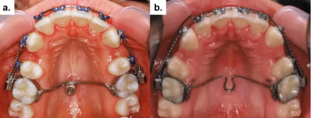
Fig. 1.Anchorage design in the palatal implant group (a) and in the dental anchorage group (b) during canine retraction
3.1.3. Assessed measurements
The main outcome measures were the duration of the orthodontic treatment phases and
cephalometric analysis of maxillary first molar and incisor movement. The mesiodistal widths of the extracted first premolars were registered on the dental casts using a digital gauge.
Blinding of the operator and the patient was not possible in this study, but the cephalometric analysis was carried out anonymously, using an opaque marker in the approximate position of an implant in both groups as described by Benson et al. (Benson et al. 2007). So the assessors were unaware to which treatment group the assessed radiograph belonged. The duration of the orthodontic treatment, as well as each phase of the treatment in both groups were measured.
The beginning of the treatment was defined as T0, start of the canine retraction as T1 and the end of the canine retraction as T2. Front retraction started also as T2, while T3 denoted the end of the front retraction and start of the finishing phase. End of treatment was marked as T4. The orthodontic check-up intervals were 3-4 weeks. At all treatment stages digitalized lateral cephalograms were taken and computerized cephalometric Ricketts-english-modified analysis using the orthodontic diagnostic software FR-WIN® (Computer Konkret AG,
Falkenstein, Germany) were carried out. The landmarks, lines and planes are described and well-known. The main cephalometric measures were analyzed in this study in the following way:
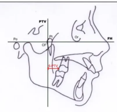
a. Upper 6 to PTV – The distance between the Pterygoid Vertical (PTV) line and the most distal point of the upper first permanent molar’s crown. The PTV line is perpendicular to the Frankfurt Horizontal and goes through the PT point. (Figure 2.) b. Upper 1 to APo – represents the maxillary incisor inclination. The angle is formed by
the long axis of the upper centrals with a line between point A on the maxilla and Pogonion on the mandible.
c. Inter-incisal angle – relates the angular position of the long axes of the upper and lower central incisors to each other.
d. Facial axis – the angle formed by the intersection of the facial axis and basion-nasion line. It indicates the growth pattern of the mandible. An important clinical application of this measurement is that this angle shows little, if any, change (± 1 degree) with growth. Another clinical significance of the facial axis is that the maxillary 1st
permanent molar erupts downward and forward along the facial axis. Any deviation of this path can only be caused by orthodontic treatment. (Analyzed only in T0 and T4.)
Fig. 2. 6-PTV distance on the lateral cephalogram
3.1.4. Statistical analysis
In order to minimise methodological errors, the cephalometric measurements were repeated twice for each lateral cephalogram and the mean of the two measurements used.
We used the Mann-Whitney-Wilcoxon test to compare continuous variables in the two groups. The statistical significance level was set at P < 0.05.
3.2. Histological and histomorphometric examination of the immediate loadibility of palatal implants
3.2.1. Subjects
During the randomized clinical trial, after extending the number of patients who get palatal implants by one, 16 adolescents (mean age: 14,2 ± 1,3 years) – not participating in the clinical study – were chosen for the examination, in which we studied the immediate loadability of palatal implants with histological methods after their removal. In the examined group of three girls and five boys the loading of the implant occurred within 72 hours of the implantation i.e.
“immediately” (n=8, mean age 14,15 ± 1,2 years). In the control group of five girls and three boys (n=8, mean age 14,3 ± 1,5 years) loading of the palatal implant after the presumed osseointegration took place only after 12 weeks following the producer’s recommendation.
Placing the palatal implant, making the dental impression and applying the implant as well as the explantation matched the process described above in all cases.
3.2.2. Sample preparation
Samples were fixed in a 4% paraformaldehyde solution at 4°C. The samples were then prepared in accordance with the procedure described by Donath and Breuner [10]. After desiccating the explant in a series of increasing alcohol concentrations and embedding the sample in cold-polymerised methyl methacrylate (Technovit® 9100, Heraeus-Kulzer, Kulzer division, Werheim, Germany), the sample cubes were cut along the longest diameter of the implant using a precision band saw (Exakt Apparatebau GmbH, Norderstedt, Germany).
Using a precision splicer (Exakt Apparatebau GmbH, Norderstedt, Germany), the plane- ground outer surface of the sample blocks was spliced to both sides of a plane-parallel Perspex mount using cyanoacrylate (Technovit® 7210 VI-C) and again cut into approx.
180 µm slices using the band saw. The sections were, where possible, cut through the implant in such a way that it was possible to visualise the full length of the implant.
The so-obtained sections were further reduced to 90 to 130 µm using a grinding machine (Exakt Apparatebau GmbH, Norderstedt) and irradiated in a Faxitron® cabinet x-ray machine at 13 - 14 kV tube voltage and 2.5 mA for 2.5 minutes. In order to achieve the highest possible resolution, the distance between the object and the film was close to zero. The acquired microradiographs were digitised using a scanner and saved as 12-bit grey-scale uncompressed jpg files. In order to carry out histological analysis, it was necessary to reduce the sections further to a thickness of 30 µm using the grinding procedure described above.
3.2.3. Histomorphometry
The thin sections were stained using toluidine blue O to allow better microscopic analysis of the bone implant contact. Mineralised hard tissues are visualised as unstained or pale blue;
cells, cell nuclei, osteoid seams, collagen fibres and soft tissues are visualised in various shades of blue.
The samples were analysed using image processing software (Bioquant Osteo, BIOQUANT Image Analysis Corporation, Nashville, USA). Microscope images were captured using a high-resolution digital camera and transferred to the computer workstation. Using colour gradation, the software automatically measures contact regions for various tissues and materials and displays them in false colour. The area of the bone in apposition to the implant was determined visually and the bone-implant contact percentage was then calculated using a saved algorithm.
In order to minimise methodological errors, the measurements were repeated twice for each implant and the mean of the two measurements were used.
3.2.4. Statistical analysis
Bio-statistical analysis was performed using version 7 of statistics software application Axum® (Insightful Corporation, Seattle, WA, USA). For metrical data, descriptive statistics show the arithmetic mean and, as a measure of variability, the standard deviation. A t-test for unrelated samples with a significance level of 0.05 (95%) was used for an exploratory comparison of the results between the two study groups.
4. Results
4.1. Randomized Clinical Trial
There was no difference in the size of the premolars measured in both groups. In the PI group it was 7.32 (±0.33) mm and in the DA group 7.23 (±0.59) mm.
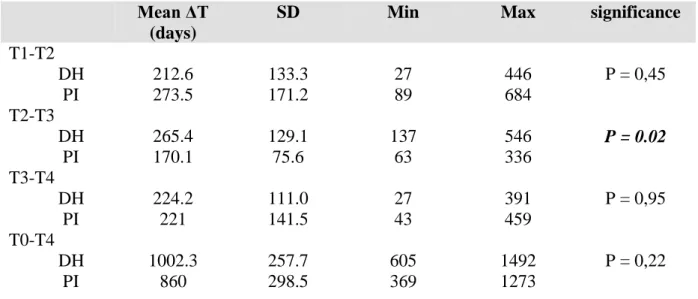
Table 1 shows the results of the descriptive statistics of the duration of the orthodontic treatment step by step. There is a non-significant difference between the two groups in the canine retraction time, which was about 9 months in the PI versus 7 months in the DA group.
But a significant difference (P = 0.02) was found in the duration of the front retraction (T2- T3). The front retraction took 5 and a half months in the PI group and nearly 9 months in the DA group. The total treatment time (T0-T4) in the PI group was 860 days on average, which
is 2 years and 9 months, while in the control group the treatment took 1000 days, i.e. 5 months more, but this difference was statistically not significant.
The results of the upper molar mesial movement are shown in Table 2. The changes in the upper 6-Ptv distance showed no difference during the canine retraction phase between the groups, but there was a significant difference during the front retraction (P = .0 039).
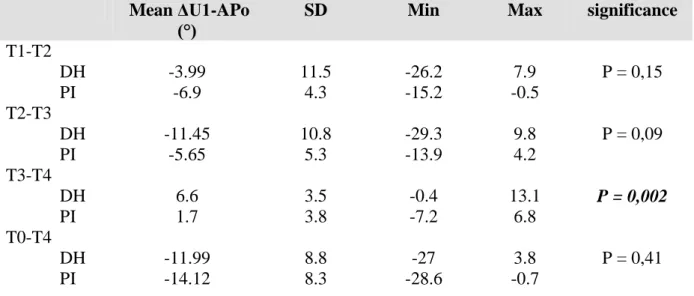
In Table 3, we present the maxillary incisor movement - the tipping of the centrals related to the A-Pogonion plane. During the first phase the incisors in the DA group tipped less - nearly not at all - distally, than in the PI group. In contrast to this, in the front retraction period the distal tipping of incisors was more than double than in the PI group, although this difference was not statistically significant (P = 0.1). However, the same changes in the angulations of the central incisors during finishing (T3-T4) were significantly different (P = 0.002).
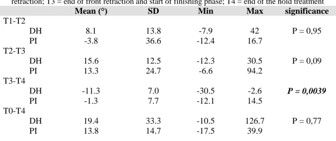
Table 4 shows the results of interincisal angle’s change. No difference was found during the canine retraction and front retraction phases between the groups, but there was a significant difference during finishing (P = 0.039). During the whole treatment no significant difference was detected.
There was no change in the facial axis in the groups during the whole treatment (Table 5).
Table 1: Descriptive statistics of the orthodontic treatment duration (ΔT)
T0 = start of treatment; T1 = start of canine retraction; T2 = end of canine retraction and start of front retraction;
T3 = end of front retraction and start of finishing phase; T4 = end of the hold treatment
Mean ΔT (days)
SD Min Max significance
T1-T2 DH PI
212.6 273.5
133.3 171.2
27 89
446 684
P = 0,45 T2-T3
DH PI
265.4 170.1
129.1 75.6
137 63
546 336
P = 0.02
T3-T4 DH PI
224.2 221
111.0 141.5
27 43
391 459
P = 0,95 T0-T4
DH PI
1002.3 860
257.7 298.5
605 369
1492 1273
P = 0,22
Table 2: Descriptive statistics of the upper molar mesial movement (ΔU6-PTV)
T0 = start of treatment; T1 = start of canine retraction; T2 = end of canine retraction and start of front retraction;
T3 = end of front retraction and start of finishing phase; T4 = end of the hold treatment
Mean ΔU6-PTV (mm)
SD Min Max significance
T1-T2 DH PI
1.48 1.57
1.56 1.06
0 0
5.1 3.29
P = 0,049 T2-T3
DH PI
1.26 0.59
0.93 0.74
0 0.2
3.6 2.2
P = 0,039
T3-T4 DH PI
0.89 1.52
0.77 1.69
0 0
2.1 5.5
P = 0,55 T0-T4
DH PI
4.28 4.19
1.5 2.7
1 0
6.6 10.5
P = 0,51
Table 3: Descriptive statistics of the maxillary incisor movement (ΔU1-APo)
T0 = start of treatment; T1 = start of canine retraction; T2 = end of canine retraction and start of front retraction; T3 = end of front retraction and start of finishing phase; T4 = end of the hold treatment
Mean ΔU1-APo (°)
SD Min Max significance
T1-T2 DH PI
-3.99 -6.9
11.5 4.3
-26.2 -15.2
7.9 -0.5
P = 0,15 T2-T3
DH PI
-11.45 -5.65
10.8 5.3
-29.3 -13.9
9.8 4.2
P = 0,09 T3-T4
DH PI
6.6 1.7
3.5 3.8
-0.4 -7.2
13.1 6.8
P = 0,002
T0-T4 DH PI
-11.99 -14.12
8.8 8.3
-27 -28.6
3.8 -0.7
P = 0,41
Table 4: Descriptive statistics of the interincisal angle´s change
T0 = start of treatment; T1 = start of canine retraction; T2 = end of canine retraction and start of front retraction; T3 = end of front retraction and start of finishing phase; T4 = end of the hold treatment
Mean (°) SD Min Max significance
T1-T2 DH PI
8.1 -3.8
13.8 36.6
-7.9 -12.4
42 16.7
P = 0,95 T2-T3
DH PI
15.6 13.3
12.5 24.7
-12.3 -6.6
30.5 94.2
P = 0,09 T3-T4
DH PI
-11.3 -1.3
7.0 7.7
-30.5 -12.1
-2.6 14.5
P = 0,0039
T0-T4 DH PI
19.4 13.8
33.3 14.7
-10.5 -17.5
126.7 39.9
P = 0,77
Table 5: Descriptive statistics of the facial axis change T0 = start of treatment; T4 = end of the active treatment
Mean (°) SD Min Max significance
T0-T4 DH PI
-0.29 0.04
1.7 1.9
-3 -2.8
2.8 3.8
P = 0,86
4.2. Histomorphometry
Of 16 implants, all were positionally stable after insertion and healed with no complications and with no significant peri-implant inflammation or mucosal hyperplasia. Implant insertion and explantation after conclusion of the treatment were graded as not very unpleasant by the majority of patients. With the use of adequate analgesia, all patients reported that the
intervention was predominantly pain-free.
The 16 implants examined histomorphologically all showed adequate osseointegration. The bone remodelling process was largely complete and the bone-implant interface showed lamellar bone. In the immediate loading group, peri-implant bone contact up to the shoulder of the implant was demonstrable for four out of the eight implants. In two cases there was vertical bone loss to the depth of the second screw thread. This was also seen in one case in the control group.
Histomorphometric analysis of the osseointegrated implants showed higher bone-implant contact rates (BIC) in the group which received conventional treatment with a mean value of 73.1% and a standard deviation of 19.8%.
The figure for bone-implant contact (BIC) in the immediate loading group was 55 ± 21.6%.
The t-test for unrelated samples showed that the difference in bone-implant contact values between the two groups (p=0.1661) was not statistically significant.
5. Conclusions
Based on our results, it is now proven that palatal implants can also be used in adolescents for the enhancement of posterior anchorage. In this randomized clinical trial significant
difference can be detected in regards to the movement of individual teeth and tooth sets during the treatment phases. However, considering the end results, there is no significant difference between the dental anchorage method fixed to the palatal implant and the traditional intraoral one. At the same time, the application of the palatal implant has some advantages, as it eliminates the undesired side effects of the dental anchorage, and hereby seemingly shortens the treatment period as well. The treatment period can be further reduced by placing the implants in an immediate (within 72 hours) indirect anchorage unit.
It turned out during the in vitro examination that there is enough bone support available in the sutura palatina mediana distalis in the middle of the palatum durum for a 4mm long implant, since after the removal of the palatal implant the creation of a high degree of bone integration was histologically verifiable in all cases. Our histomorphometric studies confirm the clinical observation that, according to the previous protocol, the three-month healing period does not indicate any advantage in view of the stability of the implant, the successfulness of the treatment, or the patients’ conformity.
Based on our experience we arrived at the conclusion that the definition of “maximal anchorage” should be reformulated in accordance with the new skeletal anchorage devices.
Further studies are necessary to prove the clinical benefits of the palatal implant, preferably with larger sample size and probably with another indication field. Such a promising indication field for example is the correction of anchorage loss with bilateral molar distalization, during which the immediately loaded palatine implant serves as direct anchorage.
6. New statements
Previous research has only shown in certain cases the clinical use of the palatal implant as anchorage. Although some randomized clinical trials integrating adults have been published (Benson et al. 2007, Sandler et al. 2008, Spivalovsky et al. 2010) that compares the palatal implant to a strongly compliance-dependent extraoral device (headger). Also, an RCT (Feldman et al. 2008) comparing four different anchorage methods that involve 120 patients was published. This was the first such EBM II. level research – starting in 2000 – that uses traditional intraoral anchorage in case of adolescents taking into consideration the maximal anchorage indication in the control group.
We did not manage to prove the original hypothesis saying that PI ensures a higher level of anchorage than a traditional anchorage protocol. Although significant difference can be detected in the movement of individual teeth and tooth sets during the treatment phases, considering the end results, there is no such significant difference between the effectiveness of the dental anchorage method fixed to the palatal implant and the traditional intraoral one.
However, the application of the palatal implant has some advantages, as it eliminates the undesired side effects of the dental anchorage, and hereby seemingly shortens the treatment period as well, especially if we use the immediate loading of the PI, which – as our studies indicate – does not have any negative influence on osseintegration.
7. Publication list
7.1. Catalogue of the most significant scientific publications connected to the PhD theses in peer-reviewed journals
Borsos G, Vokó Z, Gredes T, Kunert-Keil C, Vegh A. (2012) Tooth movement using palatal implant supported anchorage compared to conventional dental anchorage. Ann Anat, 194(6):
556-60. IF: 1,861
Borsos G, Jianu R, Vegh A. (2011) Comparison of bone-borne and tooth tissue-borne
anchorage during maxillary canine retraction in growing patients: a randomised clinical trial.
TMJ, 61:(1-2): 98-101
Borsos G, Rudzki-Janson I, Stockmann P, Schlegel KA, Végh A. (2008) Immediate loading of palatal implants in still-growing patients: a prospective, comparative, clinical pilot study. J Orofac Orthop, 69(4): 297-308
Végh A, Jianu R, Borsos G, Ogodescu A. (2006) The use of palatal implant for posterior anchorage in extraction cases by adolescent patients. Case report. TMJ, 56: 3-7
7.2. Catalogue of the most significant scientific publications not closely related to the PhD theses in peer-reviewed journals:
Gedrange T, Gredes T, Gredes M, Allegrini M.R.F, Borsos G, Vegh A, Salles MB, Heinemann F, Dominiak M, Allergrini JR. S. (2009) Comparative Animal Study on Hard Tissue Integration and Bone Formation of Different Nobel Biocare Implants. J Physiol Pharmacol, 60(Suppl 8): 38-56 IF: 1,489
Gedrange T, Mai R, Mack F, Zietek M, Borsos G, Vegh A, Spassov A, Gredes T. (2008) Evaluation of shape and size changes of bone and remodelled bone substitute after different fixation methods. J Physiol Pharmacol, 59(Suppl 5): 87-94.
Nagy Krisztián, Nagy A. Péter, Borsos Gabriella, Végh András, Maurice Y. Mommaerts, Becse Miklós. (2004) A distractiós osteogenesis szerepe az arcközép sebészetében (TPD műtéttechnika és esetismertetés) Fül-,Orr-, Gégegyógyászat, 50(3): 235-260
Hably C, Borsos G, Bartha J. (1994) Different effects of naproxen on the organ blood flows in normo- and hypervolemic anaesthetized rats. Acta Physiol Hung, 82(3): 267-79.
7.3. Catalogue of scientific publications related to the PhD theses in not peer-reviewed journals
Borsos G, Jianu R, Végh A. (2007) Disto-palatinale Positionierung des Gaumenimplantates bei Jugendlichen. Kieferorthopaediesche Nachrichten, Nr.11.: 6-7
Borsos G, Végh A. (2005) A disztopalatinálisan behelyezett osszeointegrált szájpadimplantátum. Implantológia, 2 évf. 1: 50-53
Végh A, Borsos G. (2004) Az ortodonciai célból ideiglenesen a palatumba helyezett osseointegrált implantátum és annak klinikai alkalmazása. Implantológia, 1 évf. 2: 32-36 Borsos G, Végh A. (1999) Implantátumok alkalmazása az orthodonciában. Dental Hírek,3 évf. 6. 10-11
7.4. Citable congress abstracts published in peer-reviewed journals
Borsos G, Gedrange T, Kunert-Keil C, Vegh A. (2012) Osseointegrated Palatal Implant Supported versus Conventional Dental Anchorage in Adolescents. Eur J Orthod, 34:(5) p.
e81.
Borsos G, Gedrange T, Végh A. (2010) Palatal Implant Supported Versus Conventional Dental Anchorage in Adolescents. Randomized Controlled Trial. Eur J Orthod, 32:(6) p. e5.
Borsos G, Gedrange T, Végh A. (2010) Osseointegrated Palatal Implant Supported Versus Conventional Dental Anchorage in Adolescents. Randomized Controlled Trial. World J Orthod, 11:(Suppl.)
Vegh A, Borsos G. (2008) Immediate loading of orthodontic palatal implants with masticatory forces J Dent Res 87(Spec Iss B):1223. (www.dentalresearch.org)
Borsos G, Vegh A. (2008) Early versus conventional loading of palatal implants with masticatory forces ABSTRACTS OF LECTURES AND POSTERS • EUROPEAN ORTHODONTIC SOCIETY, 84th Congress Lisbon, Portugal 2008, 10–14 June Eur J Orthod, 30: e52
Borsos G, Schlegel KA, Rudzki-Janson I, Vegh A. (2007) Osseointegrated palatal implant versus dental anchorage in adolescents. RCT study. ABSTRACTS OF LECTURES AND POSTERS • EUROPEAN ORTHODONTIC SOCIETY, 83rd Congress Berlin, Germany 2007, 20–24 July Eur J Orthod , 29: e5– e6
Vegh A, Schlegel KA, Borsos G. (2006) Palatal implant anchorage vs. tooth-connected anchorage: a randomized controlled study J Dent Res, 85(Spec Iss C): 0463,
(www.dentalresearch.org).
Borsos G, Schlegel KA, Vegh A. (2006) Stability of palatinal implant supported vs. tooth- connected anchorage in adolescents. ABSTRACTS OF LECTURES AND POSTERS • EUROPEAN ORTHODONTIC SOCIETY, 82nd Congress Vienna, Austria 2006, 4–8 July Eur J Orthod, 28: e188 – e189
Borsos G, Végh, A. (2006) Comparison of the Palatal Implant and a Conventional Tooth- Connected Anchorage System in Adolescents Fogorv. Szle. 99: 244-245.
Borsos G, Vegh A. (2005) Maxillary canine retraction using bone-borne versus tooth-borne anchorage in growing patients World Journal of Orthod, 6: 83-84.
Vegh A, Borsos G. (2005) How to apply palatal implants in difficult anchorage cases World J Orthod, 2005; 6: 327
Borsos G, Végh, A. (2003) Clinical Investigation method of Conventional Dental Anchor Versus Palatal Implant Used in Adolescent Extraction Orthodontic Therapy with Maximal Anchorage Indication Fogorv. Szle. 99: 239.
Végh A, Borsos, G.: (2003) Method of Use and Comparative Evaluation of Anchorage Implants in Palate Fogorv. Szle. 99: 239.