MODERN TECHNIQUES FOR EXTERNAL BEAM RADIATION THERAPY OF PROSTATE CANCER:
NEW METHODS AND CLINICAL RESULTS
PhD thesis Jorgo Kliton
Pathologic Sciences Doctoral School Semmelweis University
Supervisor: Prof. Csaba Polgár, MD, DSc Ágoston Péter, MD, PhD
Official reviewers: Bellyei Szabolcs, MD, PhD, med. habil.
Bánfi Gergely, MD, PhD
Head of the Complex Examination Committee: Prof. Péter Nyirády, MD, DSc Members of the Complex Examination Committee: Csilla Katona, MD, PhD
András Horváth, MD, PhD
Budapest 2019
1
Introduction
Based on the available evidence, treatment options for non-metastatic prostate cancer include: radical prostatectomy, external beam radiation therapy (EBRT), brachytherapy (BT), combination of the above and in selected cases active surveillance. Depending on the risk group, radiation therapy may be supplemented by hormone therapy that reduces the effect of circulating androgens on tumour cells.
In almost every Hungarian radiotherapy department three-dimensional, conformal radiation planning (3D-CRT) has been replaced by inverse planning and intensity modulated radiation therapy (IMRT). With IMRT the prescribed dose follows more closely the shape of the target volume, thereby improving the target coverage and reducing the dose to the organs at risk. More precise radiation planning requires more accurate dose delivery. In modern radiotherapy centres, the so-called image guided radiotherapy (IGRT) has become a part of clinical practice in recent years. IGRT is a radiation treatment method that involves repeated imaging in the treatment room, in order to obtain information about patient positioning and the exact 3-dimensional location of the target volume. The use of IGRT improves biochemical tumor control and allows higher dose delivery to the clinical target, without increasing toxicities to surrounding normal tissues. Various technical solutions have been developed for the implementation of IGRT, the most common is kilovoltage (kV) or megavoltage (MV) cone beam CT (CBCT).
In patients with prostate cancer undergoing IGRT a very precise setup correction can be made with gold markers implanted into the prostate and detected by X-rays. Gold markers are usually implanted by urologists or radiologists under ultrasound control, through the rectum, similar to a prostate biopsy. Due to the nature of the procedure, rectal bleeding, inflammation of the prostate or infection may occur. For gold marker based IGRT electronic portal imaging device (EPID) is used. The position of the markers during treatment corresponds to the position of the prostate. The method is simple to implement and does not require CBCT, so it can be used on less advanced linear accelerators with an appropriate software.
Several randomized trials have shown improved biochemical control with dose escalation for prostate cancer. The rapid technical progress makes it possible to escalate the irradiation dose and to increase the fractional dose. The α/β ratio for prostate cancer is suggested to be lower than that of surrounding normal tissues, and may be as low as 1.5Gy.
Consequently the therapeutic outcome using external beam radiotherapy is expected to be
2
improved with hypofractionation when higher biologically effective dose (BED) is delivered than with conventional external beam radiotherapy. As α/β ratio for rectum and urinary bladder is estimated to be 3 Gy and 5-10 Gy, respectively, the low α/β ratio of prostate cancer theoretically allows dose escalation with hypofractionation without increasing late toxicity.
Beyond the advantages in terms of tumour control and late toxicity, the use of large dose per fraction is preferred by patients and may have important implications for cost-effectiveness by shortening the overall treatment time. Several contemporary phase III, randomized trials with mature data have confirmed similar tumour control and late toxicity among various hypofractionated regimens to conventionally fractionated external beam radiotherapy.
Independently of the patients risk groups, except one trial, the treatment target volume was the prostate gland and +/- seminal vesicles without pelvic lymph nodes. The rational of elective whole pelvis radiotherapy (WPRT) in patients with high risk of subclinical lymph node involvement (Roach equation ≥15%) is the possibility to improve loco-regional control.
Large, contemporary retrospective trials confirmed a statistically significant benefit in biochemical control with WPRT compared to prostate (+/- irradiation of seminal vesicles) irradiation, only. However, currently there is insufficient evidence to recommend routine use of WPRT for high risk patients. Three randomized trials have shown no benefits for WPRT with respect to disease-free or overall survival.
It has been hypothesized that, by using transperineal gold marker implantation compared to conventional transrectal approach, the risk of infection, prostate inflammation and rectal bleeding can be significantly reduced. The clinical results and the toxicity of moderate hypofractionation are similar to those of conventional fractionation, but the overall treatment time is shorter, therefore, we aimed to introduce moderate hypofractionation into clinical practice. In our high-risk patients under seventy years of age, elective pelvic lymph node radiation treatment was considered to be expedient due to the potential cure of occult metastases. Therefore, a simultaneous integrated "boost" technique was performed, treating the pelvic region by conventional fractionation, with a 2,5 Gy and 2,05 Gy fraction dose of the prostate and seminal vesicles for moderate hypofractionation.
In my dissertation I introduce the implementation of the gold marker based IGRT in Hungary, analyse the side effects caused by the implantation and determine the safety margin required for the application of the method. I describe the introduction of moderately hypofractionated external beam radiation therapy with SIB technique for prostate cancer patients in Hungary and report the results with the treatment of the first consecutive 162 patients.
3
Aims
1. To implement the gold marker implantation for prostate radiotherapy and to review the peri- and postoperative complications and side effects of the intervention in patients with prostate cancer.
2. To introduce the gold marker based IGRT and to determine the CTV-PTV safety margin for external beam prostate irradiation.
3. To implement IG-IMRT using SIB to the prostate with moderate hypofractionation in prostate cancer patients.
4. To evaluate 30-month clinical outcomes, acute and late toxicities of the first 162 patients treated with SIB technique.
Materials and methods
Gold marker implantation
About 30 minutes before the procedure every patient received 100 mg contramal and 5 mg metoclopramid intramuscular injection. Patients were laid down in lithotomy position. In each case three radiopaque, gold-plated markers (Best Medical International, Springfield, VA, USA, 1.0 mm x 3.0) were implanted transperineally into the prostate. Under transrectal ultrasound guidance using transversal and longitudinal views, and a template, through the perineum 2 fiducial markers were inserted bilaterally into the base of the prostate gland and the third marker was placed into the apex. The treatment planning CT scan was performed at least 1 week after the marker insertion to allow for any oedema or haemorrhage to settle. To evaluate the tolerance and the peri- or postoperative complications, every patient prior to implantation received a questionnaire about the pain, dysuria, urinary frequency, nycturia, rectal bleeding, haematuria, haematospermia, pain duration after the implantation, fever and inflammation symptoms caused by the implantation. The pain caused by the intervention was scored on a 1-10 scale, where 1 was a very weak and 10 was an unbearable pain. The survey inquired about the comparison of the pains caused by implantation and previous prostate biopsy.
4 CTV-PTV safety margin
In order to calculate the CTV-PTV margin in gold marker based IGRT 34 patients were enrolled. For patient set-up 4 points were tattooed on the skin of the patients, which were marked with metal markers during the planning CT. Based on these tattoos, during treatment planning a so-called reference point was determined. Patient positioning was made by adjusting the patient's tattooed reference skin points with the side and longitudinal lasers.
Based on the treatment plan, the table was moved according to the difference between the reference point and the isocentre in the x, y and z directions to match the lasers point to the isocentre. Digitally reconstructed X-ray images (DRRs) were created from anterior (0 degree) and lateral (90 degrees) directions according to the planning CT and sent to the linear accelerator’s control computer. Immediately before the irradiation, kV or MV X-rays were taken, showing the gold markers' position in the prostate.
a, b,
Figure 1. 0-degree, kilovoltage (kV) image taken with an Electronic Portal Imaging Device (EPID) fused with a Digitally Reconstructed X-ray (DRR) image before (a) and after (b) correction. The blue contour represent the 3 gold markers on the DRR image and the red contour show the prostate planning volume (PTV-PROS).
Subsequently, two pairs of images were manually matched and the inaccuracy of prostate positioning was determined based on the position of the markers in three directions.
The set-up inaccuracy was corrected by manually shifting the treatment couch. After manual image alignment, the prostate positioning errors in the lateral (LAT), longitudinal (LONG), and vertical (VERT) directions were recorded. We determined systematic (Σ) and random (σ) error in the target volume setting for the patient population (p = 34). The required CTV-PTV safety margin (SM) was calculated in the LAT, LONG and VERT directions using the van Herk formula:
SM = 2,5 x Σpop+ 0,7 x σpop
5
Moderate hypofractionation with simultaneously integrated “boost” technique
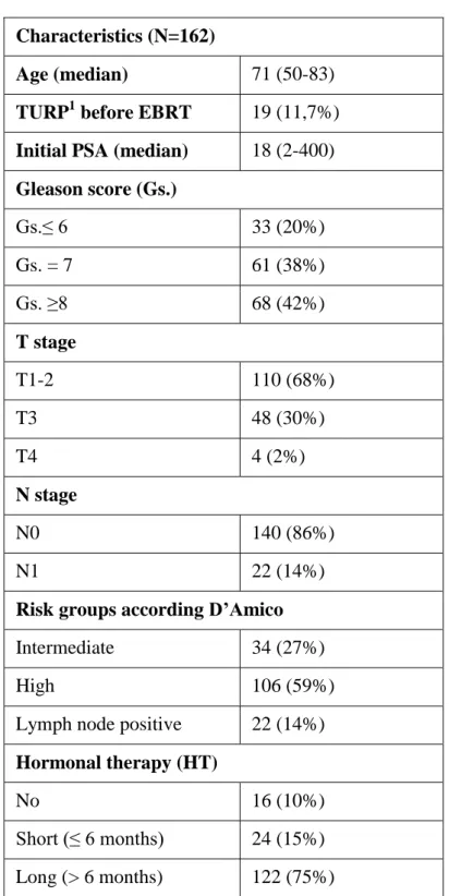
Between January 2015 and March 2017, 162 patients with histologically confirmed adenocarcinoma of the prostate were enrolled in this trial. The main characteristics of the patients are shown in Table 1.
Table 1. Patient, tumour and treatment characteristics Characteristics (N=162)
Age (median) 71 (50-83)
TURP1 before EBRT 19 (11,7%) Initial PSA (median) 18 (2-400) Gleason score (Gs.)
Gs.≤ 6 33 (20%)
Gs. = 7 61 (38%)
Gs. ≥8 68 (42%)
T stage
T1-2 110 (68%)
T3 48 (30%)
T4 4 (2%)
N stage
N0 140 (86%)
N1 22 (14%)
Risk groups according D’Amico
Intermediate 34 (27%)
High 106 (59%)
Lymph node positive 22 (14%) Hormonal therapy (HT)
No 16 (10%)
Short (≤ 6 months) 24 (15%) Long (> 6 months) 122 (75%)
1TURP: transurethral resection of the prostate; 2EBRT: external beam radiotherapy
6
Target volumes were defined according to risk groups published by D’Amico et al.All patients participating in this study were intermediate or high risk or node positive. High risk and node positive patients received whole pelvic irradiation. According to our institutional protocol in high risk patients older than 70 years, considering the toxicity/benefit ratio, prophylactic WPRT was omitted, in the lack of high level of evidence proving better biochemical control or disease free survival after WPRT. Intermediate risk patients with bulky disease or unfavourable histological parameters received neoadjuvant and concomitant hormone therapy for 6 months (short-term hormone therapy). In high risk patients neoadjuvant-concomitant-adjuvant hormone therapy was given for 24-36 months (long-term hormone therapy).
For intermediate risk 2 clinical target volumes were defined. The prostate clinical target volume (CTV_pros) included the whole prostate gland. The prostate and seminal vesicles CTV (CTV_psv) was generated by 5 mm expansion of CTV_pros in all directions except posteriorly at the prostate-rectum interface + proximal 1 cm of the seminal vesicles.
For high risk patients CTV_pros was the same as above. CTV_psv was defined by 5 mm expansion of CTV_pros in all directions except posteriorly + proximal 2 cm of seminal vesicles (in case of cT3b the entire seminal vesicles were included). Whole pelvis clinical target volume (CTV_pelv) consisted of CTV_psv + common iliac (under L5-S1 space), external iliac, presacral and obturator lymph nodes. The rectum, bladder and hip joints were contoured as organs at risk (OARs).
Image guidance was performed using fiducial markers when pelvic irradiation was not required and with mega- or kilovoltage cone beam CT (MV or kV CBCT) in case of WPRT.
According to our institutional analysis, PTV margins with the use of fiducials and CBCT were 5 mm and 8 mm in every direction, respectively.
Intermediate risk patients received 70 Gy to PTV_pros in 2.5 Gy/fraction (EQD2=80 Gy) and 57.4 Gy to PTV_psv in 2.05 Gy/fraction (EQD2=58.2 Gy). High risk and lymph node positive patients received 70 Gy to PTV_pros, 57.4 Gy to PTV_psv and 50.4 Gy to the pelvic lymph nodes using 1.8 Gy/fraction.
Biochemical relapse free survival (bRFS) and cancer specific survival (CSS) curves were calculated using the Kaplan-Meier method. Spearman rank order and Chi-squared tests were used to evaluate the correlations between pelvic irradiation, method of image guidance, transurethral resection of prostate (TURP), hormone therapy, different dosimetric parameters of rectum, bladder, hip joints and acute GI, GU toxicities. Kaplan-Meier method and Gehan- Wilcoxon tests were used to evaluate the effects of pelvic irradiation, method of image
7
guidance, TURP, hormone therapy on toxicity free survival. Logistic linear and Cox regressions were used in multivariate analysis to evaluate the prognostic factors of acute and late toxicities.
Results
Between November 2011 and April 2016 gold markers were implanted into the prostate gland of 300 intermediate and high risk prostate cancer patients. The total implantation procedure required approximately 10 minutes. Despite only the first patient received a prophylactic antibiotic, none of the patients developed fever or infection. Based on the questionnaires none experienced dysuria or rectal bleeding. Neither urinary frequency nor nycturia worsened after implantation. Among the 300 patients studied, 12 (4%) had haematospermia, 43 (14%) haematuria, which lasted for an average of 3.4 and 1.8 days, respectively. Haemorrhage stopped spontaneously, no specific treatment was required. The average pain score was 4.6 (range: 0-9) on a 1-10 scale. Of 300 patients 87 (29%) felt any pain after the intervention, which took an average of 1.5 days (range: 1-5), but none of the patients needed analgesics due to that pain. Compared to the pain felt during biopsy 105 patients (35%) reported less, 80 patients (27%) more, and 94 patients (31%) equal amount of pain. Twenty-one patients, who had a biopsy performed under general anaesthesia, did not answer this question. During the treatments no significant marker migration (>0.5 cm) was observed. The characterization of marker migration is out of the scope of this study.
CTV-PTV safety margin with gold marker based IGRT
To determine the CTV-PTV safety margin, the first 34 gold marker implanted prostate cancer patients were enrolled for definitive external radiotherapy alone. 2010 pre-treatment verifications were performed and a total of 6030 errors were evaluated. Prostate positioning inaccuracies based on gold markers were calculated from the collected data, separated into systematic and random errors. According to our calculations determined by systematic and random errors using the Van Herk's formula, the size of the CTV-PTV safety margin was 0.5 cm in the LAT, VERT and LONG directions.
8
Results after moderately hypofractionated external radiation therapy with SIB technique Of 162 patients, 156 (96%) completed the moderately hypofractionated radiotherapy with SIB technique as planned. Seventy-eight patients (48.2%) were treated with WPRT, 84 patients (51,8%) with no WPRT. Six patients (4%) received only 27 fractions (67.5 Gy to the prostate in 2.5 Gy fractions). Median follow-up time was 30months (range: 21-45). IGRT was performed with fiducial markers, kV CBCT or MV CBCT in 33%, 61% and 6% of the patients, respectively. “Step and shoot” IMRT and VMAT techniques were applied in 6% and 94%, respectively.
At a median follow-up of 30 months crude rate of biochemical relapse free (Figure 2), cancer specific and overall survival was 98%, 99%and 95%, respectively No in-field lymph node recurrence was observed. Three patients (2%) developed multiplex bone metastases, currently one of them is receiving chemotherapy, 2 patients (1%) died due to disease progression. Six (4%) patients died from non-prostate cancer disease.
Acute toxicities are reported in Table 2.
Table 2. Acute toxicities after hypofractionation with SIB technique (n=162).
Toxicity Acute genitourinary n (%)
Acute gastrointestinal n (%)
Grade 0 18 (11) 44 (27)
Grade 1 50 (31) 82 (51)
Grade 2 92 (57) 34 (21)
Grade 3 2 (1) 2 (1)
Grade 4 0 (0) 0 (0)
In our patients, severe early side effects were rare, most of acute toxicity resolved spontaneously or with the administration of medications. Acute grade 2 and 3 GI adverse events occurred in 21% and 1%, respectively. The bleeding proctitis (GI grade 3) passed spontaneously and did not require surgical intervention. Grade 2 and grade 3 GU toxicity was reported in 92 (57%) and 2 (1%) cases, respectively. Grade 3 GU was a complete retention of urine, followed by transurethral resection of prostate (TURP) six months after treatment.
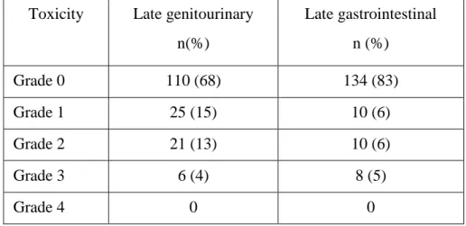
Cumulative late side effects are shown in Table 3.
9
Table 3. Cumulative late toxicities after hypofractination with SIB technique (n=162).
Toxicity Late genitourinary n(%)
Late gastrointestinal n (%)
Grade 0 110 (68) 134 (83)
Grade 1 25 (15) 10 (6)
Grade 2 21 (13) 10 (6)
Grade 3 6 (4) 8 (5)
Grade 4 0 0
In our study, the incidence of cumulative drug-dependent grade 2 GU and GI late toxicities were 13% and 6%, respectively. Late grade 3 UG and GI side effects occurred in 4% (6 cases) and 5% (8 cases), respectively. Grade 3 genitourinary toxicity in 3 patients was urinary retention, which required TURP surgery, and partial incontinence, as a consequence.
One patient underwent 3 times endourethral incision due to urethral stricture. Two patients had to undergo bladder rinsing due to radiogenic bleeding cystitis and had to use anti- haemorrhagic drugs, and since then they have had no complaints. Of the patients with late grade 3 GI toxicity (bleeding proctitis), 1 had to undergo surgery for persistent rectal bleeding. After surgery, the bleeding ceased, and at the last control the patient was free of complaints. In the other five patients, despite medication, bleeding symptoms are still present.
Two patients developed faeces incontinence due to radiation treatment.
In general, urinary toxicity resolved spontaneously or with the administration of non- steroidal anti-inflammatory or α1 blocker medications. Six patients (4%) presented grade 3 late GU toxicity and 8patients (5%) grade 3 late GI toxicity. None of the patients experienced acute or late grade 4 side effects.
Significant correlation was found between acute GI and cumulative late GI and between acute GU and cumulative late GU toxicities (p<0.05).We found no correlation between acute or late toxicity and the use of WPRT, previous urological surgery or modality of IGRT. No correlation was found between WPRT vs. no WPRT, method of image guidance, TURP vs. no TURP, length of hormone therapy or the different dosimetric parameters of rectum, bladder, hip joints and acute GI, GU toxicities. For other parameters significant effects on toxicity were not found. In multivariate analysis no significant predictor was found for acute or late GI or GU toxicities.
10
Conclusions
1. As part of a prospective study, first in Hungary we introduced the gold marker implantation for image-guided external beam radiation therapy of prostate cancer. Since then the method has been routinely applied in our clinical routine.
2. During implantation rate of perioperative and postoperative complications were low, and no permanent damage was observed. Haematospermia and haematuria was uncommon, symptoms stopped spontaneously and no specific treatment was required. No patient experienced fever, infection or sepsis, without the use of prophylactic antibiotics. Based on the questionnaires none experienced dysuria or rectal bleeding. Because of the pain, the intervention did not have to be postponed, none of the patients needed analgesics due to the pain. Compared to the pain felt during biopsy 2/3 of patients reported less or equal amount of pain.
3. Using the daily IGRT with gold markers, the CTV-PTV safety margin was reduced from 8 mm to 5 mm. Target volume reduction may decrease the incidence of side effects.
4. In 2015 we were the first in Hungary to perform moderate hypofractionation with SIB technique for prostate radiotherapy. Since then this fractionation scheme has been routinely used in our department.
5. With moderate hypofractionation, the biochemical relapse-free survival of 162 of our patients was 98% at a median follow-up of 30 months, which corresponds to the published data. Despite elective pelvic irradiation of high-risk patients, the incidence of acute and late GI side effects was acceptable. Acute grade 3 UG, GI toxicity was observed in 0.6%. Due to late grade 2 GU and GI toxicities, 13% and 6% of patients were treated with drug medication.
Because of grade 3 late GU and GI toxicity, 4% and 5% of patients underwent invasive medical treatment. No Grade 4 side effects occurred. The rate of radiogenic side effects caused by our treatments are similar to literature data.
6. Significant correlation was found between acute GI and cumulative late GI and between acute GU and cumulative late GU toxicities. The pelvic irradiation did not increase the rate of GI side effects. No correlation was found between method of image guidance, previous urologic surgery or length of hormone therapy and frequency and severity of GI, GU toxicities.
11
List of publications
Publications related to the theses:
1. Jorgo K, Ágoston P, Major T, Polgar Cs. (2017) Transperineal gold marker implantation for image-guided external beam radiotherapy of prostate cancer. A single institution, prospective study. Strahlenther Onkol, 193:452–458. IF: 2,459 2. Jorgo K, Polgar Cs, Major T, Stelczer G, Herein A, Pocza T, Gesztesi L. Agoston P.
(2019) Acute and late toxicity after moderate hypofractionation with simultaneous integrated boost (SIB) radiation therapy for prostate cancer. A single institution, prospective study. Pathol Oncol Res, (Epub ahead of print) DOI: 10.1007/s12253-
019-00623-2 IF : 2,433
3. Jorgo K, Ágoston P, Major T, Polgár Cs. (2012) Kilovoltos CT-vel végzett betegbeállítás vizsgálata prosztatarákos betegek képvezérelt külső besugárzásakor.
Magy Onkol, 56:193-198.
4. Jorgo K, Ágoston P, Szabó Z, Major T, Polgár Cs. (2014) A prosztatába ültetett aranymarkerek alkalmazásának bevezetése prosztatarákos betegek képvezérelt sugárkezeléséhez. A beültetés okozta mellékhatások ismertetése. Magy Onkol, 58:182- 187.
5. Major T, Agoston P, Jorgo K, Polgár C. (2012) Képvezérelt sugárterápia klinikai alkalmazása daganatos betegek külső besugárzásánál. Magy Onkol, 56:258-65.
Other publications:
1. Major T, Agoston P, Fröhlich G, Baricza K, Szabo Z, Jorgo K, Herein A, Polgar Cs.
(2014) Loose versus stranded seeds in permanent prostate brachytherapy: dosimetric comparison of intraoperative plans. Phys Med, 30:909-913. IF: 2,403 2. Major T, Polgár C, Jorgo K, Stelczer G, Ágoston P. (2017) Dosimetric comparison between treatment plans of patients treated with low-dose-rate vs. high-dose-rate interstitial prostate brachytherapy as monotherapy: Initial findings of a randomized clinical trial. Brachytherapy, 16:608-615. IF:2,227
12
3. Küronya Z, Sükösd F, Varga L, Bíró K, Gyergyay F, Géczi L, Nagyiványi K, Jorgo K, Szarvas T, Kovács Á, Laczó I, Varga Z, Pósfai B, Pepó J, Maráz A. (2019) ERG expression can predict the outcome of docetaxel combined with androgen deprivation therapy in metastatic hormone-sensitive prostate cancer. Urol Oncol, 37:289.e1-289.e9
IF: 2,863
4. Jorgo K, Polgár Cs, Major T, Stelczer G, Tenke P, Kovács G, Ágoston P. (2017) Izominvazív hólyagrák képvezérelt sugárkezelése intravesicalisan befecskendezett lipiodolos jelöléssel. A hólyagmegtartó kezelés új lehetősége. Orv Hetil, 158(51):
2041–2047 IF: 0,322
5. Jorgo K, Ágoston P, Jánváry L, Gesztesi L, Stelczer G, Kontra G, Major T, Polgár Cs.
(2019) Kis és közepes kockázatú prosztatarákos betegek sztereotaxiás sugárkezelése CyberKnife gyorsítóval: Korai radiogén mellékhatások. Magy Onkol, 63:52–59.
6. Ágoston P, Jorgo K, Mátrai Z, Polgár C. (2014) A végtag és a törzsfelszín lágyrész szarkómáinak sugárkezelése. Magy Onkol, 58:65-76.
7. Ágoston P, Jorgo K, Mátrai Z, Polgár C. (2014) A sugárterápia szerepe a retroperitoneális lágyrész szarkómák kezelésében. Magy Onkol, 58:77-82.
8. Herein A, Ágoston P, Szabó Z, Jorgo K, Markgruber B, Pesznyák C, Polgár C, Major T. (2015) Intraoperatív és posztimplantációs dozimetria összehasonlítása permanens implantációs prosztata-brachyterápiával kezelt betegeknél. Magy Onkol, 59:148-53.