doi:10.1002/ejhf.711
Prevention of the development of heart failure with preserved ejection fraction by the
phosphodiesterase-5A inhibitor vardenafil in rats with type 2 diabetes
Csaba Mátyás
1* , Balázs T. Németh
1, Attila Oláh
1, Marianna Török
1, Mihály Ruppert
1, Dalma Kellermayer
1, Bálint A. Barta
1, Gábor Szabó
2,
Gábor Kökény
3, Eszter M. Horváth
4, Beáta Bódi
5, Zoltán Papp
5, Béla Merkely
1, and Tamás Radovits
11Experimental Research Laboratory, Heart and Vascular Center, Semmelweis University, Városmajor u. 68,1122, Budapest, Hungary;2Department of Cardiac Surgery, University of Heidelberg, Heidelberg, Germany;3Institute of Pathophysiology, Semmelweis University, Budapest, Hungary;4Department of Physiology, Semmelweis University, Budapest, Hungary; and5Division of Clinical Physiology, Faculty of Medicine, University of Debrecen, Debrecen, Hungary
Received 20 July 2016; revised 21October 2016; accepted 9 November 2016 ; online publish-ahead-of-print19 December 2016
Aims Heart failure with preserved ejection fraction (HFpEF) has a great epidemiological burden. The pathophysiological role of cyclic guanosine monophosphate (cGMP) signalling has been intensively investigated in HFpEF. Elevated levels of cGMP have been shown to exert cardioprotective effects in various cardiovascular diseases, including diabetic cardiomyopathy. We investigated the effect of long-term preventive application of the phosphodiesterase-5A (PDE5A) inhibitor vardenafil in diabetic cardiomyopathy-associated HFpEF.
...
Methods and results
Zucker diabetic fatty (ZDF) rats were used as a model of HFpEF and ZDF lean rats served as controls. Animals received vehicle or10 mg/kg body weight vardenafil per os from weeks 7 to 32 of age. Cardiac function, morphology was assessed by left ventricular (LV) pressure–volume analysis and echocardiography at week 32. Cardiomyocyte force measurements were performed. The key markers of cGMP signalling, nitro-oxidative stress, apoptosis, myocardial hypertrophy and fibrosis were examined. The ZDF animals showed diastolic dysfunction (increased LV/cardiomyocyte stiffness, prolonged LV relaxation time), preserved systolic performance, decreased myocardial cGMP level coupled with impaired protein kinase G (PKG) activity, increased nitro-oxidative stress, enhanced cardiomyocyte apoptosis, and hypertrophic and fibrotic remodelling of the myocardium. Vardenafil effectively prevented the development of HFpEF by maintaining diastolic function (decreased LV/cardiomyocyte stiffness and LV relaxation time), by restoring cGMP levels and PKG activation, by lowering apoptosis and by alleviating nitro-oxidative stress, myocardial hypertrophy and fibrotic remodelling.
...
Conclusions We report that vardenafil successfully prevented the development of diabetes mellitus-associated HFpEF. Thus, PDE5A inhibition as a preventive approach might be a promising option in the management of HFpEF patients with diabetes mellitus.
...
Keywords Vardenafil • cGMP • Diabetic cardiomyopathy • Diastolic dysfunction • Cardiomyocyte stiffness
*Corresponding author. Tel:+361458 6810, Fax:+361458 6842, Email: csaba.matyas@gmail.com
© 2016 The Authors.European Journal of Heart Failurepublished by John Wiley & Sons Ltd on behalf of European Society of Cardiology.
This is an open access article under the terms of the Creative Commons Attribution-NonCommercial-NoDerivs License, which permits use and
Introduction
Heart failure (HF) is a complex clinical syndrome characterized by specific clinical signs and symptoms and it is one of the most com- mon causes leading to hospitalization.1Three main forms of HF are determined by the value of left ventricular (LV) ejection frac- tion (EF) including HF with preserved EF (HFpEF; LVEF≥50%).1In general, HFpEF is associated with diastolic dysfunction character- ized by prolonged LV isovolumic relaxation, increased LV stiffness, increased LV end-diastolic pressure and slow LV filling.2To date, no pharmacological treatment has been shown to effectively reduce HFpEF-associated morbidity and mortality.1
Many diseases lead to the development of HF, such as atheroscle- rosis, hypertension, cardiomyopathies, valvular diseases, arrhyth- mias, etc.1Furthermore, different co-morbidities such as diabetes mellitus (DM) and obesity are often observed in HFpEF patients and they play an important role in the progression and out- come of HF.1,2 Therefore, the presence of these co-morbidities must be taken into account in the prevention or treatment of HFpEF.
Diabetic cardiomyopathy is a distinct disease entity that develops in DM regardless of the presence of coronary artery disease and hypertension.3 Several key processes can be attributed to the development of diabetic cardiomyopathy including myocardial fibrosis, hypertrophy, cardiac (mainly dias- tolic) dysfunction, increased nitro-oxidative stress, apoptosis, and inflammation.3
The nitric oxide (NO)–soluble guanylate cyclase (sGC)–cyclic guanosine monophosphate (cGMP)–protein kinase G (PKG) axis has been described as an important regulator of cardiac contractility.4 In brief, under physiological conditions NO is pro- duced by the endothelial cells and activates sGC as a gaseous transmitter in its target cells such as cardiomyocytes and vas- cular smooth muscle cells. In response to this, sGC produces cGMP, the key regulator of the downstream effector PKG enzyme.4 Essential regulators of this system are the phosphodi- esterases (PDEs) as they are able to degrade cGMP to 5′-GMP.4 Phosphodiesterase-5A (PDE5A) is specific for cGMP molecules4 and has been described to be upregulated in different types of HF and in diabetic cardiomyopathy in particular.5,6 Theoretically, the above-mentioned upregulation of PDEs coupled with the enhanced nitro-oxidative stress3 could notably contribute to the impaired cGMP–PKG signalling in the myocardium of HFpEF patients.7,8
Many pharmacological interventions have been proposed to modulate NO signalling in the diabetic myocardium, including PDE inhibitors.6 Vardenafil, a highly selective PDE5A inhibitor is an on-demand treatment for erectile dysfunction and it displays the highest potency compared with its comparators.9 Restora- tion of the impaired cGMP signalling by the PDE5A inhibitor var- denafil has been proven cardioprotective in different myocardial pathologies.10–12
Based upon this, we investigated, in the present study, whether long-term application of the PDE5A inhibitor vardenafil, started in the prediabetic phase,13could prevent the development of HFpEF in an animal model of type 2 DM (T2DM). ...
Methods
For details see the Supplementary material online,Methods S1.
Animals
The investigation conformed to the EU Directive 2010/63/EU and the Guide for the Care and Use of Laboratory Animals used by the US National Institutes of Health (NIH Publication No.
85–23, revised1996). The experimental protocol was reviewed and approved by the institutional ethics committee (permission number:
22.1/1162/3/2010). The Zucker diabetic fatty (ZDF) rat was used as an animal model of HFpEF.14
Study protocol
Seven-week-old ZDF diabetic (fa/fa) and ZDF lean (+/?) rats (Charles River, Sulzfeld, Germany) were randomized into four groups:
vehicle-treated controls (ZDFLean; n=8), vardenafil-treated con- trols (ZDFLean+Vard; n=7), vehicle-treated diabetic (ZDF; n=7), and vardenafil-treated diabetic (ZDF+Vard; n=8). Rats were fed Purina #5008 diet (Charles River) and waterad libitum. Everyday per os drug treatment [10 mg/kg body weight (BW) vardenafil dissolved in 0.01mol/L citrate buffer] or vehicle (0.01mol/L citrate buffer) administration via drinking water was initiated at the age of 7 week and continued until the end of the experimental period. Functional measurements were performed at the age of 32 weeks. The BW of the animals was measured every 2 days and the dose of vardenafil was adjusted accordingly.
Echocardiography
Echocardiography was performed as described previously.15 The LV anterior (AW) and posterior wall (PW) thicknesses and LV internal diameter (ID) in end-diastole (d) and in end-systole (s) were measured and relative wall thickness (RWT), LVmass, LVmass/tibia length (TL, cm), LVmass index (LVmass/BW) were calculated.
Invasive haemodynamics
Invasive haemodynamic investigation was performed as described earlier5 with a 2 F microtip pressure-conductance microcatheter (SPR-838; Millar Instruments, Houston, TX, USA) system under isoflurane anaesthesia (1–2%). Heart rate (HR), mean arterial blood pressure (MAP), EF, cardiac output (CO), stroke work (SW), maximal slope of systolic pressure increment (dP/dtmax) and diastolic pressure decrement (dP/dtmin), time constant of LV pressure decay (TauW) were calculated. The slope (Ees) of the LV end-systolic pressure–volume relationships (ESPVR) and preload recruitable stroke work (PRSW) were used as load-independent indices of contractility. The slope of the LV end-diastolic pressure–volume relationship (EDPVR) was determined as an index of LV diastolic stiffness. TL and heart weight (HW, g) were measured.
Force measurement in permeabilized left ventricular cardiomyocytes
Permeabilized rat LV cardiomyocytes were mounted in a mechan- ical apparatus to measure isometric force and sarcomere length (SL). Maximal active force (Fmax) was determined in the presence
of a saturating Ca2+concentration [pCa 4.75; pCa= −lg(Ca2+)], and Ca2+-independent passive force (Fpassive) was measured in relaxing solu- tion (pCa 9.0) during release–restretch manoeuvres. Both Fmaxand Fpassivewere routinely recorded at a SL 2.3μm, while Fpassivewas also registered for a range of SLs (between1.9μm and 2.5μm).
Biochemistry
Blood glucose (BG) level was determined by a digital blood glucose meter (Accu-Chek® Sensor; Roche, Mannheim, Germany). Plasma cGMP was measured by using a cGMP enzyme immunoassay kit (Amersham cGMP EIA Biotrak System; GE Healthcare, Chalfont St Giles, UK). Plasma total nitrite/nitrate levels (NO bioavailability) were determined by Nitric Oxide Colorimetric Assay Kit (#K262–200;
Biovision, Milpitas, CA, USA).
Quantitative real-time polymerase chain reaction
LV mRNA samples were used for quantitative real-time polymerase chain reaction (qRT-PCR) experiments. Myocardial hypertrophy marker atrial natriuretic factor (ANF), fibrotic remodelling markers fibronectin-1(Fn1), collagen1a1(Col1a1) and 3a1(Col3a1), markers related to oxidative stress,16 such as catalase and thioredoxin-1 and sarcoplasmic reticulum calcium ATPase 2 (SERCA2a), phospho- lamban (PLB) and PLB/SERCA2a ratios were investigated (see the Supplementary material online, Table S1). Data were normalized to glyceraldehyde-3-phosphate dehydrogenase (GAPDH).
Western blot
Western blot experiments were performed from LV samples. We examined PDE5A, PKG, vasodilator-stimulated phosphoprotein (VASP) and phospho-VASP (p-VASP) [p-VASP/VASP ratio (marker of PKG activity)], cleaved caspase-3, total/cleaved poly (ADP-ribose) poly- merase (PARP1), phospholamban (PLB), and phospho-phospholamban (p-PLB) (see the Supplementary material online, Table S2). After development, band densities were quantified and values were adjusted to𝛼-tubulin.
Histology and immunohistochemistry
Myocardial sections were deparaffinized and stained with haema- toxylin and eosin (H&E), Masson’s trichrome (MT) or PicroSirius. Car- diomyocyte diameter was measured as described previously.5Fibrotic remodelling was evaluated on MT and PicroSirius stained sections.
PicroSirius area was assessed on red, green and blue (RGB) stacked images by thresholding with Image J (NIH, Bethesda, MD, USA).
Immunohistochemistry for 3-nitrotyrosine (3-NT) and cGMP were also performed (see the Supplementary material online,Table S2).
Terminal deoxynucleotidyl transferase dUTP nick-end labelling assay
Terminal deoxynucleotidyl transferase dUTP nick-end labelling (TUNEL) assay (DeadEnd™ Colorimetric TUNEL System;
Promega, Mannheim, Germany) was performed to detect DNA fragmentation. ...
Statistics
Data are presented as mean±SEM. Normal distribution was tested by the Shapiro–Wilks method. Two-way analysis of variance (ANOVA) with the factors ‘T2DM’ and ‘Vardenafil’ was performed (see the Sup- plementary material online, Table S3). A Tukey honestly significant difference (HSD) post hoctest was used to examine intergroup dif- ferences. Pearson or Spearman test was used for correlation analysis appropriately depending on data distribution. A P-value <0.05 was deemed significant.
Results
Basic characteristics
The BW of the animals did not differ statistically at the end of the study period (Table1). Both ZDF and ZDF+Vard animals had significantly elevated BG levels throughout the study period (see the Supplementary material online,Figure S1).
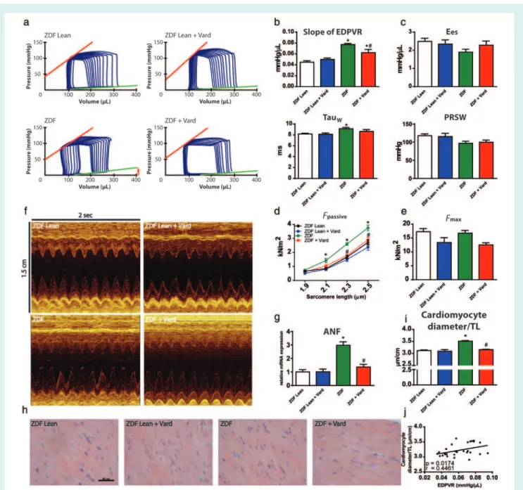
Vardenafil prevented type 2 diabetes mellitus-associated left ventricular dysfunction in vivo
Neither HR nor MAP differed among the groups (Table 1). The slope of EDPVR (LV stiffness parameter) and TauWshowed signif- icant increase in ZDF (Figure1a,b). Vardenafil treatment markedly improved the slope of EDPVR, while TauW tended to decrease in ZDF rats (Figure1b). Factorial ANOVA revealed significant dif- ferences in EDPVR and TauW between T2DM and non-diabetic animals (see the Supplementary material online,Table S3). Despite the marked diastolic dysfunction in T2DM, conventional systolic parameters, such as EF, CO, dP/dtmax, and SW did not differ among our study groups (Table 1). Moreover, reliable load-independent systolic parameters Ees and PRSW remained unchanged (Figure1c).
Vardenafil prevented type 2 diabetes mellitus-associated stiffening of LV cardiomyocytes
The value of Fpassive (at different SLs; a marker of cardiomyocyte stiffness) increased significantly in ZDF rats (Figure1d). Vardenafil prevented the diabetes-associated increase of Fpassive (Figure1d), however, it had no effect on Fmax(Figure1e).
Vardenafil decreased myocardial hypertrophy in Zucker Diabetic Fatty rats
Although HW and HW/BW ratios were not different, HW/TL ratio increased significantly in ZDF compared with ZDFLean rats (Table1). The HW/TL ratio of ZDF+Vard rats tended to decrease compared with ZDF rats (Table1). In addition, echocardiography revealed signs of myocardial hypertrophy in ZDF rats, indicated by the significantly elevated LVAW and LVPW in ‘s’ and ‘d’, increased
Table1 Basic characteristics and haemodynamic parameters in the study groups
Variable ZDFLean ZDFLean+Vard ZDF ZDF+Vard
. . . . Basic characteristics
BW (g) 421±9 419±12 395±25 405±28
HW (g) 1.46±0.03 1.55±0.02 1.55±0.03 1.51±0.04
HW/BW (g/kg) 3.46±0.05 3.71±0.13 3.98±0.25 3.79±0.24
HW/TL (g/cm) 0.346±0.007 0.364±0.005 0.389±0.007* 0.377±0.010
Echocardiography†
LVAWs (mm) 2.53±0.04 2.58±0.15 2.90±0.08* 2.48±0.05#
LVAWd (mm) 1.73±0.03 1.76±0.03 1.88±0.01* 1.72±0.03#
LVIDs (mm) 4.91±0.21 4.84±0.16 4.50±0.14 5.04±0.19
LVIDd (mm) 8.08±0.20 7.83±0.15 7.99±0.32 8.08±0.29
LVPWs (mm) 2.63±0.07 2.69±0.10 3.07±0.10* 2.93±0.11
LVPWd (mm) 1.72±0.08 1.91±0.07 2.12±0.10* 2.02±0.10
RWT 0.41±0.02 0.47±0.01 0.50±0.03* 0.47±0.02
LVmass (g) 0.98±0.02 1.01±0.05 1.15±0.05 1.09±0.07
LVmass/TL (g/cm) 0.232±0.005 0.245±0.118 0.299±0.010* 0.271±0.017
LVmass index (g/kg BW) 2.53±0.09 2.66±0.13 3.23±0.23* 2.99±0.23
Haemodynamic parameters
HR (b.p.m.) 326±4 327±8 310±6 319±8
MAP (mmHg) 96±3 96±3 99±2 105±2
EF (%) 65±2 66±2 61±2 64±4
CO (mL/min) 67±5 70±5 55±5 58±9
dP/dtmax(mmHg/s) 9426±453 9061±270 8478±234 9994±634
dP/dtmin(mmHg/s) −9799±549 −9706±424 −9039±639 −9463±1084
SW (mmHg.μL) 19378±1125 20567±1231 17279±1392 17848±2576
BW, body weight; HW, heart weight, TL, tibia length; LV, left ventricular; AW, anterior wall thickness; PW, posterior wall thickness; LVID, LV internal diameter; RWT, relative wall thickness; HR, heart rate; MAP, mean arterial pressure; EF, ejection fraction; CO, cardiac output; dP/dtmaxand dP/dtmin, maximal and minimal slope of dP/dt; SW, stroke work.
*P<0.05 vs. ZDFLean;#P<0.05 vs. ZDF.
†The ‘s’ and ‘d’ after the acronyms indicate end-systolic and end-diastolic, respectively.
RWT, LVmass/TL, and LVmass index (Table1). All of these param- eters tended to decline in ZDF+Vard rats, while LVAWs and LVAWd were reduced markedly in response to vardenafil treat- ment when compared with ZDF rats (Table 1). In addition to the robust hypertrophy observed on echocardiography (Figure1f), significant elevation of ANF (Figure1g), histological evaluation of H&E sections (Figure 1h) along with the robust increase in car- diomyocyte diameter/TL (Figure1i) supported cardiac hypertrophy.
Vardenafil significantly reduced the gene expression level of ANF (Figure1g) and decreased cardiomyocyte diameter/TL (Figure1i).
The slope of EDPVR correlated robustly with the hypertrophy marker cardiomyocyte diameter/TL (Figure1j).
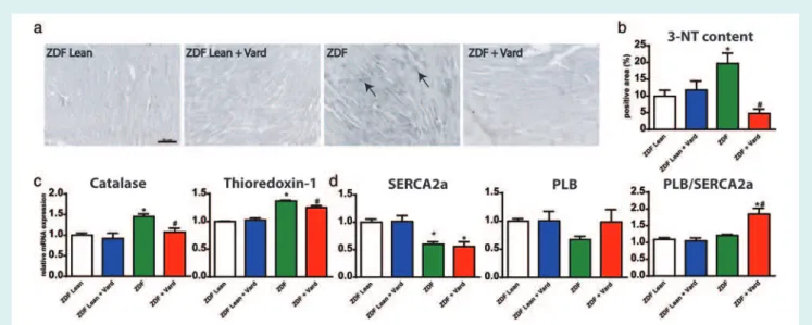
Vardenafil reduced alterations associated with myocardial nitro-oxidative stress in type 2 diabetes mellitus
Type 2 DM was associated with markedly elevated 3-NT content of the left ventricle (Figure 2a,b), however, vardenafil preven- tion effectively reduced it (Figure 2a,b). In accord with this, we observed significant upregulation of different antioxidant enzymes, including catalase and thiorexodin-1in the ZDF group (Figure 2c).
Nevertheless, as a result of chronic drug treatment catalase and ...
thioredoxin-1levels declined significantly in ZDF rats (Figure 2C).
Moreover, SERCA2a was markedly downregulated in ZDF rats regardless of treatment (Figure 2d). The PLB gene expression tended to decrease in the ZDF group (Fig.2D), although, in the ZDF+Vard group, it did not show any difference when compared with the ZDFLean group (Figure 2d). Despite the unchanged PLB/
SERCA2a ratio in T2DM (Figure 2d), vardenafil treatment markedly increased the ratio of PLB/SERCA2a in ZDF animals (Figure 2c).
Vardenafil suppressed myocardial fibrotic remodelling in type 2 diabetes mellitus
Masson trichrome and PicroSirius staining revealed fibrotic remod- elling of the myocardium in ZDF (Figure 3a–d), the extent of which correlated robustly with the slope of EDPVR (Figure 3e).
Fibronectin-1 was markedly overexpressed in T2DM (Figure 3f).
Both Col1a1and Col3a1mRNAs were also significantly downreg- ulated in ZDF rats (Figure 3g). Prevention by vardenafil effectively reduced the fibrotic remodelling of the myocardium (Figure 3a-d) and significantly reduced Fn1gene expression (Figure 3f) in T2DM.
Interestingly, Col1a1and Col3a1gene expressions were unaltered by vardenafil in the ZDF+Vard group compared with the ZDF group (Figure 3g).
Figure1 The effect of vardenafil on the haemodynamic alterations and on myocardial hypertrophy in heart failure with preserved ejection fraction animals. (a) Representative left ventricular (LV) pressure–volume (P-V) loops. The arrow indicates the increase of the slope of end-diastolic pressure–volume relationship (EDPVR). (b) Graphs represent the value of the slope of (EDPVR) and TauW. (c) Graphs of the slope (Ees) of the LV end-systolic P-V relationship and the value of preload-recruitable stroke work (PRSW). (d) Fpassive(cardiomyocyte stiffness marker) at different sarcomere lengths. (e) Fmaxin the study groups. (f) Representative M-mode echocardiography images at the mid-papillary level on short axis view. (g) Relative gene expression of atrial natriuretic factor (ANF). (h) Representative haematoxylin-eosin stained sections.
Bar: 50μm, Magnification: 200×. (i) Quantification of cardiomyocyte diameter/tibia length (TL). (j) Correlation analysis between cardiomyocyte diameter/tibia length (TL) and the slope of EDPVR. Study groups are defined in the text. *P<0.05 vs. ZDFLean;#P<0.05 vs. ZDF.
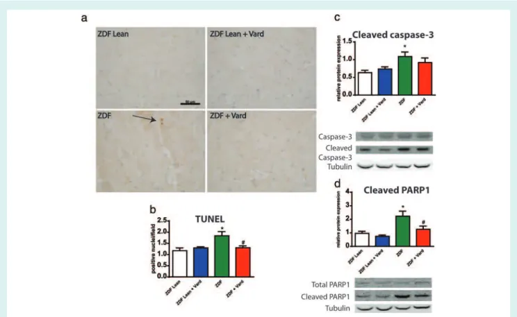
Phosphodiesterase-5A inhibition prevented cardiomyocyte apoptosis in Zucker Diabetic Fatty rats
Evidence for increased cardiomyocyte apoptosis was shown by TUNEL assay (Figure 4a,b), and demonstrated by markedly ...
risen cleaved caspase-3 and cleaved PARP1 band densities (Figure 4c,d). However, vardenafil prevented the above alter- ations by significantly decreasing the number of TUNEL-positive nuclei (Figure 4b) and cleaved PARP1 band density (Figure 4d).
Cleaved caspase-3 band density was not significantly differ- ent in ZDF+Vard group compared with the ZDFLean group (Figure 4c).
Figure 2 Phosphodiesterase-5A inhibition reduces the extent of cardiac nitro-oxidative stress in heart failure with preserved ejection fraction.
(a) Representative images of 3-nitrotyrosine (3-NT) stained sections. Arrows indicate the grey coloured 3-NT positive area. Bar: 50μm, Magnification: 200×. (b) Quantification of 3-NT positive area in the experimental groups. (c) Relative gene expression levels of catalase and thioredoxin-1. (d) Gene expression levels of sarcoplasmic reticulum calcium ATPase 2 (SERCA2a), phospholamban (PLB) and the ratio of PLB/SERCA2a are shown. A detailed description of the study groups is available in the text. *P<0.05 vs. ZDFLean;#P<0.05 vs. ZDF.
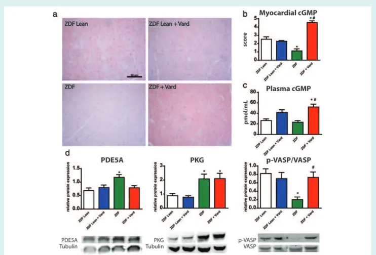
Vardenafil prevented the disturbances of myocardial cyclic guanosine
monophosphate–protein kinase G signalling in Zucker Diabetic Fatty rats
The PDE5A–cGMP–PKG axis significantly deteriorated in T2DM, as demonstrated by the markedly lower cGMP staining intensity of the myocardium (Figure 5a,b), by the increased protein levels of PDE5A and PKG (Figure 5d) and by the lower p-VASP/VASP ratio (as a marker of impaired PKG activity;Figure 5d). Myocardial PDE5A levels in the ZDF+Vard group did not differ from the healthy controls (Figure 5d). Vardenafil effectively increased the cGMP staining intensity of the ZDF group myocardium (Figure 5a,b).
Furthermore, vardenafil elevated the plasma cGMP content in ZDF rats (Figure 5c) and restored the ratio of p-VASP/VASP (Figure 5d).
Interestingly, the plasma cGMP level showed a strong tendency toward elevation in ZDFLean+Vard group (Figure 5d). Plasma total nitrite/nitrate levels and p-PLB/PLB ratios were not different among the groups (see the Supplementary material online,Figure S2).
Discussion
The main findings of the present study are that PDE5A inhibi- tion with long-term vardenafil application (i) effectively prevents the development of HFpEF (characterized by increased myocardial stiffness and worsened diastolic function), (ii) reduces the patho- physiological features of T2DM-associated diabetic cardiomyopa- thy, and (iii) restores the activity of cGMP–PKG axis by increasing myocardial as well as plasma cGMP levels.
Heart failure with preserved EF is characterized by the clini- cal signs of HF, however, cardiac systolic function measured by EF ...
is preserved (LVEF ≥50%) with a concomitant decrease in dias- tolic function (increased stiffness, decreased relaxation and slow LV filling).1,2The importance of co-morbidities and the subsequent deterioration of the NO-cGMP-PKG signalling has been proposed in the development of HFpEF by Paulus and Tschope.8The pres- ence of co-morbidities (especially obesity and T2DM) leads to an increased level of reactive oxygen species (ROS), decreased NO bioavailability, and lower cGMP levels, with subsequent deactiva- tion of the main effector, PKG enzyme. In line with this finding, van Heerebeeket al.7 found lower myocardial PKG activity in the myocardium of HFpEF patients.
The restoration of NO-cGMP-PKG axis has been proven to be cytoprotective in different cardiovascular diseases6,17,18 including diabetic cardiomyopathy.5,6,12 Phosphodiesterase-5A inhibitors block one of the main regulator of cGMP degradation thereby pre- serving and/or increasing intracellular cGMP concentration.4The- oretically, blocking the PDE5A in pathological LV remodelling could provide a useful tool in the management of HF patients. The above idea led to a clinical trial investigating the cardioprotective effects of sildenafil in HFpEF patients (RELAX study).19Despite the promis- ing preclinical data, sildenafil showed no improvements in exercise capacity or on the clinical outcomes in advanced HFpEF patients.19 However, cGMP plasma levels were not significantly different at the end of the study period between the study groups. In light of this, one can speculate that PDE5A inhibition might have been ineffective and it could have contributed to the negative results.19The above data suggest that improving cGMP signalling is a promising avenue of research; however, the result of the RELAX trial raises impor- tant questions about the appropriate pharmacological approach.
In line with this, Franssen and Gonzalez Miqueo20reported that the initial phase of HFpEF is presumably predominated by the
Figure 3 Protective effects of vardenafil on myocardial fibrosis in heart failure with preserved ejection fraction. (a) Representative images and (b) semiquantitative scoring of Masson’s trichrome stained sections. Arrows indicate interstitial fibrosis of the myocardium. Bar: 50μm, Magnification: 200×. (c) Representative images and (d) quantification of PicroSirius stained myocardium. Bar: 50μm, Magnification: 200×.
(e) Correlation analysis between PicroSirius positive area and the slope of end-diastolic pressure–volume relationship (EDPVR). (f) Gene expression of fibronectin-1(Fn1), (g) collagen1a1and 3a1(Col1a1; Col3a1). A detailed description of the study groups is available in the text.
*P<0.05 vs. ZDFLean;#P<0.05 vs. ZDF
Figure 4 The effects of vardenafil on myocardial DNA fragmentation and apoptosis. (a) Representative images of terminal deoxynucleotidyl transferase dUTP nick-end labelling (TUNEL) assay. Bar: 50μm, Magnification: 200×. (b) Quantification of TUNEL positive nuclei/field. (c) Graphs and representative western blot bands of cleaved caspase-3 (17 kDa) and (d) cleaved poly (ADP-ribose) polymerase (PARP1; 85 kDa) levels in the myocardium. A detailed description of the study groups is available in the text. *P<0.05 vs. ZDFLean;#P<0.05 vs. ZDF.
dysfunction of cardiomyocytes, thus it could be useful to improve cGMP signalling to reduce cardiomyocyte stiffness. In contrast, sildenafil has been shown to exert anti-remodelling effect in T2DM diabetic cardiomyopathy in humans.21 Moreover, Koka et al.22 showed beneficial effects of tadalafil on systolic performance and mitochondrial function indb/dbmice. Nevertheless, they did not report how PDE5A inhibitors affect diastolic dysfunction in T2DM.
Accordingly, we believe that instead of treating the already developed HFpEF, effective pharmacological prevention by PDE5A inhibitors might be more appropriate in the management of HFpEF patients with co-morbidities such as obesity and DM. Thus, in the present study, we investigated the effects of PDE5A inhi- bition in a preventive manner (from the pre-diabetic state) on the development of HFpEF (mainly on diastolic function) in the ZDF rat.
Several studies have focused on the investigation of cardiac func- tion in HFpEF. Previous data showed that diastolic dysfunction can be determined in the HFpEF animal model ZDF rat.23,24In accord with the literature we observed a significant increase in LV stiff- ness and prolonged relaxation time by pressure–volume analysis in our model. In addition, cardiomyocyte stiffness (as shown by increased Fpassive) was also evident in T2DM. However,in vivoand in vitrosystolic performance was preserved, fulfilling the criteria for HFpEF in ZDF rats. Interestingly, we did not observe any difference ...
in HR and MAP. Vardenafil effectively prevented diastolic dysfunc- tion bothin vivo(decreased LV stiffness, improved relaxation time) and at the sarcomeric level (decreased cardiomyocyte Fpassive) in ZDF rats. Hypophosphorylation of the PEVK-domain of titin might play a role in the observed phenomena.25
We found lower myocardial cGMP level coupled with increased protein expression of PDE5A (a possible contributor to the low myocardial cGMP content) and PKG enzymes in the heart of HFpEF animals. Although PKG protein levels were increased, PKG activity (as reflected by the p-VASP/VASP ratio) showed significant impairment in the diabetic myocardium. Interestingly, plasma cGMP levels remained unchanged in ZDF. One can speculate that this might be a consequence of the observed compensatory upregula- tion of ANF and subsequent activation of particulate GC in other organs. Thus preserved plasma cGMP is seen as a sign of overspill of cGMP from different tissues.5Vardenafil effectively restored the activity of the cGMP–PKG axis, as shown by increased plasma/
cardiac cGMP concentrations and p-VASP/VASP ratio.
Pathological remodelling of the myocardium in diabetic car- diomyopathy is a well-known phenomenon and is character- ized by fibrosis, hypertrophy, increased nitro-oxidative stress, and cardiomyocyte apoptosis.3 Hyperglycaemia can directly lead to the accumulation of ROS and to the development of severe nitro-oxidative stress in DM.3 Several mechanisms have been
Figure 5 Modulatory effects of vardenafil on the myocardial NO-cGMP signalling in T2DM. (a) Representative images. Bar: 50μm, Magnification: 200×. (b) Quantification of cGMP immunohistochemistry in the study groups. (c) Plasma cGMP levels in the experimental groups. (d) Graphs and representative western blot bands of phosphodiesterase-5A (PDE5A,130 kDa), protein kinase G (PKG, 75 kDa) and phospho-vasodilator-stimulated phosphoprotein (p-VASP) and VASP (50 kDa) are shown. A detailed description of the study groups is available in the text. *P<0.05 vs. ZDFLean;#P<0.05 vs. ZDF.
described to play a decisive role in DM-associated nitro-oxidative stress such as the upregulation of NADPH-oxidases and NO synthases.3Moreover, in nitro-oxidative stress peroxynitrite is gen- erated when ROS directly reacts with NO thus it contributes to the decreased NO bioavailability.26 Peroxynitrite is a highly reactive molecule that directly deteriorates different cellular ele- ments, enzymes, myofibrillar proteins, and DNA.26In agreement with this, we observed hyperglycaemia at an early age which increased gradually during the study. We also found increased nitro-oxidative stress as well as an upregulation of the different antioxidant enzymes in the LV myocardium of ZDF animals. How- ever, plasma nitrite/nitrate levels (reflecting NO bioavailability) were not diminished. In addition, SERCA2a gene expression was significantly lower, which might reflect the disturbance of intracellu- lar Ca2+homeostasis and could contribute to the prolonged relax- ation time in T2DM.27Vardenafil, however, significantly affected the DM-associated nitro-oxidative stress as it prevented an increase of 3-NT staining and the elevation of catalase and thioredoxin-1 in the ZDF group myocardium. The protective feature of PDE5A inhibition is probably attributed to its antioxidative effects28and to ...
the enhancement of cGMP signalling.12Moreover, vardenafil signif- icantly increased the ratio of PLB/SERCA2a gene expression which might have contributed to the observed improved diastolic function in the ZDF+Vard group.
Not only peroxynitrite but ROS also directly propagates DNA fragmentation and apoptosis in DM leading to the loss of cardiomyocytes.5,26 In addition to the increased rate of apopto- sis, several pathological processes play role in the development of myocardium hypertrophy and fibrosis (both interstitial and replacement types), including the dysregulation of the transform- ing growth factor𝛽(TGF-𝛽) signalling,3,5 fibroblast proliferation,3 and disturbance of the matrix metalloproteinases (MMPs).29 Corresponding to this, our DM model developed HFpEF char- acterized by increased apoptosis. Moreover, our experiments revealed massive cardiac hypertrophy not only by echocardiog- raphy but also by the post-mortem analysis of the myocardium (increased HW/TL, cardiomyocyte diameter/TL, and ANF gene expression). In addition to the development of concentric hyper- trophy, fibrotic remodelling was present in the left ventricle of our ZDF animals (higher MT score, PicroSirius area and Fn1gene
expression). Interestingly, Col1a1and Col3a1mRNA levels were significantly reduced in T2DM; however, in agreement with data in the literature,5,29 this might be the consequence of a negative feedback mechanism. Through the improved cGMP signalling, vardenafil effectively reduced myocardial apoptosis (via the inhi- bition of PARP cleavage), cardiomyocyte hypertrophy, and fibrotic remodelling of the myocardium. Our results are in line with the data of previous studies that reported antihypertrophic effects of the enhancement of cGMP signalling.5,30 In the background of improved fibrosis a regulatory cross-talk between the enhanced PKG signalling and the key members of cardiac remodelling such as TGF-𝛽signalling,5,12 microvascular inflammation, endothelin-1, angiotensin II, and aldosterone8 can be assumed. It is notable that diastolic dysfunction (slope of EDPVR) correlated with both cardiac hypertrophy and fibrosis. Although many contributing factors have been identified in the progression of HFpEF, we still lack a treatment for proper patient management. A signifi- cant point of intervention might be to improve the myocardial NO-cGMP-PKG signalling. However, from previous clinical trials it seems rather that a pharmacological prevention could contribute significantly to the improvement of HFpEF. To our knowledge, this is the first study reporting the preventive cardioprotective effects of vardenafil on diastolic function in an animal model of HFpEF. The presence of co-morbidities in HFpEF patients has to be taken into account when planning the pharmacological management of patients. In agreement with this, early initiated pharmacological prevention with the PDE5A inhibitor varde- nafil might be a therapeutic alternative for patients with DM and HFpEF.
Study limitations
Our study is limited to young, male rats. Although the p-VASP/VASP ratio was considered as a marker to estimate PKG activity, direct measurement of PKG activity is the gold stan- dard method as VASP phosphorylation could also be influenced by other PKs. Involvement of cGMP–cAMP crosstalk and PKA activation as a subsidiary mechanism in the observed effects of vardenafil cannot be ruled out. Our present work focused on the effects of preventive therapy by vardenafil in T2DM. However, the determination of the optimal time-point of the pharma- ceutical intervention might be an important aspect of future investigations.
Supplementary Information
Additional Supporting Information may be found in the online version of this article:
Figure S1.Blood glucose values in the study groups.
Figure S2. Plasma total nitrate/nitrite level and phospholamban assay.
Method S1.Expanded methods.
Table S1.TaqMan gene expression assays used.
Table S2.Antibodies used in the study.
Table S3.Results of two-way analysis of variance. ...
Acknowledgements
The technical assistance of Lilla Szabó, Anna Meltzer, Alex Ali Say- our, Tímea Fischinger, Henriett Biró, Gábor Fritz, Krisztina Fazekas, and Viktória Gregor is acknowledged. The authors thank Bayer HealthCare (Wuppertal, Germany) for providing vardenafil. The scientific advice of Prof. Miklós Kellermayer and Dr Hedvig Tor- dai (Department of Biophysics and Radiation Biology, Semmelweis University, Budapest, Hungary) is gratefully acknowledged.
Funding
This work was supported by the Hungarian Scientific Research Fund (OTKA-PD100245 (TR), OTKA-K109083 (ZP)) and by the János Bolyai Research Scholarship of the Hungarian Academy of Sci- ences (T.R.). B.T.N. received a fellowship from the European Social Fund in the framework of TÁMOP 4.2.4. A/1-11-1-2012-0001
‘National Excellence Program’. C.M. was supported by the schol- arship of Human Resource Support Office (National Talent Pro- gramme; NTP-NFTÖ-16-0081).
Conflict of interest:none declared.
References
1. Ponikowski P, Voors AA, Anker SD, Bueno H, Cleland JG, Coats AJ, Falk V, Gonzalez-Juanatey JR, Harjola VP, Jankowska EA, Jessup M, Linde C, Nihoy- annopoulos P, Parissis JT, Pieske B, Riley JP, Rosano GM, Ruilope LM, Ruschitzka F, Rutten FH, van der Meer P. 2016 ESC Guidelines for the diagnosis and treat- ment of acute and chronic heart failure: The Task Force for the diagnosis and treatment of acute and chronic heart failure of the European Society of Cardiol- ogy (ESC) Developed with the special contribution of the Heart Failure Associ- ation (HFA) of the ESC.Eur J Heart Fail2016;18:891–975.
2. Zouein FA, de Castro Bras LE, da Costa DV, Lindsey ML, Kurdi M, Booz GW. Heart failure with preserved ejection fraction: emerging drug strategies.
J Cardiovasc Pharmacol2013;62:13–21.
3. Huynh K, Bernardo BC, McMullen JR, Ritchie RH. Diabetic cardiomyopathy:
mechanisms and new treatment strategies targeting antioxidant signaling path- ways.Pharmacol Ther2014;142:375–415.
4. Zhao CY, Greenstein JL, Winslow RL. Roles of phosphodiesterases in the regulation of the cardiac cyclic nucleotide cross-talk signaling network.J Mol Cell Cardiol2016;91:215–227.
5. Matyas C, Nemeth BT, Olah A, Hidi L, Birtalan E, Kellermayer D, Ruppert M, Korkmaz-Icoz S, Kokeny G, Horvath EM, Szabo G, Merkely B, Radovits T. The soluble guanylate cyclase activator cinaciguat prevents cardiac dysfunction in a rat model of type-1diabetes mellitus.Cardiovasc Diabetol2015;14:145.
6. Das A, Durrant D, Salloum FN, Xi L, Kukreja RC. PDE5 inhibitors as therapeutics for heart disease, diabetes and cancer.Pharmacol Ther2015;147:12–21.
7. van Heerebeek L, Hamdani N, Falcao-Pires I, Leite-Moreira AF, Begieneman MP, Bronzwaer JG, van der Velden J, Stienen GJ, Laarman GJ, Somsen A, Verheugt FW, Niessen HW, Paulus WJ. Low myocardial protein kinase G activity in heart failure with preserved ejection fraction.Circulation2012;126:830–839.
8. Paulus WJ, Tschope C. A novel paradigm for heart failure with preserved ejection fraction: comorbidities drive myocardial dysfunction and remodeling through coronary microvascular endothelial inflammation.J Am Coll Cardiol 2013;62:
263–271.
9. Bischoff E. Potency, selectivity, and consequences of nonselectivity of PDE inhibition.Int J Impot Res. 2004;16(Suppl1):S11–S14.
10. Szabo G, Radovits T, Veres G, Krieger N, Loganathan S, Sandner P, Karck M. Var- denafil protects against myocardial and endothelial injuries after cardiopulmonary bypass.Eur J Cardiothorac Surg2009;36:657–664.
11. Loganathan S, Radovits T, Hirschberg K, Korkmaz S, Barnucz E, Karck M, Szabo G. Effects of selective phosphodiesterase-5-inhibition on myocardial contractil- ity and reperfusion injury after heart transplantation.Transplantation2008;86:
1414–1418.
12. Radovits T, Bomicke T, Kokeny G, Arif R, Loganathan S, Kecsan K, Korkmaz S, Barnucz E, Sandner P, Karck M, Szabo G. The phosphodiesterase-5 inhibitor vardenafil improves cardiovascular dysfunction in experimental diabetes mellitus.
Br J Pharmacol2009;156:909–919.
13. Ellis CG, Goldman D, Hanson M, Stephenson AH, Milkovich S, Benlamri A, Ellsworth ML, Sprague RS. Defects in oxygen supply to skeletal muscle of prediabetic ZDF rats.Am J Physiol Heart Circ Physiol2010;298:H1661–H1670.
14. Conceicao G, Heinonen I, Lourenco AP, Duncker DJ, Falcao-Pires I. Ani- mal models of heart failure with preserved ejection fraction. Neth Heart J 2016;24:275–286.
15. Radovits T, Olah A, Lux A, Nemeth BT, Hidi L, Birtalan E, Kellermayer D, Matyas C, Szabo G, Merkely B. Rat model of exercise-induced cardiac hypertrophy:
hemodynamic characterization using left ventricular pressure–volume analysis.
Am J Physiol Heart Circ Physiol2013;305:H124–H134.
16. Varga ZV, Giricz Z, Liaudet L, Hasko G, Ferdinandy P, Pacher P. Interplay of oxidative, nitrosative/nitrative stress, inflammation, cell death and autophagy in diabetic cardiomyopathy.Biochim Biophys Acta2015;1852:232–242.
17. Radovits T, Arif R, Bomicke T, Korkmaz S, Barnucz E, Karck M, Merkely B, Szabo G. Vascular dysfunction induced by hypochlorite is improved by the selective phosphodiesterase-5-inhibitor vardenafil.Eur J Pharmacol2013;710:110–119.
18. Kukreja RC, Ockaili R, Salloum F, Yin C, Hawkins J, Das A, Xi L. Cardioprotection with phosphodiesterase-5 inhibition – a novel preconditioning strategy.J Mol Cell Cardiol2004;36:165–173.
19. Redfield MM, Chen HH, Borlaug BA, Semigran MJ, Lee KL, Lewis G, LeWinter MM, Rouleau JL, Bull DA, Mann DL, Deswal A, Stevenson LW, Givertz MM, Ofili EO, O’Connor CM, Felker GM, Goldsmith SR, Bart BA, McNulty SE, Ibarra JC, Lin G, Oh JK, Patel MR, Kim RJ, Tracy RP, Velazquez EJ, Anstrom KJ, Hernandez AF, Mascette AM, Braunwald E. RELAX Trial Effect of phosphodiesterase-5 inhibition on exercise capacity and clinical status in heart failure with preserved ejection fraction: a randomized clinical trial.JAMA2013;309:1268–1277.
20. Franssen C, Gonzalez Miqueo A. The role of titin and extracellular matrix remodelling in heart failure with preserved ejection fraction. Neth Heart J 2016;24:259–267.
21. Giannetta E, Isidori AM, Galea N, Carbone I, Mandosi E, Vizza CD, Naro F, Morano S, Fedele F, Lenzi A. Chronic inhibition of cGMP phosphodi- esterase 5A improves diabetic cardiomyopathy: a randomized, controlled clin- ical trial using magnetic resonance imaging with myocardial tagging.Circulation 2012;125:2323–2333. ...
22. Koka S, Aluri HS, Xi L, Lesnefsky EJ, Kukreja RC. Chronic inhibition of phospho- diesterase 5 with tadalafil attenuates mitochondrial dysfunction in type 2 diabetic hearts: potential role of NO/SIRT1/PGC-1alpha signaling.Am J Physiol Heart Circ Physiol2014;306:H1558–H1568.
23. Radovits T, Korkmaz S, Matyas C, Olah A, Nemeth BT, Pali S, Hirschberg K, Zubarevich A, Gwanmesia PN, Li S, Loganathan S, Barnucz E, Merkely B, Szabo G. An altered pattern of myocardial histopathological and molecular changes underlies the different characteristics of type-1 and type-2 diabetic cardiac dysfunction.J Diabetes Res2015;2015:728741.
24. Franssen C, Chen S, Unger A, Korkmaz HI, De Keulenaer GW, Tschope C, Leite-Moreira AF, Musters R, Niessen HW, Linke WA, Paulus WJ, Hamdani N. Myocardial microvascular inflammatory endothelial activation in heart failure with preserved ejection fraction. JACC Heart Fail 2016;
4:312–324.
25. Hamdani N, Franssen C, Lourenco A, Falcao-Pires I, Fontoura D, Leite S, Plettig L, Lopez B, Ottenheijm CA, Becher PM, Gonzalez A, Tschope C, Diez J, Linke WA, Leite-Moreira AF, Paulus WJ. Myocardial titin hypophosphorylation importantly contributes to heart failure with preserved ejection fraction in a rat metabolic risk model.Circ Heart Fail2013;6:1239–1249.
26. Pacher P, Beckman JS, Liaudet L. Nitric oxide and peroxynitrite in health and disease.Physiol Rev2007;87:315–424.
27. Zarain-Herzberg A, Garcia-Rivas G, Estrada-Aviles R. Regulation of SERCA pumps expression in diabetes.Cell Calcium2014;56:302–310.
28. Dias-Junior CA, Neto-Neves EM, Montenegro MF, Tanus-Santos JE. Hemody- namic effects of inducible nitric oxide synthase inhibition combined with sildenafil during acute pulmonary embolism.Nitric Oxide2010;23:284–288.
29. Van Linthout S, Seeland U, Riad A, Eckhardt O, Hohl M, Dhayat N, Richter U, Fischer JW, Bohm M, Pauschinger M, Schultheiss HP, Tschope C. Reduced MMP-2 activity contributes to cardiac fibrosis in experimental diabetic cardiomyopathy.
Basic Res Cardiol2008;103:319–327.
30. Takimoto E, Champion HC, Li M, Belardi D, Ren S, Rodriguez ER, Bedja D, Gabrielson KL, Wang Y, Kass DA. Chronic inhibition of cyclic GMP phospho- diesterase 5A prevents and reverses cardiac hypertrophy.Nat Med2005;11:
214–222.