Citation:Freiwan, M.; Kovács, M.G.;
Kovács, Z.Z.A.; Sz ˝ucs, G.; Dinh, H.;
Losonczi, R.; Siska, A.; Kriston, A.;
Kovács, F.; Horváth, P.; et al.
Investigation of the Antiremodeling Effects of Losartan, Mirabegron and Their Combination on the Development of Doxorubicin-Induced Chronic Cardiotoxicity in a Rat Model.Int. J. Mol. Sci.2022,23, 2201.
https://doi.org/10.3390/ijms23042201 Academic Editor: Ryuji Okamoto Received: 26 January 2022 Accepted: 12 February 2022 Published: 16 February 2022 Publisher’s Note:MDPI stays neutral with regard to jurisdictional claims in published maps and institutional affil- iations.
Copyright: © 2022 by the authors.
Licensee MDPI, Basel, Switzerland.
This article is an open access article distributed under the terms and conditions of the Creative Commons Attribution (CC BY) license (https://
creativecommons.org/licenses/by/
4.0/).
Article
Investigation of the Antiremodeling Effects of Losartan, Mirabegron and Their Combination on the Development of Doxorubicin-Induced Chronic Cardiotoxicity in a Rat Model
Marah Freiwan1,2 , Mónika G. Kovács1,2 , Zsuzsanna Z. A. Kovács1,2, Gerg ˝o Sz ˝ucs1,2 , Hoa Dinh1,2 , Réka Losonczi1,2, Andrea Siska3 , András Kriston4,5,6, Ferenc Kovács4,5,6, Péter Horváth4,5,6, Imre Földesi3, Gábor Cserni7, LászlóDux2,8,*, Tamás Csont1,2,†and Márta Sárközy1,2,*,†
1 MEDICS Research Group, Department of Biochemistry, Albert Szent-Györgyi Medical School, University of Szeged, H-6720 Szeged, Hungary; marah.mf.94@gmail.com (M.F.);
kovacs.monika.gabriella@med.u-szeged.hu (M.G.K.); kovacs.zsuzsanna@med.u-szeged.hu (Z.Z.A.K.);
szucs.gergo@med.u-szeged.hu (G.S.); dinhhoaqa@gmail.com (H.D.); losonczireka1997@gmail.com (R.L.);
csont.tamas@med.u-szeged.hu (T.C.)
2 Interdisciplinary Center of Excellence, University of Szeged, H-6720 Szeged, Hungary
3 Department of Laboratory Medicine, Albert Szent-Györgyi Medical School, University of Szeged, H-6720 Szeged, Hungary; siska.andrea@med.u-szeged.hu (A.S.); foldesi.imre@med.u-szeged.hu (I.F.)
4 Synthetic and Systems Biology Unit, Biological Research Centre, Eötvös Loránd Research Network, H-6726 Szeged, Hungary; kriston.andras@single-cell-technologies.com (A.K.);
kovacs.ferenc@single-cell-technologies.com (F.K.); peter.horvath@brc.hu (P.H.)
5 Single-Cell Technologies Ltd., H-6726 Szeged, Hungary
6 Institute for Molecular Medicine Finland (FIMM), University of Helsinki, FIN-00014 Helsinki, Finland
7 Department of Pathology, Albert Szent-Györgyi Medical School, University of Szeged, H-6720 Szeged, Hungary; cserni.gabor@med.u-szeged.hu
8 Muscle Adaptation Group, Department of Biochemistry, Albert Szent-Györgyi Medical School, University of Szeged, H-6720 Szeged, Hungary
* Correspondence: dux.laszlo@med.u-szeged.hu (L.D.); sarkozy.marta@med.u-szeged.hu (M.S.)
† These authors contributed equally to the work.
Abstract: Despite the effectiveness of doxorubicin (DOXO) as a chemotherapeutic agent, dose- dependent development of chronic cardiotoxicity limits its application. The angiotensin-II receptor blocker losartan is commonly used to treat cardiac remodeling of various etiologies. The beta-3 adrenergic receptor agonist mirabegron was reported to improve chronic heart failure. Here we investigated the effects of losartan, mirabegron and their combination on the development of DOXO- induced chronic cardiotoxicity. Male Wistar rats were divided into five groups: (i) control; (ii) DOXO- only; (iii) losartan-treated DOXO; (iv) mirabegron-treated DOXO; (v) losartan plus mirabegron- treated DOXO groups. The treatments started 5 weeks after DOXO administration. At week 8, echocardiography was performed. At week 9, left ventricles were prepared for histology, qRT-PCR, and Western blot measurements. Losartan improved diastolic but not systolic dysfunction and ameliorated SERCA2a repression in our DOXO-induced cardiotoxicity model. The DOXO-induced overexpression ofIl1andIl6was markedly decreased by losartan and mirabegron. Mirabegron and the combination treatment improved systolic and diastolic dysfunction and significantly decreased overexpression ofSmad2 andSmad3in our DOXO-induced cardiotoxicity model. Only mirabegron reduced DOXO-induced cardiac fibrosis significantly. Mirabegron and its combination with losartan seem to be promising therapeutic tools against DOXO-induced chronic cardiotoxicity.
Keywords:onco-cardiology; doxorubicin-induced chronic cardiotoxicity; heart failure; cardiac fi- brosis; diastolic dysfunction; angiotensin II receptor blocker; beta-3 adrenoceptor agonist; cardiac inflammation; TGF-β/SMAD signaling pathway; sarcoendoplasmic reticulum calcium ATPase 2a
Int. J. Mol. Sci.2022,23, 2201. https://doi.org/10.3390/ijms23042201 https://www.mdpi.com/journal/ijms
1. Introduction
Cancer and cardiovascular diseases (CVDs) are the leading causes of morbidity and mortality worldwide [1,2]. Due to advances in early diagnosis and treatment of cancer pa- tients, long-term cancer survivors are one of the largest growing populations accessing the healthcare system [3,4]. After recurrent malignancies, CVDs are the second leading cause of morbidity and mortality in cancer survivors [3]. Cancer therapies, particularly chemo and radiotherapy, have many recognized side effects on the cardiovascular system [3,5,6].
In early and late chronic stages, chemotherapy-induced cardiotoxicity commonly man- ifests in decreased left ventricular ejection fraction (LVEF), leading to heart failure (HF) symptoms [3,6].
Anthracyclines, including doxorubicin (DOXO), are essential drugs in chemothera- peutic regimens in different cancers, such as leukemias, lymphomas, soft tissue sarcomas, and solid malignancies (i.e., breast, ovary, prostate, stomach, thyroid, liver, and small cell lung cancers) [7,8]. Although anthracyclines are effective and commonly used chemothera- peutic agents, their application could be limited by the dose-dependent development of cardiotoxicity. Anthracyclines-induced cardiotoxicity can manifest in acute, early chronic, and late chronic forms. Acute toxicity is usually reversible and predominantly presents supraventricular arrhythmias, transient left ventricular dysfunction, and electrocardio- graphic changes in less than 1% of patients immediately after treatment [6]. Notably, acute cardiac dysfunction may lead to early or late chronic cardiotoxicity characterized by systolic dysfunction [9]. Early chronic cardiotoxic signs occur within the first year of treatment, while late effects present after several years (median of 7 years after treatment) [3,10,11].
In the case of DOXO, the risk for developing chronic cardiotoxicity is 5% at a cumulative dose of 400 mg/m2, 26% at a dose of 550 mg/m2, and 48% at a dose of 700 mg/m2 in humans [6]. Patients under 18 or over 65 years, suffering from cardiovascular comorbidi- ties such as hypertension, left ventricular hypertrophy, coronary artery disease, diabetes mellitus, or prior radiation exposure, are at higher risk for developing DOXO-induced chronic cardiotoxicity [3,6].
The basic mechanisms underlying DOXO-induced chronic cardiotoxicity have not yet been fully understood. In cancer cells, DOXO was shown to bind to topoisomerase-2α, causing deoxyribonucleic acid (DNA) double-strand break and cell death [12,13]. In car- diomyocytes, DOXO was reported to target topoisomerase-2β, also leading to DNA double- strand breaks and the death of cardiomyocytes. DOXO-bound topoisomerase-2βcan bind to promoters of antioxidative genes and peroxisome proliferator-activated receptor-gamma coactivator 1 (PGC1), which are needed for the expression of antioxidant enzymes and the elements of the mitochondrial electron transport chain [14]. Thus, topoisomerase-2β may be able to account for the three hallmarks of DOXO-induced cardiotoxicity, includ- ing (i) cardiomyocyte death mainly by apoptosis, (ii) generation of reactive oxygen and nitrogen species (ROS/RNS), and (iii) mitochondrial damage [12,13]. Another accepted theory is that DOXO forms an anthracycline-iron complex, which then induces lipid perox- idation, protein oxidation, and DNA damage by ROS production that results in contractile impairment, irreversible myocardial damage, and fibrosis [7,15]. At the same time, other mechanisms have been proposed, such as tissue inflammation, extracellular matrix re- modeling, myofilament dysfunction, and disturbance in intracellular calcium ion (Ca2+) homeostasis [7,15].
Although DOXO effectively kills tumor cells, there is currently no sufficiently effec- tive agent to prevent or treat DOXO-induced chronic cardiotoxicity without diminishing antitumor effects of DOXO or promoting secondary malignancy [3]. The renin-angiotensin- aldosterone system (RAAS) was reported to be overactivated in cardiovascular pathologies, including hypertension, cardiac hypertrophy, and heart failure leading to elevated nitro- oxidative stress, inflammation, apoptosis, and fibrosis [16]. Among the inhibitors of RAAS overactivation, angiotensin-II receptor blockers (ARBs) are widely used drugs to prevent the progression of chronic heart failure in various comorbidities [17,18]. ARB losartan showed cardioprotective effects against experimental DOXO-induced cardiotoxicity [19,20].
Indeed, based on the results of clinical trials, inhibition of RAAS overactivation with ARBs has also shown beneficial effects on the development of DOXO-induced chronic cardiotoxicity [13].
The beta-3 adrenoreceptor (β3AR) agonist mirabegron is used in urology to treat hyper- active bladder syndrome [21]. In preclinical models, theβ3AR agonists attenuated cardiac fibrosis and improved cardiac contractility via coupling ofβ3AR to the eNOS/cGMP path- way in cardiomyocytes [22–24]. Moreover, the antioxidant effects of theβ3AR signaling and the down-regulation of the angiotensin II type 1 receptor (AT1) in response toβ3AR stimulation may protect the heart from elevated nitro-oxidative stress and the consecutive pro-inflammatory and fibrotic processes [23,25–28]. Our group recently showed moderate antifibrotic effects of mirabegron in a rat model of uremic cardiomyopathy independently of theβ3AR/eNOS pathway [29]. Bundgaard reported that mirabegron significantly in- creased LVEF in a subset of patients with less than 40% starting LVEF compared to patients given placebo [30]. This result may suggest that mirabegron could have beneficial effects on heart failure with reduced ejection fraction (HFrEF). Indeed, the antiremodeling effects of theβ3AR agonist mirabegron are being investigated in HFrEF patients in clinical trials (trial numbers: NCT03926754 and NCT02775539). However, the antiremodeling effects of mirabegron have not been studied in DOXO-induced chronic cardiotoxicity. Therefore, in our present study, we aimed at investigating and comparing the potential antiremod- eling effects of the widely-used ARB losartan, theβ3AR agonist mirabegron, and their combination on the development of DOXO-induced chronic cardiotoxicity in a rat model.
2. Results
Our present study aimed to use a DOXO-induced chronic cardiotoxicity model pre- senting similar cardiac pathology to those seen in tumor survivor patients treated with DOXO. The treatments with losartan, mirabegron, and their combination started after the DOXO-administration, similar to the conventionally scheduled heart failure therapeutic regimens in clinical practice [6,31]. Our experimental protocol is presented in Figure1.
Altogether nine animals died in the DOXO groups (n= 1 in the DOXO-only group at week 4,n= 2 in the losartan treated DOXO group (one animal at week 4 before the start of losartan treatment and one animal 1 day before the termination at week 9),n= 4 in the mirabegron-treated DOXO group (one animal was excluded and terminated earlier due to its poor echocardiographic result and body weight at week 4, one animal before the start of mirabegron treatment due to abdominal ulceration at week 4 and two animals during the mirabegron treatment at weeks 7 and 8), andn= 2 in the losartan plus mirabegron- treated DOXO group (one animal was excluded and terminated earlier due to its poor echocardiographic results at week 4 and one animal during the losartan plus mirabegron treatment at week 6)).
2.1. Early Echocardiographic Signs of Systolic Dysfunction Developed in the DOXO Groups before Starting the Treatments at Week 4
Echocardiography was performed at week 4 to assess the effects of DOXO on cardiac morphology and function before starting the treatments with losartan, mirabegron, and their combination (Figure1, Table1). There were no significant differences in most of the measured morphologic and functional parameters between the DOXO and control groups (Table1). Only the left ventricular end-systolic diameter was significantly higher in the DOXO groups compared to the control group indicating an early sign of DOXO-induced chronic cardiotoxicity assessed by echocardiography (Table1). Additionally, fractional shortening, diastolic septal wall thickness, and systolic anterior wall thickness showed a statistically non-significant decrease in the DOXO groups compared to the control group (Table1).
Figure 1.Experimental protocol. Male Wistar rats (n= 50, 350–400 g) were divided into one control and four doxorubicin (DOXO)-treated groups (ip.1.5 mg/kg in 6 cycles; cumulative dose: 9 mg/kg).
From the 5th week after the last cycle of DOXO administration, rats were treated via oralgavage daily until the end of the experiments as follows: (i) control group treated with tap water (n= 8, 2 mL/kg/day), (ii) DOXO-only group treated with tap water (n= 11, 2 mL/kg/day), (iii) DOXO group treated with losartan (L,per os20 mg/kg/day,n= 10) dissolved in tap water, iv) DOXO group treated with mirabegron (M,per os30 mg/kg/day,n= 10) dissolved in tap water, and (v) DOXO group treated with the combination of losartan (per os20 mg/kg/day) and mirabegron (per os 30 mg/kg/day) dissolved in tap water (n= 11, 2 mL/kg/day). Cardiac morphology and function were assessed by transthoracic echocardiography (Echo) at weeks 4 and 8 under isoflurane anesthesia.
At week 9, an invasive blood pressure (BP) measurement was performed under pentobarbital anesthe- sia, then blood was collected from the abdominal aorta to measure routine laboratory parameters, and hearts, lungs, and tibias were isolated. Left and right ventricles were separated, and left ventricular samples were prepared for histology, qRT-PCR, and Western blot (WB) measurements.
Table 1.Effects of DOXO on cardiac morphology and function assessed by transthoracic echocardio- graphy before starting the treatments with losartan, mirabegron, and their combination at week 4.
Parameter (Unit) Groups
Control DOXO DOXO + L DOXO + M DOXO + L + M
SWTs (mm) 3.57±0.06 3.43±0.09 3.35±0.19 3.53±0.15 3.34±0.16
SWTd (mm) 2.17±0.04 1.92±0.09 1.90±0.09 1.99±0.09 1.98±0.14
PWTs (mm) 3.1±0.11 2.82±0.10 2.97±0.15 3.16±0.12 3.09±0.10
PWTd (mm) 1.76±0.05 1.73±0.09 1.86±0.07 1.91±0.05 1.83±0.02
Table 1.Cont.
Parameter (Unit) Groups
Control DOXO DOXO + L DOXO + M DOXO + L + M
AWTs (mm) 3.34±0.11 3.05±0.13 3.09±0.15 3.21±0.16 3.04±0.12
AWTd (mm) 2.01±0.08 1.84±0.08 1.84±0.09 1.88±0.07 1.82±0.09
IWTs (mm) 3.09±0.1 2.88±0.17 2.93±0.11 3.01±0.23 2.97±0.07
IWTd (mm) 1.81±0.07 1.85±0.15 1.81±0.1 1.94±0.13 1.86±0.08
LVEDD (mm) 7.02±0.26 7.07±0.21 7.10±0.19 6.72±0.24 6.86±0.31
LVESD (mm) 3.04±0.17 3.55±0.23 * 3.62±0.25 * 3.41±0.08 * 3.43±0.31 *
FS (%) 57±1 50±2 49±3 53±2 51±3
EF (%) 90±1 85±1 84±3 88±1 86±2
E (m/s) 0.90±0.04 0.92±0.06 0.93±0.02 0.91±0.08 0.92±0.05
e’ (m/s) 0.089±0.009 0.076±0.008 0.066±0.007 0.064±0.009 0.085±0.014
E/e’ 10±2 13±1 15±2 15±3 13±3
IVRT (ms) 17±0.64 18±0.81 16±0.65 16±0.95 17±0.7
IVCT (ms) 16±0.67 15±0.67 15±0.47 16±0.99 15±0.78
HR (1/min) 365±9 368±9 363±11 382±15 381±7
Values are presented as mean±S.E.M., *p< 0.05 vs. control group (n= 8–9, ANOVA on ranks in cases of IWTd, PWTd, E, e’, and E/e’, and one-way ANOVA for the other parameters, Holm-Sidakpost hoctest).
AWT: anterior wall thickness, d: diastole, DOXO: doxorubicin, E-velocity: early ventricular filling velocity, e’- velocity: diastolic septal mitral annulus velocity, EF: ejection fraction, FS: fractional shortening, HR: heart rate, IVCT: isovolumic relaxation time, IVRT: isovolumic contraction time, IWT: inferior wall thickness, L: losartan, LVEDD: left ventricular end-diastolic diameter, LVESD: left ventricular end-systolic diameter, M: mirabegron, PWT: posterior wall thickness, s: systole, SWT: septal wall thickness.
2.2. DOXO-Treated Groups Presented Lower Body Weight and Higher Serum Cholesterol Levels Irrespective of the Treatments with Losartan, Mirabegron, and their Combination at Week 9
There was no significant difference in body weight before the start of the DOXO administration between the groups (Table2). After the last cycle of DOXO administration (i.e., week 0, Figure1), the body weight was significantly lower of the DOXO-treated animals compared to the controls (Table2). Nine weeks after the last cycle of DOXO administration, the body weight was significantly lower in the DOXO groups compared to the control group irrespective of treatments with losartan, mirabegron, or their combination (Table2).
Table 2. Effects of losartan, mirabegron, and their combination on routine laboratory and clinical parameters in our DOXO-induced chronic cardiotoxicity model.
Parameter (Unit) Groups
Control DOXO DOXO + L DOXO + M DOXO + L + M
Body weight before DOXO (g) 362±10 357±8 360±11 364±9 358±9
Body weight after DOXO at week 0 (g) 394±8 353±11 * 355±9 * 367±9 * 363±9 *
Body weight at week 9 (g) 451±7 372±19 * 360±13 * 351±18 * 379±11 *
Serum carbamide (mmol/L) 7.76±0.53 9.07±0.80 7.93±0.74 9.66±1.82 9.23±1.03
Serum creatinine (µmol/L) 36±1.91 36±3.59 35±2.17 32±3.44 27±1.96
Serum cholesterol (mmol/L) 1.75±0.07 8.91±1.08 * 6.14±0.93 * 6.01±1.07 * 6.96±1.20 * Serum triglyceride (mmol/L) 0.69±0.06 2.94±0.30 * 2.71±0.50 * 1.19±0.30# 1.79±0.24 *
SBP (mmHg) 148±5 146±8 119±5 *# 153±8 128±6
DBP (mmHg) 108±4 111±5 79±5 *# 116±8 92±4
MBP (mmHg) 122±4 123±7 98±4 *# 131±8 106±5
Values are presented as mean±S.E.M., *p< 0.05 vs. control group, #p< 0.05 vs. DOXO-only group (n= 10–11 for body weight before and after DOXO administration,n= 6–9 for body weight and serum parameters at week 9, andn= 5–8 for blood pressure parameters at week 9, ANOVA on ranks in cases of serum carbamide and creatinine levels, and one way ANOVA for the other parameters, Holm-Sidakpost hoctest). DOXO: doxorubicin, DBP: diastolic blood pressure, L: losartan, M: mirabegron, MBP: mean arterial blood pressure, SBP: systolic blood pressure.
At week 9, there was no significant difference in routine renal functional parameters, including serum carbamide and creatinine levels, between the groups (Table2). At week 9, serum cholesterol and triglyceride levels, and blood pressure were measured as cardio-
vascular risk factors. Interestingly, serum cholesterol levels were significantly higher in all DOXO groups, irrespective of treatments (Table1). Notably, serum cholesterol levels showed a trend of decreasing in response to losartan (p= 0.07) and mirabegron (p= 0.08) compared to the DOXO-only group (Table2). Serum triglyceride levels were significantly higher in the DOXO-only group compared to the control group (Table2). Mirabegron significantly reduced the serum triglyceride level compared to the DOXO-only group.
However, losartan or the combination treatment failed to markedly improve the serum triglyceride levels compared to the DOXO-only group (Table2). There were no significant differences in the systolic, diastolic, and mean arterial blood pressure values between the DOXO-only and the control groups (Table2). Due to its antihypertensive effects, losartan markedly decreased the systolic, diastolic, and mean arterial blood pressure compared to the values of the control or the DOXO-only groups (Table2). Mirabegron did not sig- nificantly change the blood pressure parameters compared to the control or DOXO-only groups (Table2). Notably, the combination treatment showed a trend toward a decrease in the systolic (p= 0.13), diastolic (p= 0.08), and mean (p= 0.07) arterial blood pressure compared to the DOXO-only group (Table2).
2.3. Echocardiographic Signs of the DOXO-Induced Chronic Cardiotoxicity Were Alleviated by Mirabegron and the Combination Treatment but Not by Losartan at Week 8
Echocardiography was performed at week 8 to assess the effects of losartan, mirabegron, or their combination on the DOXO-induced pathologic changes in cardiac morphology and function (Figures1–3, Table3). The systolic septal, posterior, anterior, inferior, and diastolic septal wall thicknesses were significantly smaller in the DOXO-only group compared to the control group (Figure2a–d, Table3). There was no significant change in the left ventricular end-diastolic diameter; however, the left ventricular end-systolic diameter was markedly increased in the DOXO-only group compared to the control group (Figure2e,f).
Consequently, fractional shortening and ejection fraction was significantly reduced in the DOXO-only group compared to the control group, indicating the development of systolic dysfunction in response to DOXO (Figure2, Table3). Additionally, another systolic parameter, the isovolumic contraction time, was significantly prolonged in the DOXO-only group compared to the control group (Table3). There were no significant differences in the diastolic posterior, anterior and inferior wall thicknesses between the DOXO-only and control groups. Notably, the heart rate tended to decrease (p= 0.07) in the DOXO-only group compared to the control group (Table3).
Comparing the losartan-treated DOXO-group to the control group, the systolic septal and anterior wall thicknesses, fractional shortening, and ejection fraction were significantly reduced, and the left ventricular end-systolic diameter was significantly increased, similar to the results of the DOXO-only group (Table3, Figure2). It should be mentioned that the isovolumic contraction time was significantly reduced by losartan compared to the DOXO-only group (Table2). There were no significant differences in the diastolic septal, posterior, anterior, and systolic posterior and inferior wall thicknesses, left ventricular end-diastolic diameter, and heart rate between the losartan-treated DOXO and DOXO-only or control groups (Table2, Figure3).
In response to mirabegron, the systolic septal, anterior and inferior wall thicknesses were significantly increased compared to those in the DOXO-only group (Table3, Figure2).
Accordingly, there were no significant differences in the left ventricular end-systolic di- ameter, fractional shortening, ejection fraction, and isovolumic contraction time between the mirabegron-treated DOXO and control groups (Table3, Figure 2). In response to mirabegron, the heart rate and the other measured wall thicknesses were not significantly different from those in the DOXO-only or control groups (Table3).
Figure 2. Effects of losartan, mirabegron, and their combination on morphological changes and systolic dysfunction in DOXO-induced chronic cardiotoxicity assessed by echocardiography at week 8. Values are presented as mean±S.E.M., *p < 0.05 vs. control group, #p < 0.05 vs. DOXO-only group (n= 6–9, one-way ANOVA, Holm-Sidakpost hoctest). (a) Representative M-mode images, (b) systolic septal wall thickness (SWTs), (c) systolic inferior wall thickness (IWTs), (d) left ventricular end-systolic diameter (LVESD), (e) left ventricular end-diastolic diameter (LVEDD), (f) fractional shortening (FS), and (g) ejection fraction (EF). DOXO: doxorubicin, L: losartan, M: mirabegron.
There were no significant differences in most wall thicknesses, left ventricular end- systolic and end-diastolic diameters, fractional shortening, ejection fraction, and isovolumic contraction time in the losartan plus mirabegron-treated DOXO group compared to the control group or the DOXO-only group (Table3, Figure2). However, it should be mentioned that fractional shortening and ejection fraction showed decreasing tendencies (p= 0.13 andp= 0.14, respectively) in the losartan plus mirabegron-treated group compared to the control group. Accordingly, the isovolumic contraction time tended to increase in the losartan plus mirabegron-treated group compared to the control group (p= 0.06 using unpairedt-test). Notably, systolic inferior wall thickness and heart rate were significantly reduced, and the systolic posterior and diastolic septal wall thicknesses showed a tendency of decrease in the losartan plus mirabegron-treated DOXO group compared to those in the control group (Table3, Figure2).
Figure 3.Effects of losartan, mirabegron, and their combination on diastolic dysfunction in DOXO- induced chronic cardiotoxicity assessed by echocardiography at week 8. Values are presented as mean±S.E.M., *p < 0.05 vs. control group, #p < 0.05 vs. DOXO-only group (n= 6–9, one-way ANOVA or ANOVA on ranks in the case of E/e’, Holm-Sidakpost hoctest). (a) representative pulse wave Doppler (PWD) and tissue Doppler (TD) images, (b) mitral valve early flow velocity (E) to septal mitral annulus velocity (e’) ratio (E/e’), (c) mitral valve early flow velocity (E), (d) septal mitral annulus velocity (e’), and (e) isovolumic relaxation time (IVRT). DOXO: doxorubicin, L:
losartan, M: mirabegron.
Table 3.Effects of losartan, mirabegron, or their combination on DOXO-induced cardiac morphologic and functional changes assessed by echocardiography at week 8.
Parameter (Unit)
Groups
Control DOXO DOXO + L DOXO + M DOXO + L + M
SWTd (mm) 2.10±0.04 1.69±0.09 * 1.76±0.1 2.32±0.25# 1.83±0.1
PWTs (mm) 3.10±0.09 2.52±0.13 * 2.79±0.18 3.01±0.22 2.72±0.18
PWTd (mm) 1.79±0.12 1.69±0.11 1.90±0.10 1.92±0.12 1.67±0.13
AWTs (mm) 3.04±0.10 2.57±0.12 * 2.61±0.19 * 3.21±0.17# 3.31±0.22#
AWTd (mm) 1.71±0.07 1.60±0.06 1.63±0.16 2.03±0.16# 2.02±0.14#
IWTd (mm) 1.75±0.07 1.57±0.13 1.65±0.1 1.84±0.16 1.64±0.1
IVCT (ms) 16±2 21±1 * 16±1# 18±2 20±1
HR (1/min) 371±11 347±12 349±15 343±12 335±10#
Values are presented as mean±S.E.M., *p< 0.05 vs. control group, #p< 0.05 vs. DOXO-only group (n= 8–9, ANOVA on ranks in cases of PWTd and IVCT, and one-way ANOVA for the other parameters, Holm-Sidak post hoctest). AWT: anterior wall thickness, d: diastole, DOXO: doxorubicin, HR: heart rate, IVCT: isovolumic contraction time, IWT: inferior wall thickness, L: losartan, M: mirabegron, PWT: posterior wall thickness, s: systole, SWT: septal wall thickness.
In the DOXO-only group, the septal mitral annulus velocity e’ was significantly smaller, and the early flow velocity E was unchanged, leading to markedly higher E/e’ compared to those of the control group, indicating diastolic dysfunction (Figure3a–d). Another parameter of the diastolic function, the isovolumic relaxation time, was significantly pro- longed in the DOXO-only group compared to the control group (Figure3e). In response to losartan, the E, e’, and their ratio were not significantly different from those of the control or DOXO-only groups (Figure3a–d). Notably, losartan significantly reduced the isovolumic relaxation time compared to the DOXO-only group (Figure3e). The treatment by mirabegron did not affect the E significantly, whereas it markedly increased the e’ and reduced the E/e’ compared to the values of the DOXO-only group (Figure4e). In contrast, the isovolumic relaxation time remained significantly longer in the mirabegron-treated DOXO group than in the control group (Figure3e). In response to losartan plus mirabegron treatment, E was markedly reduced compared to the control group (Figure3a,c). The treatment by losartan plus mirabegron significantly increased the e’ and reduced the E/e’
compared to the values of the DOXO-only group (Figure3b,d). The isovolumic relaxation time was not significantly different between the losartan plus mirabegron-treated DOXO groups and the control or DOXO-only groups (Figure3e).
Figure 4.Effects of losartan, mirabegron, and their combination on DOXO-induced histologic changes at week 9. Values are presented as mean±S.E.M., *p< 0.05 vs. control group, #p< 0.05 vs. DOXO- only group (n= 6–9, one-way ANOVA in the case of cardiomyocyte cross-sectional area and ANOVA on ranks in the case of LV collagen, Holm-Sidak post hoc test). (a) Representative hematoxylin- eosin (HE)-stained sections at 40×and 100×magnifications, (b) cardiomyocyte cross-sectional area, (c) representative picrosirius red and fast green (PSFG)-stained sections at 20×magnification, (d) left ventricular (LV) collagen content. DOXO: doxorubicin, L: losartan, M: mirabegron.
2.4. DOXO-Indued Heart Weight Loss Was Alleviated by Mirabegron at Week 9
At weeks 9, hearts, lungs, and tibias were isolated, then left and right ventricles were separated, and the organ weights and tibia lengths were measured (Table4). Tibia lengths were not significantly different between the groups (Table4). However, the tibia lengths in the DOXO-only (p= 0.081) and losartan-treated DOXO groups (p= 0.065) showed a trend to a decrease compared to the control group (Table4). Heart weight, as well as left and right ventricular weights, were significantly lower in the DOXO-only group compared to those of the control group (Table4). Right ventricular weight was not significantly different between the losartan-treated DOXO and control groups (Table4). In response to losartan, the heart weight and left ventricular weight remained significantly smaller as compared to those of the control group (Table4).
Table 4. Effects of losartan, mirabegron, and their combination on tibia length, heart, and lung weights in our DOXO-induced chronic cardiotoxicity model at week 9.
Parameter (Unit)
Groups
Control DOXO DOXO + L DOXO + M DOXO + L + M
Tibia length (cm) 4.13±0.02 4.06±0.03 4.04±0.04 4.07±0.05 4.09±0.03
Heart weight (mg) 1167±20 972±43 * 989±52 * 1076±75 1011±27 *
LV weight (mg) 838±15 693±31 * 690±36 * 768±55 698±22 *
RV weight (mg) 218±12 178±12 * 190±17 181±14 184±4 *
Lung weight (mg) 1581±29 1497±44 1615±60 1665±106 1684±52
Values are presented as mean±S.E.M., *p< 0.05 vs. control group (n= 6–9, ANOVA on ranks in the cases of heart and RV weights, and one-way ANOVA for the other parameters, Holm-Sidakpost hoctest). L: losartan, LV:
left ventricle, M: mirabegron, RV: right ventricle.
In response to mirabegron, heart weight, as well as left and right ventricular weights, were not markedly different from the values of the control group (Table4). In contrast, the combination of losartan and mirabegron resulted in significantly smaller heart weights and left and right ventricular weights than those of the control group (Table4). Lung weight showed a decreasing trend in the DOXO-only group compared to the control group (p= 0.13). Lung weights in the DOXO groups treated with losartan, mirabegron and their combination were not significantly different from those of the control group (Table4).
2.5. DOXO-Indued Cardiac Fibrosis Was Significantly Reduced Only by Mirabegron at Week 9 To characterize the potential anti-remodeling effects of losartan, mirabegron, or their combination in DOXO-induced cardiotoxicity, cardiomyocyte cross-sectional areas were measured on hematoxylin-eosin-stained sections, and fibrosis was quantified on picrosirius red and fast green-stained sections (Figure4a–d).
There was no significant difference in the cardiomyocyte cross-sectional areas between the DOXO-only and control groups (Figure 4a,b). Interestingly, losartan significantly reduced the cardiomyocyte cross-sectional area compared to the control group (Figure4a,b).
Mirabegron and the combination of losartan and mirabegron did not significantly affect the cross-sectional areas compared to those of the control or DOXO-only groups (Figure4a,b).
In the DOXO-only group, the interstitial collagen content of the left ventricles was significantly increased compared to the control group, indicating the development of LV fibrosis (Figure4c,d). In response to mirabegron, the LV collagen content was significantly smaller than the DOXO-only group (Figure4c,d). In response to losartan or the combination treatment, the LV collagen content was not significantly different from the control or the DOXO-only group (Figure4c,d). Notably, the LV collagen content showed a trend to a decrease in response to losartan plus mirabegron compared to the DOXO-only group (p= 0.085, Figure4c,d).
LV expressions of the collagenases matrix metalloprotease-2 (Mmp2) and matrix metalloprotease-9 (Mmp-9) were significantly increased in the DOXO-only group compared to the control group (Table5). In response to losartan, theMmp9remained significantly
overexpressed, and theMmp2expression was not significantly different from the control group (Table5).
Table 5.The effects of losartan, mirabegron, and their combination on left ventricular expression of matrix metalloprotease-2 and -9, natriuretic peptides A and B, and apoptotic markers at the mRNA level in our DOXO-induced chronic cardiotoxicity model at week 9.
Relative Gene Expression
Groups
Control DOXO DOXO + L DOXO + M DOXO + L + M
Mmp2/Ppia 1.04±0.07 1.71±0.22 * 1.19±0.15 1.29±0.18 1.12±0.28
Mmp9/Ppia 0.75±0.06 1.5±0.17 * 1.55±0.28 * 1.27±0.22 0.92±0.08 #
Nppa/Ppia 0.26±0.05 1.49±0.28 * 0.78±0.15 # 1.59±0.27 * 0.82±0.16
Nppb/Ppia 0.75±0.12 1.14±0.12 * 0. 82±0.09 1.43±0.12 * 0.73±0.13 #
Bax/Ppia 0.89±0.19 1.69±0.34 0.97±0.29 0.71±0.08 0.68±0.12
Bcl2/Ppia 0.91±0.08 1.01±0.14 0.83±0.1 0.80±0.05 0.80±0.19
Bax/Bcl2 0.96±0.18 1.63±0.13 * 1.26±0.26 0.87±0.15 # 1.21±0.34
Values are presented as mean±S.E.M., *p< 0.05 vs. control group, #p< 0.05 vs. DOXO-only group (n= 5–9, one- way ANOVA, Holm-Sidak post hoc test). DOXO: doxorubicin, L: losartan, M: mirabegron,Bax: BCL2-associated X apoptosis regulator,Bcl2: B-Cell CLL/lymphoma 2 apoptosis regulator,Mmp2: matrix metalloprotease-2,Mmp9:
matrix metalloprotease-9,Nppa: atrial natriuretic peptide A,Nppb: atrial natriuretic peptide B,Ppia: peptidylprolyl isomerase A (house-keeping gene for normalization).
In response to mirabegron,Mmp2andMmp9expressions were not markedly different from those of the control group (Table5). The combination of losartan and mirabegron reduced theMmp9expression significantly and resulted in a decreasing tendency inMmp2 expression as compared to the DOXO-only group (Table5).
The heart failure markers natriuretic peptides A and B (NppaandNppb, respectively) were significantly overexpressed in the DOXO-only group and remained significantly higher in the mirabegron-treated DOXO group compared to the control group (Table5).
Losartan significantly reducedNppaoverexpression, and the combination of losartan plus mirabegron markedly decreasedNppbexpression compared to the DOXO-only group (Table5).
The left ventricular expression of the apoptotic markerBaxshowed a trend to increase in the DOXO-only group compared to the control group (p = 0.066) (Table5). There was no significant difference in theBaxexpression between the control and the losartan, mirabegron, or losartan plus mirabegron-treated DOXO groups (Table5). There was no significant difference in the left ventricular expression of the antiapoptoticBcl2between the groups (Table5). Interestingly, theBax/Bcl2ratio was significantly increased in the DOXO-only group compared to the control group (Table5). Only mirabegron, but not losartan or the combination treatment, significantly decreased theBax/Bcl2ratio compared to the control group (Table5). These results are in accordance with the heart weight, left ventricular weight, and fibrosis results (Table4and Figure4).
2.6. Overexpression of Smad2 and Smad3 Were Ameliorated by Treatments with Losartan, Mirabegron and their Combination in DOXO-Indued Chronic Cardiotoxicity
To further characterize the potential antiremodeling effects of losartan, mirabegron and their combination, left ventricular expression of several elements of the TGF-β/SMAD fibrotic pathway was measured at the transcript or the protein level in DOXO-induced chronic cardiotoxicity (Figure5a–f). Left ventricular expressions of the connective tissue growth factor (Ctgf), transforming growth factor-beta receptor type II (TGFβRII), mothers against decapentaplegic homolog 2 and 3 (Smad2andSmad3), and collagen 1a1 (Col1a1) were significantly increased in the DOXO-only group compared to those of the control group (Figure5a,c–f).
Figure 5.The effects of losartan, mirabegron, and their combination on the left ventricular expression of several elements of the TGF-β/SMAD fibrotic pathway in our DOXO-induced chronic cardiotox- icity model at week 9. (a) Left ventricular mRNA expressions of connective tissue growth factor (Ctgf) and (b) transforming growth factor-beta (Tgfb); (c) left ventricular protein expression and cropped representative Western blot images of (c) transforming growth factor-beta receptor type II (TGFβRII); (d) left ventricular mRNA expressions of mothers against decapentaplegic homolog 2 (Smad2); (e) mothers against decapentaplegic homolog 3 (Smad3), and (f) collagen 1a1 (Col1a1).
Values are presented as mean±S.E.M., *p < 0.05 vs. control group, #p < 0.05 vs. DOXO-only group (n= 5–9 for qPCR andn= 5–6 for Western blot results, ANOVA on ranks in the cases ofTgfb and TGFβRII expressions, and one-way ANOVA for the other parameters, Holm-Sidak post hoc test). DOXO: doxorubicin, GAPDH: glyceraldehyde 3-phosphate dehydrogenase (loading control in Western blot measurements), L: losartan, M: mirabegron.Ppia: peptidylprolyl isomerase A was used as the housekeeping gene for normalization in qPCR measurements. Western blot images were captured with the Odyssey CLx machine and exported with Image Studio 5.2.5 software. The full-length Ponceau-stained membranes and Western blots are presented in Supplementary Figure S1.
There was no significant difference in the left ventricular mRNA expression of trans- forming growth factor-beta (Tgfb) between the groups (Figure5b). In response to losartan, the left ventricular expressions ofCtgf, TGFβRII,Smad3, andCol1a1were not significantly different from those of the control group (Figure5a,c,e,f). Moreover, theSmad2 over- expression was significantly decreased by losartan compared to the DOXO-only group (Figure5d). In response to mirabegron, left ventricular expressions ofCtgf and TGFβRII remained significantly higher than in the control group (Figure5a,c). Smad2andSmad3 overexpression were significantly reduced by mirabegron compared to the DOXO-only group (Figure5d,e). There was no significant difference in theCol1a1expression between the mirabegron-treated DOXO and control groups (Figure5f). In response to losartan
plus mirabegron, the expressions ofCtgf,Tgfb, TGFβRII, andCol1a1were not significantly different from those of the control group (Figure5a–c,f). In addition, the combination of losartan plus mirabegron significantly reduced theSmad2andSmad3expressions compared to those of the DOXO-only group (Figure5d,e).
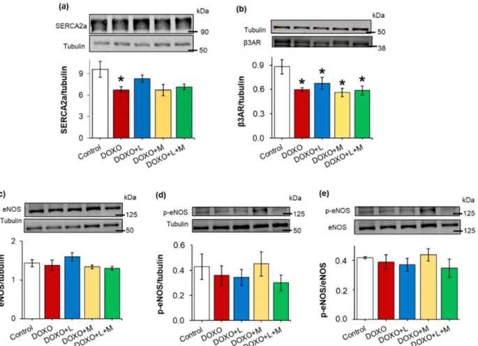
2.7. DOXO-Indued Repression of SERCA2a Was Ameliorated by Losartan at Week 9
Decreased sarcoendoplasmic reticulum calcium ATPase 2a isoform (SERCA2a) ex- pression and disturbed calcium homeostasis are related to impaired diastolic relaxation and reduced systolic function [32]. In our present study, the left ventricular expression of the SERCA2a protein was significantly decreased in the DOXO-only group compared to the control group (Figure6a). Losartan markedly increased the left ventricular SERCA2a expression compared to the DOXO-only group (Figure6a). In cases of the mirabegron and the combination-treated groups, the SERCA2a expression showed a trend to decrease compared to the control group (p= 0.062 andp= 0.054, respectively) (Figure6a).
Figure 6.Effects of losartan, mirabegron, or their combination on the left ventricular expression of SERCA2a,β3AR, eNOS and p-eNOS in DOXO-induced cardiotoxicity at week 9. Left ventricular expression and cropped representative images of (a) sarcoendoplasmic reticulum calcium ATPase 2a (SERCA2a); (b) beta-3 adrenoceptor (β3AR); (c) endothelial nitric oxide synthase (eNOS); (d) phospho- eNOS (p-eNOS), and (e) p-eNOS/eNOS ratio. Values are presented as mean±S.E.M., *p< 0.05 vs.
control group (n= 5–6, ANOVA on ranks in the cases of SERCA2a and p-eNOS expressions, and one-way ANOVA for the other parameters, Holm-Sidak post hoc test). Tubulin was used as a loading control. DOXO: doxorubicin, L: losartan, M: mirabegron. Images were captured with the Odyssey CLx machine and exported with Image Studio 5.2.5 software. The full-length Ponceau-stained membranes and Western blots are presented in Supplementary Figures S2–S5.
2.8. Left Ventricularβ3AR Protein Levels were Decreased in all DOXO-Treated Groups Irrespective of Treatments with Losartan, Mirabegron, and their Combination
Myocardial overexpression of theβ3AR and dysfunction in the eNOS-mediated path- ways were reported in heart failure of different etiologies [21]. In contrast, theβ3AR protein level was significantly decreased in the DOXO-treated groups irrespective of treat- ments with losartan, mirabegron, and their combination, compared to the control group (Figure6b). There were no significant differences in the eNOS and p-eNOS levels and the p-eNOS/eNOS ratios among the groups (Figure6c–e).
2.9. Left Ventricular Expression of the Inducible Nitric Oxide Synthase was Increased in All DOXO-Treated Groups Irrespective of Treatments with Losartan, Mirabegron, and their Combination
The left ventricular expressions of neuronal (Nos1) and inducible (Nos2) nitric oxide synthase were measured at the transcript level by qPCR (Table6). NOS1 is localized in the myocardial sarcoplasmic reticulum and modulates cardiac function and intracellular Ca2+ fluxes [33]. However, no significant difference was found in the left ventricular Nos1expression between the groups in our present study (Table6). Inducible nitric oxide synthase (Nos2) and NADPH oxidase 4 (Nox4) are major sources of elevated nitro-oxidative stress in the heart [34].
Table 6.The effects of losartan, mirabegron, and their combination on left ventricular expression of redox enzymes at the mRNA level in DOXO-induced chronic cardiotoxicity at week 9.
Relative Gene Expression
Groups
Control DOXO DOXO + L DOXO + M DOXO + L + M
Nos1/Ppia 0.80±0.17 0.68±0.07 0.7±0.16 0.64±0.22 0.65±0.05
Nos2/Ppia 0.48±0.04 1.00±0.17 * 0.75±0.10 * 0.98±0.2 * 0.64±0.06 *
Nox4/Ppia 0.96±0.15 1.24±0.21 1.20±0.39 0.24±0.10 *# 0.38±0.11#
Sod1/Ppia 1.05±0.05 0.97±0.06 0.96±0.13 0.78±0.15 0.62±0.09 *
Sod2/Ppia 0.94±0.05 0.88±0.06 0.80±0.09 0.82±0.09 0.51±0.07 *#
Sod3/Ppia 1.21±0.15 1.09±0.2 0.96±0.21 0.54±0.17 * 0.41±0.09 *#
Cat/Ppia 0.46±0.04 0.43±0.03 0.48±0.08 0.37±0.04 0.24±0.04 *#
Values are presented as mean±S.E.M., *p < 0.05 vs. control group, #p < 0.05 vs. DOXO-only group (n= 5–9, ANOVA on ranks in the cases ofNos1, Nos2, Nox4, andSod2 expressions, and one-way ANOVA for the other parameters, Holm-Sidak post hoc test). DOXO: doxorubicin, L: losartan, M: mirabegron,Cat: catalase, Nos1: neuronal nitric oxide synthase,Nos2: inducible nitric oxide synthase,Nox4: NADPH-oxidase isoform 4, Sod1: superoxide dismutase 1 (cytoplasmic),Sod2: superoxide dismutase 2 (mitochondrial),Sod3: superoxide dis- mutase 3 (extracellular),Ppia: peptidylprolyl isomerase A was used as the house-keeping gene for normalization.
Here,Nos2was significantly overexpressed in all DOXO groups irrespective of treat- ments with losartan, mirabegron, and their combination (Table6). There was no significant difference in theNox4expression at the mRNA level between the DOXO-only and the control groups. Losartan did not affect theNox4expression; however, mirabegron and the combination of losartan plus mirabegron markedly reduced theNox4expression compared to those of the control or DOXO-only groups (Table6). The left ventricular expressions of the free radical eliminating superoxide dismutase isoforms (Sod1, Sod2, andSod3) were similar in the control, DOXO-only, and losartan-treated DOXO groups (Table6). In re- sponse to mirabegron, the extracellularSod3isoform was repressed compared to the control group (Table6). In response to losartan plus mirabegron, allSodisoforms were repressed compared to the control group (Table6). The left ventricular expression of the hydrogen peroxide eliminating catalase (Cat) was also similar in the control, DOXO-only, and losartan- treated DOXO groups (Table6). Notably, losartan treatment resulted in a trend toward decreasingCatexpression compared to the control group (Table6). Moreover, the losartan plus mirabegron treatment significantly reduced theCatexpression compared to the values of the control or DOXO-only groups (Table6).
2.10. Left Ventricular Overexpression of Inflammatory Markers Il1, Il6 and Tnf Were Ameliorated by Losartan and Mirabegron in DOXO-Induced Chronic Cardiotoxicity at Week 9
Inflammatory processes triggered by the over-activation of RAAS are reported to be major contributors to the development of cardiac remodeling and fibrosis in heart failure [35]. In our DOXO-induced chronic cardiotoxicity model, there was no significant difference in the left ventricular expression of the alternative angiotensin II activator chymase (Cma), angiotensinogen (Agt), and angiotensin II type 1 receptor (Agtr1) between the DOXO-only and control groups (Figure7a–c). Notably, theAgt(p= 0.095) andAgtr1 (p= 0.074) expressions showed a trend towards decreasing response to mirabegron and a significant reduction in response to losartan plus mirabegron compared to the control group (Figure7a–c). The inflammatory markers interleukin-1 (Il1), interleukin-6 (Il6), and tumor necrosis factor-alpha (Tnf) were significantly overexpressed in the DOXO-only group compared to those of the control group (Figure7d–f). Losartan significantly reduced the overexpression ofIl1andIl6compared to the values of the DOXO-only group (Figure7d–f).
Mirabegron reduced the overexpression of all measured inflammatory markers compared to the DOXO-only group (Figure7d–f).
Figure 7.Effects of losartan, mirabegron, or their combination on the left ventricular expression of selected elements of the tissue renin-angiotensin-aldosterone system and inflammatory markers in DOXO-induced cardiotoxicity at week 9. Left ventricular expressions of (a) chymase (Cma); (b) an- giotensinogen (Agt); (c) angiotensin II receptor type 1 (Agtr1); (d) interleukin-1 (Il1); (e) interleukin-6 (Il6), and (f) tumor necrosis factor-alpha (Tnf). Values are presented as mean±S.E.M., *p < 0.05 vs. control group, #p < 0.05 vs. DOXO-only group (n= 5–6, ANOVA on ranks in the cases of Il6 and Tnf expressions, and one-way ANOVA for the other parameters, Holm-Sidak post hoc test).
Peptidylprolyl isomerase A (Ppia) was used as the housekeeping gene for normalization DOXO:
doxorubicin, L: losartan, M: mirabegron.
In response to losartan plus mirabegron,Il6showed a decreasing tendency, whereas Il1andTnfexpressions were not significantly different from those of the DOXO-only group (Figure7d–f).
3. Discussion
Here we report that mirabegron and its combination with losartan improved the systolic and diastolic dysfunction and reducedSmad2andSmad3overexpression in our DOXO-induced chronic cardiotoxicity model. Only mirabegron improved the DOXO- induced LV fibrosis markedly. Losartan failed to ameliorate the systolic dysfunction;
however, it improved the diastolic dysfunction and prevented the SERCA2a repression in our DOXO-induced cardiotoxicity model. LV overexpression ofIl1bandIl6was significantly reduced by losartan and mirabegron. Mirabegron and its combination with losartan seem to be promising therapeutic tools against systolic and diastolic dysfunction in DOXO-induced chronic cardiotoxicity.
Systolic and diastolic dysfunction resulting in heart failure are severe and well-known adverse effects of DOXO treatment [6,31]. In the present study, the mortality rate [36], echocardiographic parameters, heart rate [37,38], blood pressure [39,40], serum lipid [41,42], autopsy, and histologic findings in our DOXO-induced chronic cardiotoxicity model are consistent with the literature data on preclinical models [43] and similar to those seen in tumor survivor patients treated with DOXO [3,6,31]. Among the nine animals that died in the DOXO groups, five rats died before the start of the treatments or should be excluded due to poor systolic function (LVEF < 40%) assessed by echocardiography. During the treatments with losartan, mirabegron, and their combination, four animals died. These cases might be the consequences of DOXO treatment and not the side effects of the drugs administered in this study. To decide if the used drugs have severe side effects in DOXO- induced cardiotoxicity, more parameters, including arrhythmias, should be tested in more doses and follow-up time points with higher sample numbers in the groups.
In the cardio-oncology practice, secondary prevention of chemotherapy-induced chronic cardiotoxicity starts by using drugs applied in standard HF therapy when symp- toms, increase in cardiac biomarkers, or systolic dysfunction develops [6,31]. In our present study, 4 weeks after the last DOXO cycle, early signs of systolic dysfunction (i.e., increased LV end-systolic diameter and decreased FS) were detected in the DOXO groups. There- fore, in our present study, treatments by losartan, mirabegron, and their combination started from week 5, mimicking the conventionally scheduled therapeutic regimens in chemotherapy-induced cardiotoxicity in tumor survivor patients. It should be mentioned that cancer patients at increased risk for chronic cardiotoxicity, defined by a history or risk factors of CVDs, previous cardiotoxicity, or exposure to cardiotoxic agents, may benefit from primary preventive HF strategies when the HF medication starts during or before the chemotherapy [6,31]. Our present study might better mimic the clinical situation when children or young adults without severe risk factors for CVDs are diagnosed with tumors and treated with DOXO. At the endpoint of our present study, the DOXO-induced LV wall thinning, systolic dysfunction, and cardiac fibrosis were associated with the overex- pression of selected elements of the fibrotic TGF-β/SMAD signaling pathway (i.e.,Ctgf, TGFβRII,Smad2, Smad3,andCol1a1) and molecular markers of apoptosis (i.e.,Bax, and Bax/Bcl2ratio), cardiac remodeling (i.e.,Mmp2andMmp9), heart failure, (i.e.,Nppaand Nppb), inflammation (i.e.,Il1, Il6,andTnf) and nitro-oxidative stress (i.e.,Nos2). Moreover, the DOXO-induced diastolic dysfunction was accompanied by a reduced left ventricular SERCA2a level. These molecular findings are also in accordance with the literature data on DOXO-induced cardiotoxicity models [7,15,44–47]. Several DOXO-induced chronic cardiotoxicity models, particularly if using higher DOXO doses, may also develop severe kidney failure [43]. In our previous study, mirabegron significantly worsened the renal function in a rat model of chronic kidney disease [29]. Therefore, we aimed at using a DOXO-induced chronic cardiotoxicity model, which does not develop severe renal failure and consequently does not worsen the DOXO-induced heart failure (i.e., type 4 cardio-renal
syndrome [48]). Indeed, the serum carbamide and creatinine levels were not significantly increased in the DOXO-treated groups compared to the control group in our present study.
According to the latest guideline of the European Society of Cardiology (ESC), RAAS inhibitors, including ARBs, are recommended for pharmacological therapy of HFrEF pa- tients to reduce the risk of HF hospitalization and CV death [31]. Moreover, the latest ESC position paper on cancer treatments and cardiovascular toxicity stated that patients who develop asymptomatic LV dysfunction or HF during cancer therapy are likely to profit from the angiotensin-converting enzyme (ACE) inhibitors or ARBs and beta-blocker treatment similar to the general HF population [6]. Since our DOXO-induced cardiotoxicity model developed bradycardia, we avoided the administration of beta-blockers alone or in combi- nation with ARBs. The ARB losartan showed antiremodeling and cardioprotective effects in our rat models of radiation-induced heart disease [49] and uremic cardiomyopathy [29], or DOXO-induced chronic cardiotoxicity models used by others [19,50,51]. In contrast, in our hands, losartan failed to significantly improve the morphologic parameters (i.e., systolic wall thicknesses, LV end-systolic diameter, cardiomyocyte cross-sectional area) and the sys- tolic dysfunction (i.e., reduced FS and EF) in DOXO-induced chronic cardiotoxicity in the present study. A potential explanation for the lack of significant antiremodeling effects of losartan could be that losartan failed to significantly reduce the LV fibrosis and showed only a tendency to decrease in cardiac collagen content at the endpoint in our DOXO-induced cardiotoxicity model. Indeed, the LV expressions of selected elements of the TGF-β/SMAD fibrotic pathway (i.e.,Ctgf,TGFβRII,Smad3, andCol1a1) were not significantly different in the losartan-treated DOXO group compared to the DOXO-only or control groups. In contrast to the results of Zong et al., using higher DOXO doses (ip. 6×2.5 mg/kg) and shorter follow-up time (i.e., 6 weeks) to induce chronic cardiotoxicity,Agtr1afailed to be overexpressed in the left ventricles of our DOXO-induced cardiotoxicity model [50].
This fact might provide another explanation for the lacking antiremodeling effects of the ARB losartan in our DOXO-induced cardiotoxicity model. Notably, losartan significantly shortened the heart rate-independent diastolic function parameter isovolumetric relaxation time (IVRT) in our DOXO-induced chronic cardiotoxicity model. SERCA was shown to determine the magnitude of myocyte Ca2+cycling [52]. The early diastolic reuptake of Ca2+
into the sarcoplasmic reticulum, in part, determines the velocity at which the left ventri- cle relaxes (i.e., IVRT) [52]. Accordingly, losartan prevented the repression of SERCA2a associated with shorter IVRT in our DOXO-induced cardiotoxicity model. Moreover, it was reported that cytokines, particularly IL6, induced reciprocal expression of SERCA and natriuretic peptides at the mRNA level in cultured rat ventricular myocytes [53]. Indeed, in our present study, losartan significantly reduced DOXO-induced LV overexpression of inflammatory markers (i.e.,Il1andIl6) and natriuretic peptides (i.e.,NppaandNppb) in our DOXO-induced chronic cardiotoxicity model. In contrast, the mitral annulus velocity e’ and consequently the E/e’ ratio failed to be significantly improved by losartan in our DOXO model. Notably, cardiac fibrosis was shown to worsen myocardial relaxation pa- rameters, including e’ and E/e’ [54]. Since losartan failed to significantly improve the LV fibrosis, and probably, as a consequence, the e’ and E/e’ were not different from those in the DOXO-only group.
Mirabegron is aβ3AR agonist recently used in the treatment of overactive bladder syndrome in urology practice [21]. Interestingly,β3AR agonists showed beneficial effects on the symptoms of HF [21,55]. In healthy atrial and ventricular myocytes, the expression of β3AR is considered low but more abundant in non-cardiomyocytes, including endothelial cells [21,56,57]. In contrast toβ1- andβ2ARs, cardiacβ3AR expression increases in chronic ischemia and heart failure, which is considered a counterregulatory mechanism to prevent chronic adrenergic overactivation [21,56,58–60]. Most studies investigating the effects of β3AR agonists in HF demonstrated that β3AR agonists attenuated cardiac fibrosis and improved cardiac contractility via the β3AR/eNOS/cGMP signaling pathway as the main mechanism [21,55]. In contrast, the left ventricular β3AR was significantly repressed in all DOXO groups independent of treatments, and the eNOS and p-eNOS
levels and their ratios were similar in all groups in our present experiment. However, mirabegron significantly improved the morphologic (i.e., left ventricular wall thicknesses and end-systolic diameter), systolic (i.e., FS and EF), and several diastolic (i.e., e’ and E/e’) parameters in our DOXO-induced cardiotoxicity model. Importantly, in our present study, mirabegron was the only treatment that significantly reduced DOXO-induced cardiac fibrosis and the apoptosis markerBax/Bcl2ratio. In our previous study in a rat model of uremic cardiomyopathy, mirabegron had a moderate antifibrotic effect associated with improved diastolic function independently of theβ3AR/eNOS signaling pathway [29].
Indeed,β3AR agonists were shown to have beneficial effects independently of coupling the β3AR to eNOS in the heart and other tissues. These mechanisms include, e.g., antifibrotic effects via downregulation of the AT1 receptor [25–27,29], and CTGF [61], antioxidant and anti-inflammatory properties [62,63]. Therefore, we investigated the changes in the left ventricular expression of selected molecular markers of the RAAS, fibrosis, nitro-oxidative stress, and inflammation in response to losartan, mirabegron, and their combination in our DOXO-induced cardiotoxicity model. Interestingly, in response to mirabegron, the left ventricular expressions of the RAAS-associatedAgt and Agtr1afailed to change, and the expressions of nitro-oxidative stress-associatedNos2andNox4, and the fibroticCtgf and TGFβRII remained high in our DOXO-induced cardiotoxicity model. In contrast, mirabegron significantly decreasedSmad2andSmad3expressions compared to the DOXO- only group, and theCol1a1expression was not markedly different from the control group.
Therefore, we speculate here that mirabegron exerts its antiremodeling effect via inhibiting SMAD2/3-mediated fibrotic mechanisms independently of theβ3AR/eNOS signaling pathway in our DOXO-induced chronic cardiotoxicity model.
To investigate if the beneficial effects of losartan and mirabegron are additive, the combination of losartan and mirabegron was administered in a group of DOXO-treated animals. In summary, the combination treatment preserved the systolic function (i.e., FS and EF) 8 weeks after the DOXO administration. However, it should be mentioned that both FS and EF tended to decrease, and the isovolumic contraction time showed an increasing tendency in the combination treatment group compared to the control group. However, the combination treatment improved the diastolic parameters e’ and E/e’, similarly to the effects of mirabegron alone in our DOXO-induced cardiotoxicity model. Notably, the combination treatment failed to prevent the repression of SERCA2a and significantly reduce the overexpression of the inflammatory markers (i.e.,Il1andIl6) compared to the DOXO-only group. In response to the combination treatment, the cardiac collagen content and the expressions ofCol1a1, CtgfandSmad2were not significantly different compared to those of the control group, whereasSmad3, Agtr1, Nox4, Sod2,andNppbexpressions were significantly reduced compared to the DOXO-only group. The combination treatment significantly repressed the LV overexpression of the superoxide eliminatingSod2andSod3 and the hydrogen-peroxide eliminatingCat. This might be a consequence of the significant repression of the nitro-oxidative stress markersNox4andSod2compared to those of the DOXO-only group. Angiotensin-II is known to increase the nitro-oxidative stress and inflammation via AT1 (Agtr1) receptors by increasing NADPH-oxidase (Nox4) [64].Agtr1 was also significantly repressed in response to the combination treatment in our present study. Losartan and mirabegron seem to have a potentiating effect on reducing the AT1 receptor-mediated nitro-oxidative stress in DOXO-induced cardiotoxicity. However, this speculation should be proven by further experiments investigating the levels of ROS and RNS as well as RAAS and nitro-oxidative stress-associated proteins.
Our study is not without limitations. We intended to test and compare the effects of chronic administration of the ARB losartan, theβ3AR agonist mirabegron, and their combination on cardiac remodeling in DOXO-induced chronic cardiotoxicity in rats. We used only single doses of losartan and mirabegron, which are comparable to the human therapeutic doses and used in heart failure models of other etiologies. We tested the effects of these aforementioned drugs only in secondary prevention. It could not be excluded that losartan might have beneficial effects if its administration starts in primary prevention
(i.e., during or before the DOXO treatment). DOXO-induced chronic cardiotoxicity is a well-known complication most commonly manifested as left ventricular systolic dysfunc- tion with reduced ejection fraction. Nevertheless, significant differences exist between the DOXO administration and pathomechanisms in the model used vs. that in patients. The time period between the consecutive DOXO cycles was shorter compared to the human protocols due to the lifespan of the rats. Although female gender is a risk factor in the development of DOXO-induced cardiotoxicity in humans [3,6,65], the standardization of the results independent of female sex hormonal effects is easier in males. In our present study, only healthy male rats without cancer, CVDs, or other comorbidities were used in order to investigate the isolated DOXO effects on the heart. In future studies, animal models of both sexes representing tumor development and receiving multimodality oncotherapy (i.e., radio- and chemotherapy) would be more suitable to mirror the clinical scenario of onco-cardiology patients as closely as possible. Moreover, there are still many unknown mechanisms, including the exact role ofβ3AR in the development of DOXO-induced chronic cardiotoxicity. We demonstrated here that LVβ3AR expression decreased, and theAgtr1expression failed to change in DOXO-induced chronic cardiotoxicity 9 weeks after DOXO administration in our rat model. However, it could not be excluded that LV β3AR orAgtr1expressions might change at the protein level in earlier or later HF phases of DOXO-induced cardiotoxicity. The time-dependent and mechanistic investigation of the cardiacβ3AR expression in the development of DOXO-induced chronic cardiotoxicity was out of the scope of the present study. Notably, the precise mechanisms and functional role of theβ3AR in HF induced by different cardiovascular diseases, including diabetes mellitus, acute myocardial infarction, chronic kidney disease, or other chemotherapy-induced heart failure forms, are not fully discovered. We focused mainly on the potential protective effects of losartan, mirabegron, and their combination in DOXO-induced chronic cardiotoxicity, investigating the well-known markers of cardiac remodeling and the effects caused by losartan and mirabegron. The deep mechanistic insight of the cardioprotective effects caused by losartan and mirabegron was out of the scope of our present descriptive study.
We found antiremodeling effects of mirabegron and hypothesized that mirabegron could have antifibrotic effects in DOXO-induced chronic cardiotoxicity independently of the β3AR-eNOS-mediated pathway, but probably associated with its effects causing repression on the elements of the fibrotic TGF-β/SMAD2/3 pathway. However, further mechanistic studies, including the inhibition of the TGF-β/SMAD2/3 pathway and investigation of the expression of phosphorylated SMAD2 and SMAD3, as well as endogenous negative regula- tors of this pathway such as SMAD6 and SMAD7, are needed to explore the antiremodeling effects of mirabegron in DOXO-induced chronic cardiotoxicity.
4. Materials and Methods 4.1. Ethics Approval
This investigation conformed to the EU Directive 2010/63/EU and was approved by the regional Animal Research Ethics Committee of Csongrád County (Csongrád county, Hungary; project license: XV./57/2020, date of approval: 12 February 2020). All institu- tional and national guidelines for the care and use of laboratory animals were followed.
4.2. Animals
A total of 50 male Wistar rats (350–400 g, 8–10 weeks old) were used in the experiments.
The animals were housed in pairs in individually ventilated cages (Sealsafe IVC system, Buguggiate, Italy) in a temperature-controlled room (22±2◦C; relative humidity 55±10%) with a 12 h:12 h light/dark cycle. Standard rat chow and tap water were supplied ad libitum during the experiment.