Epigenomic and transcriptomic approaches in the post-genomic era: path to novel targets for
diagnosis and therapy of the ischaemic heart?
Position Paper of the European Society of
Cardiology Working Group on Cellular Biology of the Heart
Cinzia Perrino
1, Albert-Laszl o Barab asi
2,3,4,5, Gianluigi Condorelli
6,7, Sean Michael Davidson
8, Leon De Windt
9, Stefanie Dimmeler
10,11, Felix Benedikt Engel
12,
Derek John Hausenloy
13,14,15,16,17,18, Joseph Addison Hill
19, Linda Wilhelmina Van Laake
20,21, Sandrine Lecour
22, Jonathan Leor
23,24, Rosalinda Madonna
25,26, Manuel Mayr
27, Fabrice Prunier
28, Joost Petrus Geradus Sluijter
29, Rainer Schulz
30, Thomas Thum
31, Kirsti Ytrehus
32, and Pe´ter Ferdinandy
33,34,35*
1Department of Advanced Biomedical Sciences, Federico II University, Via Pansini 5, 80131 Naples, Italy;2Center for Complex Networks Research and Department of Physics, Northeastern University, Boston, MA, USA;3Center for Cancer Systems Biology (CCSB) and Department of Cancer Biology, Dana-Farber Cancer Institute, Boston, MA, USA;4Center for Network Science, Central European University, Budapest, Hungary;5Department of Medicine, and Division of Network Medicine, Brigham and Womens Hospital, Harvard Medical School, 75 Francis Street, Boston, MA 02115, USA;6Department of Cardiovascular Medicine, Humanitas Research Hospital and Humanitas University, Rozzano, Italy;7Institute of Genetic and Biomedical Research, National Research Council of Italy, Rozzano, Milan, Italy;8The Hatter Cardiovascular Institute, Institute of Cardiovascular Science, University College London, London, UK;9Department of Cardiology, CARIM School for Cardiovascular Diseases, Maastricht University, 6229 ER Maastricht, The Netherlands;10Institute for Cardiovascular Regeneration, University Frankfurt, Frankfurt, Germany;11German Center for Cardiovascular Research (DZHK), RheinMain, Germany;12Experimental Renal and Cardiovascular Research, Department of Nephropathology, Institute of Pathology, Friedrich-Alexander-Universit€at Erlangen-Nu¨rnberg (FAU), Erlangen, Germany;13The Hatter Cardiovascular Institute, University College London, London, UK;
14The National Institute of Health Research University College London Hospitals Biomedical Research Centre, London, UK;15Cardiovascular and Metabolic Disorders Program, Duke- National University of Singapore, Singapore;16National Heart Research Institute Singapore, National Heart Centre Singapore, Singapore;17Yong Loo Lin School of Medicine, National University Singapore, Singapore;18Barts Heart Centre, St Bartholomew’s Hospital, London, UK;19Departments of Medicine (Cardiology) and Molecular Biology, UT Southwestern Medical Center, Dallas, TX, USA;20Division of Heart and Lungs, Hubrecht Institute, University Medical Center Utrecht, Utrecht, The Netherlands;21UMC Utrecht Regenerative Medicine Center and Hubrecht Institute, Utrecht, The Netherlands;22Hatter Cardiovascular Research Institute, University of Cape Town, Cape Town, South Africa;23Neufeld Cardiac Research Institute, Tel-Aviv University, Tel-Aviv, Israel;24Tamman Cardiovascular Research Institute, Sheba Medical Center; Sheba Center for Regenerative Medicine, Stem Cell, and Tissue Engineering, Tel Hashomer, Israel;25Center of Aging Sciences and Translational Medicine – CESI-MeT, “G. d’Annunzio” University, Chieti, Italy; Institute of Cardiology, Department of Neurosciences, Imaging, and Clinical Sciences, “G. d’Annunzio” University, Chieti, Italy;26The Texas Heart Institute and Center for Cardiovascular Biology and Atherosclerosis Research, Department of Internal Medicine, The University of Texas Health Science Center at Houston, Houston, TX, USA;27King’s British Heart Foundation Centre, King’s College London, London, UK;28Department of Cardiology, Institut MITOVASC, University of Angers, University Hospital of Angers, Angers, France;29Cardiology and UMC Utrecht Regenerative Medicine Center, University Medical Center Utrecht, Utrecht, The Netherlands;30Institute of Physiology, Justus Liebig University Giessen, Giessen, Germany;
31Institute of Molecular and Translational Therapeutic Strategies, Hannover Medical School, Hannover, Germany;32Department of Medical Biology, Faculty of Health Sciences, UiT The Arctic University of Norway, Tromsø, Norway;33Department of Pharmacology and Pharmacotherapy, Semmelweis University, Budapest, Hungary;34Cardiovascular Research Group, Department of Biochemistry, University of Szeged, Szeged, Hungary; and35Pharmahungary Group, Szeged, Hungary
Received 20 September 2016; revised 12 February 2017; editorial decision 5 April 2017; accepted 27 April 2017; online publish-ahead-of-print 29 April 2017
Abstract Despite advances in myocardial reperfusion therapies, acute myocardial ischaemia/reperfusion injury and conse- quent ischaemic heart failure represent the number one cause of morbidity and mortality in industrialized societies.
Although different therapeutic interventions have been shown beneficial in preclinical settings, an effective cardio- protective or regenerative therapy has yet to be successfully introduced in the clinical arena. Given the complex
* Corresponding author. E-mail: peter.ferdinandy@pharmahungary.com
VCThe Author 2017. Published by Oxford University Press on behalf of the European Society of Cardiology.
This is an Open Access article distributed under the terms of the Creative Commons Attribution Non-Commercial License (http://creativecommons.org/licenses/by-nc/4.0/), which permits non-commercial re-use, distribution, and reproduction in any medium, provided the original work is properly cited. For commercial re-use, please contact
journals.permissions@oup.com
.. ..
.. ..
.. ..
.. ..
.. ..
.. ..
.. ..
.. ..
.. ..
.. ..
.. ..
.. ..
.. ..
.. ..
.. ..
.. ..
..
pathophysiology of the ischaemic heart, large scale, unbiased, global approaches capable of identifying multiple branches of the signalling networks activated in the ischaemic/reperfused heart might be more successful in the search for novel diagnostic or therapeutic targets. High-throughput techniques allow high-resolution, genome-wide investigation of genetic variants, epigenetic modifications, and associated gene expression profiles. Platforms such as proteomics and metabolomics (not described here in detail) also offer simultaneous readouts of hundreds of pro- teins and metabolites. Isolated omics analyses usually provide Big Data requiring large data storage, advanced com- putational resources and complex bioinformatics tools. The possibility of integrating different omics approaches gives new hope to better understand the molecular circuitry activated by myocardial ischaemia, putting it in the context of the human ‘diseasome’. Since modifications of cardiac gene expression have been consistently linked to pathophysiology of the ischaemic heart, the integration of epigenomic and transcriptomic data seems a promising approach to identify crucial disease networks. Thus, the scope of this Position Paper will be to highlight potentials and limitations of these approaches, and to provide recommendations to optimize the search for novel diagnostic or therapeutic targets for acute ischaemia/reperfusion injury and ischaemic heart failure in the post-genomic era.
䊏 䊏 䊏 䊏 䊏 䊏 䊏 䊏 䊏 䊏 䊏 䊏 䊏 䊏 䊏 䊏 䊏 䊏 䊏 䊏 䊏 䊏 䊏 䊏 䊏 䊏 䊏 䊏 䊏 䊏 䊏 䊏 䊏 䊏 䊏 䊏 䊏 䊏 䊏 䊏 䊏 䊏 䊏 䊏 䊏 䊏 䊏 䊏 䊏 䊏 䊏 䊏 䊏 䊏 䊏 䊏 䊏 䊏 䊏 䊏 䊏 䊏 䊏 䊏 䊏 䊏 䊏 䊏 䊏 䊏 䊏 䊏 䊏 䊏 䊏 䊏 䊏 䊏 䊏 䊏 䊏 䊏 䊏 䊏 䊏 䊏 䊏 䊏 䊏 䊏 䊏 䊏 䊏 䊏 䊏 䊏 䊏 䊏 䊏 䊏 䊏 䊏 䊏 䊏 䊏 䊏 䊏 䊏 䊏 䊏 䊏 䊏 䊏 䊏 䊏 䊏 䊏 䊏 䊏 䊏 䊏 䊏 䊏 䊏 䊏 䊏 䊏 䊏 䊏 䊏 䊏 䊏 䊏 䊏 䊏 䊏 䊏 䊏 䊏 䊏 䊏 䊏 䊏 䊏 䊏 䊏 䊏 䊏 䊏 䊏 䊏 䊏 䊏 䊏 䊏 䊏 䊏 䊏 䊏 䊏 䊏 䊏 䊏 䊏 䊏 䊏 䊏 䊏 䊏 䊏 䊏 䊏 䊏 䊏 䊏 䊏 䊏 䊏 䊏 䊏 䊏 䊏 䊏 䊏 䊏 䊏 䊏 䊏 䊏 䊏 䊏 䊏 䊏 䊏 䊏 䊏 䊏 䊏 䊏 䊏 䊏 䊏 䊏 䊏 䊏 䊏 䊏 䊏 䊏 䊏 䊏 䊏
Keyword Big Data
•
Omics•
Multiomics•
Tailored medicine•
Bioinformatics•
Network analysis1. Introduction
Myocardial ischaemia and reperfusion and its consequence heart failure are the leading causes of death and disability in industrialized societies.1 Despite intensive research in the last three decades, there is still no car- dioprotective drug on the market that can alleviate myocardial ischaemia and reperfusion injury thereby reducing infarct size.2–4Likewise, cardiac regeneration has the therapeutic potential to prevent the development of post-ischaemic heart failure.5 The lack of successful translation of promising therapeutic strategies to the clinical arena includes several fac- tors such as the use of hypothesis-driven, biased target discovery6,7and neglecting the interfering effects of common cardiovascular risk factors and their routine medications with cardioprotective pathways.4,8 Therefore, for successful development of cardioprotective therapies for acute myocardial ischaemia/reperfusion injury and ischaemic heart fail- ure, new ways to discover valid targets must be used.
Since the completion of the Human Genome Project (HGP)9a tre- mendous effort has been undertaken through Genetics, Genomics, and Functional Genomics studies to determine how this information might be linked to a healthy or unhealthy cardiac phenotype, particularly in myocardial ischaemia/reperfusion and the failing ischaemic heart.6 Despite great expectations, so far the clinical translation of basic
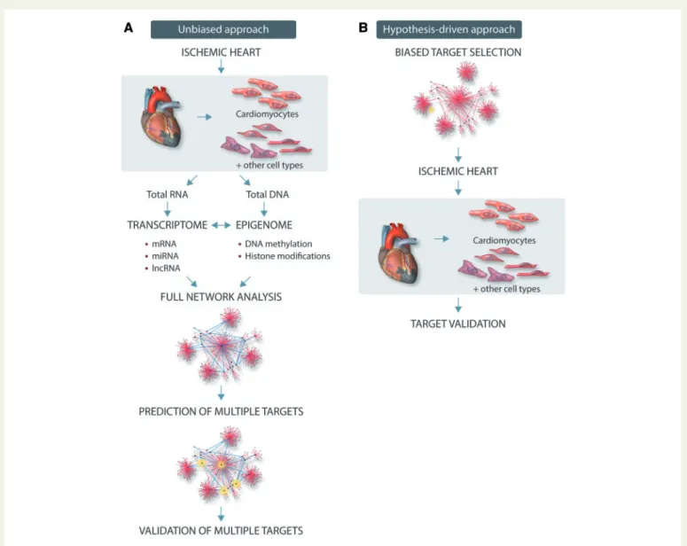
discoveries in the areas of genetics has produced disappointing results (previously addressed by a Scientific Statement from the American Heart Association,10and not discussed here). Several additional mecha- nisms related to epigenomic, transcriptomic, proteomic, and metabolo- mic regulation might be crucial to determine the pathological phenotype of myocardial ischaemia/reperfusion injury and ischaemic heart failure (for a glossary of terms, please seeTable1). The combination of omics techniques (Figure1)11,12might be extremely powerful and effective to improve our understanding of the molecular mechanisms of myocardial ischaemia/reperfusion, facilitate the development of ‘tailored’ care, and in turn ameliorate the outcome of these patients. For example, a com- bined proteomics and metabolomics analysis of murine hearts revealed a reduction in succinate after preconditioning.13Succinate was subse- quently implicated in reperfusion injury through mitochondrial ROS gen- eration.14In addition, mass spectrometry is the method of choice for the analysis of post-translational modifications involved in epigenomic regulation, including histone modifications and the identification of histone-associated proteins.15,16 Although neither proteomics nor metabolomics can currently analyse the entire proteome or metabo- lome of mammalian tissues, with the rapid advances in mass spectrome- try instrumentation the coverage is becoming increasingly more comprehensive.17–19
...
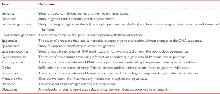
Table 1Glossary of terms
Term Definition
Genetics Study of specific, individual genes, and their role in inheritance.
Genomics Study of genes, their functions, and biological effects.
Functional genomics Study of changes in gene products (transcripts, proteins, metabolites) and how these changes mediate normal and abnormal biological function.
Comparative genomics The study to compare the genes in one organism with those of another.
Epigenetics The study of processes that lead to heritable changes in gene expression without changes in the DNA sequence.
Epigenomics Study of epigenetic modifications across the genome.
Epitranscriptomics Study of post-transcriptional RNA modifications not involving a change in the ribonucleotide sequence.
Gene expression The study of mechanisms translating information encoded by a gene into RNA structures or proteins.
Transcriptomics The study of the complete set of RNA transcripts that are produced by the genome under specific conditions.
Omics Suffix added to the names of many fields to denote studies undertaken on a large or genome-wide scale.
Proteomics The study of the complete set of translated proteins within a biological sample under particular circumstances.
Metabolomics Quantitative study of all intermediary metabolites in a given biological state.
Phenome The whole set of phenotypic entities in an organisms.
Diseasome All molecular or phenotype-based relationships between diseases observed in an organism.
.. ..
.. ..
.. ..
.. ..
.. ..
.. ..
.. ..
.. ..
.. ..
.. ..
.. ..
.. ..
.. ..
.. ..
.. ..
.. ..
.. ..
.. ..
.. ..
.. ..
.. ..
.. ..
.. ..
.. ..
.. ..
.. ..
.. ..
.. ..
.. ..
.. ..
.. ..
.. ..
.. ..
.. ..
.. ..
.. ..
.. ..
.. ..
.. ..
.. ..
.. ..
.. ..
.. ..
.. ..
.. .
Many of the stress signalling pathways activated by myocardial ischae- mia and reperfusion culminate in the nucleus, leading to alterations in cardiac gene expression.20–22A wide range of molecular players are involved in the regulation of gene expression at multiple levels, including transcriptional regulatory proteins binding specific DNA motifs in the control regions of the genes, and epigenetic modifications, inducing changes of gene expression without alterations in the gene sequence.
A variety and continuously increasing number of epigenetic mechanisms modulating gene expression has been described (reviewed in23–27), including ATP-dependent chromatin remodelling, ncRNA-based mecha- nisms, covalent histone modifications, and DNA methylation. Moreover, myocardial ischaemia/reperfusion injury and heart failure develop due to a combination of the individual genetic make-up and interaction with car- diovascular risk factors and co-morbidities accumulating during ageing, that also affect cardiac gene expression profile and phenotype.6,8
How variations in global cardiac gene expression profile can directly contribute to disease progression, the elucidation of the molecular sig- nalling pathways underlying gene expression and the ensuing phenotype is extremely complex. Microarray or sequencing-based techniques now allow high-resolution, genome-wide investigation of both epigenomic and transcriptomic landscapes, and the integration of results from these approaches, with subsequent robust bioinformatics platforms to analyse such ‘Big Data’ are emerging as promising strategies to identify key regu- latory networks and signalling hubs in response to cardiac injury (reviewed in6,12,24,26,28
). However, the simple description of gene expression modifications is not sufficient to demonstrate the mechanistic role of all the modulated genes in myocardial ischaemia/reperfusion pathophysiology. Better understanding how gene expression profile affects cardiac phenotype, global cardiac protein expression including post-translational modifications (i.e. proteomics) as well as their effect on global cell metabolism (i.e. metabolomics) should be studied as well.
Along with these bright prospects, such innovations also represent a new and great challenge. Thus, the aim of this Position Paper is to discuss promises and pitfalls of high-throughput epigenomic and transcriptomic
(proteomics and metabolomics are not described here in detail) profiling in myocardial ischaemia/reperfusion and ischaemic heart failure (proteo- mics and metabolomics are not described here in detail), and to provide recommendations to optimize the potentialities of these tools and iden- tify novel diagnostic and therapeutic targets in the ‘post-genomic’ era.
2. Epigenomics and transcriptomics in the ischaemic heart
2.1 Epigenomics
Although a large number of studies have investigated the role of epige- netic mechanisms in cardiac development, postnatal maturation and adverse cardiac remodelling in response to pressure overload,29–31only few studies have specifically investigated genome-wide epigenetic modifi- cations in cardiomyocytes induced by myocardial ischaemia or in ischae- mic heart failure. It has been recently demonstrated, using a combination of chromatin immunoprecipitation followed by sequencing (ChIPSeq) and microarray transcriptome profiling, that ischaemic preconditioning promotes epigenetic repression of the Mtor gene and induction of autophagy by enhancing dimethylation on lysine 9 on histone H3.32 In cardiac samples from end-stage heart failure patients, differences in DNA methylation and distinct patterns of histone H3 methylation (lysine 26) could also be identified.33,34Methylation differences in several genes related to heart disease or with unknown function were also identified in samples from patients with idiopathic dilated cardiomyopathy.35In addi- tion, satellite repeat elements were also found significantly hypomethy- lated in end-stage cardiomyopathic hearts compared to healthy normal controls.36These findings collectively suggest an important role of epige- netic modifications in ischaemic heart failure. However, it is still not clear whether and when epigenomic modifications might be involved in the development and progression of cardiac dysfunction promoting heart failure, and whether their modulation might ameliorate the phenotype.
Work over the past decade has revealed important roles for reversi- ble protein acetylation in cardiac development and disease.22,27As small molecule inhibitors of histone deacetylases are available and approved for clinical use, there is much interest in repurposing these compounds for cardiovascular indications in hopes of translating this biology into the clinical domain.37,38Although such compounds are already used in clini- cal trials or daily practice in cancer patients, their potential cardiac effects have been poorly described.39Future studies will be needed to address their effects in the ischaemic heart.
2.2 Transcriptomics
Transcriptomics studies in animal models of myocardial ischaemia/reperfu- sion and ischaemic heart failure have clearly demonstrated the importance of altered expression of genes involved in cardiac metabolism, cell growth and survival, inflammation, cytoskeleton structure, and extracellular matrix remodelling (recently reviewed in40). However, only a few studies per- formed global gene expression profiling in ischaemic heart failure. Some studies identified gene expression profiles differentiating between ischae- mic and non-ischaemic cardiomyopathy41,42and others compared tran- scriptomics of failing and non-failing human tissue samples.43However, Kuneret al.44revealed poor separation of human cardiomyopathies of ischaemic and non-ischaemic aetiologies by gene expression profile.
ncRNA expression profile revealed a dynamic regulation of myocardial ncRNAs in failing human heart and remodelling with mechanical circulatory support.45 Although there is an increasing interest in protecting the Figure 1Schematic representation of a full omics approach.
.. ..
.. ..
.. ..
.. ..
.. ..
.. ..
.. ..
.. ..
.. ..
.. ..
.. ..
.. ..
.. ..
.. ..
.. ..
.. ..
.. ..
.. ..
.. ..
.. ..
.. ..
.. ..
.. ..
.. ..
.. ..
.. ..
.. ..
.. ..
.. ..
.. ..
.. ..
.. ..
.. ..
.. ..
.. ..
.. ..
.. ..
.. ..
.. ..
.. ..
.. ..
.. ..
.. ..
.. ..
.. .
ischaemic heart by ischaemic conditioning and related cardioprotective mechanisms,3only a small fraction of conditioning studies assessed global gene expression profile of the heart in response to ischaemic conditioning and most of these studies are rather descriptive. The first studies in the lit- erature using the pioneer DNA-chips showed that cardioprotection by ischaemic preconditioning affected gene expression profile of the heart in rabbits and rats more than was previously thought.46,47These results have been confirmed by several papers showing that ischaemic conditioning trig- gers a cardioprotective gene expression profile in the heart at the tran- script level.6 Moreover, changes in the expression of the post- transcriptional regulators of gene expression, i.e. ncRNAs, have recently been shown by ischaemic conditioning in rat hearts.48However, little is known about whether remote ischaemic conditioning may also affect the global gene expression profile of the heart. Nevertheless, the study of Simkhovichet al.47showed that regional ischaemia led to changes in the gene expression profile in the remote non-ischaemic area of the heart. So far one paper has confirmed that remote ischaemic preconditioning leads to alteration of gene expression profile in the heart in mice.49In line with this assumption, extracellular vesicles, potential carriers of microRNAs (miR), have been shown to mediate the cardioprotective effect of remote ischaemic preconditioning in rat hearts.50
Identification of global gene expression profile of the heart due to ischae- mic conditioning stimuli may help to identify key cardioprotective pathways to be targeted, e.g. by gene therapy (see for a recent review5). As an exam- ple, by systematic comparisons looking at the direction of miR expression changes due to ischaemia/reperfusion with or without conditioning stimuli, potential cardioprotective miR targets termed ‘protectomiRs’ such as miR- 125b*, miR-139-3p, miR-320, miR-532-3p, and miR-188 have been identi- fied.48Similarly to myocardial ischaemia/reperfusion, changes in the global cardiac gene expression profile including miRNAs has been shown during the development of heart failure in different preclinical animal models51–53 as well as in human endomyocardial biopsies of heart failure patients.54,55 Moreover, transcriptomic studies have identified some miRNA clusters (miR15, miR17/92, miR 302/367) and individual miRs (including miR-99/
Let-7c, miR-100/Let-7a, miR-199a, and miR-590), that control cardiomyo- cyte proliferation and may exert potent cardiac regeneration of the adult myocardium56–58(reviewed in reference59).
2.3 Influence of risk factors, comorbidities, and their pharmacological treatment
Several risk factors, ageing, comorbidities, and their drug treatments have been shown to modulate gene expression profile of the normal and dis- eased heart thereby modulating cellular signalling by either direct modifi- cation of transcription factors, post-transcriptional, and/or post- translational modifications or epigenetic mechanisms.6–8,60–65Most of the studies determined specific epigenetic patterns or transcriptomics in the whole heart tissue or in circulating blood cells,6–8,57–62but little is known on the global gene expression or epigenetic regulation in the different cell types of cardiac tissues. One study examined the gene expression profile of patients with new-onset heart failure due to idiopathic dilated cardio- myopathy, and revealed important sex-specific differences, i.e. 35 overex- pressed and 16 down-regulated transcripts in men vs. women.66
Long-term effects of hyperlipidemia and obesity on cardiac gene expression have been studied in some experimental models. Expression of numerous genes was altered in rat hearts on cholesterol-enriched chow for 2 months,67 in high-fat chow fed mice68, and in dogs after 9 weeks of high fat diet.69In dogs fed a high-fat chow for 9–24 weeks, a time-dependent decrease in expression of several genes implicated in
obesity-related cardiac pathologies, such as hypertrophy and fibrosis was found.69Expression of several miRs is altered in the ventricles of rats fed a cholesterol-enriched chow.70In this study, one of the most robust down-regulation was found with respect to miR-25, which might be responsible for the increased oxidative- and nitrosative stress observed in the heart in this model.70However, a more complex bioinformatic analysis of the miRNA omics data is missing from this study.
Unfortunately no relevant human data are available on the effect of car- diovascular risk factors on the global gene expression pattern of the nor- mal or ischaemic heart. Although a study showed that the gene expression pattern of the human atrium is significantly altered in obe- sity,71so far data are unavailable from ventricular samples that would be relevant for myocardial ischaemia/reperfusion injury or ischaemic heart failure. For more details on cardiac transcriptomic changes in response to risk factors, see recent reviews.6,8
Data mining analysis has suggested an important role for epigenetic factors in the pathogenesis of type 2 diabetes.72However, most epige- netic studies addressing the mechanisms and effects of diabetes were performed in blood cells, endothelial cells, smooth muscle cells, or pan- creatic cells,73–75while limited information is currently available in car- diac myocytes. Significant divergence in myocardial gene expression profile has been observed in the hearts of the diabetic Otsuka Long- Evans Tokushima fatty rats when compared to the non-diabetic con- trols76and in Zucker diabetic fatty compared to control lean rats.77In type 1 diabetic rats, transcriptomic changes were responsible for altered myocardial metabolic substrate utilization that may account for the exag- gerated progression of post-infarction remodelling.78Moreover, altera- tions in miR expression profile in the heart of type 1 diabetic rats coupled with bioinformatic target prediction suggested that the affected miRs make a significant contribution to the myocardial transcriptomic profile involved in hypertrophy and fibrosis.79,80
Currently used drugs to treat comorbidities of myocardial ischaemia may modify the response of the heart to ischaemia/reperfusion injury via several mechanisms including changes in cardiac gene expression pattern.8For example, excessive use of nitrate inducing nitrate tolerance also modifies gene expression profile of the heart and leads to disruption of cardioprotective pathways.81Statins are potent cholesterol-lowering drugs that are widely used in clinical practice for primary and secondary prevention of coronary heart disease. Although some rodent studies showed alterations in cardiac gene expression profile due to treatment by different statins82,83 and despite the abundance of clinical studies investigating the effects of statins, little is known regarding the effects of this class of drugs or other widely used anti-hyperlipidemic drugs on car- diac gene expression.
2.4 Role of mitochondria?
Although most DNA is nuclear, mitochondria also contain a small amount of mitochondrial DNA (mtDNA). Mitochondria have been shown to ‘interfere’ with epigenetic modifications of nuclear DNA by several mechanisms.81,84 Moreover, mtDNA mutations have been recently associated with coronary heart disease.82,85The possibility that mtDNA might directly undergo epigenetic modifications (in particular DNA methylation since mammalian mtDNA lacks histones) has been questioned and extensively investigated for several years.86 Although mtDNA contains a lower number of CpG dinucleotides compared to nuclear DNA and lacks CpG islands, it has been recently proposed that epigenetic modifications of cytosines in mtDNA might be much more frequent than previously believed.87Mitochondria-specific oligonucleo- tide microarrays have been developed to analyse the expression levels
.. ..
.. ..
.. ..
.. ..
.. ..
.. ..
.. ..
.. ..
.. ..
.. ..
.. ..
.. ..
.. ..
.. ..
.. ..
of genes important for mitochondrial function encoded by nuclear or
.
mtDNA in differentin vitroandin vivomouse models and in humans, mostly utilized in hypoxia, toxicogenomics, and aging.87,88Future studies will be needed to determine whether and how changes in cardiac mtDNA methylation or mitochondrial transcriptomics patterns are associated with myocardial ischaemia and its comorbidities.
3. Prospects and pitfalls of
epigenomic and transcriptomic profiling in the ischaemic heart
3.1 Prospects of unbiased omics approaches
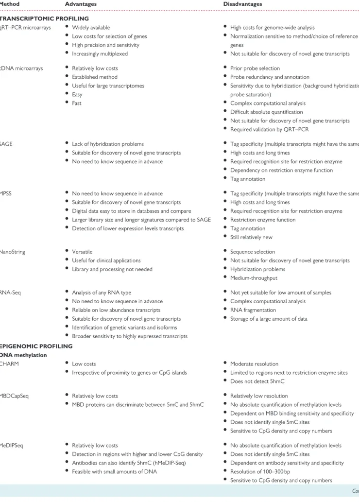
Epigenomic and transcriptomic profiling provide quantitative informa- tion about epigenetic changes, gene expression and splicing variants, and allow to study variations in the heart throughout disease progres- sion and in response to environmental changes or treatments. As global approaches, they provide large quantities of data that can be
used for unbiased assessment of pathophysiological processes with- out a priori assumption (Figure 2). Newer techniques such as ribo- some sequencing (Ribo-seq), which report only those transcripts that are being actively translated, might allow a more nuanced interroga- tion of the transcriptional landscape.89Correlation and causality asso- ciations obtained from bioinformatic analyses of these data might represent a great opportunity for researchers to gain important novel insights into the mechanisms underlying myocardial ischaemia and reperfusion. In contrast to other approaches targeting a putative, sin- gle molecular target, this strategy might be more helpful to identify multiple key targets determining cardiac dysfunction in response to ischaemia and reperfusion. Once identified crucial networks in pre- clinical studies and/or in patient’s samples, more focused analyses of specific pathways or targets might offer the potential for rapid diag- nostics and prognostics, drug safety testing and patient selection, and stratification for clinical trials. In addition to single-centre experiences, international consortia have been developed to share datasets, resources and protocols, providing a large amount of freely available information that is likely to exponentially increase in the near future (see for example reference90).
Figure 2Schematic representation of unbiased experimental omics approach (A) or hypothesis-driven traditional approach (B).
.. ..
.. ..
.. ..
.. ..
.. ..
.. ..
.. ..
.. ..
.. ..
.. ..
.. ..
.. ..
.. ..
.. ..
.. ..
.. ..
.. ..
.. ..
.. ..
.. ..
.. ..
.. ..
.. ..
.. ..
.. ..
.. ..
.. ..
.. ..
.. ..
.. ..
.. ..
.. ..
.. ..
.. ..
.. ..
.. ..
.. ..
.. ..
.. ..
.. ..
.. ..
.. ..
.. ..
.. ..
.. .
Thus, authors of this Position Paper believe that compared to hypothesis-driven investigations, a non-hypothesis-driven research approach through omics methodologies have a strong potential to speed up the discovery process and give broader insight into signalling hubs activated by myocardial ischaemia/reperfusion injury together with its co-morbidities and their routine medications.
3.2 Technical and experimental pitfalls
Despite these promising aspects, there are multiple pitfalls when dealing with epigenomic and transcriptomic analysis in the ischaemic heart.
Major technical problems are the heterogeneity within biological speci- mens, the quality of RNA and DNA (integrity, DNA contamination), and the insufficient availability of clinical samples from patients with myocar- dial ischaemia and reperfusion of ischaemic heart failure. Indeed, access to human cardiac tissues is often limited and, when possible, it usually involves samples from end-stage heart failure explanted hearts. These hearts have usually undergone multiple treatments and present relevant comorbidities that might directly or indirectly affect gene expression and epigenetic patterns. Cardiac tissue samples often contaminated by blood may also alter their omics readouts. Moreover, fresh healthy hearts are not easily available making the comparison with appropriate control groups often extremely challenging.
Given the relative inaccessibility of cardiac tissue, alternative sources have been considered, including human cell-based samples such as whole blood or white blood cells, or cell-free DNA samples from the plasma.
The rationale supporting the use of these samples is that cardiac cells may release epigenetically modified DNA or that blood cells may have undergone changes similar to cardiomyocytes. Although future investiga- tions focused on this topic might be extremely interesting and valuable, at the present time the mechanisms underlying this potential ‘mirroring’
are not clearly demonstrated, and therefore studies focusing on cardiac tissue and cells are recommended.
Preclinical studies using animal models have been extremely helpful and valuable as they allow the investigation of the progress of mechanisms of adverse remodelling towards end-stage heart failure, and comparisons of genomic expression across species often identifies patterns of evolutionary conserved transcriptional profiles among mammalian species. However, species-specific expression patterns have also been described,91and even in the presence of high-degree homology between species, there might be small but significant differences in the activated intracellular pathways.92 Moreover, the role of genetic background even in the same species is still not clearly established yet.93A vast array of heart failure-related genetically modified mouse models has been produced since the techniques became available.94 Cardiac-specific inducible and conditional gene targeting in mice has greatly enlarged our understanding of the function of individual genes or even gene variants.94However, the potential importance of adap- tations in the heart to the altered expression of a single gene may modify the global gene expression pattern of the heart.
Whatever the experimental model used, since cell-specific gene expres- sion and epigenetic profiles have been consistently reported, it would be ideal to focus and profile specific cell types, rather than the whole tissue, in which the underlying changes might be small and diluted by the presence of non-affected cells. In this regard, it would be crucial to define the target cell type, and describe the methods used to isolate, characterize, culture and eventually treat the cells. Studies have been performed with purified populations of cardiomyocytes obtained using different techniques25,29,30 or isolating cardiomyocyte nuclei from the heart by magnetic or fluorescent-assisted sorting.95The isolation procedure of a specific cell- type and cell culturing might also cause changes in RNA expression and
epigenetic profiles, thus affecting the analysis.96Single-cell techniques to perform transcriptomic analysis are already available and have been used to study gene expression during cardiogenesisin vitro,96,97even if they are not yet in widespread use. Such technologies may also provide insights into the cellular heterogeneity of tissues as recently elegantly shown for cardiac fibroblasts.98Single cell approaches also might be especially useful when myocardial ischaemia is treated by cell-based therapies, often after genetic manipulation of the transplanted cells (see for review reference5).
From a temporal standpoint, most studies so far have analysed static expression data, such as snapshots in different disease states. This approach might fail to detect disease-causing events that are transient.
By using this kind of analysis, it is difficult to determine the changes directly or indirectly initiated by myocardial ischaemia. In addition, static expression data limit the use of reverse engineering approaches to infer a network of gene interactions. In contrast, a dynamic approach describ- ing changes in expression profiles at different time points during the course of a disease would allow network-based analysis and provide novel biological hypotheses.99 Yet, only very few temporal data sets exist, and the majority of these datasets are limited to blood samples or specimens of patients of different age and conditions. Longitudinal stud- ies describing heart disease in individuals appear rather impossible, how- ever, longitudinal studies using animal models appear promising to infer a network of gene interactions, elucidate underlying mechanisms of heart diseases, and to develop novel therapies.
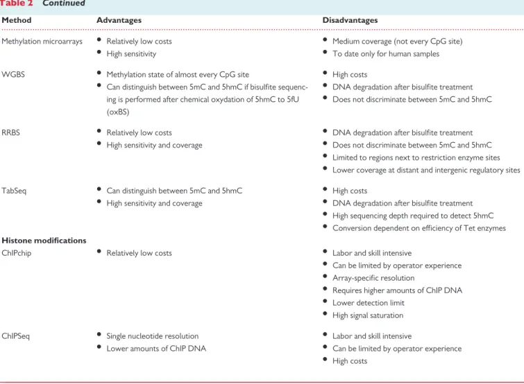
The number and diversity of transcriptomic and epigenomic profiling techniques is so wide that it can be challenging to select the best one.
Although a complete review of all possible methodologies is beyond the scope of this Position Paper, major advantages and disadvantages of the most commonly used techniques for transcriptomics and epigenomic profiling are reported inTable2. Different results may be obtained by using different technologies, and even with the same technology, variabil- ity might start in the wet lab, using different myocardial portions, sample storage conditions, processing methods, and timing. For all these rea- sons, for high-throughput sequencing could be considered running in duplicates of biological samples. In addition, when RNA is prepared and subsets are enriched or depleted (e.g. poly-A or ribo-minus selection), preparation of libraries, sequencing methods (e.g. strand specific sequencing), as well as the depth of the sequencing and bioinformatics analysis can yield strikingly different results depending on, e.g. the map- ping to the reference genome and annotations.100Such technical details may become major obstacles when comparing independent studies, which not only use different experimental conditions and platforms, but also varying bioinformatics approaches making potential meta-analyses difficult or useless. Re-analysing raw data may help to overcome at least the variability introduced by the bioinformatics analysis, but may not avoid heterogeneity introduced by the wet lab. Thus, in order to reduce variability, robust and streamlined methods, gold-standard instrumenta- tions, and only widely accepted protocols should be employed to obtain optimal starting samples. Taken together, although generation of omics data could be better harmonized by more standard techniques, novel bioinformatics models may still allow analysis of disease-specific molecu- lar networks from rather heterogeneous data sets as well.
Authors of this Position Paper believe that cardiac tissue samples and isolation of specific cell types should always be preferentially used to derive information on epigenomic and transcriptomic targets in the ischaemic heart whenever possible. Although we recognize the relative inaccessibility of human cardiac tissues in the daily clinical practice, cardiac tissue samples should be preferentially used to derive information on epi- genomic and transcriptomic targets related to myocardial ischaemia
...
Table 2Advantages and disadvantages of major genome-wide transcriptomic and epigenomic profiling techniques
Method Advantages Disadvantages
TRANSCRIPTOMIC PROFILING qRT–PCR microarrays • Widely available
• Low costs for selection of genes
• High precision and sensitivity
• Increasingly multiplexed
• High costs for genome-wide analysis
• Normalization sensitive to method/choice of reference genes
• Not suitable for discovery of novel gene transcripts cDNA microarrays • Relatively low costs
• Established method
• Useful for large transcriptomes
• Easy
• Fast
• Prior probe selection
• Probe redundancy and annotation
• Sensitivity due to hybridization (background hybridization, probe saturation)
• Complex computational analysis
• Difficult absolute quantification
• Not suitable for discovery of novel gene transcripts
• Required validation by QRT–PCR
SAGE • Lack of hybridization problems
• Suitable for discovery of novel gene transcripts
• No need to know sequence in advance
• Tag specificity (multiple transcripts might have the same tag)
• High costs and long times
• Required recognition site for restriction enzyme
• Dependency on restriction enzyme function
• Tag annotation
MPSS • No need to know sequence in advance
• Suitable for discovery of novel gene transcripts
• Digital data easy to store in databases and compare
• Larger library size and longer signatures compared to SAGE
• Detection of lower expression levels transcripts
• Tag specificity (multiple transcripts might have the same tag)
• High costs and long times
• Required recognition site for restriction enzyme
• Restriction enzyme function
• Tag annotation
• Still relatively new
NanoString • Versatile
• Useful for clinical applications
• Library and processing not needed
• Sequence selection
• Not suitable for discovery of novel gene transcripts
• Hybridization problems
• Medium-throughput
RNA-Seq • Analysis of any RNA type
• No need to know sequence in advance
• Reliable on low abundance transcripts
• Suitable for discovery of novel gene transcripts
• Identification of genetic variants and isoforms
• Broader sensitivity to highly expressed transcripts
• Not yet suitable for low amount of samples
• Complex computational analysis
• RNA fragmentation
• Storage of a large amount of data
EPIGENOMIC PROFILING DNA methylation
CHARM • Low costs
• Irrespective of proximity to genes or CpG islands
• Moderate resolution
• Limited to regions next to restriction enzyme sites
• Does not detect 5hmC MBDCapSeq • Relatively low costs
• MBD proteins can discriminate between 5mC and 5hmC
• Relatively low resolution
• No absolute quantification of methylation levels
• Dependent on MBD binding sensitivity and specificity
• Does not identify single 5mC sites
• Sensitive to CpG density and copy numbers
MeDIPSeq • Relatively low costs
• Detection in regions with higher and lower CpG density
• Antibodies can also identify 5hmC (hMeDIP-Seq)
• Feasible with small amounts of DNA
• No absolute quantification of methylation levels
• Does not identify single 5mC sites
• Dependent on antibody sensitivity and specificity
• Resolution of 100–300 bp
• Sensitive to CpG density and copy numbers
Continued
.. ..
.. ..
.. ..
.. ..
.. ..
.. ..
.. ..
.. ..
.. ..
.. ..
.. ..
.. ..
.. ..
.. ..
.. ..
.. ..
.. ..
whenever possible. Alternative and more accessible sources of human biological material (whole blood, serum, white cells, plasma, urine, etc.) can be used only if the mechanisms underlying the potential mirroring of the heart in these samples are clearly demonstrated and validated.
3.3 Target identification: importance of bioinformatics
Target discovery is the crucial initial step in biomarker and drug develop- ment. Undoubtedly, a critical aspect in the search of novel targets in the post-genomic era is probably no longer data generation, but rather data analysis and interpretation. The search, identification, validation, and interpretation of novel disease-associated targets will require innova- tions in high-throughput technologies, biostatistics and bioinformatics, and will necessitate interdisciplinary expertise and teamwork of clini- cians, biologists, biochemists, and bioinformaticians. In this context, final data interpretation usually develops in a multidisciplinary, shared envi- ronment. Accordingly, bioinformatics has undoubtedly gained wider prominence over the last decade in myocardial ischaemia/reperfusion and other research areas. Bioinformatics areas of interest include the development of new algorithms and statistics to assess relationships among members of large data sets, the ability to analyse and interpret dif- ferent types of sequences, domains, and structures, and the capacity to
develop or implement tools enabling efficient access and management of different types of information. A large number of methods, including pat- tern recognition algorithms, network analysis, and other artificial intelligence-based algorithms have been applied to bioinformatics research and will not be discussed here in detail.12,101–103In general, all approaches are characterized by specific advantages or limitations for target identification and, in order to apply a ‘tailored’ bioinformatics approach to the research plan, it is usually necessary to accurately pre- determine and focus on the elements to be investigated, on the interac- tions between the different elements, on the mathematical models to describe these interactions and on statistics to be used to analyse the data.99Thus, combining biological concepts with computer tools or stat- istical methods it is possible to discover, select and prioritize targets.
Importantly, ‘targets’ should be broadly considered molecular entities (such as genes, proteins, RNA, and their modifications) as well as biologi- cal phenomena (such as molecular functions and signalling pathways).
The complex features induced by myocardial ischaemia are difficult to
‘capture’ using single omic data modality, addressing only one mechanism of disease. There is now significant interest in the use of multiple high- throughput omic data for the identification of novel molecular subtypes of a disease. Integration of expression data with epigenetic datasets is a prom- ising strategy to improve accuracy of the inferred networks, enable the ...
Table 2 Continued
Method Advantages Disadvantages
Methylation microarrays • Relatively low costs
• High sensitivity
• Medium coverage (not every CpG site)
• To date only for human samples WGBS • Methylation state of almost every CpG site
• Can distinguish between 5mC and 5hmC if bisulfite sequenc- ing is performed after chemical oxydation of 5hmC to 5fU (oxBS)
• High costs
• DNA degradation after bisulfite treatment
• Does not discriminate between 5mC and 5hmC
RRBS • Relatively low costs
• High sensitivity and coverage
• DNA degradation after bisulfite treatment
• Does not discriminate between 5mC and 5hmC
• Limited to regions next to restriction enzyme sites
• Lower coverage at distant and intergenic regulatory sites
TabSeq • Can distinguish between 5mC and 5hmC
• High sensitivity and coverage
• High costs
• DNA degradation after bisulfite treatment
• High sequencing depth required to detect 5hmC
• Conversion dependent on efficiency of Tet enzymes Histone modifications
ChIPchip • Relatively low costs • Labor and skill intensive
• Can be limited by operator experience
• Array-specific resolution
• Requires higher amounts of ChIP DNA
• Lower detection limit
• High signal saturation ChIPSeq • Single nucleotide resolution
• Lower amounts of ChIP DNA
• Labor and skill intensive
• Can be limited by operator experience
• High costs
qRT–PCR,quantitative real-time PCR; SAGE,serial analysis of gene expression; MPSS, massively parallel signature sequencing; RNASeq, RNA-sequencing; CHARM, comprehensive high-throughput arrays for relative methylation; MBDCapSeq, methyl-CpG-binding domain (MBD) capture by affinity purification followed by sequencing; MeDIPSeq, followed by sequencing; WGBS, whole genome bisulfite sequencing; RRBS, reduced representation bisulfite sequencing; TabSeq, tet-assisted bisulfite sequencing; ChIPchip, chromatin immuno precipitation followed by microarrays; ChIPSeq, chromatin immuno precipitation followed by sequencing.
.. ..
.. ..
.. ..
.. ..
.. ..
.. ..
.. ..
.. ..
.. ..
.. ..
.. ..
.. ..
.. ..
.. ..
.. ..
.. ..
.. ..
.. ..
.. ..
.. ..
.. ..
.. ..
.. ..
.. ..
.. ..
.. ..
.. ..
.. ..
.. ..
.. ..
.. ..
.. ..
.. ..
.. ..
.. ..
.. ..
..
discovery of new insights and create a global perspective from which novel diagnostic, prognostic or therapeutic targets in the ischaemic heart could be discerned. Despite the abundance of genome-wide transcriptomic and epigenomic datasets, there is a surprising shortage of statistical tools aimed at their specific integrative analysis. Such integrative tools might be crucial for the discovery of novel subnetworks (disease module) and/or relevant molecular pathways. Combination of these methods usually entails specific difficulties, and therefore establishing the concepts and fundaments of data integration is clearly needed. Traditional database techniques are not focused on the problems that arise in the search and finding of data in large, not pre-organized repositories, for linking what is found to other data, or for reusing and repurposing the data without significant effort.
Moreover, different omics data have different distributions and variation patterns that configure data heterogeneity. In addition to the specific varia- tion patterns in each layer of information, inter-layer regulatory co- variations can be identified, increasing complexity. While multiple pro- grams and web services are available for the analysis of single omics data, the majority of these tools has a command-line interface, performs only one specific task and typically requires file conversions. To better explore and integrate multiple omics data modalities (for example epigenetic, tran- scriptional, proteomic, and metabolomic modifications), novel and more sophisticated bioinformatics methods, that can seamlessly account for the networked nature of the subcellular environment, and that are simple to use by the general research community would be needed.
Authors of this Position Paper believe that in order to fully realize the scientific value of the vast amount of omics data, it is essential to fill the gap between data generation and investigators’ ability to retrieve and interpret the data. Most network and clustering methods proposed to identify molecular targets were developed for single omic data types and may not be appropriate when more than one omic data type is collected in the same study subjects. To better explore and integrate multiple omics data modalities, novel and sophisticated bioinformatics methods would be needed, allowing automated, systematic, and unbiased identifi- cation of targets. This approach will need more financing and more col- laboration between cardiovascular and bioinformatic research groups, ideally hiring biomedical scientists with strong bioinformatic expertise, a species hardly found due to gaps in biomedical education.
3.4 Data availability, storage, and sharing
Collaborative research is usually the rule in this field, and it requires implementation of an integrative framework with standardization of data formats and possible access to the data from multiple web-based user positions. Distinct data warehousing approaches have been used, and which is more useful is yet to be determined. Shared warehousing possi- bilities and software components might be extremely helpful for success- ful collaborative work. Moreover, since several heterogeneous databases are currently available and more are under steady develop- ment, a standardized interface might represent an important target of
research for developers. In general, more advanced and powerful com- puting will be needed in the future for large-scale omics analysis in terms of hardware and development of novel software. Additional issues regarding security and appropriate control of access to data, privacy pro- tection, protection by backup, ethical issues, real-time availability and eventually data mobility will also likely be a matter of research and dis- cussion in this field. These next-generation post-genomic challenges will need to be addressed in the future by computer developers, researchers, and the general population of patients who might benefit from the potential applications of these studies.
Various datasets derived from different detection systems are in need of harmonization as most profiling studies are performed using dissimilar plat- forms, requiring re-analysis of existing raw datasets, and after harmonization of datasets. Since availability of data is crucial for validation of the bioinfor- matics approach, authors of this Position Paper recommend that publishers and researchers commit to make freely available all relevant information, raw data sets, and their analysis. Details of protocols are essential allowing investigators to compare or pool data sets. As soon as technologies become state-of-the-art, the development of standard operating proce- dures (SOPs) might be helpful to provide guides for the research commun- ity. Moreover, acquired data should be stored and managed in open- source, web-based platforms for data-intensive biomedical research.
3.5 Target validation
Afterin silicoidentification of key gene/RNA/protein targets by different bioinformatic tools analysing epigenomic and transcriptomic data, exper- imental validation of these predicted molecular targets at the protein level is essential. Usingin vitrocellular systems (genome editing, transfec- tions, etc.) for target validation has become a relatively easy approach.70 However, target validationin vivoin the target disease models preferably in the presence of relevant co-morbidities, obviously cannot be spared.6,8 An obvious pitfall of such in vivo validation is that the translational value of routinely usedin vivodisease models of myocardial ischaemia/reperfusion and heart failure is not sufficient (e.g. lack of comorbidities and co-medications), and the cost of such target validation is relatively high. Nevertheless, an increasing number of credible data on cellular signalling in health and disease in multiplex preclinical disease models and from human samples will lead to creation of an accurate dis- ease module, a subnetwork of the full human interactome, integrating knowledge from DNA, epigenome, transcriptome, proteome, and metabolome.104 Since the network analysis approach of Big Data is somewhat tolerant to input of data with heterogenous quality, this will allow us to integrate data in the ischaemic heart in the context of the ‘dis- easome’, allowing us to explore its relationship to common cardiac comorbidities and other diseases.104
Authors of this Position Paper believe that predicted targets identified by bioinformatics approaches from omics studies should be always vali- dated in order to avoid generation of purely descriptive data and Table 3Major recommendations
• Unbiased bioinformatic analysis of full epigenomic and transcriptomic profiles of the ischaemic heart (preferably including proteomic and metabolomic data) may lead to identification of novel molecular targets;
• Cardiac tissue samples should be used for omics assays and cell-specific analysis should be attempted;
• User friendly bioinformatic tools should be developed for target prediction from large omics data;
• Omics data should be stored in open-access databases to enable their analysis by the global scientific community;
• Experimental validation of predicted targets should be performed in relevant models of the ischaemic heart.