Possibilities for protecting cardiac and vascular function against ischemia-reperfusion injury
Ph.D. Thesis
Enikő Kriebitzsch-Barnucz
Doctoral School of Basic Medicine Semmelweis University
Supervisor: Prof. Gábor Szabó, MD, med. habil.
Official reviewers:
Zsuzsanna Miklós, M.D., Ph.D.
Tamás Csont, M.D., med. habil.
Head of the Final Examination Committee:
Prof. Emil Monos, M.D., D.Sc.
Members of the Final Examination Committee:
Tamás Ivanics, M.D., med. habil.
Csaba Csonka, M.D., med. habil.
Budapest 2017
2 1. INTRODUCTION
The difficulty of reducing ischaemia-reperfusion (I/R) injury and hypoxia is a major problem in cardiology and cardiovascular surgery, and is also related to problems that occur during organ transplantation. The aim of cardioprotection during and after I/R injury in a clinical setting is to reduce the infarct size, prevent severe cardiac arrhythmias and increase the contractile function of the intact myocardium. Cardiovascular protection during decreased blood and oxygen supply is extensively discussed and studied in pathophysiology and clinical research. Therapeutic strategies have improved over recent years; at present, drug research and operative developments have resulted in better outcomes and prognoses even for patients with complicated disorders. However, the development and investigation of new cardioprotective drugs is necessary because cardiovascular ischaemia-related disorders are some of the leading causes of death in the developed world. This thesis therefore focuses on cardiac and vascular protection against I/R injury. It attempts to identify potential targets for novel therapeutic options to reduce ischaemia- and reperfusion-related organ damage, function loss of implanted organs and mortality.
1.1. Hypoxic adaptation mechanisms
Mammals have oxygen sensing mechanisms that help cells adapt to hypoxia. In the course of evolution, some cell groups specialised to sense O2 tension, including the carotid body, pulmonary artery, adrenal chromaffin cells, mitochondria via reactive oxygen species, energy state and cytosolic redox state. Hypoxia-inducible factor (HIF)-1 is a heterodimeric transcription factor consisting of the consecutively expressed HIF-β (also called ARNT) and a regulatory HIF-α subunit (mainly post-translationally regulated), which regulates after dimerisation. The regulation of HIF-1α is regulated via prolil hydroxlases (PHD). Under normoxic condition PHD leads to degradation of HIF-1α, in contrast under hypoxic conditions HIF- 1α can be expressed and translocate into the nucleus, were they bind to hypoxia responsible element and induce expression of several adaptive genes. The HIF-1α in animal infarct models improved myocardial perfusion, maintained ventricular function and cardiac
3
HIF-1α overexpression which reduced the myocardial infarct size and promoted post-ischaemic function and capillarisation. This suggests that HIF-1α activation/stabilisation is beneficial in cardiac ischaemic syndromes. The mechanism of how HIF activation can protect cells against I/R injury has not been completely explored, although it involves several pathways such as antioxidant, angiogenesis, cell death and antiapoptotic pathways. Drugs, such as dimethyloxalilglycin (DMOG), which can induce HIF stabilisation under normoxic conditions, could be one of the next generation treatments for myocardial infarction, stroke, renal or liver injury, peripheral vascular disease or severe anaemia. In this study, we investigated the potential beneficial effects of treatment with DMOG in the model of vascular ischemic injury.
1.2. Role of zinc in cytoprotection during I/R injury
Disruption of zinc homeostasis is associated with severe pathophysiological conditions such as decreased erythrocyte copper–
zinc superoxide dismutase, increased low-density lipoprotein cholesterol, decreased high-density lipoprotein cholesterol, decreased methionine and leucine encephalins, decreased glucose clearance and abnormal cardiac function. However it leads to oxidative stress, which is the main and common point in the pathophysological processes. Copper- or iron-binding sites of macromolecules such as DNA, peptides or proteins, nucleotides or glucose behave as centres for production of hydroxyl radicals via Fenton reaction. The acute protective role of zinc ions for myocardial tissue is mainly due to changes in redox homeostasis: a decrease in generation of .OH from H2O2 due to antagonism of redox-active metals such as iron and copper. The other mechanism is the stabilisation of sulfhydryl, where zinc protects several enzymes (e.g. delta-aminolevulinate dehydratase, dihydroorotase and tubulin). Numerous publications over the last 10–20 years have reported on the protective effects of zinc. Several authors have also published data on different zinc complexes such as chloride salt or zinc complexed with carnosine, histidinate or aspartate. The fact remains that the one common component of all of these studies is the presence of zinc. In this study, we investigated the potential beneficial effects of Q50, an
4
iron-chelating and zinc-complexing agent that belongs to the 8- hydroxyquinoline family. Therefore, Q50 may be a good candidate as a therapeutic agent because of its iron-chelating potential. In addition, it acts on intracellular sources of zinc, forming a protective complex.
2. OBJECTIVES
The objectives of this study were to describe the pathophysiological changes to the vascular system and myocardium during and after I/R injury using rodent models. The use of antioxidants and effect of the HIF stabilisation under myocardial or vascular ischaemic conditions were investigated. This may help to improve our understanding of the role of these pathways and form the basis for future developments.
The first aim of this study was to focus on the role of the HIF in vascular cold ischaemic storage and warm reperfusion injury. We treated isolated rat aortic rings with DMOG (due to PHD inhibition induced HIF stabilisation) and simulated reperfusion injury in an organ bath by adding hypochlorite. In addition to the vascular functional measurements, we investigated the cellular and molecular changes.
The second aim of this study was to investigate the activity and characteristics of the newly developed iron-chelating and zinc- complexing agent Q50. This study was carried out in rodent models of regional and global myocardial I/R. Regional myocardial ischaemia was induced in the rodent model by ligation of the left anterior descending (LAD) coronary artery. Global myocardial ischaemia was induced by orthotopic heart transplantation. Cellular and molecular changes to the heart were investigated after cardiac functional measurements were performed.
5 3. METHODS
3.1. Effects of prolyl hydroxylase inhibition on vascular function
3.1.1. Animals
Sprague Dawley rats (male, 250–350 g) were used in the experiments. The rats were randomly assigned to different groups.
3.1.2. Experimental groups
The aortic segments for the organ bath experiments were randomised into three groups: 1) control group, the aortic rings were immediately mounted in the organ bath; 2) Sodium hypochlorite (NaOCl) group, the aortic rings were preserved in saline at 4°C for 24 h after explantation; 3) DMOG group, the aortic rings were stored at 4°C for 24 h in saline or in 10−4 M DMOG-supplemented saline.
Vascular smooth muscle cells (VSMC) were divided into three groups: 1) control group, without cold ischaemia and warm reperfusion; 2) NaCl group, cells were stored at 4°C in saline for 24 h followed by 6 h warm reperfusion in a normal medium at 37°C; 3) DMOG (10−3M) group, cells were stored at 4°C in DMOG- supplemented saline for 24 h followed by 6 h warm reperfusion in a normal medium at 37°C.
3.1.3. Model of in vitro cold ischaemic storage and warm reperfusion-induced vascular injury
After 24 h of cold storage, we investigated in vitro vascular functions in an organ bath. Therefore, the aortic rings were investigated in a similar manner, with additional exposure to NaOCl for 30 min and rinsed before phenylephrine pre-contraction. The different preservation solutions were aerated with nitrous oxide to reduce the oxygen concentration, simulating hypoxic conditions.
3.1.3.1. Preparation and in vitro assessment of aortic rings vascular function
Rats were anaesthetised with an intraperitoneal pentobarbital injection. After a bilateral thoracotomy, the thoracic aorta was removed and immediately placed in a cold Krebs–Henseleit solution (KHS). After dissecting the fat and connective tissue, 4 mm length
6
segments of the aorta were placed in test tubes with different solutions. Isolated aortic rings were mounted on stainless steel hooks in individual organ baths containing 25 ml of KHS at 37°C. The aortic rings were placed under a resting tension of 2 g and equilibrated for 60 min. At the beginning of each experiment, maximal contraction forces in response to KCl were determined, and the aortic rings were washed until the resting tension was obtained again. Aortic preparations were pre-constricted with phenylephrine (10−6 M), the α-adrenergic receptor agonist, until a stable plateau was obtained. Relaxation responses were examined by adding cumulative concentrations of endothelium-dependent dilator acetylcholine (10−9– 10−4 M). To test the relaxation responses of VSMC, a direct nitric oxide-donor, sodium nitroprusside (SNP, 10−10−10−5 M), was used.
Contractile responses to phenylephrine are expressed as a percentage of the maximal contraction induced by KCl. Vasorelaxation and its maximum (Rmax) were expressed as a percentage of the contraction induced by phenylephrine.
3.1.4. Cold ischaemic storage and warm reperfusion injury on VSMC
VSMC were isolated from rat aortas, re-suspended in a base medium, plated and incubated in 6-well plates. The medium was changed for a saline or DMOG-supplemented saline solution, incubated for 24 h and stored for hypothermic ischaemia at 4°C. After the cold storage, the complete cell culture medium was added, and reperfusion was simulated by further incubation at 37°C for 6 h.
3.1.5. Aortic and VSMC mRNA expression by quantitative real- time polymerase chain reaction
The expression of HO-1 in aortic rings and VSMC was determined.
mRNA was isolated from aortic rings after 0, 2, 4 and 6 h of warm reperfusion. Sample quantifications were normalised to a glyceraldehyde-3-phosphate dehydrogenase (GAPDH) expression using a pool of all cDNAs from the control group.
3.1.6. Statistical analyses
Statistical analyses were performed using Origin v. 7.0 (OriginLab Corporation). Data distribution was tested for normality using the
7
Shapiro–Wilk test. Normally distributed data are expressed as mean
± standard error of the mean (SEM). Two groups were compared using Student’s t-test and more than two groups (e.g. PCR, immunohistochemical scores and VSMC assay) were compared using one-way analysis of variance (ANOVA) with Bonferroni correction for multiple post hoc comparisons. P < 0.05 was considered to be statistically significant.
3.2. Q50 in the I/R rat models
3.2.1. Rat model of myocardial I/R injury: surgical preparation of regional I/R
Rats were anaesthetised with sodium pentobarbital. An intratracheal tube was inserted to artificially ventilate the animals. The chest was opened via a left thoracotomy followed by a pericardiotomy. A single silk suture was passed around the LAD coronary artery, and the ends were pulled through a small pledget to form a snare and then tightened. After 45 min of ischaemia, reperfusion was achieved by releasing the snare. Sham-operated animals were subjected to the same surgical procedures, except that the suture around the LAD coronary artery was not tied.
3.2.1.1. Experimental groups
The rats were randomised into four groups: 1) Sham rats received the vehicle but no tightening of the coronary suture; 2) Sham + Q50 rats received Q50 and the ligature was placed around the LAD but without occlusion; 3) I/R rats were treated with the vehicle and subjected to I/R; 4) I/R + Q50 rats were given Q50 and subjected to I/R, undergoing 45 min of myocardial ischaemia, followed by 24 h of reperfusion. Vehicle or Q50 were given as an intravenous bolus 5 min before the onset of reperfusion.
3.2.1.2. In vivo haemodynamic parameters
After 24 h of reperfusion, the rats were anaesthetised with sodium pentobarbital, tracheotomised, intubated and artificially ventilated.
To assess cardiac function, a left ventricular (LV) pressure-volume analysis was performed with a pressure-volume catheter.
8
3.2.2. Rat model of global myocardial ischemia: heterotopic heart transplantation
Transplantations were performed in an isogenic Lewis to Lewis rat strain. In brief, the donor rats were intraperitoneally anesthetised with a mixture of ketamine and xylazine and then heparinised.
Cardiac arrest was induced by administering a Custodiol® solution.
After 1 h of ischaemia, the hearts were implanted intra-abdominally, anastomosing end-to-side the aorta and pulmonary artery of the donor heart with the abdominal aorta and inferior vena cava of the recipient, respectively. After completion of the anastomoses, the vessels were released and the heart was perfused in situ.
3.2.2.1. Experimental groups
The rats were randomly divided into four groups: 1) control, heart explanted without any treatment; 2) control + Q50, Q50 administered 1 h prior to explantation; 3) I/R, donor rats received vehicle 1 h prior to explantation, hearts were then subjected to 1 h ischaemia and transplanted; 4) Q50 + I/R, Q50 treatment of the donor animals 1 h prior to explantation, hearts were then subjected to 1 h ischaemia and transplanted. Vehicle or Q50 were intravenously given.
3.2.2.2. Haemodynamic measurements
After 1 h of reperfusion, rats were intraperitoneally anesthetised with a mixture of ketamine and xylazine. A balloon catheter was introduced into the left ventricle via the apex to determine LV function using a Millar micromanometer at different LV volumes.
LV pressure-volume relationships were constructed using these data.
At the end of the experiment hearts were removed for further analysis.
3.2.2.3. Determination of high-energy phosphate levels
Heart tissue was homogenised and using enzyme kinetic assay ATP, ADP and AMP contents were expressed as micromoles per gram of dry weight. The energy charge potential was calculated as [ATP + 0.5 (ADP)]/[ATP + ADP + AMP].
9
3.2.2.4. Quantitative real-time PCR and Western blotting
The mRNA and protein expression of cytochrome-c oxidase, SOD-1 and MMP-2 (data not shown) was determined. Sample quantifications were normalised to the GAPDH expression.
3.2.3. In vitro cardiac myocyte protection studies
H9c2 rat embryonic cardiac muscle cells were cultured in Dulbecco’s Modified Eagle’s Medium and treated with and without Q50 30 min after exposure to H2O2. On the following day, H9c2 rat embryonic cardiac muscle cells were post-treated (30 min after H2O2
treatment) with Q50 or a solvent of the compound. The absolute control group did not receive the H2O2 treatment. The H2O2
concentration used here to elicit cell injury (100 µmol/L) was previously optimised for H9c2 cells according to their sensitivity to oxidative stress. Cells were dynamically monitored over 24 h by measuring the electrical impedance every 5 min with real-time cell electronic sensing assay.
3.2.4. Statistical analyses
All data are expressed as mean ± SEM. For the heart transplantation haemodynamic parameters, Student’s t-test was used to analyse differences between the groups. In all other cases, means were compared between groups by one-way ANOVA with Bonferroni correction for multiple post hoc comparisons. P < 0.05 was considered statistically significant.
4. RESULTS
4.1. Effects of prolyl-hydroxylase inhibition on vascular function
4.1.1. Endothelium-dependent vasorelaxation of aortic rings In NaOCl group, endothelial dysfunction was indicated by reduced Rmax to acethylcholine and right shift of concentration response curves of aortic segments to acethylcholine compared with the control group. Treatment with DMOG significantly improved acethylcholine-induced vasorelaxation. Contractile responses to high K+-induced depolarization was significantly reduced at high concentrations of DMOG compared with those in the control group.
10
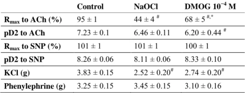
Table 1. Contractile responses and vasorelaxation ability in the three groups.
Values of maximal relaxation (Rmax) and pD2 to acethylcholine and sodium nitroprusside in the control, NaOCl-exposed and DMOG-treated aortic rings. Values represent mean ± SEM. Significance (P < 0.05): # vs. control; * vs. NaOCl.
4.1.2. Effects of DMOG on relative haem-oxygenase-1 mRNA- expression
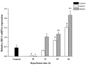
At the beginning of the warm reperfusion, expression of HO-1 in the NaOCl group was significantly reduced compared with that in the control. From the second hour of ‘reperfusion’, the aortic segments treated with the prolyl-hydroxylase inhibitor showed a significantly increased level of an inducible HO form compared with the NaOCl treated aortic rings.
VSMC after 24 h cold storage followed by 6 h warm reperfusion relative mRNA-expression of HO-1 was significantly higher in the DMOG group than in the NaCl group (data not shown).
Control NaOCl DMOG 10−4 M Rmax to ACh (%) 95 ± 1 44 ± 4 # 68 ± 5 #,*
pD2 to ACh 7.23 ± 0.1 6.46 ± 0.11 6.20 ± 0.44 # Rmax to SNP (%) 101 ± 1 101 ± 1 100 ± 1 pD2 to SNP 8.26 ± 0.06 8.11 ± 0.06 8.33 ± 0.10 KCl (g) 3.83 ± 0.15 2.52 ± 0.20# 2.74 ± 0.20# Phenylephrine (g) 3.25 ± 0.15 3.45 ± 0.15 3.10 ± 0.16
11
0.0 0.5 1.0 1.5 2.0
2.5 *
*
#
#
#
#*
Control t2 t4 t6
Reperfusion time (h)
Relative HO-1 mRNA-expression
Control NaOCl DMOG
t0
# *
Figure 1. HO-1 mRNA-expression. Relative expression of HO-1 in aortic segments compared with the expression of GAPDH after 24 h of cold ischaemic storage followed by 0, 2, 4 and 6 h of warm reperfusion in the control, NaOCl and DMOG groups. After the second hour, the DMOG treated group had significantly higher HO-1 mRNA levels. Values represent mean ± SEM.
4.2. Effect of Q50 treatment on regional and global myocardial I/R injury
Rats subjected to LAD occlusion and reperfusion showed no observed difference in the area at risk between the vehicle and Q50 treated rats, indicating that a comparable degree of ischaemia was induced in both groups. Post-ischaemic treatment with Q50 did not reduce the myocardial infarct size compared with that in the I/R group.
4.2.1. Effect of Q50 on cardiac function after myocardial infarction
Cardiac parameters of myocardial infarcted rats showed no significant difference in heart rate, LV end-diastolic pressure, stroke volume, cardiac output, stroke work or slope of EDPVR values in the groups compared with those of the controls. However, increased end- systolic and end-diastolic volumes in myocardial infarcted rats were
12
significantly reduced after post-ischaemic treatment with Q50. In the I/R group, decreased LV load-dependent (dP/dtmax) and decreased load-independent (slope of dP/dtmax/end-diastolic volume relationship and maximum time-varying elastance) contractility parameters were significantly increased after post-ischaemic treatment with Q50. Moreover, the ejection fraction was significantly increased in the I/R + Q50 group compared with that in the I/R group. Systolic and diastolic blood pressures and mean arterial pressure were significantly reduced in I/R, I/R + Q50 and sham + Q50 groups compared with those in the sham operated rats. When compared with the sham group, rats with myocardial infarction showed significantly decreased LV end-systolic pressure, PRSW, dP/dtmin and impaired cardiac relaxation, as reflected by a prolonged Tau. Post-ischaemic treatment with Q50 did not significantly restore these parameters.
13
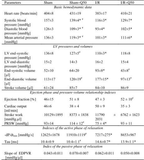
Table 2. Cardiac functions in the four groups. LV: left-ventricular; PRSW:
preload recruitable stroke work; dP/dtmin: maximal slope of the diastolic pressure decrement, Tau: time constant of left-ventricular pressure decay; EDPVR: end- diastolic pressure-volume relationship. *P<0.05 vs. sham, #P<0.05 vs. I/R.
Parameters Sham Sham+Q50 I/R I/R+Q50
Basic hemodynamic data
Heart rate [beats/min] 404±8 431±19 383±17 410±21 Systolic blood
pressure [mmHg]
157±3 139±4* # 116±3* 129±7*
Diastolic blood pressure [mmHg]
126±3 109±3* # 93±4* 102±5*
Mean arterial pressure [mmHg]
136±3 119±3* # 101±3* 111±6*
LV pressures and volumes LV end-systolic
pressure [mmHg]
136±8 127±5# 110±3* 118±8
LV end-diastolic pressure [mmHg]
15±2 14±3 16±2 15±4
End-systolic volume [µl]
52±10 64±20 93±8* 43±8#
End-diastolic volume
[µl] 113±17 120±19# 177±15* 97±13#
Stroke volume [µl] 61±24 85±7 84±10 86±9
Ejection phase and pressure-volume relationship indexes
Ejection fraction [%] 46±15 51 ± 8 47 ± 3 52 ± 10# Cardiac output
[ml/min]
46±6 38 ± 4 50 ± 9 35 ± 3
Stroke work [mmHg.µl]
10129±1895 8373 ± 1838 11790 ± 2031
6762 ± 1623
PRSW [mmHg] 93±14 114 ± 12# 75 ± 4* 93 ± 11
Indexes of the active phase of relaxation
-dP/dtmin [mmHg/s] 12625±1678 11910±1119# 7217±275* 8653±967 Tau [ms] 10.4±0.9 10.4±1.1# 14.6±0.7* 13.9±1.1*
Index of the passive phase of relaxation Slope of EDPVR
[mmHg/µl]
0.043±0.011 0.070±0.007 0.062±0.011 0.050±0.008
14
4.2.2. Effect of Q50 after heart transplantation
4.2.2.1. Effect of Q50 on graft function after heart transplantation
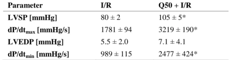
At 1 h of reperfusion, LV systolic pressure and dP/dtmax significantly increased in the Q50 treated group compared with those in the I/R group, indicating improved myocardial contractility. Moreover, Q50 treatment resulted in a significant increase in dP/dtmin values compared with the I/R group, reflecting improved myocardial relaxation. LV end-diastolic pressure, as a marker of the standardised balloon catheter measurements, did not show major differences.
Table 3. Effects of Q50 on graft function after heart transplantation. Left- ventricular (LV) parameters 1 h after reperfusion. Q50 treatment resulted in a significant increase in LVSP, dP/dtmin and dP/dtmax values compared with the I/R group. Values represent the mean ± SEM, P < 0.05: * vs. I/R.
Parameter I/R Q50 + I/R
LVSP [mmHg] 80 ± 2 105 ± 5*
dP/dtmax [mmHg/s] 1781 ± 94 3219 ± 190*
LVEDP [mmHg] 5.5 ± 2.0 7.1 ± 4.1 dP/dtmin [mmHg/s] 989 ± 115 2477 ± 424*
4.2.2.2. Effect of Q50 on graft myocardial high-energy phosphate contents after heart transplantation
After heart transplantation, myocardial high-energy phosphate contents, ATP and ADP levels, were preserved by Q50 pre- conditioning compared with the I/R group. The energy charge potential as an indicator of the myocardial energy level showed a significant improvement in Q50 pre-treated rats compared with that in the I/R group.
15
Table 4. Effect of Q50 on myocardial ATP, ADP and AMP contents in a rat model of heart transplantation. I/R: ischaemia/reperfusion, ATP: adenosine triphosphate; ADP: adenosine diphosphate; AMP: adenosine monophosphate. *P <
0.05 vs. other groups.
Parameter Control I/R Q50 + I/R
ATP [µmol/g] 6.58 ± 1.12 1.86 ± 0.41* 6.66 ± 0.63 ADP [µmol/g] 3.48 ± 0.16 2.05 ± 0.42* 5.01 ± 0.43 AMP [µmol/g] 1.91 ± 0.22 2.07 ± 0.22 2.59 ± 0.61 Energy charge potential 0.69 ± 0.07 0.49 ± 0.04* 0.85 ± 0.08
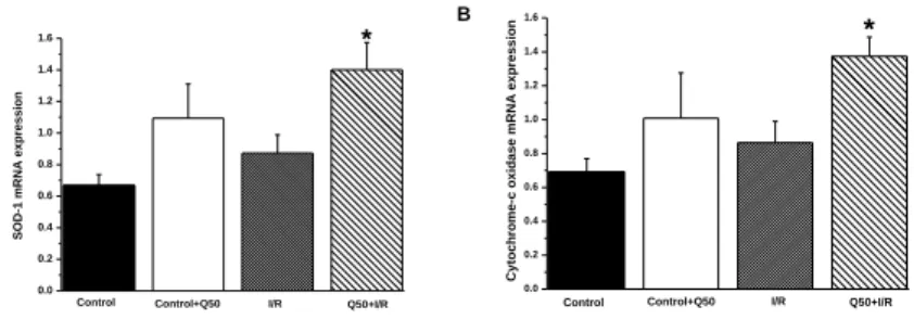
4.2.2.3. Effect of Q50 on graft gene and protein expression after heart transplantation
Quantitative real-time PCR from myocardium RNA extracts revealed that relative mRNA expression for SOD-1 and cytochrome-c oxidase remained unchanged in control, control Q50 and I/R groups.
However, their expressions were significantly upregulated after a Q50 treatment compared with that in the control group. Furthermore, densitometric analysis of bands after heart transplantation showed that a Q50 treatment significantly upregulated the protein expression of SOD-1 compared with the control and I/R groups and increased cytochrome-c oxidase protein level compared with the controls (data not shown).
0.0 0.2 0.4 0.6 0.8 1.0 1.2 1.4 1.6
Control+Q50
SOD-1 mRNA expression
Control I/R Q50+I/R
*
A
0.0 0.2 0.4 0.6 0.8 1.0 1.2 1.4 1.6
Control+Q50
Cytochrome-c oxidase mRNA expression
Control I/R Q50+I/R
B *
Figure 2. Effects of Q50 on gene expression after heterotopic heart transplantation. (A) SOD-1, (B) cytochrome-c oxidase mRNA expression in the myocardium. SOD-1 and cytochrome-c oxidase were significantly up-regulated in the Q50 pre-treated I/R group compared with those in the control group. Values represent mean ± SEM, P < 0.05: * vs. control.
16
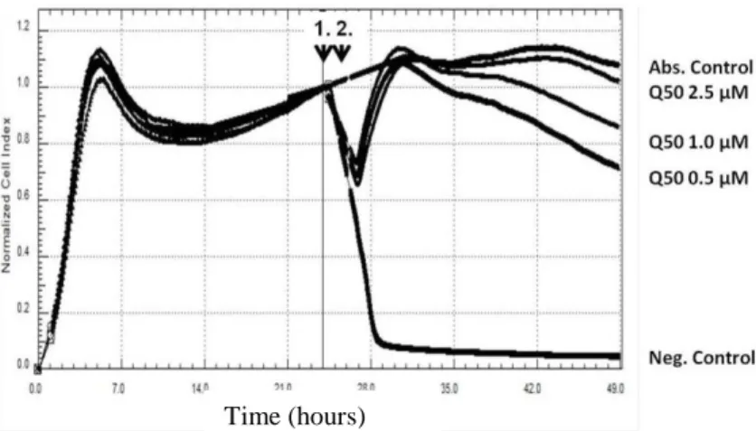
4.2.3. Cytoprotective effect of Q50 post-treatment of H9c2 cells Cells were attached and grown overnight; they were then subjected to H2O2 induced oxidative stress. After 30 min, Q50 was added to the wells at given concentrations. Normalisation of the cell index was calculated at the time of H2O2 application. Exposure of H9c2 cells to 100 μM H2O2 resulted in a rapid decrease of the cell index, whereas the cell index of the absolute control cells that did not receive H2O2
treatment continued to slightly increase. Post-treatment of Q50 exerted a dramatic dose-dependent cytoprotective effect after H2O2
stress: concentrations as low as 0.5 μM maintained the cell index near absolute control levels after the initial 3 h of the experiments.
The cell index also remained markedly elevated during the course of the entire experiment.
Figure 3. Cytoprotective effect of the Q50 post-treatment of H9c2 cells after oxidative stress. H9c2 rat embryonal cardiac muscle cells were subjected to oxidative stress (100 μM H2O2; arrow 1). After 30 min, Q50 was added to the wells (arrow 2). Curves in the figure are measurements of single wells. The normalised cell index shows the relative viability of cells per well. The black vertical line in the middle of the graph indicates the time of normalisation of the cell index, which is the time of H2O2 application.
Time (hours)
17 5. CONCLUSIONS
Exploring research areas of experimental cardiology and heart surgery for better understanding of myocardial and vessel protection under ischaemic and hypoxic conditions is necessary to improve our knowledge regarding the circumstances of these conditions.
The first aim of this study focused on the role of the HIF in vascular cold ischaemic storage and warm reperfusion injury. Therefore, we treated isolated rat aortic rings with DMOG and simulated reperfusion injury in an organ bath by adding hypochlorite. We found that HIF stabilisation leads to an improvement in vasorelaxation, which was mainly mediated via HO-1.
Our second aim was to investigate the activity and characteristics of the newly developed iron-chelating and zinc-complexing agent Q50 in rodent models of regional and global myocardial I/R. In rats with regional myocardial ischaemia induced by ligation of the LAD coronary artery, we found that treatment with Q50 showed improved contractility, although the size of myocardial infarct was not influenced. Rats with global myocardial ischaemia from an orthotopic heart transplantation that were treated with Q50 showed better LV function and increased ATP levels compared with those in the control groups.
18
6.BIBLIOGRAPHY OF THE CANDIDATE’S PUBLICATIONS PUBLICATIONS CONNECTED WITH THE Ph.D. THESIS:
1. Barnucz E, Veres G, Hegedűs P, Klein S, Zöller R, Radovits T, Korkmaz S, Horkay F, Merkely B, Karck M, Szabó G. (2013) Prolyl-hydroxylase inhibition preserves endothelial cell function in a rat model of vascular ischemia reperfusion injury. J Pharmacol Exp Ther, 345(1):25-31. IF: 3.855 2. Korkmaz S, Barnucz E*, Loganathan S, Li S, Radovits T, Hegedus P, Zubarevich A, Hirschberg K, Weymann A, Puskás LG, Ózsvári B, Faragó N, Kanizsai I, Fábián G, Gyuris M, Merkely B, Karck M, Szabó C, Szabó G. (2013) Q50, an iron-chelating and zinc-complexing agent, improves cardiac function in rat models of ischemia/reperfusion-induced myocardial injury. Circ J, 77(7):1817-1826. IF: 3.685 * shared first authorship OTHER PUBLICATIONS:
1. Veres G, Hegedűs P, Barnucz E, Schmidt H, Radovits T, Zöller R, Karck M, Szabó G. (2015) TiProtec preserves endothelial function in a rat model.
J Surg Res, 200(1):346-355.
2. Loganathan S, Korkmaz-Icöz S, Radovits T, Li S, Mikles B, Barnucz E, Hirschberg K, Karck M, Szabó G. (2015) Effects of soluble guanylate cyclase activation on heart transplantation in a rat model. J Heart Lung Transplant, 34(10):1346-1353.
3. Veres G, Hegedűs P, Barnucz E, Zöller R, Klein S, Schmidt H, Radovits T, Korkmaz S, Karck M, Szabó G.(2015) Endothelial dysfunction of bypass graft: direct comparison of in vitro and in vivo models of ischemia- reperfusion injury. PLoS One. 15;10(4):e0124025.
4. Veres G, Hegedűs P, Barnucz E, Zöller R, Klein S, Radovits T, Korkmaz S, Karck M, Szabó G. (2015) Graft preservation with heparinized blood/saline solution induces severe graft dysfunction. Interact Cardiovasc Thorac Surg, 20(5):594-600.
5. Korkmaz S, Atmanli A, Li S, Radovits T, Hegedűs P, Barnucz E, Hirschberg K, Loganathan S, Yoshikawa Y, Yasui H, Karck M, Szabó G.
Superiority of zinc complexof acetylsalicylic acid to acetylsalicylic acid in preventing postischemic myocardial dysfunction. (2015) Exp Biol Med (Maywood). 240(9):1247-1255.
6. Radovits T, Korkmaz S, Mátyás C, Oláh A, Németh BT, Páli S, Hirschberg K, Zubarevich A, Gwanmesia PN, Li S, Loganathan S, Barnucz E, Merkely B, Szabó G. (2015) An altered pattern of myocardial
19
histopathological and molecular changes underlies the different characteristics of type-1 and type-2 diabetic cardiac dysfunction. J Diabetes Res, 2015:728741.
7. Rylova SN, Barnucz E, Fani M, Braun F, Werner M, Lassmann S, Maecke HR, Weber WA. Does imaging αvβ3 integrin expression with PET detect changes in angiogenesisduring bevacizumab therapy? (2014) J Nucl Med, 55(11):1878-1884.
8. Veres G, Hegedűs P, Barnucz E, Zöller R, Radovits T, Korkmaz S, Kolonics F, Weymann A, Karck M, Szabó G. Addition of vardenafil into storage solution protects the endothelium in a hypoxia-reoxygenation model. (2013) Eur J Vasc Endovasc Surg. 46(2):242-248.
9. Radovits T, Arif R, Bömicke T, Korkmaz S, Barnucz E, Karck M, Merkely B, Szabó G. Vascular dysfunction induced by hypochlorite is improved by the selective phosphodiesterase-5-inhibitor vardenafil. (2013) Eur J Pharmacol, 15;710(1-3):110-119.
10. Li S, Korkmaz S, Loganathan S, Weymann A, Radovits T, Barnucz E, Hirschberg K, Hegedüs P, Zhou Y, Tao L, Páli S, Veres G, Karck M, Szabó G. Acute ethanol exposure increases the susceptibility of the donor hearts to ischemia/reperfusion injury after transplantation in rats. (2012) PLoS One, 7(11):e49237.
11. Weymann A, Schmack B, Okada T, Soós P, Istók R, Radovits T, Straub B, Barnucz E, Loganathan S, Pätzold I, Chaimow N, Schies C, Korkmaz S, Tochtermann U, Karck M, Szabó G. (2013) Reendothelialization of human heart valve neoscaffolds using umbilical cord-derived endothelial cells. Circ J, 77(1):207-216.
12. Korkmaz S, Loganathan S, Mikles B, Radovits T, Barnucz E, Hirschberg K, Li S, Hegedüs P, Páli S, Weymann A, Karck M, Szabó G.
(2013) Nitric oxide- and heme-independent activation of soluble guanylate cyclase attenuates peroxynitrite-induced endothelial dysfunction in rat aorta.
J Cardiovasc Pharmacol Ther, 18(1):70-77.
13. Hirschberg K, Tarcea V, Páli S, Barnucz E, Gwanmesia PN, Korkmaz S, Radovits T, Loganathan S, Merkely B, Karck M, Szabó G. (2013) Cinaciguat prevents neointima formation after arterial injury by decreasing vascular smooth muscle cell migration and proliferation. Int J Cardiol, 31;167
20
14. Loganathan S, Radovits T, Korkmaz S, Hirschberg K, Barnucz E, Weymann A, Bömicke T, Arif R, Karck M, Szabó G.(2012) Enhancement of myocardial and vascular function after phosphodiesterase-5 inhibition in a rat model. Thorac Cardiovasc Surg, 60(4):247-254.
15. Kemecsei P, Miklós Z, Bíró T, Marincsák R, Tóth BI, Komlódi-Pásztor E, Barnucz E, Mirk E, Van der Vusse GJ, Ligeti L, Ivanics T.(2010) Hearts of surviving MLP-KO mice show transient changes of intracellular calcium handling. Mol Cell Biochem, 342(1-2):251-260.
16. Hirschberg K, Radovits T, Korkmaz S, Loganathan S, Zöllner S, Seidel B, Páli S, Barnucz E, Merkely B, Karck M, Szabó G.(2010) Combined superoxide dismutase mimetic and peroxynitrite scavenger protects against neointima formation after endarterectomy in association with decreased proliferation and nitro-oxidative stress. Eur J Vasc Endovasc Surg, 40(2):168-175.
17. Korkmaz S, Radovits T, Barnucz E, Hirschberg K, Neugebauer P, Loganathan S, Veres G, Páli S, Seidel B, Zöllner S, Karck M, Szabó G.(2009) Pharmacological activation of soluble guanylate cyclase protects the heart against ischemic injury. Circulation, 120(8):677-686.
18. Korkmaz S, Radovits T, Barnucz E, Neugebauer P, Arif R, Hirschberg K, Loganathan S, Seidel B, Karck M, Szabó G. (2009) Dose-dependent effects of a selective phosphodiesterase-5-inhibitor on endothelial dysfunction induced by peroxynitrite in rat aorta. Eur J Pharmacol, 615(1- 3):155-162.
19. Radovits T, Korkmaz S, Loganathan S, Barnucz E, Bömicke T, Arif R, Karck M, Szabó G. (2009) Comparative investigation of the left ventricular pressure-volume relationship in rat models of type 1 and type 2 diabetes mellitus. Am J Physiol Heart Circ Physiol, 297(1):H125-133.
20. Radovits T, Bömicke T, Kökény G, Arif R, Loganathan S, Kécsán K, Korkmaz S, Barnucz E, Sandner P, Karck M, Szabó G. (2009) The phosphodiesterase-5 inhibitor vardenafil improves cardiovascular dys- function in experimental diabetes mellitus. Br J Pharmacol, 156(6):909-919.
21. Loganathan S, Radovits T, Hirschberg K, Korkmaz S, Barnucz E, Karck M, Szabó G. (2008) Effects of selective phosphodiesterase-5-inhibition on myocardial contractility and reperfusion injury after heart transplantation.
Transplantation, 27;86(10):1414-1418.