Cardiovascular effects of ghrelin
Doctoral Thesis
Balázs Sax, MD
Semmelweis University
Doctoral School of Basic Medical Sciences
Tutor: Dr. Violetta Kékesi associate professor, Ph.D.
Opponents:
Dr. Ágnes Végh professor, D.Sc.
Dr. Tóth-Heyn Péter assistant professor, Ph.D.
Chair of examination committee:
Dr. Rudolf de Châtel professor, D.Sc.
Members of examination committee:
Dr. István Szokodi associate professor, Ph.D.
Dr. Éva Palik assistant professor, Ph.D.
Budapest
2013
1 INTRODUCTION
Physiological and pathological cardiac function is regulated by several peptides produced (also) in the myocardium including natriuretic peptides, endothelins and angiotensin. Recently, several peptides of primarily non-cardiac origin were also proven to possess potentially significant cardiovascular effects. Examples to these latter are relaxin and also ghrelin.
Our studies involved in the thesis all aimed to describe different aspects of the role of the gastrointestinal hormone ghrelin in cardiovascular regulation. Ghrelin is the endogenous ligand of the growth hormone secretagouge receptor (GHS-R). It is mainly produced by the oxintic glandular cells of the gastric mucosa.
However, it is also produced by several other tissues including the myocardium and vascular smooth muscle. The peptide circulates in the systemic plasma in two major forms: the acylated (active) and des-acyl (inactive) ghrelin. Human myocardial cells are expressing ghrelin, both forms of ghrelin receptor and the activating enzyme (ghrelin-O-acyl-transferase, GOAT) creating the possibility of the existence of a locally acting paracrine ghrelin system in the heart.
Ghrelin receptor subtype 1a is a G-protein coupled receptor with seven transmembrane domains mediating most of the biological effects of ghrelin. Subtype 1b (GHS-R1b) is a splice variant of the receptor gene with five transmembrane domains. Its physiological relevance remains largely unknown to date.
Effects of ghrelin on growing and metabolism
Firstly discovered effect of ghrelin was its potent growth hormone secretagogoue activity. It also has a direct proliferating effect on several tissues including the myocardium. Ghrelin is a potent orexigenic peptide affecting feeding behavior, increasing food intake, gastric acid secretion and gastrointestinal motility. Its systemic plasma level is the highest before meals and decreases postprandially. Ghrelin level has a negative correlation with body mass index which refers to a negative feedback mechanism. One
potential field of therapeutic application of ghrelin might be the orexigenic effect in patients with cachexia due to heart failure or malignant diseases. Ghrelin has a bi-directional interaction with carbohydrate homeostasis by increasing plasma glucose level, stimulating glucagon and suppressing insulin secretion; whereas intravenous insulin infusion is able to decrease the plasma level of ghrelin in healthy subjects.
Effects of ghrelin on intact circulation
Ghrelin possesses widespread cardiovascular effects. In healthy humans ghrelin decreases blood pressure and increases stroke volume and cardiac index without affecting heart rate. The blood pressure decreasing effect is partially mediated by the central nervous system through nucleus tractus solitarii and vagal nerve.
Ghrelin in ischemic heart disease
Ghrelin is largely involved in different cardioprotective mechanisms. In experimental models of myocardial ischemia ghrelin was able to decrease the worsening of ejection fraction and early myocardial remodeling through anti-inflammatory and sympathicolytic activity. Ghrelin decreases post-infarction apoptosis, necrosis and thus mortality. Treatment of cardiomyocyte cell lines with ghrelin enhances proliferation. On vascular endothelial cell lines ghrelin also induces proliferation and migration resulting in angiogenesis. Human investigations showed that low ghrelin levels may carry higher risk for ischemic heart disease.
Ghrelin in heart failure
In a rat model of heart failure ghrelin treatment was able to increase left ventricular contractility, cardiac index and improve cardiac cachexia. In a small cohort of heart failure patients ghrelin bolus decreased blood pressure and increased cardiac index and stroke volume. Chronic treatment with ghrelin also decreased sympathetic activity and left ventricular dilation, increased ejection fraction and left ventricular mass, and improved muscle force. In terminal heart failure circulating ghrelin level rises and normalizes
after cardiac transplantation. Low ghrelin and a compensating high GHS-R1a expression was found in explanted hearts of transplant recipients.
Direct vascular effect of ghrelin
Direct vascular effect of ghrelin is differing between vascular territories and species and depends on vascular tone. Relaxing effect was described on human brachial artery in vivo and on isolated mammary artery in vitro. Similarly, vasodilation was observed on rat mesenterial bed after preconstrition. Unlike in rats, ghrelin has a tone-dependent constricting effect on human isolated mesenterial arterioles and also causes constriction on guinea pig renal arteries and smooth muscle cells. Ghrelin treatment decreased coronary blood flow in pig hearts and increased vascular resistance in isolated rat heart and isolated, perfused rat coronary arteries.
Pericardial fluid
Only a few methods are available to investigate the changes in the myocardial interstitium in vivo, especially in humans. Myocardial interstitium is the microenvironment of myocytes and reflects the myocardial production, degradation and metabolism of biologically active agents. Pericardial fluid is mainly a myocardial transsudate that constantly communicates with the myocardial interstitium thus it may mirror the changes of myocardial homeostasis. Pericardial fluid was proven to contain several endogenous regulating agents (adenine nucleosides, natriuretic peptides, cathecolamines, endothelin, growth factors, etc.) in a concentration much higher than systemic plasma referring to an increased myocardial production of these substances.
Pericardial concentrations of the above agents may further increase in ischemic heart disease. Despite its myocardial production and the cardiovascular effects of ghrelin, its pericardial levels has not been studied so far. According to the above, pericardial ghrelin concentration may be more closely related to the myocardial concentration and its changes in certain pathological conditions than systemic plasma ghrelin levels.
2 AIMS
The focus of my work was the role of ghrelin in cardiovascular regulation. In order to achieve this, I studied the local and systemic concentrations of the peptide in ischemic heart disease and heart failure and also its direct effect on coronary arterioles and the receptorial mechanism involved.
Knowing the widespread cardioprotective effects of ghrelin we hypothesized that local myocardial ghrelin concentration may change in ischemic heart disease. As direct measurement of myocardial ghrelin is nearly impossible in patients with chronic myocardial ischemia we aimed to measure the pericardial and plasma active (acylated) and total ghrelin concentrations of patients with ischemic or valvular heart disease undergoing open heart surgery. We examined the correlations of ghrelin concentrations with functional and morphological cardiac parameters, anthropometrical and metabolic data.
Accord to the controversial data available in the literature on the direct vascular effects of ghrelin, we aimed to investigate the presence of ghrelin receptors (GHS-R1a and GHS-R1b) in the coronary system and myocardium and to clarify the effect of ghrelin on the contractile state of intramural coronary resistance arterioles in isolated canine coronary samples. We also of examined if GHS-R1a ghrelin receptors were involved in the mediation of this effect.
To examine the changes in systemic ghrelin concentration during the development of experimental heart failure, we aimed to set a reliable, well reproducible large animal model of heart failure. This right ventricular tachypacing model served to investigate the change of plasma ghrelin and (as a control) proANP concentrations and their correlations with heart failure related echocardiographical parameters during the development of severe heart failure.
3 METHODS
3.1 Human pericardial study 3.1.1. Patient groups
heart disease patients undergoing open heart surgery:
o elective coronary artery bypass grafting (ISCH, n=54) o valve replacement (as a control group with negative
coronarogram) (VHD, n=41)
exclusion criteria: acute myocardial infarction, acute inflammatory or tumorous disease, renal or liver failure
demographical data: age (ISCH vs. VHD: 63 vs. 66 years) and BMI (28 vs 27 kg/m2) did not differ between groups
significant difference was observed between groups in the percentage of male sex (ISCH vs. VHD: 80 vs. 37%), prevalence of hypertension (87 vs. 14 %) and diabetes (33 vs.
10%; all p<0.05).
3.1.2 Sampling, biochemical and echocardiography measurements intraoperative pericardial and central venous blood sampling treatment of samples with EDTA and aprotinin
centrifugation, storage at - 80oC until measurements
measurement of active (acylated) and total (acylated+des-acyl) ghrelin (radioimmunoassay), plasma insulin (ELISA), serum lipid, glucose and other standard laboratory parameters
echocardiography parameters: left ventricular end diastolic (LVEDD) and end systolic (LVESD) diameter, intraventricular septal wall (SWT) and posterior wall (PWT) thickness, tricuspid annular plane systolic excursion (TAPSE) and left ventricular ejection fraction (EF)
HOMA-A insulin resistance index and left ventricular mass was calculated from the above parameters
3.2 In vitro canine coronary studies 3.2.1 Immunohistochemistry
fixation (formalin 4%, pH 7.4) and paraffination of canine myocardial samples
validation of anti-human GHS-R1a and 1b receptor antibodies on canine heart samples (Western blotting)
ghrelin receptor detection (immunohistochemistry) with the validated anti-human antibodies on canine myocardial samples o positive control: canine hypothalamus neurons
o negative control: omitting primary antibody 3.2.2 Isolated canine coronary arterioles
isolation of intramural coronary arterioles from healthy canine hearts (n=20; inner diameter: 219±18 µm at a constant 50 mmHg intraluminary pressure)
cannullation of the vessels on both sides (stereomicroscopy) incubation in normal Krebs-Ringer solution (KR; pH 7.4; 37oC;
bubbling with 5%CO2, 20%O2, 75%N2 carbogen gas)
measurement of changes in vessel diameter (videomicroscopy) testing endothelial function (10 µM acetylcholine)
3.2.3 Experimental protocol
first series of experiments (n=6-12):
o dose-response curve of ghrelin on coronary arterioles with spontaneous vascular tone (1-3-10-30-100-300 nM ghrelin) o dose-response curve of ghrelin on coronary arterioles with
elevated vascular tone after preconstriction with the thromboxane analogue U46619 (10-7 – 10-6 M)
second series of experiments (n=8):
o effect of ghrelin on preconstricted coronary arterioles (30- 100-300 nM ghrelin) before and under the blockade of GHS- R1a receptor (50 m D-Lys3-GHRP-6)
3.3 In vivo large animal model of heart failure 3.3.1 Set-up of the large animal model
pacemaker (PM) implantation in dogs with two right ventricular electrodes, PM set to DDD mode (n=13)
120/min pacing with 250 ms AV time resulting in 240/min regular heart rate
severe heart failure (dyspnoe, ascites, loss of apetite, pulmonary congestion) developed in 22±4 days
weekly echocardiography control with temporally switched off pacemaker: chamber diameters, wall thicknesses, mitral and tricuspid regurgitation, left ventricular ejection fraction and TAPSE was evaluated
blood sample collection and functional staging was performed weekly
3.3.3 Biochemical measurements
venous blood samples: EDTA+aprotinin, storage at -80°C plasma acylated ghrelin and proANP measurements (ELISA) routine laboratory tests (blood count, renal function, electrolytes, liver function, bilirubin)
3.4 Statistical analysis
expression of data: mean±S.E.M.
normality testing: Shapiro-Wilk test human studies:
o Mann-Whitney U, Wilcoxon, Spearman tests o Fisher’s exact test
isolated arteriole experiments:
o Student’s paired t-test
large animal model of heart failure:
o Student’s paired t-test Pearson’s correlation test level of significance: p<0.05
4 RESULTS
4.1 Results of the human pericardial study
4.1.1 Metabolical and echocardiography parameters
Comparing the metabolical parameters of the two patient groups, serum total cholesterol level was significantly lower in ischemic heart disease patients (ISCH vs. VHD: 4.4 0.2 vs. 5.1 0.2; p<0.05), which might be the consequence of the higher prevalence of taking statins in the group (35/54 vs. 16/41, p<0.05). Serum triglycerides and parameters of carbohydrate metabolism (plasma glucose, plasma insulin, HOMA-A parameter of insulin resistance) did not differ significantly.
Regarding echocardiography parameters, mildly but significantly lower ejection fraction (55 1 vs. 62 1; p<0.01) and larger end systolic diameter (36 1 vs. 32 1; p<0.01) was found in the ischemic patient group. However, these values were still within normal limits.
No significant difference could be observed in left ventricular mass and wall thickness parameters or in right ventricular morphological and functional parameters.
4.1.2 Pericardial and plasma ghrelin concentration
Significantly higher pericardial active ghrelin concentration was measured in the ischemic heart disease group, almost twice of that in the valvular heart disease group (Table 1). Plasma ghrelin concentrations did not differ significantly. Both pericardial and plasma active ghrelin concentration were found to be more than one magnitude lower than the respective total ghrelin levels in both groups.
In the valvular heart disease patient group pericardial active and total ghrelin concentrations were found to be significantly lower than the respective plasma concentrations. No such difference could be observed in the ischemic heart disease group. These data may indicate a relative active and total ghrelin surplus in chronic myocardial ischemia. In order to more closely evaluate this latter, a
pericardial-to-plasma ratio was calculated in both groups. These ratios were found to be significantly higher in the ischemic group for both active and total ghrelin as compared to the valvular heart disease control group (ISCH vs. VHD, active ghrelin pericardial-to- plasma ratio: 0.85 0.03 vs. 0.44 0.07, p<0.01; total ghrelin pericardial-to-plasma ratio: 0.97 0.05 vs. 0.88 0.03, p<0.01).
Ghrelin concentration (pg/ml, mean SEM)
ISCH (n=54)
VHD (n=41)
Acylated ghrelin
plasma 39 3 41 4**
pericardial fluid 32 3 ## 16 2
Total ghrelin
plasma 683 24 701 39**
pericardial fluid 668 29 600 38
TABLE 1 Pericardial and plasma acylated (active) and total ghrelin concentration of patients with ischemic (ISCH) or valvular (VHD) heart disease. Mean S.E.M, **p<0.01 plasma vs. pericardial fluid within patient group (Wilcoxon’s test); ## p<0.01 ischemic vs.
valvular heart disease patients (Mann-Whitney test)
4.1.3 Correlation of ghrelin concentrations with metabolical and echocardiography parameters
Significant negative correlation was observed between plasma total ghrelin level and body mass index (Table 2). Plasma insulin concentration and HOMA-A index showed a negative correlation with both plasma and pericardial total ghrelin and also with pericardial active ghrelin concentration.
Pericardial active and total ghrelin levels negatively correlated with left ventricular posterior wall thickness. Left ventricular mass (calculated using wall thickness parameters and left ventricular end diastolic diameter) was found to be negatively correlated with plasma total ghrelin concentration.
Neither left ventricular ejection fraction and diameters, nor right ventricular parameters had any significant correlation with pericardial or plasma ghrelin concentrations.
All patients BMI plasma insulin
HOMA-
A index PWT LVM Plasma
acylated ghrelin
R -0.159 -0.192 -0.192 -0.252 -0.119 p 0.128 0.071 0.071 0.060 0.381 Pericardial
acylated ghrelin
R 0.000 -0.223 -0.233 -0.306 -0.146 p 0.997 0.047 0.038 0.032 0.316 Plasma
total ghrelin
R -0.223 -0.288 -0.326 -0.243 -0.275 p 0.030 0.006 0.002 0.069 0.038 Pericardial total
ghrelin
R 0.003 -0.371 -0.395 -0.354 -0.249 p 0.973 0.000 0.000 0.007 0.061 TABLE 2 Correlations of ghrelin concentrations with metabolical and echocardiography parameters in the whole patient population.
Significant correlations are highlighted with bold. (R: Spearman correlation coefficient; p: level of significance; BMI: body mass index; PWT: posterior wall thickness; LVM: left ventricular mass)
4.2 Results of in vitro canine coronary experiments 4.2.1 Immunohistochemistry
Immunostaining with anti-GHS-R1a in canine left ventricular samples is shown in the upper row of Figure 1. Myocardium shows mild immunopositivity for GHS-R1a. Prominent immunopositivity can be observed for the receptor in the walls of various size coronary arterioles.
GHS-R1b immunopositivity is shown in the middle row of Figure 1. Myocardial GHS-R1b positivity is more pronounced as compared to the 1a subtype. Similarly to GHS-R1a, 1b receptor subtype also shows a significant positivity in the walls of arterioles.
FIGURE 1 Both receptor subtypes show significant immunopositivity in the wall of canine intramural coronary arterioles of various size on adjacent sections. Myocardial immunopositivity is more pronounced for GHS-R1b. Negative controls: primary antibody was omitted. Lines represent 50 m.
4.2.2 Isolated canine coronary arterioles
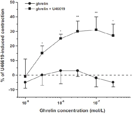
Ghrelin failed to change vessel diameter of arterioles with low spontaneous vascular tone. However, after preconstriction with the thromboxane analogue U46619 ghrelin induced dose-dependent vasoconstriction of the arterioles. U46619 itself caused 25±2%
decrease in diameter (n=20; p<0.01). Ghrelin caused a further vasoconstriction up to 31±9% of the effect of U46619.
FIGURE 2 Diameter responses to ghrelin of coronary arterioles with spontaneous or elevated (U46691) vascular tone. Values are given as the percentage of the maximal constriction induced by U46619. (mean S.E.M, *p<0.05, **p<0.01 elevated vs. spontaneous tone; n=6: 1-10 nmol/l and n=9-12: 30-300 nmol/l)
In a second series of experiments the blockade of GHS-R1a receptor with its specific antagonist (50 mol/L D-Lys3-GHRP-6) did not abolish the vasoconstriction caused by ghrelin on arterioles with elevated vascular tone. As seen in Figure 3, there was no difference in ghrelin (30-300 nM) induced vasoconstriction under the blockade of GHS-R1a.
FIGURE 3 Effect of the GHS-R1a antagonist D-Lys3-GHRP-6 (50 mol/l) in the ghrelin induced vasoconstriction on canine coronary arterioles with elevated vascular tone (U46619, 10-7-10-6 mol/l).
Ghrelin induced vasoconstriction is given as the percentage of the constriction induced by U46619. (mean S.E.M, n=8, not significant)
4.3 Results of the large animal heart failure model
4.3.1 Changes in echocardiography and laboratory parameters during the development of heart failure
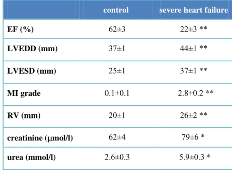
Sustained rapid right ventricular pacing (240/min) induced severe heart failure (similar to NYHA IV stage in human) in 22±4 days. A significant reduction in left ventricular ejection fraction, increase in mitral regurgitation, left ventricular end diastolic and end systolic diameter and mild right ventricular dilation could be observed.
Renal function showed significant worsening during the course of heart failure development (Table 3).
control severe heart failure
EF (%) 62±3 22±3 **
LVEDD (mm) 37±1 44±1 **
LVESD (mm) 25±1 37±1 **
MI grade 0.1±0.1 2.8±0.2 **
RV (mm) 20±1 26±2 **
creatinine ( mol/l) 62±4 79±6 *
urea (mmol/l) 2.6±0.3 5.9±0.3 *
TABLE 3 Changes of echocardiography parameters and renal function during the development of heart failure during rapid right ventricular pacing in dogs. (mean±SEM, *p<0.05 and **p<0.01 vs.
control; Student’s t-test, n=13; EF: ejection fraction; LVEDD: left ventricular end diastolic diameter; LVESD: left ventricular end systolic diameter; RV: right ventricular diameter).
4.3.2 Changes of plasma ghrelin and proANP concentration Plasma concentration of proANP showed significant, tenfold elevation during the development of severe heart failure (mean±SEM: 6284±1067 vs. 801±167 pg/ml; p<0.01). Ghrelin plasma concentration also increased during right heart rapid pacing (Figure 4). Until the development of severe heart failure an approximately twofold increase could be measured (1586±220 vs.
905±132 pg/ml; p<0.01). Ghrelin and proANP levels showed a significant positive correlation in the course of heart failure development when evaluating all measured values (R=0.37; p<0.03).
control severe heart failure 0
2000 4000 6000 8000 10000 12000 14000
proANP (pg/ml)
**
control severe heart failure 0
500 1000 1500 2000 2500 3000
active ghrelin (pg/ml)
**
FIGURE 4 Changes of plasma proANP and ghrelin concentration during the development of severe heart failure in the large animal model of rapid right ventricular pacing. (**p<0.01 vs. control;
n=13)
4.3.3 Correlation of ghrelin and proANP with cardiac parameters When evaluating all data from weekly controls, proANP plasma concentration showed negative correlation with ejection fraction (R=-0.49; p<0.01) and positive correlation with the grade of mitral regurgitation (R=0.52; p<0.01), left ventricular end diastolic (R=-0.48; p<0.01) and end systolic (R=-0.52; p<0.01) diameter.
Plasma concentration of ghrelin had a significant positive correlation with left ventricular end diastolic diameter (R=0.41; p=0.02).
5 CONCLUSIONS
1. Human pericardial fluid contains both acylated (active) and des- acyl (inactive) ghrelin in a concentration comparable to plasma.
2. Pericardial concentration of active ghrelin in patients with myocardial ischemia is higher than that in non-ischemic valvular heart disease patients which may refer to an increased myocardial ghrelin production in chronic myocardial ischemia.
3. Pericardial active ghrelin concentration is negatively influenced by insulin resistance and cardiac hypertrophy in heart disease patients.
4. Presence of both ghrelin receptors (GHS-R1a and GHS-R1b) can be detected in the myocardium and coronary artery walls in canine samples.
5. Pharmacological doses of ghrelin induce dose-dependent vasoconstriction in isolated canine coronary arterioles with elevated vascular tone which is not mediated by GHS-R1a ghrelin receptor.
6. Significant elevation of plasma active ghrelin concentration in severe heart failure and its correlation with proANP concentration in the large animal (canine) model of ventricular tachypacing induced heart failure suggest the possible role of ghrelin as a biomarker of heart failure.
6 LIST OF PUBLICATIONS
Publications related to the thesis:
1. Sax B, Nadasy GL, Turi K, Hirschberg K, Furjesz D, Nagy A, Merkely B, Szabo G, Monos E, Kekesi V. (2011) Coronary vasoconstrictor effect of ghrelin is not mediated by growth hormone secretagogue receptor 1a type in dogs. Peptides, 32:362-7. IF: 2,434
2. Sax B, Merkely B, Turi K, Nagy A, Ahres A, Hartyanszky I Jr, Huttl T, Szabolcs Z, Cseh K, Kekesi V. (2013) Characterization of pericardial and plasma ghrelin levels in patients with ischemic and non-ischemic heart disease. Regul Pept, 186:131- 6. IF (2012): 2.056
Publications not related to the thesis:
3. Toma I, Sax B, Nagy A, Entz L jr., Rusvai M, Juhasz-Nagy A, Kekesi V. (2006) Intrapericardial angiotensin II stimulates endothelin-1 and atrial natriuretic peptide formation of the in situ dog heart. Exp Biol Med (Maywood), 231:847-51. IF 2.845 4. Nagy A, Sax B, Entz L jr., Barat E, Toma I, Becker D, Merkely
B, Kekesi V. Comparison of elimination and cardiovascular effects of adenine nucleosides administered intrapericardially or intravenously in anesthetized dog. J Cardiovasc Pharmacol, 2009;54:341-7. IF: 2.826
5. Kovacs A, Apor A, Nagy A, Vago H, Toth A, Nagy AI, Kovats T, Sax B, Szeplaki G, Becker D, Merkely B. Left ventricular untwisting in athlete’s heart: key role in early diastolic filling?
Int J Sports Med, 2013 (DOI: 10.1055/s-0033-1349076).
IF (2012): 2.433