1Department of Nuclear Medicine;
2Department of Family Medicine;
3Department of Rheumatology and Immunology, 4Second Department of Internal Medicine and Cardiology Centre, University of Szeged, Hungary.
Zsuzsanna Besenyi, MD Gergely Ágoston, MD, PhD Rita Hemelein, MD Annamária Bakos, MD Ferenc T. Nagy, MD, PhD Albert Varga, MD, PhD László Kovács, MD, PhD László Pávics, MD, PhD, Dsc Please address correspondence to:
Dr Zsuzsanna Besenyi,
Department of Nuclear Medicine, University of Szeged,
6720 Szeged Korányi fasor 6, Hungary.
E-mail:
besenyi.zsuzsanna@med.u-szeged.hu Received on August 22, 2018; accepted in revised form on November 5, 2018.
Clin Exp Rheumatol 2019; 37 (Suppl. 119):
S88-S96.
© Copyright CliniCaland
ExpErimEntal rhEumatology 2019.
Key words: systemic sclerosis, heart involvement, FDG-PET/CT, strain echocardiography, myocardial inflammation
Competing interests: none declared.
ABSTRACT
Objective. Primary cardiac manifesta- tion is a common complication of sys- temic sclerosis (SSc) with poor progno- sis. The aim of the current study was to detect potential myocardial inflam- mation present in asymptomatic SSc patients by 18F-FDG-PET/CT and to investigate its relationship with early signs of myocardial dysfunction as de- tected by 2D speckle tracking echocar- diography (2DSTE).
Methods. Sixteen consecutive patients with SSc and 9 control patients with- out clinical evidence of cardiac in- volvement were enrolled in the study.
On 18F-FDG-PET acquired images blood-pool normalised SUV ratio and heterogenity index (HI: standard devi- ation of SUV divided with mean SUV) were calculated. Within 24 hours all SSc patients underwent 2DSTE strain analysis.
Results. Eight of 16 SSc patients were found to be visually PET-positive and showed significantly higher myocar- dial 18F-FDG SUV ratio (1.78±0.74 vs. 0.98±0.03; p<0.05) and heterogen- ity index (0.13±0.02 vs. 0.05±0.02;
p<0.001) as compared to the control group. FDG-PET/CT derived values did not differ significantly between visually PET-negative (8/16) and con- trol patients (SUV ratio: 0.98±0.05 vs. 0.98±0.03; HI: 0.05±0.01 vs.
0.05±0.02). Global left ventricular longitudinal strain values did not dif- fer significantly between PET-positive and negative patients (17.18±3.49%
vs. 17.59±3.65%).
Conclusion. Myocardial inflammation, as a potential sign of early cardiac in- volvement can be detected by 18-FDG- PET/CT in a considerable percentage of systemic sclerosis patients present- ing without cardiac symptoms.
Introduction
Systemic sclerosis (SSc) is a rare, chronic, progressive systemic connec- tive tissue disease characterised by mi- crovascular dysfunction, immune-me- diated inflammation and fibrosis with multi organ involvement. Cardiac in- volvement is common for patients with SSc, both in diffuse (DcSSc) and lim- ited cutaneous forms (LcSSC) of dis- ease with an estimated clinical preva- lence of 15-35% (1, 2). Cardiovascular disease in SSc may be direct (myocar- ditis, heart failure, coronary artery dis- ease, valvular and pericardial disease, conduction disturbances) and indirect (pulmonary artery hypertension (PAH) and renal crisis). When heart involve- ment becomes clinically evident, it ap- pears as a bad prognostic factor with a patient mortality rate of up to 70% at 5 years (3). In the majority of patients (up to 80%), however cardiovascular disease is subclinical (4, 5) for variable duration. Therefore preclinical identi- fication and monitoring of cardiac in- volvement is pivotal for adequate early management of these patients.
Routine transthoracic echocardiogra- phy (TTE) is the first line modality of choice in assessment of cardiac involve- ment (6). Echocardiography deforma- tion imaging, such as speckle-tracking echocardiography (STE) for measure- ment of strain and strain rate, have been developed for a more accurate depiction of regional contractility and early de- tection of myocardial dysfunction (7).
Indeed several recent publications have verified the promising role of STE for early detection of myocardial dysfunc- tion in SSc patients (8-11).
Although diffuse myocardial fibrosis re- mains the pathologic hallmark of direct myocardial involvement (12), the pres- ence of inflammation is often found in
18F-FDG-PET/CT in patients with systemic sclerosis without cardiac symptoms: a pilot study
Z. Besenyi
1, G. Ágoston
2, R. Hemelein
3, A. Bakos
1,
F.T. Nagy
4, A. Varga
2, L. Kovács
3, L. Pávics
1biopsies of SSc patients suggesting that cardiac inflammation may be more com- mon than originally appreciated. Moreo- ver, the fibrotic process may be second- ary to chronic inflammation of the heart (13). 18F-fluorodeoxyglucose(FDG)- positron emission tomography/com- puted tomo-graphy (PET/CT) is a non- invasive molecular imaging technique that is highly sensitive in quantitative evaluation of metabolically active pro- cesses such as inflammation. 18-FDG- PET/CT is generally gaining signifi- cance in the detection and monitoring of cardiac inflammation and infection (14, 15). We hypothesised that 18F- FDG-PET/CT may also be positive in asymptomatic SSc patients with poten- tial subclinical myocarditis.
The aim of the current study was to de- tect potential myocardial inflammation present in asymptomatic SSc patients by 18F-FDG-PET/CT. We also sought to investigate the relationship between pathological myocardial findings on 18F-FDG-PET/CT and clinical indi- ces of SSc disease and early signs of myocardial dysfunction as detected by 2DSTE.
Patients and methods Study population
Sixteen consecutive patients affected with SSc but without overt cardiovas- cular involvement were enrolled in the current prospective study. Inclusion cri- teria for the SSc group were: 1) age>18 and <85 years; 2) a previous diagnosis of SSc according to the ACR EULAR guidelines for SSc classification (16).
Exclusion criteria were: 1) inability to provide informed consent; 2) known history of coronary artery disease, electrocardiographic signs of myocar- dial ischaemia, left ventricular ejection fraction <55%, regional wall motion abnormalities, left ventricular hypertro- phy, significant valvular heart disease, pericardial effusion, severe pulmonary hypertension (pulmonary artery sys- tolic pressure >40Hgmm). To avoid un- necessary radiation exposure to healthy subjects, 9 persons (5 male, 4 female;
age 46.55±18.05 years) who underwent FDG-PET/CT examination for various other diagnostic reasons were enrolled as controls. Control subjects did not
have SSc, nor had evidence of active in- flammatory disease or cardiac disease, complied otherwise with the general exclusion criteria, and underwent the same FDG-PET/CT image acquisition and analysis protocol as the study pa- tients. All subjects gave informed con- sent. The study was approved by the Local Ethical Committee for Clinical Research at the University of Szeged (ref. no.: 3647).
Laboratory and clinical assessment All SSc patients underwent compre- hensive rheumatologic and cardio- vascular evaluation including, but not limited to, the assessment of: disease duration, EUSTAR disease activity score (17), Framingham score (18), presence of gastrointestinal involve- ment, pulmonary involvement, digital ulcers, prior immunosuppressive treat- ment, cardiovascular risk factors and current medication. General laboratory workup including determination of dis- ease specific autoantibodies and mark- ers of inflammation were performed according to local practice guidelines.
Creatinine-kinase (CK) as a marker of gross skeletal muscle/myocardial in- jury was also ascertained (Table I).
18F-FDG-PET/CT image acquisition protocol
To suppress physiological glucose up- take in the myocardium patients were instructed to take low-carbohydrate, high-fat, high-protein diet for 24 hours followed by a minimum of 6 hours fast- ing before PET/CT scan examinations according to previously published car- diac FDG-PET/CT guidelines (19). A mean activity of 481.98±80.95 MBq (6,5 MBq/kg) FDG was administered intravenously. Blood glucose levels were checked (<8 mmol/L) beforehand.
All scans were performed on an inte- grated whole-body PET/CT system (GE Discovery ST 4, GE Healthcare, Amer- sham, UK). Imaging of the cardiac re- gion in 2D, non-gated mode was carried out 60 minutes after the administration of the radioisotope. PET scan data col- lection was completed by low-dose CT (120 kV and 70 mAs) acquisition. After imaging of the cardiac region, a routine whole-body acquisition was performed
in 3-dimensional mode, extending from the skull base to the upper third of the thighs. Images were reconstructed us- ing a standard ordered subset expecta- tion maximisation algorithm with CT for attenuation correction.
18F-FDG-PET/CT image analysis protocol
FDG-PET images were visually evalu- ated for the presence of FDG uptake in the heart by consensus reading of two experienced PET/CT specialists. Based on uptake, FDG-PET images were clas- sified into 4 patterns: “none,” “diffuse,”
“focal,” and “focal on diffuse.” Quan- titative evaluation of FDG uptake was performed by PMOD version 3.704 software (PMOD Technologies Zu- rich) on attenuation corrected 2D PET images. Reorientation of axial cardiac images was performed. Seventeen seg- ments cardiac model was used accord- ing to the scientific statement from the American Heart Association (20).
Body weight standardised uptake value (SUV) in g/ml was calculated. Defini- tion of left ventricular volume of inter- est (VOI) was performed in a semi-au- tomated fashion. Blood pool VOI was contoured in the left ventricular cavity.
Mean global, segmental myocardial and blood pool SUV were calculated.
To avoid misinterpretation due to dif- fering patient metabolic characteristics, SUV values were divided by blood pool SUV to create normalised SUV ratios (21). To further specify pathophysi- ological uptake a coefficient of vari- ation as a metric of image heterogen- ity was also determined. Average and standard deviation of SUV values were calculated from SUV values in the 17 myocardial segments. The coefficient of variation (heterogenity index; HI) was calculated as the standard devia- tion of SUV divided by the average of SUV (22). Widespread diffuse myocar- dial FDG uptake exceeding liver uptake was considered as failed inhibition of physiological myocardial glucose up- take and patients were excluded from further analysis (23).
Echocardiography
All patients with SSc underwent com- prehensive echocardiography within 24
hours after PET/ CT scanning. Echo- cardiographic examinations were per- formed in all patients at rest using com- mercially available ultrasound machine (Vivid S70, GE Medical Systems, Hort- en, Norway). Left ventricular (LV) end- diastolic and end-systolic diameters, interventricular septum diastolic (IVS), and posterior wall thickness (PW), left atrial (LA) volumes, maximum LA size measurements were performed. Early (E) and late (A) mitral inflow velocity, E/A ratio, and deceleration time were obtained. Systolic (S’), early diastolic (E’), and late diastolic (A’) velocities were also measured by tissue Doppler imaging (TDI). Pulmonary artery sys- tolic pressure was estimated (24). All measurements were performed accord- ing to the recommendations of the Eu- ropean Association of Cardiovascular Imaging/American Society of Echo- cardiography (25). 2D speckle tracking echocardiography (2DSTE) analyses were performed on standard echocar- diographic grey-scale images. The loops were recorded with a frame rate between 60 and 80 frames per second.
LV longitudinal strain was determined from the 3 apical (2-, 3-, and 4-cham- ber) views. The 2D images were ana- lysed using dedicated software package (EchoPac PC, version, GE Vingmed, Horton, Norway). During analysis, the endocardial border was manually traced at end systole, and the region of interest width was adjusted to include the entire myocardium. The software then auto- matically tracks and accepts segments of good tracking quality and rejects poorly tracked segments while allow- ing the observer to manually override its decisions on the basis of visual as- sessments of tracking quality. Peak lon- gitudinal strain values were determined in 17 segments by Bull’s eye method (Fig. 1). Based on a guideline published by the American Society of Echocardi- ography and a meta-analysis of normal values, global longitudinal systolic peak strain (GLPS) values below 19.7% were considered pathological (25, 26).
Statistical analysis
Data are presented as mean ± standard deviation (SD) unless otherwise stat- ed. Comparisons between FDG-PET/
Table I. Clinical characteristics of systemic sclerosis patients.
All SSc patients DcSSc LcSSc n=16 n=8 n=8
Age (year) 59 ± (44-74) 62 ± (45-74) 57± (44-62)
Sex (male/female) 2 M/14 F 1M/7F 1 M/7 F
Disease duration (years) 5.56 ± 6.22 5.38 ± 6.56 5.75 ± 5.43 EUSTAR disease activity score 2.88 ± 1.77 3.75 ± 1.35 2 ± 1.38
Framingham score 2.81 ± 3 2.83 ± 2.25 2.79 ± 3.76
ANA positive (n) 11 5 6
ACA positive (n)* 6 0 6
anti-Scl70 positive (n) 4 4 0
DLCO (%) 49 ± 22 46 ± 27 52 ± 18
Pulmonary involvement (n) 10 6 4
Digital ulcers (n) 8 2 6
Gastrointestinal involvement (n) 13 7 6
Hypertension (n) 7 4 3
Hyperlipidaemia (n) 7 4 3
Diabetes (n) 3 1 2
Immunosuppressive treatment in the past (n) 8 6 2
Immunosuppressive treatment in the last 6 months (n) 4 4 0 Corticosteroid (up to 4 mg/day) treatment at the time 3 3 0 of PET/CT examination (n)
Beta-blockers (n) 4 3 1
ACE-inhibitors/ARB (n) 5 4 1
Calcium Channel Inhibitors (n) 6 3 3
Statins (n) 5 3 2
Platelet aggregation inhibitor (n) 6 2 4
CK, U/l 99 ± 54.65 105.25 ± 63.96 86.5 ± 33.07
CRP, mg/L 19.40 ± 29.68 23.31 ± 39.08 15.50 ± 18.07
ESR, mm/h 28.10 ± 15.53 31.87 ± 15.44 24.32 ± 15.67
WBC, G/L 8.40 ± 2.10 8.85 ± 2.44 7.94 ± 1.75
ACA: anti-centromere antibody; ACE: angiotensin converting enzyme; ANA: antinuclear antibodies;
anti-Scl70: anti-topoisomerase; ARB: angiotensin II receptor blocker; CK: creatinine-kinase; CRP: C- reactive protein; DLCO: diffusing capacity of the lung for carbon monoxide; DcSSc: diffuse cutaneous systemic sclerosis; ESR: erythrocyte sedimentation rate; LcSSc: limited cutaneous systemic sclerosis;
WBC: white blood cell count. *p<0.05.
Fig. 1. Longitudinal strain bull’s eye plot derived from two-dimensional speckle tracking in patient with systemic sclerosis. Decreased strain values in inferior and anterior segments.
CT positive and negative, DcSSc and LcSSc patients were performed using Student’s t tests or by non-parametric Mann-Whitney U-test, as appropri- ate. Comparisons between categorical variables were made with the Fisher’s exact test. All tests were two-sided and p-values <0.05 were considered statisti- cally significant. Overall and segmen- tal agreement between FDG-PET/CT and 2DSTE was assessed using Co- hen’s kappa coefficients. Correlations between the two methods were tested by Pearson or Spearman’s correlation tests, as appropriate. All analyses were performed using JMPV.14 statistical software v.14 (SAS Institute Inc., Cary, NC, USA).
Results
Clinical characteristics of the SSc pa- tient population are presented in Table I. Of the 16 patients 8 patients present- ed with limited cutaneous and 8 with diffuse cutaneous form of systemic sclerosis. Clinically expected differ- ences between the two subgroups were
related to a more frequent need for im- munosuppressive and low-dose corti- costeroid therapy in the DcSSc group, and the differences between autoanti- body positivity, although statistically significant difference was observed only with regard to anti-centromere antibody-positivity.
FDG-PET/CT
Dietary protocol to suppress normal glucose uptake was followed in all 16 SSc patients and 9 control subjects.
Low carbohydrate, high-fat, high-pro- tein diet time was: 32.5±3.14 hours, while total fasting time direct before image acquisition was: 18.32±4.74 hours. Blood glucose levels before ra- diopharmaceutical administration were 4.92±0.99 mmol/l. According to visual classification of cardiac FDG-PET im- ages in SSc patients, 8/16 none, 0/16 diffuse, 6/16 focal, 2/16 focal on dif- fuse patterns were found. In all of the control subjects, the cardiac FDG-up- take was near or equal to lower than blood pool activity meaning a “none”
pattern (Fig. 2). In SSc patients, global myocardial SUV was 3.019±0.02 g/ml and blood pool SUV was 2.1±0.56 g/
ml. In control patients, the same values were 1.81±0.26 g/ml and 1.85±0.27 g/
ml (p<0.05). Normalisation accord- ing to blood pool uptake SUV ratio in the patient population was 1.38±0.65 and 0.98±0.03 in the control group (p<0.05). Heterogenity indices of SSc and control patients were 0.095±0.04 and 0.05±0.02 (p<0.05). Of the 16 SSc patients 8 visually “PET positive”
(focal or focal on diffuse myocardial FDG uptake) patients showed signifi- cantly increased normalised SUV ratio (1.78±0.74 versus 0.98±0.03, p<0.05) and a significantly higher heterogen- ity index (0.13±0.02 vs. 0.05±0.02, p<0.001) as compared to the control group (Fig. 3). In contrast, in the 8 visually “PET-negative” SSc patients, normalised SUV ratio and heterogenity index did not differ significantly from control subjects (normalised SUV ratio:
0.98±0.05 vs. 0.98±0.03; HI: 0.05±0.01 vs. 0.05±0.02). No significant differ-
Fig. 2. FDG-PET/CT examination of a control patient without known cardiovascular disease. FDG-PET/CT fused axial (A), coronal (B) and sagittal (C) slices. The images show successful dietary suppression of physiological myocardial glucose uptake with no focal or diffuse increased uptake. Global nor- malised standard uptake ratio: 0.97, heterogenity index: 0.03.
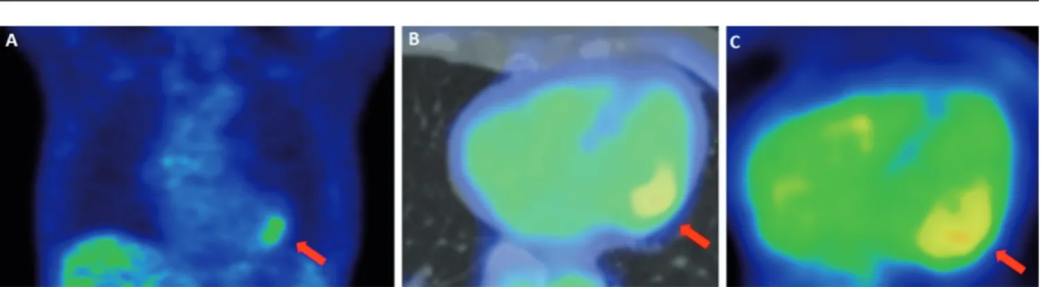
Fig. 3. FDG/PET-CT examination of a PET-positive systemic sclerosis patient presenting without clinical signs of primary cardiac involvement.
A: Coronal FDG-PET image of chest. B: Axial FDG-PET/CT fused image of the heart. C: Same level axial FDG-PET slice without CT fusion. PET images show increased focal FDG uptake (arrows) in the lateral wall in the presence of otherwise suppressed myocardial glucose uptake. Global normalised SUV ratio: 1.10, heterogenity index: 0.14.
ences were detected between the two groups in regards to clinical charac- teristics and laboratory parameters (Table II). No correlations were found between FDG-PET/CT derived values and type of SSc, disease activity scores, disease duration and laboratory indices of inflammation or cardiac involvement and echocardiographic parameters.
Echocardiography
Due to poor acoustic window, com- prehensive transthoracic echocardi- ography, global and segmental strain measurements were unsuccessful in 1 of 16 SSc patients. Echocardiography characteristics of the SSc patient popu- lation are presented in Table III. There were no significant differences be- tween DcSSc and LcSSc patients based on TTE parameters. There was also no statistically significant difference between PET positive and negative groups in regards to conventional TTE findings and, of special interest, global longitudinal peak strain (17.18±3.49 vs. 17.59±3.65). No correlations were found between GLPS values and FDG- PET/CT derived indices (global SUV, normalised global SUV and HI).
Spatial agreement
Spatial agreement between FDG-PET/
CT and 2DSTE derived segmental lon- gitudinal strain was assessed according to the 17- segment model in a total of 234 left ventricular segments. Over- all, 96/234 segments with increased FDG-uptake were found. According to 2DSTE analysis 48/234 segments had pathological low segmental longitudi- nal strain value. Overall and PET posi- tive patient spatial agreement between the two methods was poor (κ=0.04 and κ=0.021). To avoid possible orienta- tion bias for further analysis, the left ventricular bull’s eye was divided into four anatomical regions: apex (13-17 segments), septum (2-3, 8-9 segments), anterior and anterolateral wall (1,6,7,12 segments) and inferior and inferolat- eral wall (4,5,10,11 segments). Patho- logical FDG-PET/CT (20/56) and STE derived segmental longitudinal strain (21/56) regions were determined.
Overall and in patients with pathologi- cal PET findings, spatial agreement be-
tween the two methods remained to be poor (κ=0.12 and κ=0.15).
Discussion
To the best of our knowledge, this is the first study to investigate early stage myocardial involvement in SSc patients with FDG-PET/CT. We prospectively evaluated the myocardial glucose meta- bolic activity in SSc patients without clinical signs of primary cardiac in- volvement and compared it to normal control subjects. Major finding of our study is that pathological myocardial FDG uptake as a sign of inflammation can be found in a significant number of SSc patients with otherwise no clinical evidence of cardiovascular disease.
The pathological hallmark of cardiovas-
cular involvement in SSc is patchy myo- cardial fibrosis reported in up to 80% of cases in autopsy studies (12). Pathways leading to this final stage of disease are multifactorial. Recurrent episodes of ischaemia-reperfusion injury provoked by microvascular dysfunction (27) and vasospasm of small coronary arterioles, play a significant role in the develop- ment of patchy “contraction band ne- crosis” leading to fibrosis. The inflam- matory and autoimmune nature of SSc and association with skeletal myositis suggests that myocardial inflammation my also play a crucial role in the devel- opment heart disease in SSc. Historical- ly, overt myocarditis had only been re- ported in isolated cases of SSc patients with acute and severe cardiac symptoms Table II. Clinical characteristics of PET positive and PET negative systemic sclerosis patients.
PET positive PET negative n=8 n=8
Age (year) 58 (44-74) 61 (55-67)
Sex (Male/Female) 0 M/ 8 F 2 M/ 6 F
Disease duration (years) 7.65 ± 6.96 3.5 ± 4.95
EUSTAR disease activity score 2.75 ± 1.66 3 ± 1.96
Framingham score 2.13 ± 3.53 3.47 ± 3.53
DcSSc (n) 4 4
LcSSc (n) 4 4
ANA positive (n) 5 6
ACA positive (n) 4 2
anti-Scl70 positive (n) 0 4
DLCO (%) 53 ± 27 46 ± 16
Pulmonary involvement (n) 5 5
Digital ulcers (n) 4 4
Gastrointestinal involvement (n) 6 7
Hypertension (n) 3 4
Hyperlipidaemia (n) 4 3
Diabetes (n) 0 3
Immunosuppressive treatment in the past (n) 4 4
Immunosuppressive treatment in the last 6 months (n) 2 2 Corticosteroid treatment (up to 4 mg/day) at the time 0 3 PET/CT examination (n)
Beta-blockers (n) 0 4
ACE-inhibitors/ARB (n) 2 3
Calcium Channel Inhibitors (n) 4 2
Statins (n) 2 3
Platelet aggregation inhibitors (n) 2 4
CK U/l 96.6 ± 65.09 100.71 ± 51.41
CRP, mg/L 22.43 ± 38.82 16.35 ± 18.98
ESR, mm/h 31.12 ± 18.60 25.8 ± 13.03
WBC, G/L 8.6 ± 1.47 8.2 ± 2.69
EF (%) 69.87 ± 5.59 65.12 ± 7.35
GLPS (%) 17.18 ± 3.49 17.59 ± 3.65
There were no statistically significant differences between PET positive and PET negative systemic sclerosis patients.
ACA: anti-centromere antibody; ACE: angiotensin converting enzyme; ANA: antinuclear antibodies;
anti-Scl70: anti-topoisomerase; ARB: angiotensin II receptor blocker; CK: creatinine-kinase; CRP: C- reactive protein; DLCO: diffusing capacity of the lung for carbon monoxide; DcSSc: diffuse cutaneous systemic sclerosis; EF: ejection fraction; ESR: erythrocyte sedimentation rate; GLPS: global longitu- dinal peak systolic strain; LcSSc: limited cutaneous systemic sclerosis; WBC: white blood cell count.
(28-30). More recently however, in an endomyocardial biopsy study of 25 SSc patients presenting with clinical signs of cardiac involvement at least low grade inflammation was diagnosed in almost all of the patients and high grade myo- cardial inflammation was present in 20
% of the cases (31). This was confirmed by Pieroni et al. (32) who in an even larger cohort of SSc patients with newly developed symptoms of heart failure and cardiac involvement proved myo- carditis to be a common finding. Due to the poor prognosis of clinically manifest cardiovascular disease in SSc, early de- tection of myocardial involvement has become a crucial aspect of patient man- agement. Screening imaging modalities to identify early myocardial involve- ment have concentrated on the detection of fibrosis and/or consequential myocar- dial dysfunction (33, 34). More recently, advances in magnetic resonance imag- ing (MRI) technology have allowed an early detection of surrogate parameters of inflammation, indicative of myocar- dial inflammatory processes. Indeed MRI has proven to be a useful tool to detect myocardial inflammation in sev- eral forms of rheumatic disorders: rheu- matoid arthritis (35), systemic lupus erythematosus (SLE) (36), ankylosing spondylitis (37), ANCA-associated vas- culitis (38). However, MRI has its limi- tations, which are particularly apparent in chronic myocarditis with diagnostic accuracy as low as 50% (39).
Increased glucose metabolism due to high glycolytic activity of inflammatory
cells (monocyte, macrophage, lympho- cyte) is considered a hallmark of in- flammation. Thus PET using 18F-FDG is the standard reference for molecular imaging of myocardial inflammation (40). At the present time FDG-PET/CT is already clinically established in the diagnosis and follow up of cardiac sar- coidosis (41). It is also gaining accept- ance in the detection and monitoring myocarditis in general (42). 18F-FDG PET has shown showed excellent agree- ment with endomyocardial biopsy for detecting active inflammatory foci in subjects with clinically suspected active acute myocarditis (43). 18F-FDG PET may also provide useful information in addition to cardiac magnetic resonance (CMR) to distinguish between acute and chronic myocardial inflammation (44, 45). Potential feasibility of PET scan with 18F-FDG to detect and monitor active cardiac involvement and treat- ment efficacy in patients with systemic sclerosis has been reported in one case (46). In the current study we report the presence of myocardial inflammation detected by FDG-PET/CT in SSc pa- tients without cardiac symptoms. These findings are in agreement with previous reports based on CMR imaging (47, 48).
Our results are also similar to those of Perel-Winkler et al. who detected myo- cardial inflammation by 18F-FDG-PET- CT not only in symptomatic but also in asymptomatic SLE patients (49). PET positive patients were identified by in- creased FDG uptake paired with het- erogeneous distribution according to the
pathophisology of disease presentation by “patchy” immune-inflammatory foci (50). PET positive patients exhibited significantly higher FDG uptake and heterogeneity index as compared to both FDG negative and control patients. Inci- dence of subclinical myocardial inflam- mation of 50% was comparable to one CMR based investigation (53%) (47), and significantly higher than in another 10% (48). However, authors in the later study acknowledged that the low inci- dence reported by them was probably due to the lack of T1, T2 mapping and extracellular volume assessment (48).
Although the relatively low number of patients prevented conclusive compari- sons between PET-positive and -nega- tive SSc patients, some differences are apparent. It is somewhat unexpected that all the anti-Scl70-positive patients had negative PET-CT results, although this antibody is known to be associated with a higher risk of cardiopulmonary parenchymal organ involvement. It co- incides with the finding that all 3 pa- tients who were taking moderate dose of methylprednisolone (up to 4 mg) clustered in the PET-negative subgroup.
These 3 patients were also anti-Scl70 positive, and even this low dose of cor- ticosteroid therapy may have had an impact on the myocardial inflammatory process. Underlining the subclinical nature of disease presentation, no sig- nificant differences between laboratory indices of inflammation or myocardial necrosis were identified between PET positive and negative patients similarly to several other studies (10, 47, 48).
There was also no significant difference between PET positive and negative pa- tients in regards to disease activity, dis- ease duration or disease type (DcSSc vs.
LcSSc). We note that the primary aim of our work was to evaluate whether it is feasible to detect early myocardium involvement in SSc by FDG-PET/CT.
The study population was thus select- ed with a relatively strict exclusion of cardiovascular factors unrelated to SSc but otherwise represented an unselected and heterogeneous group with regard to SSc clinical manifestations. Valida- tion of clinical correlations is out of the scope of this work and requires further studies.
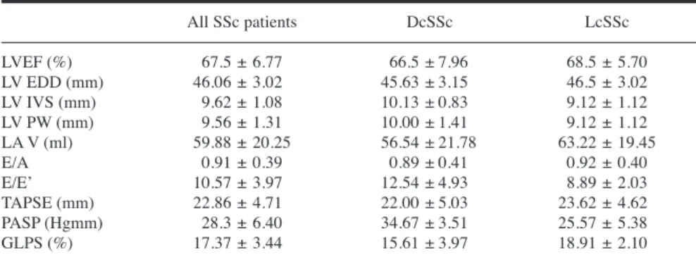
Table III. Echocardiographic parameters of systemic sclerosis patients.
All SSc patients DcSSc LcSSc
LVEF (%) 67.5 ± 6.77 66.5 ± 7.96 68.5 ± 5.70
LV EDD (mm) 46.06 ± 3.02 45.63 ± 3.15 46.5 ± 3.02
LV IVS (mm) 9.62 ± 1.08 10.13 ± 0.83 9.12 ± 1.12
LV PW (mm) 9.56 ± 1.31 10.00 ± 1.41 9.12 ± 1.12
LA V (ml) 59.88 ± 20.25 56.54 ± 21.78 63.22 ± 19.45
E/A 0.91 ± 0.39 0.89 ± 0.41 0.92 ± 0.40
E/E’ 10.57 ± 3.97 12.54 ± 4.93 8.89 ± 2.03
TAPSE (mm) 22.86 ± 4.71 22.00 ± 5.03 23.62 ± 4.62
PASP (Hgmm) 28.3 ± 6.40 34.67 ± 3.51 25.57 ± 5.38
GLPS (%) 17.37 ± 3.44 15.61 ± 3.97 18.91 ± 2.10
There were no statistically significant differences between DcSSc and LcSSc patient groups.
A: late mitral inflow velocity; DcSSc: diffuse cutaneous systemic sclerosis; E: early mitral inflow velocity; E’: early diastolic velocity; EDD: end diastolic diameter; EF: ejection fraction; GLPS: global longitudinal peak systolic strain; LA V: left atrial volume; LcSSc: limited cutaneous systemic sclero- sis, LV: left ventricular; IVS: intraventricular septum; PASP: pulmonary artery systolic pressure; PW:
posterior wall; TAPSE: tricuspid annular plane systolic excursion.
Echocardiography is an essential, first line, noninvasive method to detect car- diac involvement in patients with SSc.
Common pathological findings in overt disease may include wall motion dis- turbances, reduced ejection fraction, pericardial effusion and PAH. Recently echocardiographic deformation imag- ing modalities such as STE strain anal- ysis have been developed to provide more accurate depiction of regional contractility and dysfunction otherwise not seen with routine parameters. Sev- eral STE studies have described sub- clinical left ventricle (8), right ventricle (9) and left atrial dysfunction (10) in asymptomatic SSc patients as markers of early myocardial involvement. In the current study, we report decreased left ventricle GLPS values indica- tive of subtle myocardial dysfunction in asymptomatic SSc patients. These results are in accordance with previ- ously published findings (8, 51). We found no significant differences in left ventricular GLPS values between PET positive and negative groups. Accord- ingly, no significant correlations were found between left ventricular GLPS values and the measures of pathological glucose uptake (normalised SUV ratio, HI). We also investigated spatial agree- ment between the two methods on seg- mental and regional basis as well. Our study demonstrated poor agreement of FDG-PET/CT and left ventricular strain in regards to spatial distribution of im- aging findings. These differing results between FDG-PET/CT and 2D speckle tracking echocardiography strain analy- sis maybe explained by the two differ- ent phases of disease evolution is SSc.
In myocardial biopsy studies inflam- matory infiltrates were recognisable as a form of early phase immune modu- lated interstitial/perivascular inflamma- tion which may ultimately lead to the second phase of disease, replacement fibrosis (13, 30). The results capture the dual nature of the disease: pathologi- cal glucose uptake representing early immune mediated inflammation and lower strain values representing subtle mechanical changes caused by fibrosis.
Distinct differences between the two imaging modalities should also be noted in regards to clinical implemen-
tation. Echocardiography is a validated, readily available noninvasive screening tool to detect and follow myocardial involvement in patients with systemic sclerosis. On the other hand, routine clinical use of FDG-PET-CT is not rec- ommended at this time and before pos- sible clinical implementation further investigations are warranted to evaluate specificity, sensitivity and overall clini- cal impact (prognostic value, therapeu- tic relevance) of the method in asymp- tomatic SSC patients. Until then we suggest that asymptomatic FDG-posi- tive patients should be followed more closely with currently validated cardiac techniques (i.e. CMR).
Limitations of the study
Some limitations of the study should be noted. First: the sample size was small due to the pilot nature of the study and the rare incidence of SSc. Addition- ally, we excluded patients with overt cardiac symptoms, already diagnosed cardiovascular disease further narrow- ing the patient population pool. Second:
the study lacks a true comparator for myocardial inflammation as detected by FDG-PET/CT. Standard of reference or lack thereof is a common problem when evaluating imaging modalities for myo- carditis. Endomyocardial biopsy (EMB) is still considered the gold standard for diagnosing myocarditis. However, it is not frequently performed due to risk, cost, variable specificity, and lack of experience in some facilities (42). EMB in the current, asymptomatic patient population was not considered because of its invasive nature and potential risks.
In the recent decades CMR has gained wide acceptance both as a research tool and as a clinical diagnostic method in myocarditis. Although CMR is the most validated imaging method of myocar- ditis it still is not considered to be gold standard. In one study for example it was non-decisive or falsely negative in half of patients with biopsy-proven my- ocarditis (52). Nonetheless CMR should be considered as first choice compara- tor in future studies. Furthermore, pos- sible simultaneous assessment of car- diac inflammation and fibrosis using an integrated FDG-PET/MR examination would be particularly well suited for
comparative studies. Initial experiences with this hybrid method to detect myo- cardial involvement in cardiac sarcoido- sis (53) and in suspected myocarditis (23) have been promising. Third: patho- logical 18-FDG detected by FDG-PET/
CT is highly sensitive, but nonspecific method for visualisation of inflamma- tion. Due to lack of standardisation in regards to patient preparation, reporting of images and – as mentioned above – a true comparator, false positive diagnosis is possible. Two patients in the current study exhibiting “focal on moderately elevated” diffuse uptake could poten- tially also be interpreted as false posi- tive due to unsuccessful physiologic glucose uptake suppression. Fourth:
concomitant corticosteroid therapy at the time of FDG-PET/CT examination therapy, even in low doses, may mask potential underlying low-grade myocar- dial inflammation, potentially leading to false negative results. Fifth: the study lacks a matched, healthy control popula- tion for 2DSTE imaging. Limitations of 2DSTE analysis include intervendor and patient-specific clinical factor variabil- ity. Strain values are influenced by age, concomitant cardiovascular risk factors, haemodynamic values, and medication.
Thus, the definition of normal strain values remains problematic (54). None- theless a guide on normal values by the American Society of Echocardiography and previously published meta-analysis of normal ranges in healthy subjects al- low for a meaningful interpretation of our results (25, 26).
Conclusion
Myocardial inflammation, as a potential sign of early cardiac involvement may be detected by 18-FDG-PET/CT in a considerable percentage of systemic sclerosis patients presenting without cardiac symptoms. We hypothesise that this imaging method has the potential to be used as a new diagnostic tool for the evaluation of SSc patients. However, it is evident that before clinical implemen- tation further clinical investigations are warranted in larger patient groups to de- termine the prognostic impact of these findings and to identify specific patient risk groups in whom this examination may be of direct therapeutic relevance.
References
1. STEEN VD, MEDSGER TA JR: Severe organ involvement in systemic sclerosis with dif- fuse scleroderma. Arthritis Rheum 2000; 43:
2437-44.
2. FERRI C, VALENTINI G, COZZI F et al.:
Systemic sclerosis: demographic, clinical, and serologic features and survival in 1,012 Italian patients. Medicine (Baltimore) 2002;
81: 139-53.
3. IOANNIDIS JP, VLACHOYIANNOPOULOS PG, HAIDICH AB et al.: Mortality in systemic scle- rosis: an international meta-analysis of indi- vidual patient data. Am J Med 2005; 118: 2-10.
4. FOLLANSBEE WP, CURTISS EI, MEDSGER TA JRet al.: Physiologic abnormalities of car- diac function in progressive systemic sclero- sis with diffuse scleroderma. N Engl J Med 1984; 310: 142-8.
5. BARSOTTI S, STAGNARO C, d’ASCANIO A, DELLA ROSSA A: One year in review 2016:
systemic sclerosis. Clin Exp Rheumatol 2016; 34: (Suppl. 100): S3-13.
6. RANGARAJAN V, MATIASZ R, FREED BH: Cardiac complications of systemic sclerosis and management: recent progress. Curr Opin Rheumatol 2017; 29: 574-84.
7. GUNASEKARAN P, PANAICH S, BRIASOU- LIS A, CARDOZO S, AFONSO L: Incremental Value of Two Dimensional Speckle Tracking Echocardiography in the Functional Assess- ment and Characterization of Subclinical Left Ventricular Dysfunction. Curr Cardiol Rev 2017; 13: 32-40.
8. GUERRA F, STRONATI G, FISCHIETTI C et al.: Global longitudinal strain measured by speckle tracking identifies subclinical heart involvement in patients with systemic sclero- sis. Eur J Prev Cardiol 2018; 25: 1598-606.
9. SCHATTKE S, KNEBEL F, GROHMANN Aet al.: Early right ventricular systolic dysfunc- tion in patients with systemic sclerosis with- out pulmonary hypertension: a Doppler Tis- sue and Speckle Tracking echocardiography study. Cardiovasc Ultrasound 2010; 8: 3.
10. AGOSTON G, GARGANI L, MIGLIORANZA MH et al.: Left atrial dysfunction detected by speckle tracking in patients with systemic sclerosis. Cardiovasc Ultrasound 2014; 12:
11. 30. SPETHMANN S, DREGER H, SCHATTKE S et al.: Two-dimensional speckle tracking of the left ventricle in patients with systemic scle- rosis for an early detection of myocardial in- volvement. Eur Heart J Cardiovasc Imaging 2012; 13: 863-70.
12. D’ANGELO WA, FRIES JF, MASI AT, SHUL- MAN LE: Pathologic observations in system- ic sclerosis (scleroderma). A study of fifty- eight autopsy cases and fifty-eight matched controls. Am J Med 1969; 46: 428-40.
13. CHAMPION HC: The Heart in Scleroderma.
Rheum Dis Clin North Am 2008; 34: 181-90.
14. JUNEAU D, ERTHAL F, ALZAHRANI A et al.:
Systemic and inflammatory disorders involv- ing the heart: the role of PET imaging. Q J Nucl Med Mol Imaging 2016; 60: 383-96.
15. BIRNIE DH, NERY PB, HA AC, BEANLANDS RS: Cardiac Sarcoidosis. J Am Coll Cardiol 2016; 68: 411-21.
16. JOHNSON SR: New ACR EULAR guidelines for systemic sclerosis classification. Curr
Rheumatol Rep 2015; 17: 32.
17. VALENTINI G, IUDICI M, WALKER UA et al.:
The European Scleroderma Trials and Re- search group (EUSTAR) task force for the development of revised activity criteria for systemic sclerosis: derivation and validation of a preliminarily revised EUSTAR activity index. Ann Rheum Dis 2017; 76: 270-6.
18. D’AGOSTINO RB SR, VASAN RS, PENCINA MJ et al.: General cardiovascular risk profile for use in primary care: the Framingham Heart Study. Circulation 2008; 117: 743-53.
19. DORBALA S, DI CARLI MF, DELBEKE D et al.:
SNMMI/ASNC/SCCT guideline for cardiac SPECT/CT and PET/CT 1.0. J Nucl Med 2013; 54: 1485-507.
20. CERQUEIRA MD, WEISSMAN NJ, DILSIZIAN Vet al.: American Heart Association Writ- ing Group on Myocardial Segmentation and Registration for Cardiac Imaging. Standard- ized myocardial segmentation and nomencla- ture for tomographic imaging of the heart. A statement for healthcare professionals from the Cardiac Imaging Committee of the Coun- cil on Clinical Cardiology of the American Heart Association. Int J Cardiovasc Imaging 2002; 18: 539-42.
21. OH M, KIM JY, SHIN KH et al.: Imaging atherosclerosis in the carotid arteries with F-18-Fluoro-2-deoxy-D-glucose positron emission tomography: effect of imaging time after injection on quantitative measurement.
Nucl Med Mol Imaging 2010; 44: 261-6.
22. TAHARA N, TAHARA A, NITTA Y et al.:
Heterogeneous myocardial FDG uptake and the disease activity in cardiac sarcoidosis.
JACC Cardiovasc Imaging 2010; 3: 1219-28.
23. NENSA F, KLOTH J, TEZGAH Eet al.: Fea- sibility of FDG-PET in myocarditis: Com- parison to CMR using integrated PET/MRI.
J Nucl Cardiol 2018; 25: 785-94.
24. RUDSKI LG, LAI WW, AFILALO J et al.:
Guidelines for the echocardiographic as- sessment of the right heart in adults: a report from the American Society of Echocardiog- raphy endorsed by the European Association of Echocardiography, a registered branch of the European Society of Cardiology, and the Canadian Society of Echocardiography. J Am Soc Echocardiogr 2010; 23: 685-713.
25. LANG RM, BADANO LP, MOR-AVI Vet al.:
Recommendations for cardiac chamber quantification by echocardiography in adults:
an update from the American Society of Echocardiography and the European Associ- ation of Cardiovascular Imaging. Eur Heart J Cardiovasc Imaging 2015; 16: 233-70.
26. YINGCHONCHAROEN T, AGARWAL S, POPO- VIĆ ZB, MARWICK TH: Normal ranges of left ventricular strain: a meta-analysis. J Am Soc Echocardiogr 2013; 26: 185-91.
27. MUELLER KA, MUELLER II, EPPLER D et al.:
Clinical and histopathological features of patients with systemic sclerosis undergoing endomyocardial biopsy. PLoS One 2015; 10:
e0126015.
28. CARETTE S, TURCOTTE J, MATHON G: Severe myositis and myocarditis in progressive sys- temic sclerosis. J Rheumatol 1985; 12: 997- 29. 99.CLEMSON BS, MILLER WR, LUCK JC, FERISS
JA: Acute myocarditis in fulminant systemic
sclerosis. Chest 1992; 101: 872-4.
30. RAMALHO AR, COSTA S, SILVA F, DONATO P, FRANCO F, PÊGO GM: Autoimmune myocar- ditis in systemic sclerosis: an unusual form of scleroderma heart disease presentation.
ESC Heart Fail 2017; 4: 365-370.
31. GYLLENHAMMAR T, KANSKI M, ENGBLOM H et al.: Decreased global myocardial perfu- sion at adenosine stress as a potential new biomarker for microvascular disease in sys- temic sclerosis: a magnetic resonance study.
BMC Cardiovasc Disord 2018; 18: 16.
32. PIERONI M, DE SANTIS M, ZIZZO G et al.:
Recognizing and treating myocarditis in recent-onset systemic sclerosis heart disease:
potential utility of immunosuppressive ther- apy in cardiac damage progression. Semin Arthritis Rheum 2014; 43: 526-35.
33. THUNY F, LOVRIC D, SCHNELL F et al.:
Quantification of myocardial extracellular volume fraction with cardiac MR imaging for early detection of left ventricle involve- ment in systemic sclerosis. Radiology 2014;
271: 373-80.
34. SPETHMANN S, RIEPER K, RIEMEKASTEN G et al.: Echocardiographic follow-up of pa- tients with systemic sclerosis by 2D speckle tracking echocardiography of the left ventri- cle. Cardiovasc Ultrasound 2014; 12: 13.
35. NTUSI NAB, PIECHNIK SK, FRANCIS JM et al.: Diffuse Myocardial Fibrosis and Inflam- mation in Rheumatoid Arthritis: Insights From CMR T1 Mapping. JACC Cardiovasc Imaging 2015; 8: 526-36.
36. HINOJAR R, FOOTE L, SANGLE S et al.:
Native T1 and T2 mapping by CMR in lu- pus myocarditis: Disease recognition and re- sponse to treatment. Int J Cardiol 2016; 222:
717-26.
37. BIESBROEK PS, HESLINGA SC, KONINGS TC et al.: Insights into cardiac involvement in an- kylosing spondylitis from cardiovascular mag- netic resonance. Heart 2017; 103: 745-52.
38. GREULICH S, MAYR A, KITTERER Det al.:
T1 and T2 mapping for evaluation of myo- cardial involvement in patients with ANCA- associated vasculitides. J Cardiovasc Magn Reson 2017; 19: 6.
39. LURZ P, EITEL I, ADAM J et al.: Diagnostic performance of CMR imaging compared with EMB in patients with suspected myo- carditis. JACC Cardiovasc Imaging 2012; 5:
513-24.
40. KIRCHER M, LAPA C: Novel noninvasive nu- clear medicine imaging techniques for car- diac inflammation. Curr Cardiovasc Imaging Rep 2017; 10: 6.
41. HULTEN E, ASLAM S, OSBORNE M: Cardiac sarcoidosis—state of the art review. Cardio- vasc Diagn Ther 2016; 6: 50-63.
42. KADKHODAYAN A, CHAREONTHAITAWEE P, RAMAN SV, COOPER LT: Imaging of in- flammation in unexplained cardiomyopathy.
JACC Cardiovasc Imaging 2016; 9: 603-17.
43. OZAWA K, FUNABASHI N, DAIMON M et al.:
Determination of optimum periods between onset of suspected acute myocarditis and
18F-fluorodeoxyglucose positron emission tomography in the diagnosis of inflammatory left ventricular myocardium. Int J Cardiol 2013; 169: 196-200.
44. TAKANO H, NAKAGAWA K, ISHIO Net al.:
Active myocarditis in a patient with chron- ic active Epstein-Barr virus infection. Int J Cardiol 2008; 130: e11-3.
45. PIRIOU N, SASSIER J, PALLARDY A, SERFATY JM, TROCHU JN: Utility of cardiac FDG-PET imaging coupled to magnetic resonance for the management of an acute myocarditis with non-informative endomyocardial biopsy. Eur Heart J Cardiovasc Imaging 2015; 16: 574.
46. REDUREAU E, LAIREZ O, HITZEL A, PUGNET G: Can positron emission tomography be use- ful to manage systemic sclerosis cardiac in- volvement? J Nucl Cardiol 2017; 24: 1814-5.
47. NTUSI NA, PIECHNIK SK, FRANCIS JM et al.:
Subclinical myocardial inflammation and dif- fuse fibrosis are common in systemic sclerosis - a clinical study using myocardial T1-map- ping and extracellular volume quantification.
J Cardiovasc Magn Reson 2014; 16: 21.
48. MAVROGENI S, KOUTSOGEORGOPOULOU L, KARABELA G et al.: Silent myocarditis in systemic sclerosis detected by cardiovascular magnetic resonance using Lake Louise crite- ria. BMC Cardiovasc Disord 2017; 17: 187.
49. PEREL-WINKLER A, BOKHARI S, PEREZ- RECIO T, ZARTOSHTI A, ASKANASE A, GERALDINO-PARDILLA L: Myocarditis in systemic lupus erythematosus diagnosed by 18F-fluorodeoxyglucose positron emission to- mography. Lupus Sci Med 2018; 5: e000265.
50. SEVDALINA L: Cardiac manifestations in sys- temic sclerosis. World J Cardiol 2014; 6: 993- 1005.
51. El-SHAFIE MM, SALEM SS, MOGHAZI AA: Left ventricular myocardial ischemia in col- lagen disease associated with pulmonary hy-
pertension: an evaluation by rest-stress gated SPECT and coronary angiography. Nucl Med Commun 2011; 32: 641-8.
52. ZIZZO G, DE SANTIS M, BOSELLO Set al.:
Myocarditis in systemic sclerosis diagnosed through endomyocardial biopsy. Arthritis Rheum 2009; 60 (Suppl. 10): 1201.
53. WICKS EC, MENEZES LJ, BARNES Aet al.:
Diagnostic accuracy and prognostic value of simultaneous hybrid 18F-fluorodeoxyglucose positron emission tomography/magnetic res- onance imaging in cardiac sarcoidosis. Eur Heart J Cardiovasc Imaging 2018; 19: 757- 54. 67.COLLIER P, PHELAN D, KLEIN A: A Test in
Context: Myocardial Strain Measured by Speckle-Tracking Echocardiography. J Am Coll Cardiol 2017; 69: 1043-56.