plaques predict early restenosis after eversion carotid endarterectomy
Edit Dósa, MD, PhD,a,bKristóf Hirschberg, MD,cAstrid Apor, MD,cZsuzsanna Járányi, MD, PhD,a László Entz, MD, PhD,aGyörgy Acsády, MD, DSc,aandKálmán Hüttl, MD, PhD,cBudapest, Hungary;
and Portland, Ore
Objective:Although the association between vulnerable lesions and cardiovascular events is well established, little is known about their relationship to postsurgery restenosis. To address this issue, we initiated a prospective, nonrandom- ized study to examine the femoral plaques on both sides in patients who were undergoing eversion carotid endarterectomy (CEA) and were longitudinally followed-up for early restenosis development.
Methods:The final analysis enrolled 321 patients (189 women) with a median age of 67.0 years (interquartile range, 59.0-73.0 years), who underwent eversion CEA (2005 to 2007). Using duplex ultrasound scanning, we evaluated 321 common femoral atherosclerotic lesions on the day before CEA. A quantitative scale was used to grade the size of plaques as grade 1, one or more small plaques (<20 mm2); grade 2, moderate to large plaques; and grade 3, plaques giving flow disturbances. The plaque morphology in terms of echogenicity was graded as echolucent, 1; predominantly echolucent, 2; predominantly echogenic, 3; echogenic 4; or calcified, 5. The plaque surface was categorized as smooth, irregular, or ulcerated. The patients underwent carotid duplex ultrasound imaging at 6 weeks and at 6, 12, and 24 months after CEA.
Mann-WhitneyUtest,2test, and multivariate logistic regression were used for statistical evaluation.
Results:Internal carotid artery restenosis of>50% was detected in 33 patients (10.28%) in the operated region. Neither the size (grade 1,Pⴝ.793; grade 2,Pⴝ.540; grade 3,Pⴝ.395) nor the surface characteristics of the femoral plaques (smooth,Pⴝ.278; irregular,Pⴝ.281; ulcerated,Pⴝ.934) were significantly different between the patients with and without carotid restenosis. Echolucent-predominantly echolucent femoral lesions were an independent predictor of recurrent carotid stenosis (adjusted odds ratio, 5.63; 95% confidence interval, 2.14-10.89;P<.001).
Conclusion:Ultrasound evaluation of femoral plaque morphology before CEA can be useful for identifying patients at higher risk for carotid restenosis. ( J Vasc Surg 2010;51:345-50.)
Eversion endarterectomy of the carotid artery (CEA) is a generally accepted elective surgical method in both symp- tomatic and asymptomatic patients with significant internal carotid artery (ICA) stenosis. Despite of the best surgical care, recurrent stenosis has been reported to occur in 1% to 36% of patients.1 Stoney and String2 were the first to classify carotid restenoses into two distinct types: lesions occurringⱕ2 years after CEA were attributed to intimal hyperplasia and thereafter, to recurrent atherosclerosis.
Multiple inflammatory mediators, including growth factors and cytokines, have been postulated to play different roles to trigger the neointimal response.3Similarly, inflam- mation is involved in all stages of the atherosclerotic pro-
cess, from the initiation of the fatty streak to the final stage of plaque rupture.4Histologic findings have revealed that high-risk vulnerable lesions have a thin fibrous cap, a large lipid pool, and a macrophage dense inflammation on or beneath their surface.5,6 Ultrasound (US) images show these rupture-prone plaques are predominantly echolucent and heterogeneous, as well as ulcerated.7
Previous work has shown that the presence of femoral ath- erosclerotic lesions has a predictive value for future cardiovascular events. Moreover, the size and echogenicity of the plaques were important determinants of the vascular risk.8Novo et al9re- ported a significantly higher prevalence of femoral lesions in patients with restenosis after percutaneous coronary interven- tions than in those without recurrent lumen narrowing. A 2009 study by Sirico et al10demonstrated that in patients with periph- eral artery disease, the presence of echolucent femoral plaques was associated with a greater prevalence of echolucent carotid lesions. Much less is known about the femoral plaques in relation to recurrent carotid stenosis. The primary aim of the present study was to investigate the putative role of femoral plaque size and lesion characteristics on US imaging in patients who were undergoing carotid eversion endarterectomy and were longitu- dinally followed-up for early ICA restenosis development.
PATIENTS AND METHODS
This prospective study enrolled 402 consecutive pa- tients scheduled for CEA between June 1, 2005, and
From the Department of Cardiovascular Surgery,aand Heart Centre,c Faculty of Medicine, Semmelweis University, Budapest, Hungary; and the Blood-Brain Barrier and Neuro-Oncology Program, Oregon Health and Science University, Portland.b
Competition of interest: none.
Reprint requests: Edit Dósa, MD, PhD, Blood-Brain Barrier and Neuro- Oncology Program, Oregon Health and Science University, 3181 SW Sam Jackson Park Rd, (Mail code L603) Portland, OR 7239-3098 (e-mail:dosaedit@yahoo.com).
The editors and reviewers of this article have no relevant financial relation- ships to disclose per the JVS policy that requires reviewers to decline review of any manuscript for which they may have a competition of interest.
0741-5214/$36.00
Copyright © 2010 by the Society for Vascular Surgery.
doi:10.1016/j.jvs.2009.08.080
345
March 31, 2007. All patients were treated at the Cardio- vascular Surgery Department of Semmelweis University, Budapest, Hungary. None of these patients had prior ipsi- lateral CEA or stent implantation. The study was per- formed in accordance with the Declaration of Helsinki and was approved by the local Ethics Committee. All partici- pants provided informed consent.
Patient data. At baseline, clinical history and physical examination were evaluated with special attention to car- diovascular risk factors to determine their relationship with occurrence of carotid restenosis: age, gender, body mass index (BMI), history of smoking, and presence of hyper- tension, diabetes mellitus, and hyperlipidemia. The risk factors were assessed by a review of medical records com- bined with a patient questionnaire.
● The BMI was calculated as kg/m2.
● Smoking habit was coded as smokers and nonsmokers.
Current cigarette smoking was defined as a daily intake of more than five cigarettes. Nonsmokers included those who had never smoked, former smokers who had quit smoking forⱖ1 year before the study, and those whose daily intake was less than five cigarettes.
● Hypertension was diagnosed in patients with resting blood pressure values ⬎140/90 mm Hg measured repetitively (at least twice) and was assumed to be present in patients taking antihypertensive drugs.
● Diabetes was defined as self-reported history of diabe- tes mellitus, use of antidiabetic drugs, or a fasting blood glucose⬎5.8 mmol/L, oral glucose tolerance test results suggestive of disease, or glycated hemoglo- bin A1Clevel⬎6.2%.
● Hyperlipidemia was defined as any or all of an elevation of fasting triglycerides ⬎1.7 mmol/L, total cholesterol
⬎5.0 mmol/L, or low-density lipoprotein cholesterol
⬎3.0 mmol/L, and was considered to be present in patients receiving lipid-lowering medication.
Neurologic events were also documented and catego- rized as transient ischemic attack (TIA), minor stroke, and major stroke. A TIA was termed any neurologic deficit that resolvedⱕ24 hours without evidence of any residual neu- rologic damage thereafter. A minor stroke was classified as a new neurologic event that resulted in a slight impairment of neurologic function that either resolved completelyⱕ7 days or caused an increase in the National Institute of Health Stroke Scale (NIHSS) of ⬍4. A new neurologic deficit that persisted⬎7 days or increased the NIHSS by ⱖ4 was defined as a major stroke.11Current use of statin and antiplatelet (acetylsalicylic acid, dipyridamole, clopi- dogrel) therapy was recorded for each patient.
Baseline US evaluation. Each patient had femoral US examination the day before surgery at our department.
B-mode and color Doppler measurements were performed using a SSA-700A US machine (Aplio; Toshiba Medical Systems, Tustin, Calif) equipped with a linear array broad- band 7-4-MHz transducer. All femoral duplex US (DUS) scans were performed and analyzed by the same senior vascular radiologist. The common femoral artery (CFA)
was scanned bilaterally distal to the inguinal ligament along a section approximately 4 cm proximal and 1 cm distal to the flow divider (the site where the artery divides into the superficial and profound femoral arteries) to assess the occurrence of plaques.
Femoral plaque was defined as a focal widening⬎50%
relative to the adjacent segment, as recommended by the US Task Force on noninvasive atherosclerosis measure- ment.12Each plaque was measured in its own longitudinal view in which the plaque was biggest. A cursor on the screen was used to trace the perimeter of the plaque, and intrinsic software of the DUS scanner was used to compute the cross-sectional area of the plaque. The cross-sectional areas of all CFA plaques were then summed to give the total plaque area. The plaques were categorized as grade 1, one or more small plaques (total plaque area⬍20 mm2); grade 2, moderate to large plaques; and grade 3, plaques causing flow disturbances.13This analysis included plaques in the near wall as well as the far wall of the vessel. When the plaque size was different between the two sides, the higher grade was considered for further evaluation.
The plaque morphology in terms of echogenicity was visually judged and graded from 1 to 5 as echolucent, 1;
predominantly echolucent, 2; predominantly echogenic, 3;
echogenic, 4; or calcified, 5.14Echogenicity was standard- ized against three reference structures: flowing blood for echolucent, iliopsoas muscle for isoechogenic, and the bright far wall media-adventitia interface for echogenic plaques.15Calcified plaques had zones of acoustic shadow- ing that obscured the deeper part of the arterial wall as well as the vessel lumen. The plaque surface was categorized as smooth, irregular, or ulcerated. The height variations of the surface were determined by using the software incorpo- rated in the US machine. The surface was regarded as irregular in the case of height variations of between 0.4 and 2 mm on the contour of the plaque.
Ulceration was defined as a recess that was atⱖ2 mm deep and 2 mm long and had an area of reversed flow within the recess or a zone of low flow signal at the level of the recess.16In patients with multiple plaques, only the thickest was selected for analysis.
The US images were sent to a PACS (Picture Archiving Communication System) workstation, and 50 randomly chosen plaques were re-evaluated by the same radiologist at the end of the study. The intraobserver reproducibility for plaque size and morphology (echogenicity, surface charac- teristics) was good ( ⫽.81, ⫽.79,⫽.71, respectively).
Eversion CEA. The criteria to perform CEA were based on the recommendations by the American Heart Association.17 The procedures were performed with the patient under deep general anesthesia by four experienced vascular surgeons. After systemic heparinization, the distal internal, common, and external carotid arteries were clamped. The ICA was transected obliquely from the com- mon carotid artery (CCA). The ICA wall was everted over the atheromatous core until a distal end point was directly visualized. After the ICA had been completely endarterec- tomized, significant disease in the common and external
carotid arteries were removed. The ICA was then reanasto- mosed to the CCA. All patients were prescribed an acetyl- salicylic acid regimen (325 mg daily)ⱕ24 hours postoper- atively, which was continued indefinitely.
Follow-up. Follow-up visits were scheduled at 6 weeks, and at 6, 12, and 24 months after CEA. Outpatient carotid DUS scans were performed by another experienced radiologist who was unaware of the results of CFA plaque characterization. The common, internal, and external ca- rotid arteries on both sides were examined in the standard fashion. Representative values of peak systolic velocity (PSV) and end-diastolic velocity (EDV) were measured.
The PSV of the ICA was the primary parameter for the grading of ICA restenosis. If the degree of recurrent ICA stenosis was indeterminate according to the PSVs, then additional parameters, including the ICA/CCA PSV ratio and the ICA EDV were considered. A 50% to 69% ICA restenosis was diagnosed when ICA PSV was 125 to 230 cm/s. Additional criteria included an ICA/CCA PSV ratio of 2.0 to 4.0 and ICA EDV of 40 to 100 cm/s. Aⱖ70%
ICA restenosis was diagnosed when the ICA PSV was
⬎230 cm/s. Additional criteria included an ICA/CCA PSV ratio⬎4.0 and ICA EDV⬎100 cm/s. These criteria had been established according to the Society of Radiolo- gists in Ultrasound Consensus Conference and were inter- nally verified.18
Statistical analysis. Statistical analyses were per- formed using Statistica 8.0 software (StatSoft Inc, Tulsa, Okla). Categoric variables were expressed as numbers and percentages and were compared between groups using the
2test. Continuous variables were assessed for normality by using the Shapiro-Wilk test. Because continuous variables exhibited a non-Gaussian distribution, they were expressed as medians and interquartile ranges (IQR) and were com- pared using the nonparametric Mann-WhitneyUtest. All analyses were two-tailed and values ofPⱕ.05 were con- sidered as statistically significant.
To identify the independent predictors of early carotid restenosis, multivariate logistic regression was done with adjustment for age, gender, BMI, and follow-up time. The dependent variable wasⱖ50% ICA restenosis. In case of combined variables, patients who fulfilled the criteria of both single variables were compared with those who did not have the combined characteristics. All variables with a value of P ⱕ .05 in the univariate analysis were tested.
Intraobserver reproducibility was calculated using the Co- henstatistic.
RESULTS
Nine of the 402 patients (2.24%) did not undergo follow-up because of all-cause mortality, including stroke in three patients, acute myocardial infarction in four, and cancer in two. No plaques were found in the CFAs in 18 patients (4.48%) and they were excluded from the study because the inclusion criterion for this investigation was the presence of femoral plaque. Unilateral or bilateral femoral artery reconstruction was done in 13 patients (3.23%).
Patients with unilateral femoral artery reconstruction were
also excluded under the assumption that the revascularized artery had the higher plaque grade. The evaluation of the CFA lesions was impossible because of the marked acoustic shadow in 15 patients (3.73%). Bilateral femoral occlusive disease was present in eight patients (1.99%). Four patients (0.99%) were followed-up at other hospitals, and 14 (3.48%) were lost to follow-up.
The final analysis therefore included 321 patients (189 women) with 321 arteries. Their median age was 67.0 years (IQR, 59.0-73.0 years). Their median BMI was 25.0 kg/m2 (IQR, 21.0-26.0 kg/m2). Risk factors included smoking in 258 patients (80.37%), hypertension in 197 (61.37%), diabetes mellitus in 78 (24.30%), and hyperlipid- emia in 127 (39.56%). Preoperatively, 95 patients (29.60%) were asymptomatic, 194 (60.44%) had TIA, 26 (8.10%) had minor stroke, and 6 (1.87%) had major stroke. Statin usage in 135 patients (42.06%) and antiplatelet therapy in 220 patients (68.54%) was recorded.
Follow-up period. The median follow-up time was 24.3 months (IQR, 22.9-25.4 months). Five (3 minor strokes, 2 major strokes) nonfatal perioperative strokes (1.56%) occurred. Cardiogenic embolization caused minor strokes in two additional patients ⱕ2 years after CEA.
Nonfatal acute myocardial infarction was recorded in five patients (1.56%).
All patients underwent US examinations preoperatively and at 24 months. The cutoff point for the definition of residual stenosis was 5.79 weeks (IQR, 4.86-6.95 weeks).
Residual lesions (⬍50%) were found in 12 patients (3.74%), but they remained stable during the rest of the follow-up.
ICA restenosis of ⱖ50% was detected in 33 patients (10.28%), among them 17 (5.30%) had severe recurrent stenosis (ⱖ70%) in the operated region. Restenosis was detected at 6.13 months (IQR, 5.60-6.67 months) after CEA in eight patients, at 12.00 months (IQR, 11.42-12.65 months) in 19 patients, and in six patients thereafter (Fig).
Eleven patients with significant recurrent lumen narrowing were assigned for carotid stenting, the other patients con- tinued to receive medical treatment.
Baseline characteristics of the patients with and without carotid restenosis. Hyperlipidemia was more common in patients with recurrent carotid stenosis (P⬍ .001). In case of preoperative symptoms (TIA, stroke) the statistical analysis did not reveal a significant difference between patients with (P⫽.723) and without (P⫽.859) carotid restenosis. No significant difference was found in statin treatment between the groups of patients with and without restenosis, as well as for antiplatelet usage (P⫽ .964,P⫽.584, respectively;Table I).
Presurgical femoral plaque size in patients with and without recurrent carotid lumen narrowing. Plaques in the CFA were grade 1 in 91 patients (28.35%), grade 2 in 111 patients (34.58%), and grade 3 in 119 patients (37.07%). The femoral plaque size did not differ signifi- cantly between the groups (grade 1,P⫽.793; grade 2,P⫽ .540; and grade 3,P⫽.395, respectively;Table II).
Echogenicity and surface characteristics of the fem- oral plaques before surgery in patients with and with-
out carotid restenosis. The incidence of predominantly echolucent CFA plaques was significantly higher in patients with documented restenosis during the follow-up period (P⬍.001). There were no significant differences in surface
characteristics of the femoral lesions between the patients with and without recurrent lumen narrowing (smooth,P⫽ .278; irregular,P⫽.281; ulcerated,P⫽.934, respectively;
Table III).
Predictors of early restenosis after eversion CEA.
The univariate analysis showed that hyperlipidemia was the only cardiovascular risk factor associated with an increased incidence of ICA restenosis during the follow-up (odds ratio [OR], 3.24; 95% confidence interval [CI], 1.72-6.11;
P ⬍ .001). In this analysis, patients with echolucent or predominantly echolucent CFA lesions were merged into one group, and those with echogenic or predominantly echogenic or calcified plaques formed another group. We also merged patients with irregular or ulcerated plaque surface into one group. The reason for this regrouping was that only a few plaques were classified as echolucent, calci- fied, or ulcerated. Patients with echolucent-predominantly echolucent femoral plaques had a higher risk of ICA reste- nosis compared with those with echogenic-predominantly echogenic or calcified CFA lesions (OR, 4.99; 95% CI, 2.12-9.69;P ⬍ .001). Irregular femoral plaques only in combination with echolucency were predictors of early carotid restenosis development (OR, 3.09; 95% CI, 2.13- 7.05;P⫽.034). None of the other US features of the CFA plaques showed a relationship with recurrent ICA lumen narrowing (Table IV). The multivariate logistic regression analysis tested all variables with a value ofPⱕ.05 in the univariate analysis, and all parameters remained statistically significant in the multivariate logistic regression model (Table V).
DISCUSSION
DUS imaging is a reliable and noninvasive method of assessing the atherosclerotic plaque morphology and deter- mining the degree of stenosis. Computer-assisted off-line classification of the plaque echogenicity is an objective technology with excellent reproducibility.16 However, these measurements require separate equipment and are time-consuming; therefore, it is difficult to apply them in everyday clinical practice. In our study the plaque structure Fig. Kaplan-Meier curve shows the incidence of restenosis after
eversion carotid endarterectomy.
Table I. Baseline characteristics of the patients with and without carotid restenosis
Characteristics
Restenosis No restenosis P (n⫽33) (n⫽288) Age, median (IQR), y 67 (58-73) 67 (60-73) .883 Female gender, No. (%) 18 (54.55) 171 (59.38) .593 BMI, median (IQR), kg/m2 24 (21-25) 25 (21-26) .200 Smoking, No. (%) 25 (75.76) 233 (80.90) .481 Hypertension, No. (%) 19 (57.58) 178 (61.81) .636 Diabetes mellitus, No. (%) 7 (21.21) 71 (24.65) .662 Hyperlipidemia, No. (%) 25 (75.76) 102 (35.42) ⬍.001a Pre-op TIA, No. (%) 19 (57.58) 175 (60.76) .723 Pre-op stroke, No. (%) 3 (9.09) 29 (10.07) .859 Statin use, No. (%) 14 (42.42) 121 (42.01) .964 Antiplatelet use, No. (%) 24 (72.73) 196 (68.06) .584 BMI,Body mass index;IQR,interquartile range;TIA,transient ischemic attack.
aStatistically significant.
Table II. Presurgical femoral plaque size in patients with and without recurrent carotid lumen narrowing
Plaque size
Restenosis No restenosis P (n⫽33) (n⫽288)
Grade 1, No. (%) 10 (30.30) 81 (28.13) .793 Grade 2, No. (%) 13 (39.39) 98 (34.03) .540 Grade 3, No. (%) 10 (30.30) 109 (37.85) .395
Table III. Echogenicity and surface characteristics of the femoral plaques before surgery in patients with and without carotid restenosis
Plaque characteristics
Restenosis No restenosis P (n⫽33) (n⫽288) Echogenicity, No. (%)
Echolucent 1 (3.03) 9 (3.13) .976
Predominantly echolucent 12 (36.36) 41 (14.24) ⬍.001a Predominantly echogenic 9 (27.27) 102 (35.42) .352 Echogenic 10 (30.30) 108 (37.50) .417
Calcified 1 (3.03) 28 (9.72) .204
Surface, No. (%)
Smooth 19 (57.58) 193 (67.01) .278
Irregular 13 (39.39) 87 (30.21) .281
Ulcerated 1 (3.03) 8 (2.78) .934
aStatistically significant.
was visually judged. The visual analysis, even if considered to be subjective, was found to be a valuable method for plaque characterization.19
This study included one of the largest series of patients in whom the femoral plaque size and lesion morphology were evaluated. Patients with early carotid restenosis had a significantly higher incidence of predominantly echolucent femoral plaques than those without recurrent lumen nar- rowing. Moreover, the plaque echogenicity seemed to be superior to the surface characteristics of the atherosclerotic lesions in regard to restenosis development, because irreg- ular femoral lesions only in combination with echolucency were independent predictors of early ICA restenosis devel- opment. Vulnerable lesions were reported to develop si- multaneously at multiple sites of the vascular bed.20 In addition, trials with lipid-lowering or anti-inflammatory therapy demonstrated a reduced rate of ischemic vascular events and death, which can scarcely be conceived as a
single-plaque phenomenon.4,21Thus, echolucent femoral lesions may represent a vulnerable patient.
Because an inflammation phenomenon seems to be the key component in both the development of unstable plaques and in the formation of neointima,3,4it may be hypothesized that those systemic inflammatory factors that are involved in the destabilization of atherosclerotic lesions at the same time facilitate the restenotic process. An obser- vation of Liapis et al22 that the presence of ipsilateral echolucent carotid plaques before surgery is a predictor of restenosis after CEA is in line with our findings. This relationship was thought to be mediated by the remainders of plaques at the CEA site. In a 2008 study, Kitta et al23 showed a strong association between the presence of echolucent carotid lesions and in-stent restenosis after per- cutaneous coronary interventions. However, it should be taken into account that the pathogenesis of recurrent ste- nosis after endarterectomy differs from the in-stent rest- enotic process in many ways.
Schmidt et al8 revealed a positive correlation between femoral plaque size and future cardiovascular events in middle- aged men. The association remained present, albeit attenu- ated, after adjustment for traditional cardiovascular risk factors, including low-density lipoprotein cholesterol and triglyceride levels, systolic blood pressure, and smoking.
We could not find a significant correlation between baseline femoral plaque size and recurrent lumen narrowing after CEA. The morphology of coexisting lesions rather than size appears to be important in the development of reste- nosis.
Previous studies support the notion that inflammation is related not only to lesion structure but also to plaque size.24,25 According to our observations, we assume, that plaque size, “the amount of atherosclerosis” and echogenicity are influenced by different systemic or local inflammatory factors, or both, and those that are associated with plaque morphology may also be involved in the restenotic process.
Several reports have examined the influence of systemic factors such as age, gender, BMI, smoking, hypertension, diabetes mellitus, and hyperlipidemia on the development of recurrent stenosis after CEA, but none has succeeded in establishing a globally accepted status.26,27Our study iden- tified patients with hyperlipidemia as being at particular risk of developing restenosis. In hyperlipidemia, the rate of LDL entry into the intima increases and the LDL under- goes progressive oxidative modification in the subendothe- lial space. The aggregation of oxidized LDL induces the release of mitogens from platelets, macrophages, and endo- thelial cells, which stimulate smooth muscle cell prolifera- tion and may lead to neointima formation.28
Despite of the results of immunohistochemical and biochemical studies that confirmed the role of oxidized LDL in the restenotic process,29 the salutary effect of lipid-lowering agents on the incidence of restenosis is con- troversial. Some investigators have found a statistically sig- nificant correlation, while others have not.27,30Of note in our study, the diagnosis of hyperlipidemia was determined by an evaluation of available medical records of the patients.
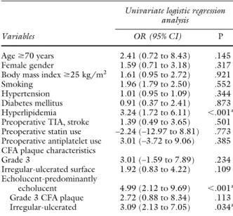
Table IV. Univariate analysis of predictors of early restenosis after eversion carotid endarterectomy
Variables
Univariate logistic regression analysis
OR (95% CI) P
Ageⱖ70 years 2.41 (0.72 to 8.43) .145
Female gender 1.59 (0.71 to 3.18) .317
Body mass indexⱖ25 kg/m2 1.61 (0.95 to 2.72) .921
Smoking 1.96 (1.79 to 2.50) .552
Hypertension 1.01 (0.95 to 1.09) .344
Diabetes mellitus 0.91 (0.37 to 2.41) .873 Hyperlipidemia 3.24 (1.72 to 6.11) ⬍.001a Preoperative TIA, stroke 1.39 (0.49 to 3.65) .501 Preoperative statin use –2.24 (–12.97 to 8.81) .773 Preoperative antiplatelet use 3.01 (–3.72 to 9.06) .385 CFA plaque characteristics
Grade 3 3.01 (–1.59 to 7.89) .234
Irregular-ulcerated surface 1.92 (0.83 to 4.22) .109 Echolucent-predominantly
echolucent 4.99 (2.12 to 9.69) ⬍.001a Grade 3 CFA plaque 2.72 (0.88 to 8.34) .113 Irregular-ulcerated 3.09 (2.13 to 7.05) .034a CFA,common femoral artery;CI, confidence interval;OR,odds ratio;TIA, transient ischemic attack.
aStatistically significant.
Table V. Multivariate analysis of predictors of early restenosis after eversion carotid endarterectomy
Variables
Multivariate logistic regression analysis
(R2⫽.152)
OR (95% CI) P
Hyperlipidemia 3.37 (1.77-6.42) ⬍.001a Echolucent-predominantly
echolucent CFA plaque 5.63 (2.14-10.89) ⬍.001a Irregular-ulcerated 2.42 (1.17-6.03) .029a CFA,Common femoral artery;CI,confidence interval;OR,odds ratio.
aStatistically significant.
Moreover, we do not know how many patients were on optimal control of their associated risk factors or were treated with statins independently of their serum lipopro- tein levels; therefore, definite conclusions about the impor- tance of traditional risk factors on the development of early recurrent lumen narrowing after surgery cannot be drawn from our investigation.
CONCLUSIONS
Patients with echolucent-predominantly echolucent fem- oral plaques, independently of their size, have an increased risk for recurrent ICA stenosis development compared with those with echogenic-predominantly echogenic or calcified athero- sclerotic lesions. However, an important question that may be asked is: If it is possible for us to examine the morphology of the carotid plaques before CEA, do we have to care about the composition of femoral lesions in regard to carotid restenosis?
The answer should be yes, but further prospective trials with analysis of both carotid and femoral lesions in the same indi- viduals are needed to reveal whether all patients or only subgroups of patients would benefit from the evaluation of CFA lesions before carotid surgery.
AUTHOR CONTRIBUTIONS Conception and design: ED, LE, GA, KH Analysis and interpretation: ED, KH Data collection: ED, AA, ZJ, KH Writing the article: ED
Critical revision of the article: K Hirschberg
Final approval of the article: ED, K Hirschberg, AA, ZJ, LE, GA, KH
Statistical analysis: K Hirschberg Obtained funding: N/A Overall responsibility: ED
REFERENCES
1. Reina-Gutiérrez T, Serrano-Hernando FJ, Sánchez-Hervás L, Ponce A, Vega de Ceniga M, Martín A. Recurrent carotid artery stenosis follow- ing endarterectomy: natural history and risk factors. Eur J Vasc Endo- vasc Surg 2005;29:334-41.
2. Stoney RJ, String ST. Recurrent carotid stenosis. Surgery 1976;80:705-10.
3. Schillinger M, Minar E. Restenosis after percutaneous angioplasty: the role of vascular inflammation. Vasc Health Risk Manag 2005;1:73-8.
4. Insull W Jr. The pathology of atherosclerosis: plaque development and plaque responses to medical treatment. Am J Med 2009;122:S3-14.
5. Naghavi M, Libby P, Falk E, Casscells SW, Litovsky S, Rumberger J, et al. From vulnerable plaque to vulnerable patient: a call for new definitions and risk assessment strategies: Part I. Circulation 2003;108:1664-72.
6. Naghavi M, Libby P, Falk E, Casscells SW, Litovsky S, Rumberger J, et al.
From vulnerable plaque to vulnerable patient: a call for new definitions and risk assessment strategies: Part II. Circulation 2003;108:1772-8.
7. Nordestgaard BG, Grønholdt ML, Sillesen H. Echolucent rupture- prone plaques. Curr Opin Lipidol 2003;14:505-12.
8. Schmidt C, Fagerberg B, Hulthe J. Non-stenotic echolucent ultrasound- assessed femoral artery plaques are predictive for future cardiovascular events in middle-aged men. Atherosclerosis 2005;181:125-30.
9. Novo G, Maniglia D, Corrado E, Muratori I, Sutera F, Evola S, et al.
Peripheral atherosclerosis is associated with the occurrence of restenosis after percutaneous coronary intervention. Coron Artery Dis 2007;18:
627-31.
10. Sirico G, Brevetti G, Lanero S, Laurenzano E, Luciano R, Chiariello M.
Echolucent femoral plaques entail higher risk of echolucent carotid
plaques and a more severe inflammatory profile in peripheral arterial disease. J Vasc Surg 2009;49:346-51.
11. Brott T, Adams HP Jr, Olinger CP, Marler JR, Barsan WG, Biller J, et al. Measurements of acute cerebral infarction: a clinical examination scale. Stroke 1989;20:864-70.
12. Redberg RF, Vogel RA, Criqui MH, Herrington DM, Lima JA, Roman MJ. 34th Bethesda Conference: Task force #3—What is the spectrum of current and emerging techniques for the noninvasive measurement of atherosclerosis? J Am Coll Cardiol 2003;41:1886-98.
13. Wendelhag I, Wiklund O, Wikstrand J. Atherosclerotic changes in the femoral and carotid arteries in familial hypercholesterolemia. Ultra- sonographic assessment of intima-media thickness and plaque occur- rence. Arterioscler Thromb 1993;13:1404-11.
14. Geroulakos G, Ramaswami G, Nicolaides A, James K, Labropoulos N, Belcaro G, et al. Characterization of symptomatic and asymptomatic carotid plaques using high-resolution real-time ultrasonography. Br J Surg 1993;80:1274-7.
15. Joakimsen O, Bønaa KH, Stensland-Bugge E. Reproducibility of ultra- sound assessment of carotid plaque occurrence, thickness, and mor- phology. The Tromsø Study. Stroke 1997;28:2201-7.
16. Sztajzel R. Ultrasonographic assessment of the morphological charac- teristics of the carotid plaque. Swiss Med Wkly 2005;135:635-3.
17. Biller J, Feinberg WM, Castaldo JE, Whittemore AD, Harbaugh RE, Dempsey RJ, et al. Guidelines for carotid endarterectomy: a statement for healthcare professionals from a special writing group of the Stroke Council, American Heart Association. Stroke 1998;29:554-62.
18. Grant EG, Benson CB, Moneta GL, Alexandrov AV, Baker JD, Bluth EI, et al. Carotid artery stenosis: gray-scale and Doppler US diagnosis—
Society of Radiologists in Ultrasound Consensus Conference. Radiol- ogy 2003;229:340-6.
19. Mayor I, Momjian S, Lalive P, Sztajzel R. Carotid plaque: comparison between visual and grey-scale median analysis. Ultrasound Med Biol 2003;29:961-6.
20. Slevin M, Wang Q, Font MA, Luque A, Juan-Babot O, Gaffney J, et al.
Atherothrombosis and plaque heterology: different location or a unique disease? Pathobiology 2008;75:209-25.
21. Stalenhoef AF. The benefit of statins in non-cardiac vascular surgery patients. J Vasc Surg 2009;49:260-5.
22. Liapis CD, Kakisis JD, Dimitroulis DA, Kostakis AG. The impact of the carotid plaque type on restenosis and future cardiovascular events: a 12-year prospective study. Eur J Vasc Endovasc Surg 2002;24:239-44.
23. Kitta Y, Obata JE, Takano H, Nakamura T, Kodama Y, Fujioka D, et al.
Echolucent carotid plaques predict in-stent restenosis after bare metal stenting in native coronary arteries. Atherosclerosis 2008;197:177-82.
24. Grønholdt ML, Sillesen H, Wiebe BM, Laursen H, Nordestgaard BG.
Increased acute phase reactants are associated with levels of lipoproteins and increased carotid plaque volume. Eur J Vasc Endovasc Surg 2001;
21:227-34.
25. Andersson J, Sundström J, Kurland L, Gustavsson T, Hulthe J, Elmgren A, et al. The carotid artery plaque size and echogenicity are related to different cardiovascular risk factors in the elderly: the Pro- spective Investigation of the Vasculature in Uppsala Seniors (PIVUS) study. Lipids 2009;44:397-403.
26. Liapis CD, Paraskevas KI. Factors affecting recurrent carotid stenosis.
Vasc Endovascular Surg 2005;39:83-95.
27. LaMuraglia GM, Stoner MC, Brewster DC, Watkins MT, Juhola KL, Kwolek C, et al. Determinants of carotid endarterectomy anatomic durability: effects of serum lipids and lipid-lowering drugs. J Vasc Surg 2005;41:762-8.
28. Aidinian G, Weiswasser JM, Arora S, Abularrage CJ, Singh N, Sidawy AN. Carotid plaque morphologic characteristics. Perspect Vasc Surg Endovasc Ther 2006;18:63-70.
29. Eto H, Miyata M, Kume N, Minami M, Itabe H, Orihara K, et al.
Expression of lectin-like oxidized LDL receptor-1 in smooth muscle cells after vascular injury. Biochem Biophys Res Commun 2006;341:591-8.
30. Perler BA. The effect of statin medications on perioperative and long- term outcomes following carotid endarterectomy or stenting. Semin Vasc Surg 2007;20:252-8.
Submitted May 31, 2009; accepted Aug 24, 2009.