Nephrol Dial Transplant (2008) 23: 3256–3262 doi: 10.1093/ndt/gfn242
Advance Access publication 1 May 2008
Original Article
Serum osteoprotegerin level, carotid-femoral pulse wave velocity and cardiovascular survival in haemodialysis patients
G´abor Speer
1,∗, Bertalan Cs. Fekete
1,∗, Taha El Hadj Othmane
1,∗, Tam´as Szab´o
2, J´ozsef Egresits
1,3, Erzs´ebet Fodor
2, Istv´an Kiss
2,3, Alexander G. Logan
4, J´anos Nemcsik
2, Andr´as Szab´o
1,
Zs´ofia K. N´emeth
1, Mikl´os Szathm´ari
1and Andr´as Tisl´er
1,211st Department of Medicine, Semmelweis University Budapest,2B. Braun Avitum Nephrological Network,3Division of Angiology and Nephrology, Department of Medicine, St Imre Teaching Hospital, Budapest, Hungary and4Samuel Lunenfeld Research Institute, University of Toronto, Toronto, Canada
Abstract
Background.Osteoprotegerin (OPG) is a marker and reg- ulator of arterial calcification, and it is related to car- diovascular survival in haemodialysis patients. The link between OPG and aortic stiffening—a consequence of ar- terial calcification—has not been previously evaluated in this population, and it is not known whether OPG-related mortality risk is mediated by arterial stiffening.
Methods.At baseline, OPG and aortic pulse wave velocity (PWV) were measured in 98 chronic haemodialysis patients who were followed for a median of 24 months. The relation- ship between OPG and PWV was assessed by multivariate linear regression. The role of PWV in mediating OPG re- lated cardiovascular mortality was evaluated by including both OPG and PWV in the same survival model.
Results.At baseline mean (standard deviation) PWV was 11.2 (3.3) m/s and median OPG (interquartile range) was 11.1 (7.5–15.9) pmol/L. There was a strong, positive, linear relationship between PWV and lnOPG (P=0.009, model R2 =0.540) independent of covariates. During follow-up 23 patients died of cardiovascular causes. In separate uni- variate survival models both PWV and lnOPG were related to cardiovascular mortality [hazard ratios 1.31 (1.14–1.50) and 8.96 (3.07–26.16), respectively]. When both PWV and lnOPG were entered into the same model, only lnOPG re- mained significantly associated with cardiovascular mor- tality [hazard ratio 1.11 (0.93–1.33) and 7.18 (1.89–27.25), respectively).
Conclusion.In haemodialysis patients OPG is strongly re- lated to PWV and OPG related cardiovascular mortality risk is, in part, mediated by increased PWV.
Correspondence and offprint requests to: Gabor Speer, 1st Department of Medicine, Semmelweis University Budapest, 2/a Kor´anyi S. u., Budapest, H-1083, Hungary. Tel:+36-30-9679-144; Fax:+36-1-313-0250; E-mail:
speerga@bel1.sote.hu
∗These authors contributed equally to this work and are considered first authors.
Keywords: cardiovascular mortality; osteoprotegerin;
pulse wave velocity; vascular calcification
Introduction
A number of cross-sectional studies reported high preva- lence of vascular calcification in haemodialysis (HD) pa- tients [1–4]. In addition, prospective studies demonstrated that vascular calcification of the intima and media of the large arteries is associated with increased risk of cardiovas- cular events and mortality in this population, independent of classical risk factors [5].
Stiffening of the wall of the aorta—as assessed for exam- ple by carotid-femoralis pulse wave velocity (PWV)—has emerged as a robust and independent predictor of all cause and cardiovascular mortality in HD patients [6,7]. Aortic stiffening leads to decreased cushioning function, faster pulse wave propagation and earlier return of the reflected wave to the heart resulting in increased central pulse pres- sure, increased systolic pressure load to the heart and de- creased diastolic pressure to boost coronary circulation [8,9]. The positive relationship between the extent of aortic calcification and PWV [10] provides a pathway that is—at least in part—plausible to explain for the cardiovascular morbid events seen in HD patients with arterial calcifica- tions.
Recent studies have indicated that osteoprotegerin (OPG), a soluble decoy receptor of the osteoclast activator RANKL, acts as an important regulatory molecule in vas- cular disease, such as arterial calcification and atheroscle- rosis [11,12]. Moreover, elevated OPG levels have been associated with the progression of vascular calcification in patients receiving long-term haemodialysis [13], and a recent analysis showed that OPG levels can, in part, ex- plain for the association between coronary artery calcifica- tion and chronic kidney disease [14]. OPG is also a novel marker of cardiovascular mortality and clinical events in
C The Author [2008]. Published by Oxford University Press on behalf of ERA-EDTA. All rights reserved.
For Permissions, please e-mail: journals.permissions@oxfordjournals.org
patients with acute myocardial infarction complicated with heart failure [15]. The link between serum OPG levels and cardiovascular survival in HD [16] and renal transplant pa- tients [17] further strengthens the role of OPG as a clinically useful prognostic marker among these patients. While the relationship between OPG and arterial calcification may provide the broad framework to explain for its effects on survival, downstream elements of a possible causal rela- tionship are yet to be identified.
We hypothesize that increased serum OPG levels in HD patients indicate arterial calcification that leads to increased PWV with consequent cardiovascular events. To test this we analysed the relationship between OPG levels and PWV in patients on maintenance HD. Furthermore, we investigated the inter-relationship of PWV and serum OPG levels on cardiovascular mortality in the same cohort during follow- up.
Subjects and methods
Patients
This was a prospective cohort study among 98 chronic HD patients with baseline cross-sectional analysis of the relationship between PWV and OPG levels, and survival analysis for cardiovascular mortality using PWV and OPG as predictor variables during follow-up.
All chronic (>3 months on HD) patients of two dialysis units of a dialysis network were invited to participate. No specific exclusion criteria were applied. Patients who gave written informed consent for participation were included and had baseline clinical assessment, laboratory and PWV measurements and then followed for a median (range) of 24 (0–31) months. Baseline demographic and clinical data were gathered by chart review, and laboratory parameters were measured prior to a midweek dialysis at the time of arterial stiffness assessment. Overt cardiovascular disease was considered to be present if the patient had a docu- mented history of myocardial infarction, revascularization procedure, stroke or peripheral artery disease. Heart failure was not included in the definition of cardiovascular dis- ease as this frequently would have been based on physician assessment only, and signs of hypervolaemia in these HD patients could have led to misclassification of heart failure.
The protocol was approved by the Ethics Committee of the dialysis network and all patients gave written informed consent to their participation.
PWV recordings
Arterial stiffness was measured as carotid-femoral PWV and carotid augmentation index (AI) using the validated PulsePen tonometer (DiaTecne, Milan, Italy) before a mid- week HD session with the patient in the supine position [18]. In each subject two sequences of measurements were performed, and their mean was used for statistical analysis.
The PulsePen device measures the time difference be- tween the R wave of the ECG and the foot of the pulse pressure wave—obtained sequentially above the carotid and the femoral arteries using a handheld tonometer—to calcu-
late pulse transit time between these two sites. The average signal of at least 10 heart cycles was used in the measure- ments at both sites for the assessment of PWV. Surface tape measurement of the distance between the carotid and femoral measurement sites was used to calculate PWV by the PulsePen software. To ensure that alterations in blood pressure and heart rate would not bias the results of PWV assessment, the software of PulsePen automatically rejected measurements in which blood pressure or heart rate changed
>5% during the time between the sequential carotid and femoral pulse wave recordings.
Carotid tonometric measurements were performed on the side contralateral to the fistula or tunnelled jugular line. The same sides were used to obtain femoral pressure waves. All femoral and carotid pulse pressure wave recordings were evaluated for quality by a single observer (TEHO). Previ- ously we evaluated the intra- and interobserver variability of PWV measurements obtained by the PulsePen device in haemodialysis patients and these were 4.8 and 7.3%, respectively.
Blood pressure and heart rate were recorded by the val- idated BpTru device (VSM Medtech, Vancouver, Canada) with two sequential measurements manually averaged.
Laboratory measurements
After the arterial stiffness measurements and prior to dialysis, blood samples were collected for laboratory mea- surements. Serum OPG levels were measured by ELISA us- ing the commercially available kit by Immundiagnostic AG Bensheim, Germany. Intra- and inter-assay coefficients of variation (CV) were below 8% and 10% with a lower detec- tion limit of 0.14 pmol/L. Other parameters were measured by standard laboratory procedures.
Statistical analysis
To assess baseline association between PWV and OPG lin- ear regression analysis was used. First, we performed uni- variate analysis using PWV as the outcome and OPG as the predictor variable. The variables considered were the ones listed in Table 1, with the exception of the carotid augmen- tation index. Next, a multivariate model was constructed that included all other variables besides OPG that showed a significant association with PWV in separate univariate models.
To evaluate the inter-relationship of PWV and OPG levels on cardiovascular mortality Cox proportional hazard regression analyses were used. Cardiovascular mortality was defined as sudden cardiac death, death related to myocardial infarction, arrhythmia, heart failure or stroke as assessed by the attending physician. Follow-up was censored at the time of death from other causes, transplan- tation, transfer to an other unit or at the end of follow-up on 30 September 2007. During the analysis, first univariate Cox proportional hazards models were used with PWV and OPG levels as predictors to evaluate their separate effects on cardiovascular mortality (Model A). Next, the analyses were repeated by including those variables into both models that showed an association with cardiovascular mortality in separate univariate models (Model B). The variables
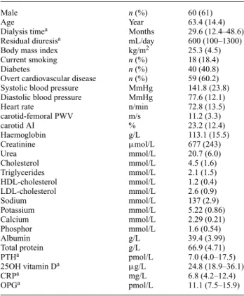
Table 1. Baseline demographic, clinical, haemodynamic and laboratory data of the participants (n=98)
Male n(%) 60 (61)
Age Year 63.4 (14.4)
Dialysis timea Months 29.6 (12.4–48.6)
Residual diuresisa mL/day 600 (100–1300)
Body mass index kg/m2 25.3 (4.5)
Current smoking n(%) 18 (18.4)
Diabetes n(%) 40 (40.8)
Overt cardiovascular disease n(%) 59 (60.2)
Systolic blood pressure MmHg 141.8 (23.8)
Diastolic blood pressure MmHg 77.6 (12.1)
Heart rate n/min 72.8 (13.5)
carotid-femoral PWV m/s 11.2 (3.3)
carotid AI % 23.2 (12.4)
Haemoglobin g/L 113.1 (15.5)
Creatinine µmol/L 677 (243)
Urea mmol/L 20.7 (6.0)
Cholesterol mmol/L 4.5 (1.6)
Triglycerides mmol/L 2.1 (1.5)
HDL-cholesterol mmol/L 1.2 (0.4)
LDL-cholesterol mmol/L 2.6 (0.9)
Sodium mmol/L 137 (2.9)
Potassium mmol/L 5.22 (0.86)
Calcium mmol/L 2.29 (0.21)
Phosphor mmol/L 1.6 (0.54)
Albumin g/L 39.4 (3.99)
Total protein g/L 66.9 (4.71)
PTHa pmol/L 7.0 (4.0–17.5)
25OH vitamin Da µg/L 24.8 (18.9–36.1)
CRPa mg/L 6.8 (4.2–12.4)
OPGa pmol/L 11.1 (7.5–15.9)
Data are mean (SD) or in the case of evidence against normal distribution (a) median (interquartile range) for continuous variables andn(%) for categorical variables.
AI: augmentation index; PWV: pulse wave velocity; PTH: parathyroid hormone; CRP: C-reactive protein; OPG: osteoprotegerin.
considered were the ones listed in Table 1, with the exception of the carotid augmentation index. Finally, both PWV and OPG were included into the same adjusted model (Model C). If OPG and PWV lie along the same pathway of future morbid cardiovascular events, one expects one or both variables to lose statistical significance.
Data are presented as mean (standard deviation) or in the case of evidence against normal distribution as median (interquartile range) and n (%) for categorical variables. As serum OPG levels were distributed non- normally, ln-transformed values were used in the analy- ses. The SAS statistical package version 6.11 was used in the analyses. P-values with a two-sided alpha of 0.05 were considered statistically significant. Hazard ratios (HR) are presented with their 95% confidence intervals in parentheses.
Results
There were 126 chronic HD patients at the two dialysis units invited to participate. Of these, 28 patients declined participation leaving 98 patients for inclusion into the study.
Baseline demographic, medical, laboratory and haemo- dynamic information of the participants are presented in Table 1. The most frequent causes for renal disease were vascular-tubulointerstitial (including hypertension) (39%),
diabetes mellitus (33%) and glomerulonephritis (13%). An- tihypertensive treatment at baseline included beta-blockers in 63%, calcium-channel blockers in 62%, angiotensin receptor blockers or angiotensin-converting enzyme in- hibitors in 53% and alpha-blockers in 30%. Of our patients 62% were on active vitamin-D therapy and 79% on calcium carbonate.
PWV measurements were successful in all participants, with a mean predialysis value of 11.2 m/s (3.3). There were 12 patients with atrial fibrillation. Their PWV (11.7 m/s) did not significantly differ from a dose with regular rhythm (11.1 m/s) and the two groups were analysed together.
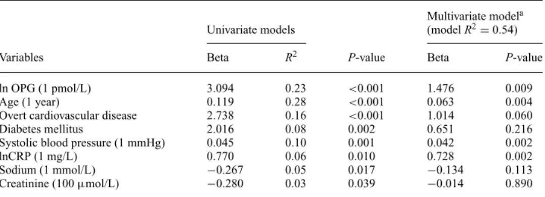
Results of the univariate and multivariate regression anal- yses for identifying significant baseline predictors of PWV are presented in Figure 1 and Table 2. In univariate analysis we found a strong positive relationship between PWV and lnOPG (β=3.09,P<0.001,R2=0.23) (Figure 1). In sepa- rate univariate models, other significant predictors of base- line PWV were age, diabetes, history of overt cardiovascu- lar disease, predialysis systolic blood pressure, predialysis creatinine, and sodium and CRP levels. The relationship be- tween PWV and lnOPG remained statistically significant after adjustment for these covariates (β=1.48,P=0.009, modelR2=0.54) (Table 2). There was no relationship be- tween the antihypertensives used and PWV. Furthermore, parameters of mineral metabolism were not significantly re- lated to PWV [calcium (P=0.190), phosphor (P=0.723), lnPTH (P=0.453) and ln25OH vitamin-D (P=0.791)]
and these were not included in the multivariate linear re- gression model.
In separate univariate models, using baseline clinical and laboratorical data as predictors, lnOPG was significantly as- sociated with age, overt cardiovascular disease, body mass index, active vitamin-D therapy, and serum triglyceride, HDL cholesterol and albumin levels. Considering these variables in a multivariate linear regression model, only age (β=0.015,P<0.001) and BMI (β= −0.026,P=0.01) were significantly related to lnOPG (modelR2=0.432).
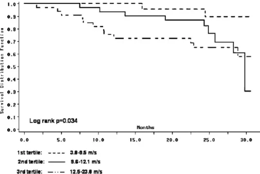
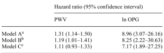
During follow-up 35 patients died (mortality rate 20.3/100 patient years) of which 23 were due to cardio- vascular causes (myocardial infarction: 7, sudden death: 5, arrhythmia: 2, heart failure: 4, stroke: 5). From the remain- ing 65 patients 6 were transplanted, 1 stopped dialysis due to improvement in renal function, 2 were transferred to other units and data of 54 patients were censored at the end of follow-up. Kaplan–Meier survival curves for cardiovascu- lar mortality using tertiles of PWV and OPG are presented in Figures 2 and 3 (log rankP-values were 0.034 and 0.0006, respectively). In separate univariate Cox proportional haz- ard models the following variables were related to cardio- vascular mortality: PWV [HR=1.31 (1.14–1.50)], lnOPG [8.96 (3.07–26.16)], age [1.07 (1.03–1.12)], overt cardio- vascular disease [3.18 (1.08–9.36)], use of angiotensin- converting enzyme inhibitor or angiotensin II receptor blocker [3.60 (1.35–11.65)] and the haemoglobin level [0.97 (0.95–0.99)]. After adjustment for age, cardiovascu- lar disease, use of angiotensin-converting enzyme inhibitor or angiotensin II receptor blocker and the haemoglobin level in separate multivariate models both PWV and lnOPG remained significantly related to cardiovascular mortality (Table 3, Model B). When however both PWV and lnOPG
Fig. 1. Linear regression between osteoprotegerin levels and carotid-femoral pulse wave velocity.
Table 2. Significant predictors of carotid-femoral pulse wave velocity in univariate and multivariate linear regression models
Multivariate modela
Univariate models (modelR2=0.54)
Variables Beta R2 P-value Beta P-value
ln OPG (1 pmol/L) 3.094 0.23 <0.001 1.476 0.009
Age (1 year) 0.119 0.28 <0.001 0.063 0.004
Overt cardiovascular disease 2.738 0.16 <0.001 1.014 0.060
Diabetes mellitus 2.016 0.08 0.002 0.651 0.216
Systolic blood pressure (1 mmHg) 0.045 0.10 0.001 0.042 0.002
lnCRP (1 mg/L) 0.770 0.06 0.010 0.728 0.002
Sodium (1 mmol/L) −0.267 0.05 0.017 −0.134 0.113
Creatinine (100µmol/L) −0.280 0.03 0.039 −0.014 0.890
aThe multivariate linear regression model considers all variables listed in the table.
were included in the same model only lnOPG remained a significant predictor of cardiovascular death (Table 3, Model C).
Discussion
In this study we demonstrated a strong relationship between PWV and OPG levels and found that the significant pre- dictive value of PWV for cardiovascular mortality is lost when OPG levels are also considered in the same model.
These findings may suggest that higher OPG levels and higher PWV are elements of the same pathway that leads from arterial calcification to cardiovascular events and that the mortality risk associated with higher OPG levels may, in part, be explained by increased PWV.
Arterial calcification is a widespread and progressive ab- normality in patients on HD that contributes to the high incidence of cardiovascular events [3]. While the exact mechanism linking arterial calcification to cardiovascular events has not been fully evaluated—and it may involve several separate processes—the positive relationship be- tween the degree of aortic calcification and PWV provides
one pathway through which arterial calcification can im- pact on cardiovascular mortality [10]. Indeed, increased carotid-femoral PWV has repeatedly been shown to be an independent predictor of cardiovascular mortality in HD patients [6,7].
Arterial calcification is not a mere consequence of the disturbance of divalent ion metabolism—frequently seen in HD patients—but rather, it is an active process regulated by paracrine and endocrine promoters and inhibitors that are also central to normal bone formation [11]. Among these factors is OPG that recently gained clinical attention.
OPG is a glycoprotein that belongs to the tumour necrosis factor (TNF) receptor superfamily, originally discovered as an inhibitor of bone resorption. OPG acts as a decoy re- ceptor of the receptor activator of the nuclear factor κB ligand (RANKL), which is a strong inducer of osteoclast differentiation [12]. OPG knockout mice develop osteo- porosis and widespread arterial calcifications implying that OPG is also an inhibitor of vascular calcification [19].
OPG is expressed in vascular cells such as coronary smooth muscle and endothelial cells. Surprisingly, in human cross- sectional and follow-up studies, higher rather than lower OPG levels were associated with the degree of coronary and
Fig. 2. Kaplan–Meier survival curves for cardiovascular mortality in tertiles of carotid-femoral pulse wave velocity.
Fig. 3. Kaplan–Meier survival curves for cardiovascular mortality in tertiles of serum osteoprotegerin levels.
aortic calcification [2,20–23], and in HD patients higher rather than lower serum OPG levels predicted progression of aortic calcification [13]. Furthermore, in a recent analy- sis of non-traditional risk factors, increasing levels of OPG diminished the association between chronic kidney disease and coronary calcification, supporting its direct relation- ship with calcifications [14]. To explain for the apparent paradox between experimental and human observational
data, it has been suggested that increased OPG levels may represent an incomplete defence mechanism against factors that promote atherosclerosis and arterial calcification [22], or alternatively, higher OPG levels may reflect an ongo- ing attempt of arterial smooth muscle cells to remodel and calcify [24]. The first of these two alternatives was also assumed in postmenopausal osteoporosis, where circulat- ing OPG levels were higher in women with an increased
Table 3. Association of carotid-femoral pulse wave velocity (PWV) and osteoprotegerin (OPG) levels with cardiovascular mortality
Hazard ratio (95% confidence interval)
PWV ln OPG
Model Aa 1.31 (1.14–1.50) 8.96 (3.07–26.16)
Model Bb 1.19 (1.01–1.41) 8.25 (2.22–30.63)
Model Cc 1.11 (0.93–1.33) 7.17 (1.89–27.25)
aSeparate univariate models.
bSeparate models adjusted for age, cardiovascular disease, use of an- giotensin converting enzyme inhibitor or angiotensin II receptor–blocker and the haemoglobin level.
cModel containing both PWV and lnOPG and adjusted for age, car- diovascular disease, use of angiotensin converting enzyme inhibitor or angiotensin II receptor–blocker and the haemoglobin level.
bone turnover and bone loss. OPG may serve as a protec- tive mechanism to slow down the increased bone resorption and subsequent bone loss [25].
While the exact role of OPG in the development and progression of vascular calcification needs further clarifi- cation, the clinical relevance of assessing OPG levels was established by studies demonstrating the predictive value of higher OPG levels for all cause and cardiovascular mortal- ity in HD and renal transplant patients [16,17]. Our finding on the relationship between OPG and PWV provides insight into downstream elements of the association of OPG with cardiovascular events. According to our hypothesis a higher OPG level is a marker of increased arterial calcification and calcification is related to arterial stiffness; therefore OPG is expected to be associated with aortic PWV. While several factors influence arterial stiffness in HD patients such as older age, increased blood pressure, presence of overt car- diovascular disease and inflammation, in our multivariate analysis the relationship between OPG and PWV remained significant after adjustment for these covariates. This find- ing is compatible with the above hypothesis.
Two previous studies reported on the analysis of the association between OPG and PWV. Kim et al. in their cross-sectional study of newly diagnosed diabetic patients and controls found a significant correlation between serum OPG levels and brachial-ankle PWV [26]. More recently, Stompor et al. reported a significant positive correlation between serum OPG levels and aortic PWV in patients on peritoneal dialysis [27]. Our results add to these findings, as none of the above studies used multivariate analysis to iden- tify an independent association between OPG and PWV, and none of them followed the patients in an attempt to eval- uate the inter-relationship of these parameters on clinical outcomes.
In the follow-up phase of our study we found that in sep- arate multivariate survival models both PWV and OPG lev- els predicted CV mortality, independent of covariates. This confirms previous studies on the prognostic value of these parameters in HD patients and provides external validity to our data. When OPG and PWV were considered in the same survival model, only OPG remained significantly related to future cardiovascular death. This result suggests that the OPG level and PWV provide overlapping prognostic infor-
mation in keeping with our hypothesis that they represent a common pathway leading to cardiovascular mortality.
Although antihypertensive drug treatment influences ar- terial stiffness [28], in our study we found no association between the use of different antihypertensive medications and PWV. Failure to find such a relationship is likely due to the cross-sectional nature of our analysis on the determi- nants of baseline PWV that does not exclude the possibility of antihypertensives impacting on PWV.
We found no association between parameters of mineral metabolism and PWV. While abnormalities in these vari- ables contribute to arterial calcification, their participation in the development of arterial stiffness in renal patients has not yet been demonstrated clearly and constantly, with some studies showing an association [10,29] while others not [30]. It may be that a snapshot of phosphate or PTH levels is insufficient to describe the abnormality in mineral metabolism, particularly if one considers the time needed to exert an effect on arterial stiffness that may not be apparent in a cross-sectional analysis.
The use of angiotensin-converting enzyme inhibitors or angiotensin receptor blockers was related to cardiovascular mortality in our survival analysis. This surprising finding is likely explained by the strong correlation between the presence of cardiovascular disease at baseline and the use of these drugs (that indicates higher baseline risk in these patients).
Two of three tertile survival curves in each figure su- perimpose after 30 months. This is likely explained by the widening confidence intervals of the survival curves due to decreasing number of patients at risk by time.
The limitations of our study need to be addressed.
First, our data do not provide an explanation for the cross-sectional relationship between OPG levels and PWV.
While the known relationship of arterial calcification to both of these factors could explain our finding, it is to be acknowledged that in our study we had no direct assessment of arterial calcification. Alternative explanation for the re- lationship of OPG with PWV such as the protective effects of OPG on the endothelial cell survival should also be con- sidered. Second, the 95% confidence interval for the hazard ratio of PWV in the survival analysis when both PWV and OPG were considered (Model C) was wide; therefore we cannot exclude the possibility that PWV is indeed related to cardiovascular mortality independent of OPG.
In summary, in this study we found significant positive relationship between serum OPG levels and aortic PWV in patients on HD and demonstrated an inter-relationship of these parameters on their effect on cardiovascular mortality.
These findings are compatible with the hypothesis that the prognostic significance of high OPG levels is, at least in part, mediated by higher PWV in these patients. Whether this relationship is dependent on the degree of aortic cal- cification and whether the same relationship holds true in other populations as well need further study.
Acknowledgements. The study was supported by research grants from the Hungarian Society of Hypertension and the Hungarian Society of Nephrology.
Conflict of interest statement. None declared.
References
1. Ketteler M, Schlieper G, Floege J. Calcification and cardiovascular health: new insights into an old phenomenon.Hypertension2006; 47:
1027–1034
2. Barreto DV, Barreto FC, Carvalho ABet al. Coronary calcification in hemodialysis patients: the contribution of traditional and uremia- related risk factors.Kidney Int2005; 67: 1576–1582
3. Caplin B, Wheeler DC. Arterial calcification in dialysis patients and transplant recipients.Semin Dial2007; 20: 144–149
4. Sigrist M, Bungay P, Taal MWet al. Vascular calcification and cardio- vascular function in chronic kidney disease.Nephrol Dial Transplant 2006; 21: 707–714
5. London GM, Gu´erin AP, Marchais SJet al. Arterial media calcifica- tion in end-stage renal disease: impact on all-cause and cardiovascular mortality.Nephrol Dial Transplant2003; 18: 1731–1740
6. London GM, Marchais SJ, Guerin APet al. Arterial stiffness: patho- physiology and clinical impact.Clin Exp Hypertens2004; 26: 689–699 7. London GM, Cohn JN. Prognostic application of arterial stiffness:
task forces.Am J Hypertens2002; 15: 754–758
8. Laurent S, Cockroft J, Van Bortel Let al. Expert consensus document on arterial stiffness: methodological issues and clinical applications.
Eur Heart J2006; 27: 2588–2605
9. Gusbeth-Tatomir P, Covic A. Causes and consequences of increased arterial stiffness in chronic kidney disease patients.Kidney Blood Press Res2007; 30: 97–107
10. Raggi P, Bellasi A, Ferramosca Eet al. Association of pulse wave velocity with vascular and valvular calcification in hemodialysis pa- tients.Kidney Int2007; 71: 802–807
11. Giachelli CM. Vascular calcification mechanisms.J Am Soc Nephrol 2004; 15: 2959–2964
12. Rogers A, Eastell R. Circulating osteoprotegerin and receptor activator for nuclear factor kappaB ligand: clinical utility in metabolic bone disease assessment.J Clin Endocrinol Metab2005; 90: 6323–6331 13. Nitta K, Akiba T, Uchida K. The progression of vascular calcification
and serum osteoprotegerin levels in patients on long-term hemodial- ysis.Am J Kidney Dis2003; 42: 303–309
14. Baber U, de Lemos JA, Khera Aet al. Non-traditional risk factors pre- dict coronary calcification in chronic kidney disease in a population- based cohort.Kidney Int2008; 73: 615–621
15. Ueland T, Jemtland R, Godang Ket al. Prognostic value of osteopro- tegerin in heart failure after acute myocardial infarction.J Am Coll Cardiol2004; 44: 1970–1976
16. Morena M, Terrier N, Jaussent Iet al. Plasma osteoprotegerin is associated with mortality in hemodialysis patients.J Am Soc Nephrol 2006; 17: 262–270
17. Hjelmesaeth J, Ueland T, Flyvbjerg Aet al. Early posttransplant serum osteoprotegerin levels predict long-term (8-year) patient survival and cardiovascular death in renal transplant patients.J Am Soc Nephrol 2006; 17: 1746–1754
18. Salvi P, Lio G, Labat Cet al. Validation of a new non-invasive portable tonometer for determining arterial pressure wave and pulse wave ve- locity: the PulsePen device.J Hypertens2004; 22: 285–293 19. Bucay N, Sarosi I, Dunstan CRet al. Osteoprotegerin-deficient mice
develop early onset osteoporosis and arterial calcification.Genes Dev 1998; 12: 1260–1268
20. Jono S, Ikari Y, Shioi Aet al. Serum osteoprotegerin levels are as- sociated with the presence and severity of coronary artery disease.
Circulation2002; 106: 1192–1194
21. Abedin M, Omland T, Ueland Tet al. Relation of osteoprotegerin to coronary calcium and aortic plaque (from the Dallas Heart Study).
Am J Cardiol2007; 99: 513–518
22. Anand DV, Lahiri A, Lim Eet al. The relationship between plasma osteoprotegerin levels and coronary artery calcification in uncompli- cated type 2 diabetic subjects.J Am Coll Cardiol2006; 47: 1850–
1857
23. Mazzaferro S, Pasquali M, Pugliese Fet al. Serum levels of calcifica- tion inhibition proteins and coronary artery calcium score: compari- son between transplantation and dialysis.Am J Nephrol2007; 27: 75–
83
24. Moe SM, Reslerova M, Ketteler Met al. Role of calcification in- hibitors in the pathogenesis of vascular calcification in chronic kidney disease (CKD).Kidney Int2005; 67: 2295–2304
25. Mezquita-Raya P, de la Higuera M, Garc´ıa DFet al. The contri- bution of serum osteoprotegerin to bone mass and vertebral frac- tures in postmenopausal women.Osteoporosis Int2005; 16: 1368–
1374
26. Kim SM, Lee J, Ryu OHet al. Serum osteoprotegerin levels are as- sociated with inflammation and pulse wave velocity.Clin Endocrinol (Oxf)2005; 63: 594–598
27. Stomp´or T, Krzanowski M, Kusnierz-Cabala B et al. Pulse wave velocity and proteins regulating vascular calcification and bone min- eralization in patients treated with peritoneal dialysis.Nephrol Dial Transplant2006; 21: 3605–3606
28. Mahmud A. Reducing arterial stiffness and wave reflection—quest for the holy grail?Artery Research2007; 1: 13–19
29. London GM, Gu´erin AP, Verbeke FHet al. Mineral metabolism and arterial functions in end-stage renal disease: potential role of 25- hydrocivitamin D.J Am Soc Nephrol2007; 18: 613–620
30. Haydar AA, Covic A, Colhoun Het al. Coronary artery calcification and aortic pulse wave velocity in chronic kidney disease patients.
Kidney Int2004; 65: 1790–1794 Received for publication: 30.1.08 Accepted in revised form: 8.4.08