ORIGINAL ARTICLE
Decreased flow-mediated dilatation with increased
arterial stiffness and thickness as early signs of atherosclerosis in polymyositis and dermatomyositis patients
Melinda Vincze&H. Dér&Gy. Kerekes&P. Szodoray&
M. Zeher&K. Dankó&P. Soltész
Received: 15 December 2013 / Revised: 19 February 2014 / Accepted: 23 February 2014
#Clinical Rheumatology 2014
Abstract Several autoimmune rheumatic diseases have been associated with accelerated atherosclerosis or other different types of vasculopathy depending on the underlying disease, leading to increased cardio- and cerebrovascular disease risk.
Polymyositis (PM) and dermatomyositis (DM), members of idiopathic inflammatory myopathies (IIMs), a group of sys- temic autoimmune diseases are also associated with elevated risk of cardiovascular diseases (CVD). Up until now, no specific data is known on the mechanisms, risk factors, or possible vasculopathy leading to increased CVD risk. The aims of the present study were to assess the flow-mediated dilatation of the brachial artery by a TensioClinic arteriograph and to measure the thickness of carotid artery intima–media, the augmentation index, and the pulse wave velocity using high-resolution ultrasonography in a cohort of PM and DM patients. We also investigated the correlation of these param- eters with the traditional risk factors of atherosclerosis and overall cardiovascular status within PM and DM patients.
Twenty-seven patients (21 females, six males) with IIMs were enrolled in this study, and 38 healthy individuals matched for sex and age served as controls. We found a decreased flow- mediated dilatation in the brachial artery (6.36 vs. 8.39 %) with increased arterial stiffness and carotid artery thickness in
our patients compared to healthy controls. We found signifi- cantly decreased flow-mediated dilatation of the brachial ar- tery (5.57 vs. 8.39 %) in DM patients. We also detected a correlation between these parameters and the traditional car- diovascular risk factors, as well as hypertriglyceridemy, hy- pertension, and peripheral arterial disease. In DM, overall, more vascular abnormalities were found than in PM. Our findings suggest that flow-mediated dilatation of the brachial artery, arterial stiffness, and carotid artery thickness measure- ments could be beneficial for predicting the CVD risk in myositis patients. Further investigations need to find the po- tential differences and role of inflammation and immune mechanisms in atherosclerotic processes in DM and PM.
Keywords Arterial stiffness . Cardiovascular risk .
Dermatomyositis . Endothelial dysfunction . Flow-mediated vasodilatation . Polymyositis
Introduction
Several systemic autoimmune, rheumatic diseases have been associated with accelerated atherosclerosis or other types of vasculopathy leading to increased cardio- and cerebrovascular disease risk. The characteristics of vasculopathy may signifi- cantly differ depending on the underlying disease. While clas- sical accelerated atherosclerosis has been associated with rheu- matoid arthritis (RA), systemic lupus erythematosus (SLE), or spondyloarthropathies (SpA); obliterative vasculopathy may rather be characteristic for systemic sclerosis (SSc) or mixed connective tissue disease (MCTD). Antiphospholipid antibod- ies have been playing an important role in vasculopathy asso- ciated with SLE, antiphospholipid syndrome (APS), RA, and MCTD [1]. In autoimmune rheumatic diseases besides the traditional risk factors, such as hypertension, diabetes, dyslipidemy, smoking, and sedentary lifestyle, the chronic Dankó K and Soltész P contributed equally to this study.
M. Vincze (*)
:
M. Zeher:
K. DankóDivision of Clinical Immunology, Department of Internal Medicine, University of Debrecen, Móricz Zs. st. 22, Debrecen H-4032, Hungary
e-mail: melinda8527@yahoo.com H. Dér
:
G. Kerekes:
P. SoltészDivision of Angiology, Department of Internal Medicine, University of Debrecen, Debrecen, Hungary
P. Szodoray
Institute of Immunology, Rikshospitalet, Oslo University Hospital, Oslo, Norway
DOI 10.1007/s10067-014-2561-y
inflammation and endothelial dysfunction also increase the CVD risk and mortality [2–6]. Although some data have been shown previously on the cardiovascular involvement and risk factors in idiopathic inflammatory myopathies (IIM), yet there have been no studies performed concerning vasculopathy in the disease.
Polymyositis (PM) and dermatomyositis (DM) are the most frequent subtypes of IIMs.
Both are associated with elevated risk of cardiovascular disease (CVD). The reported prevalence of cardiac involve- ment in patients with PM/DM varies between 3 to 100 %, based on the definition of the disease, time of the investigation in the disease course, etc. The only disease-specific CVD involvement is myocarditis which occurs rarely and could be associated with the presence of auto-antibodies, especially the anti-SRP antibody. Other cardiac involvements in IIMs are unspecific; including myocardial infarction, conduction ab- normalities, arrhythmias, mitral valve prolapse, congestive heart failure, hyperdynamic left ventricular contraction, dilat- ed and restrictive cardiomyopathies, pulmonary hypertension, and pericarditis [7]. The basic abnormality is inflammation with necrosis and fibrosis of the myocardium, similar to the pathological changes seen in the skeletal muscle. Vascular alterations in coronary arteries have also been reported such as vasculitis, intimal proliferation, media sclerosis, and microvessel disease of the heart with vasospasm angina [8].
Left ventricular failure occurs due to increased chamber stiff- ness caused by fibrosis or disturbances in calcium regulation.
Clinical assessment including history and signs of cardiac involvement is important at the time of diagnosis as well as during follow-up of myositis patients [9].
According to recent studies, the assessment of flow- mediated dilation of the brachial artery (abFMD) is one of the noninvasive approaches to measure or predict the cardiovascular disease (CVD) risk [2–6]. These investiga- tions are not commonly used during the diagnostic proce- dure of PM/DM. Moreover, the presence of endothelial dysfunction is poorly defined. Previously, only one study reported alteration of abFMD in young patients with a diagnosis of juvenile DM [10]. Another study showed that patients with DM and PM had abnormal endothelial- dependent mediated vasodilatation, assessed with re- sponse to acetylcholine infusion, but no information about abFMD were available [11].
The aims of the present study were to assess the flow-mediated dilatation of the brachial artery and to measure the thickness of carotid artery intima–media, the augmentation index, and the pulse wave velocity of the aorta in a cohort of PM and DM patients. We also examined the correlation of these parameters with the traditional risk factors of atherosclerosis, such as hyperlipidemy, hypertension, and the cardiovascular sta- tus, especially the presence of CVDs.
Materials and methods
Patients
Twenty-seven patients (21 females, six males; mean age at diagnosis: 39.48 years) with idiopathic inflammatory myopa- thies (IIMs) were enrolled in this study, that were followed up regularly at the Division of Clinical Immunology, Institute of Internal Medicine, University of Debrecen Medical and Health Science Center. Fourteen patients had primary DM (nine women and five men), and 13 persons suffered from primary PM (12 females, one male). The study was planned according to the Declaration of Helsinki and has been ap- proved by local ethics committee. The participants’written consent was obtained. We investigated the development of cardiovascular changes (hypertension, peripheral vascular, ischemic heart, and cerebrovascular diseases) during the dis- ease course, as well as traditional CVD risk factors, such as dyslipidemy.
For comparison, 38 healthy individuals matched for sex (25 females, 13 males) and age (mean: 50.3 years) served as controls. None of the controls smoked; had overt atheroscle- rosis, cardiovascular, cerebrovascular, or peripheral vascular disease; and had hypertension, dyslipidemy, or other con- founding conditions.
Five patients achieved improvement in myositis symptoms with corticosteroid monotherapy. The other patients needed second-line immunosuppressant treatment, including metho- trexate, cyclosporine A, cyclophosphamide, or azathioprine.
Thirteen patients received also intravenous immunoglobulin (IVIg) due to the serious therapy refractory symptoms. In two cases, extracorporeal plasmapheresis treatment was initiated.
One of these patients had severe dysphagia and muscle weak- ness. The other patients’had anti-synthetase syndrome with progressive alveolitis, in spite of the cyclophosphamide therapy.
Assessment of flow-mediated vasodilatation of brachial artery abFMD measurements were carried out on the patients’right arms by using the Hewlett-Packard (HP) Sonos 5500 high- resolution duplex ultrasound instrument (Soma Technology, Bloomfield, CT, USA) using a 10-MHz linear transducer and electrocardiogram gating as described earlier [12–16].
Longitudinal images were taken, based on individual anatom- ical variability, 4–7 cm proximally from the cubital fossa.
Systolic blood pressure was maintained over 50 mmHg for 5 min by inflating the cuff. Subsequently, reactive hyperemia was induced by the quick release of the cuff. We detected and digitally saved the arterial diameter at rest and then the change in the diameter due to increased flow after 60 s. Results were analyzed offline by using the AVITA software (Gtech Information Systems, Oak Brook, IL, USA) [17]. The mean
diameter was determined as the mean of three subsequent measurements performed synchronously with the R wave of the heart cycle [15,16]. Change of abFMD is presented as the percentage of increase or decrease in the arterial diameter after flow in comparison to the value at rest. All assessments were performed by the same investigator in the morning hours, in an air-conditioned room at 21 °C, and after overnight fasting of the patient and resting for 30 min. In the morning of the measurements, participants were not allowed to drink coffee, tea, or to take antioxidant vitamins. No vasoactive drugs were allowed 24 h before the assessments. (The half-life of all vasoactive drugs administered prior to the study was less than 24 h.)
Evaluation of carotid artery intima–media thickness
Carotid artery IMT was determined by a high-resolution Duplex ultrasonography using the HP Sonos 5500 instrument described above equipped with 5–10 MHz linear transducer [12,13,17]. Longitudinal and transverse section images were taken of the carotid system. If no plaques could be detected applying mediolateral transducer position, longitudinal im- ages were captured of the common carotid arteries, 10 mm proximally from the carotid bulb. Images were taken in the end-diastolic phase, and acIMT data were evaluated offline using the AVITA software [17]. acIMT was recorded as the distance between the first (lumen–intima border) and the second (media–adventitia border) echogenic lines according to the leading edge method. On both sides, 10 measurements were performed; the mean of the individual values was calcu- lated and results were presented in millimeters.
Assessment of augmentation index and pulse wave velocity in the aorta
Measurements were carried out by using a TensioClinic arteriograph system (TensioMed Kft., Debrecen, Hungary) [18]. This technique is based on the fact that the contraction of the myocardium initiates pulse waves in the aorta. The first wave becomes reflected from the aortic wall at the bifurcation;
therefore, a second, reflected wave appears as a late systolic peak. The morphology of this second, reflected wave depends on the stiffness of the large artery, the reflection time at 35 mmHg suprasystolic pressure of the brachial artery (RT S35), and the peripheral resistance-dependent amplitude.
Augmentation index (AIx) is calculated from the ampli- tudes of the first and second waves. AIx is the pressure difference between the late systolic peak pressure and the early systolic peak pressure divided by the late systolic peak pressure. The arteriograph assesses this parameter from the oscillometric data obtained from the 35-mmHg suprasystolic pressure of the brachial artery [18,19].
Pulse wave velocity (PWV) is the quotient of the jugular fossa–symphysis distance and RT S35 in meters per second.
The jugular fossa–symphysis distance is anatomically identi- cal with the distance between the aortic trunk and the bifur- cation. In order to have reproducible results, the patient needs a rest for at least 5 min before the assessment in a quiet investigation room [18,19].
Statistical analysis
For the analysis of endothelial dysfunction, independent sam- pleTtests were used. Subsequently, we performed correlation analyses. In cases of normal distribution (parametric), Pearson’s test was performed; while in cases of nonnormal distribution (nonparametric), Spearman’s test was performed.
When a significant correlation was found, the two indepen- dent variables were plotted in a coordinate system, indicating the type of correlation, the level of significance, and the value of the regression coefficient. We considered statistically sig- nificant thepvalues<0.05.
Results
The clinical investigation of CVDs in IIMs showed that 18 patients (13 women and five men) had hypertension, and eight patients (six women and two men) had ischemic heart disease (IHD) and peripheral vascular disease (PAD). None of the patients had myocardial infarction. In the IHD group, we classified the patients who have angina pectoris or ischemic signs on the electrocardiography (ECG), during echocardiog- raphy or who had positive stress ECG tests. The diagnosis of PAD was established if the individual had evidence of periph- eral arterial of venal occlusion (justified with ultrasound or angiography) or vascular ulcers.
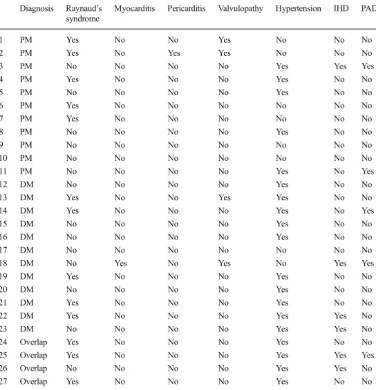
We also examined other cardiovascular diseases, such as valvulopathy, myocarditis, pericarditis, or Raynaud’s syn- drome. None of our patient had cerebrovascular diseases, including stroke or transient ischemic attack (TIA). Table1 summarizes cardiovascular manifestations found in our pa- tients with IIMs.
Flow-mediated vasodilatation of the brachial artery
Concerning abFMD, we observed decreased abFMD in myo- sitis patients (Fig.1.), although the result was not significant, while in DM, a significantly decreased abFMD was found compared to the healthy subjects (Fig.2). Subsequently, we assessed correlations between the traditional risk factors of atherosclerosis and cardiovascular diseases. We found a sig- nificantly negative correlation between the triglyceride levels and flow-mediated dilatation (R=−0.412,p=0.041).
Carotid artery intima–media thickness
Intima–media thickness of the carotid artery showed also a tendency of increased arterial thickness in myositis patients compared to healthy controls (0.61 vs. 0.59 mm; p=0.28),
which was more pronounced in DM patients (0.64 vs.
0.59 mm; p=0.07), although the difference did not reach statistical significance. We also found a significant positive correlation between the values of systolic blood pressure and the thickness of the carotid artery intima–media in IIMs Table 1 Cardiovascular mani-
festations of patients with IIMs Diagnosis Raynaud’s syndrome
Myocarditis Pericarditis Valvulopathy Hypertension IHD PAD
1 PM Yes No No Yes No No No
2 PM Yes No Yes Yes No No No
3 PM No No No No Yes Yes Yes
4 PM Yes No No No Yes No No
5 PM No No No No Yes No No
6 PM Yes No No No No No No
7 PM Yes No No No No No No
8 PM No No No No Yes No No
9 PM No No No No No No No
10 PM No No No No No No No
11 PM No No No No Yes No Yes
12 DM No No No No Yes No No
13 DM Yes No No Yes Yes No No
14 DM Yes No No No Yes No Yes
15 DM No No No No Yes No No
16 DM No No No No Yes No No
17 DM No No No No No No No
18 DM No Yes No Yes No Yes Yes
19 DM Yes No No No Yes No No
20 DM No No No No Yes No No
21 DM Yes No No No Yes No No
22 DM Yes No No No Yes Yes No
23 DM No No No No Yes Yes No
24 Overlap Yes No No No Yes No No
25 Overlap Yes No No No Yes Yes Yes
26 Overlap No No No No Yes Yes No
27 Overlap Yes No No No Yes No No
Fig. 1 Mean values and standard deviation of flow-mediated dilatation of the brachial artery in myositis patients and in healthy controls.
pindicates the level of statistical significance
Fig. 2 Mean values and standard deviation of flow-mediated dilatation of the brachial artery in dermatomyositis patients and in healthy controls.
pindicates the level of statistical significance
(Fig.3). We found significantly increased carotid IMT thick- ness in patients with hypertension compared to the healthy controls (Fig.4). Significant differences have been found both in patients with IHD + AD compared to the healthy controls.
The mean carotid IMT of IHD + PAD patients was 0.69 mm;
in the healthy controls, this value was 0.59 mm (p=0.04).
Augmentation index and pulse wave velocity in the aorta Concerning pulse wave velocity, we also found a tendency of increased arterial stiffness in the aorta both in all IIM patients (8.79 vs. 8.43 m/s;p=0.68) and in DM (8.78 vs. 8.43 m/s;p= 0.78), compared to the healthy individuals. Concerning the traditional risk factors, a significantly positive correlation has been found between the triglyceride levels and the augmenta- tion index (AIx) in myositis patients (R=0.567,p=0.029). We verified a clear significant difference in pulse wave velocity between IHD + PAD patients and the healthy individuals (Fig.5);p<0.001.
Moreover, we investigated associations between auto- antibody positivity and vascular abnormalities. One of our patients was positive for anti-Jo1 with severe interstitial lung
disease. On the grounds of pulmonary fibrosis, serious pul- monary arterial hypertension developed followed by fatal right and left ventricular failure. We had also found Jo1 positivity in five other patients. One of them had pulmonary fibrosis with slight right ventricular failure. Other cardiovas- cular diseases like IHD, PAD, and hypertension did not pres- ent in the Jo1 positive group. Five patients were positive to antiphospholipid antibodies, while two had anti-Mi2 autoan- tibodies; no specific vascular changes were found in these groups.
Concerning the therapy, we have not found any significant differences between groups. Overall, the cardiovascular changes were more frequent in patients who needed combined immunosuppressive treatment, which presumably indicated that these patients had more severe disease course, accompa- nied by increased accelerated atherosclerotic events. We also found increased acIMT (0.56 mm) and increased abFMD (6.73 %) in patients treated with IVIg comparing to other treatments.
Conclusion
In autoimmune rheumatic diseases besides the traditional risk factors, such as hypertension, diabetes, dyslipidemy, smoking, and sedentary lifestyle, the chronic inflammation and endo- thelial dysfunction also increase the CVD risk and mortality [1]. Noninvasive vascular assessments, including brachial artery FMD, carotid artery IMT, and augmentation index and/or pulse wave velocity of the aorta can reflect the cardio- vascular risk of these patients.
In APS, undifferentiated connective tissue disease (UCTD), early RA, MCTD, and systemic vasculitis impaired abFMD have been described previously [20–24]. Increased thickness of the carotid artery intima–media (acIMT) was found in patients with SLE, MCTD, and SSc compared with Fig 3 Correlation between the values of systolic blood pressure and the
thickness of carotid artery intima–media in myositis patients. A significant positive correlation was detected.Ris the regression coefficient;pmeans the level of statistical significance
Fig. 4 Mean values and standard deviation of carotid artery intima– media thickness in patients with hypertension and in healthy controls.p is the level of statistical significance
Fig. 5 Mean values and standard deviation of pulse wave velocity in patients with IHD + PAD and in healthy controls.prepresents the level of statistical significance
the healthy controls [23,25]. Surprisingly, in these studies, no correlations were found between abFMD, disease activity index, presence of autoantibodies, or organ involvement. In vasculitis, abFMD values improved after steroid therapy that suppresses inflammation, suggesting that inflammatory changes play a pivotal role in endothelial dysfunction [26].
We have shown previously that impaired abFMD is charac- teristic to UCTD patients, correlating with anti-DNA antibod- ies, suggesting the role of these autoantibodies in endothelial dysfunction [27]. In a former study, we detected impaired FMD, as well as increased carotis intima–media thickness (ccIMT), AIx, and PWV in 101 autoimmune patients in com- parison to the 36 healthy control subjects. Impaired endothelium-dependent vasodilatation and increased stiffness were observed in all patient subgroups including APS, RA, SSc, and PM. In addition, both stiffness parameters, AIx and PWV, exerted negative correlation with FMD and positive correlation with ccIMT in the patient population. AIx and PWV also positively correlated with each other [28]. We have also reported that arterial stiffness is closely associated with autoimmune inflammation in APS [29].
Our present findings underlined that similarly to other systemic autoimmune diseases, impaired abFMD was present in IIMs, increasing the risk of cardiovascular disease. We can establish that the assessments of flow-mediated dilation of the brachial artery and the arterial stiffness of the aorta and carotid artery intima–media thickness can be beneficial to predict the CVD risk in myositis patients.
Among traditional risk factors, hypertriglyceridemy, hyper- tension leading to the development of ischemic heart, or peripheral vascular diseases could be also implicated as one of the forms of vasculopathy in IIM. The elevated triglyceride levels in our patients could be explained also with sedentary life style due to muscle weakness and impaired physical activity beside the nutritional and hereditary susceptibility of hypertriglyceridemy. We have not found any major cardiovas- cular events, such as acute myocardial infarction, stroke, or critical limb ischemia in our patients.
Our findings also confirmed that in DM, more vascular abnormalities could be found than in PM. These results could arise from the differences in the pathomechanism of PM and DM. Although striated muscle is the main target of the auto- immune mechanism in both disease entities, in DM, the intramuscular small blood vessels are more dominant effec- tors’sites, while in PM, the muscle fibers are more direct targets. More recently, changes in the vascular components were proven to be the primary lesion in this disease because microtubular inclusions and microvacuoles in endomysial capillaries and MAC deposits were recognized even in other- wise normal muscle of patients with DM.
We cannot exclude that the immunosuppressant treatment could also decrease the endothelial dysfunction in chronic inflammatory diseases.
Our group is the first who measured objectively the endo- thelial dysfunction on such a large myositis population. The next goal is to assess longitudinal examination of these pa- tients considering the predictive values of these diagnostic tools and found any relation between auto-antibodies and vasculopathy in myositis. Furthermore, other risk factors, as well as the role of systemic inflammation including cytokines, chemokines, proteases, autoantibodies, adhesion receptors, and others could be responsible for vascular changes in myo- sitis. Further investigations needed to precise the role of inflammation and immune mechanisms in these changes.
“This research was organized within the following pro- gram: TÁMOP 4.2.4.A/2-11-1-2012-0001 National Excellence Program—local convergence program providing personnel support in the development and operation for stu- dents and researchers. The project was funded by the EU and the European Social Fund.”
Conflict of interest The authors have not any conflict of interest to declare.
References
1. Soltész P, Kerekes G, Dér H, Szücs G, Szántó S, Kiss E, Bodolay et al. Comparative assessment of vascular function in autoimmune rheumatic diseases: considerations of prevention and treatment.
Autoimmun Rev 2011 May;10(7):416–25
2. Verma S, Buchanan MR, Anderson TJ (2003) Endothelial function testing as a biomarker of vascular disease. Circulation 108(17):2054– 2059
3. Harris RA, Nishiyama SK, Wray DW, Richardson RS (2010) Ultrasound assessment of flow-mediated dilation. Hypertension 55(5):1075–1085
4. Inaba Y, Chen JA, Bergmann SR (2010) Prediction of future cardio- vascular outcomes by flow-mediated vasodilatation of brachial ar- tery: a meta-analysis. Int J Cardiovasc Imaging 26(6):631–640 5. Katz SD, Hryniewicz K, Hriljac I, Balidemaj K, Dimayuga C,
Hudaihed A et al (2005) Vascular endothelial dysfunction and mor- tality risk in patients with chronic heart failure. Circulation 111(3):
310–314
6. Esdaile JM, Abrahamowicz M, Grodzicky T, Li Y, Panaritis C, du Berger R, Côte R, Grover SA, Fortin PR, Clarke AE, Senécal JL (2001) Traditional Framingham risk factors fail to fully account for accelerated atherosclerosis in systemic lupus erythematosus. Arthritis Rheum 44(10):2331–2337
7. Zeki O, Robert Y, Oh SJ (2010) Polymyositis presenting with cardiac manifestations: report of two cases and review of the literature. Clin Neurol Neurosurg 112:160–163
8. Haupt HM, Hutchins GM (1982) The heart and cardiac conduction system in polymyositis and dermatomyositis: a clinicopathologic study of autopsied patients. Am J Cardiol 50:998–1006
9. Lundberg IE (2006) The heart in dermatomyositis and polymyositis.
Rheumatology 45:18–21
10. Eimer MJ, Brickman WJ, Seshadri R, Ramsey-Goldman R, McPherson DD, Smulevitz B et al (2011) Clinical status and cardio- vascular risk profile of adults with a history of juvenile dermatomy- ositis. J Pediatr 159(5):795–801
11. Gunawardena H, Harris ND, Carmichael C, McHugh NJ (2007) Microvascular responses following digital thermal hyperaemia and iontophoresis measured by laser Doppler imaging in idiopathic in- flammatory myopathy. Rheumatology (Oxford) 46(9):1483–1486 12. Kerekes G, Szekanecz Z, Dér H, Sándor Z, Lakos G, Muszbek L,
CsipőI, Sipka S, Seres I, Paragh G, Kappelmayer J, Szomják E, Veres K, Szegedi G, Shoenfeld Y, Soltész P (2008) Endothelial dysfunction and atherosclerosis in rheumatoid arthritis: a multiparametric analysis using imaging techniques and laboratory markers of inflammation and autoimmunity. J Rheumatol 35:398– 406
13. Szücs G, Tímár O, Szekanecz Z, Dér H, Kerekes G, Szamosi S, Shoenfeld Y, Szegedi G, Soltész P (2007) Endothelial dysfunction precedes atherosclerosis in systemic sclerosis—relevance for preven- tion of vascular complications. Rheumatology 46:759–762 14. Celermajer DS, Sorensen KE, Gooch VM, Spiegelhalter DJ, Miller
OI, Sullivan ID, Lloyd JK, Deanfield JE (1992) Noninvasive detec- tion of endothelial dysfunction in children and adults at risk of atherosclerosis. Lancet 340:1111–1115
15. Corretti MC, Anderson TJ, Benjamin EJ, Celemajer DS, Charbonneau F, Creager MA, Deanfield JE, Drexler H, Gerhard Herman M, Herrington D, Vallance P, Vita J, Vogel R, International Brachial Artery Reactivity Task Force (2002) Guidelines for the ultrasound assessment of endothelialdependent flow-mediated vasodilation of the brachial artery: a report of the International Brachial Artery Reactivity Task Force. J Am Coll Cardiol 39:257–265
16. Kelm M (2002) Flow-mediated dilatation in human circulation:
diagnostic and therapeutic aspects. Am J Physiol Heart Circ Physiol 282:1–5
17. Kanters SDJM, Algra A, van Leeuwen MS, Banga JD (1997) Reproducibility of in vivo carotid intima–media thickness measure- ments. Stroke 28:665–671
18. Baulmann J, Schillings U, Rickert S, Uen S, Düsing R, Cziraki A, Illyes M, Mengden T (2008) A new oscillometric method for assess- ment of arterial stiffness: comparison with tonometric and piezo- electronic methods. J Hypertens 26:523–528
19. Magometschnigg D (2005) Blood pressure and arterial stiffness. A comparison of two devices for measuring augmentation index and pulse wave velocity. Wien Med Wochenschr 155:404–410
20. Mercanoglu F, Erdogan D, Oflaz H, Kücükkaya R, Selcukbiricik F, Gül A et al (2004) Impaired brachial endothelial function in patients with primary anti-phospholipid syndrome. Int J Clin Pract 58(11):
1003–1007
21. Cugno M, Borghi MO, Lonati LM, Ghiadoni L, Gerosa M, Grossi C et al (2010) Patients with antiphospholipid syndrome display endo- thelial perturbation. J Autoimmun 34(2):105–110
22. Mosca M, Virdis A, Tani C, Ghiadoni L, Versari D, Duranti E et al (2009) Vascular reactivity in patients with undifferentiated connec- tive tissue diseases. Atherosclerosis 203(1):185–191
23. Soltesz P, Bereczki D, Szodoray P, Magyar MT, Der H, Csipo I et al (2010) Endothelial cell markers reflecting endothelial celldysfunction in patients with mixed connective tissue disease. Arthritis Res Ther 12(3):R78
24. Chatterjee Adhikari M, Guin A, Chakraborty S, Sinhamahapatra P, Ghosh A (2012) Subclinical atherosclerosis and endothelial dysfunc- tion in patients with early rheumatoid arthritis as evidenced by measurement of carotid intima-media thickness and flow-mediated vasodilatation: an observational study. Semin Arthritis Rheum 41(5):
669–675
25. Au K, Singh MK, Bodukam V, Bae S, Maranian P, Ogawa R, Spiegel B et al (2011) Atherosclerosis in systemic sclerosis: a systematic review and meta-analysis. Arthritis Rheum 63(7):2078–2090 26. Filer AD, Gardner-Medwin JM, Thambyrajah J, Raza K, Carruthers
DM, Stevens RJ et al (2003) Diffuse endothelial dysfunction is common to ANCA associated systemic vasculitis and polyarteritis nodosa. Ann Rheum Dis 62(2):162–167
27. Kerekes G, Bodolay E, Sipka S, Szomják E, Veres K, Zeher M et al (2005) Endothelial dysfunction and early atherosclerosis in non- differentiated collagenosis. Orv Hetil 146(17):791–795
28. Soltesz P, Der H, Veres K, Laczik R, Sipka S, Szegedi G, Szodoray P (2008) Immunological features of primary antiphospholipid syn- drome in connection with endothelial dysfunction. Rheumatology (Oxford) 47:1628–1634
29. Soltész P, Dér H, Kerekes G, Szodoray P, Szücs G, Dankó K, Shoenfeld Y, Szegedi G, Szekanecz Z (2009) A comparative study of arterial stiffness, flow-mediated vasodilation of the brachial artery, and the thickness of the carotid artery intima-media in patients with systemic autoimmune diseases. Clin Rheumatol 28(6):655–662. doi:
10.1007/s10067-009-1118-y, Epub 2009 Feb 18