RESEARCH
Mitochondrial dysfunction and autism:
comprehensive genetic analyses of children with autism and mtDNA deletion
Noémi Ágnes Varga1, Klára Pentelényi1, Péter Balicza1, András Gézsi1,2, Viktória Reményi1, Vivien Hársfalvi1, Renáta Bencsik1, Anett Illés1, Csilla Prekop3 and Mária Judit Molnár1*
Abstract
Background: The etiology of autism spectrum disorders (ASD) is very heterogeneous. Mitochondrial dysfunction has been described in ASD; however, primary mitochondrial disease has been genetically proven in a small subset of patients. The main goal of the present study was to investigate correlations between mitochondrial DNA (mtDNA) changes and alterations of genes associated with mtDNA maintenance or ASD.
Methods: Sixty patients with ASD and sixty healthy individuals were screened for common mtDNA mutations. Next generation sequencing was performed on patients with major mtDNA deletions (mtdel-ASD) using two gene panels to investigate nuclear genes that are associated with ASD or are responsible for mtDNA maintenance. Cohorts of healthy controls, ASD patients without mtDNA alterations, and patients with mitochondrial disorders (non-ASD) har- bouring mtDNA deletions served as comparison groups.
Results: MtDNA deletions were confirmed in 16.6% (10/60) of patients with ASD (mtdel-ASD). In 90% of this mtdel- ASD children we found rare SNVs in ASD-associated genes (one of those was pathogenic). In the intergenomic panel of this cohort one likely pathogenic variant was present. In patients with mitochondrial disease in genes responsible for mtDNA maintenance pathogenic mutations and variants of uncertain significance (VUS) were detected more frequently than those found in patients from the mtdel-ASD or other comparison groups. In healthy controls and in patients without a mtDNA deletion, only VUS were detected in both panel.
Conclusions: MtDNA alterations are more common in patients with ASD than in control individuals. MtDNA dele- tions are not isolated genetic alterations found in ASD; they coexist either with other ASD-associated genetic risk factors or with alterations in genes responsible for intergenomic communication. These findings indicate that mito- chondrial dysfunction is not rare in ASD. The occurring mtDNA deletions in ASD may be mostly a consequence of the alterations of the causative culprit genes for autism or genes responsible for mtDNA maintenance, or because of the harmful effect of environmental factors.
Keywords: Autism, Mitochondrial dysfunction, mtDNA deletion, ASD associated genetic alterations, Intergenomic communication
© The Author(s) 2018. This article is distributed under the terms of the Creative Commons Attribution 4.0 International License (http://creativecommons.org/licenses/by/4.0/), which permits unrestricted use, distribution, and reproduction in any medium, provided you give appropriate credit to the original author(s) and the source, provide a link to the Creative Commons license, and indicate if changes were made. The Creative Commons Public Domain Dedication waiver (http://creativecommons.org/
publicdomain/zero/1.0/) applies to the data made available in this article, unless otherwise stated.
Background
In recent years, the number of patients diagnosed with autism spectrum disorders (ASD) has increased with
current studies reporting a prevalence of 1% [1]. ASD shows extreme clinical heterogeneity; however, the diag- nosis of ASD according to the Diagnostic and Statistical Manual of Mental Disorders (5th edition) is based on def- icits in two areas—social communication and restricted, repetitive behaviour or interests. The patient must have deficits in both areas, and symptoms must be present from early childhood [2]. The genetic architecture of ASD
Open Access
*Correspondence: molnar.mariajudit@med.semmelweis-univ.hu
1 Institute of Genomic Medicine and Rare Disorders, Semmelweis University, Tömő Str. 25-29, Budapest 1083, Hungary
Full list of author information is available at the end of the article
is very diverse consisting of a variety of genetic altera- tions, such as chromosomal abnormalities, copy number variations, rare single nucleotide variants (SNVs), com- mon polymorphic variations, and epigenetic modifica- tions; however, only 6–15% of children with ASD have well-defined genetic syndromes [3]. Because of the devel- opment of high-throughput sequencing methods, many highly penetrant genetic causes of ASD have been iden- tified, but the underlying genetic background of 70% of cases remains unexplained [4].
Mitochondrial disease (MD) is presently one of the most recognized metabolic diseases caused by the failure of both nuclear and/or mitochondrial DNA (mtDNA).
The prevalence of mtDNA mutations responsible for MD is 1 in 5000, whereas that of nuclear mutations is 2.9 per 100,000 cases [5]. Although MD frequently results in a spectrum of disorders with multisystemic presentations, neurological symptoms are common because tissues with high-energy demands, such as neural tissue, are often the most strongly affected by mitochondrial dysfunc- tion. Even though the diagnosis of MD is increasing and becoming more frequent, the exact genetic background in many cases remains unconfirmed. Mitochondrial dys- function can be caused by either primary MD or second- ary mitochondrial damage [6]. Primary MD is because of genetic defects in mtDNA or a defect in a nuclear gene that is important for mitochondrial function. These mutations usually affect proteins involved in reactions of oxidative phosphorylation (OXPHOS). However, many disorders show similar effects in terms of mitochondrial dysfunction, but are elicited by mutations in other genes not related directly to normal mitochondrial function [7].
In other cases environmental factors, associated disor- ders or ageing are resulting in secondary alterations.
Several authors have proposed that mitochondrial dys- function may be one of the most common medical con- ditions associated with autism [8, 9]. Lombard et al. [10]
proposed that ASD may be a condition with abnormal mitochondrial function. Clinical and biochemical stud- ies have uncovered an emerging link between mitochon- drial dysfunction and neurodevelopmental disorders, including intellectual disability [11], childhood epilepsy, and ASD [9]. Furthermore, mitochondrial dysfunction has been associated with some forms of syndromic ASD [8, 11]. In many of these studies, biochemical changes, such as elevated levels of creatine kinase, lactate, pyru- vate, carnitine, ammonia, and alanine were detected in the serum of patients with ASD [11–14]. In other stud- ies, altered respiratory chain enzyme activities [15] or decreased expression of OXPHOS genes were detected in autistic brain [16], findings which indicate abnormal or altered mitochondrial function.
Damage to the OXPHOS system was found in individ- uals with ASD by Napoli et al. [17] and reviewed by Val- enti et al. [11]. Oliveira et al. [14] found that 7% (7/100) of children with ASD, who were clinically indistinguishable from other affected children with ASD, exhibited a mito- chondrial respiratory chain disorder. Weissman et al. [18]
proposed that defective mitochondrial OXPHOS may be an additional underlying pathogenic mechanism in a sub- set of individuals with autism.
Despite evidence of altered mitochondrial function in some individuals with ASD, it is not known whether mitochondrial dysfunction is a cause or an effect of ASD. Although a mitochondrial subgroup in ASD could be identified [19], findings from review articles, such as those of Palmieri and Persico [19] and Rossignol and Frye [9], found that even in this subgroup the causative genetic factor could be identified in a proportion of cases (23%). In cases of non-syndromic ASD, mitochondrial dysfunction without mtDNA alterations has been fre- quently observed [8, 9]. In a systematic review and meta- analysis, Rossignol reported that MD was present in 5%
of children with ASD [9], and in this ASD/MD subgroup, mtDNA abnormalities were found in 23% of patients [9].
These findings demonstrate that primary MD may be present in a subgroup of children with ASD.
Some studies have reported mtDNA deletions in indi- viduals with ASD [12, 20–22]. Single mtDNA deletions have a role in different paediatric and adult onset pri- mary MDs such as Kearns–Sayre syndrome, Pearson syndrome, and progressive ophthalmoplegia externa [23].
Multiple mtDNA deletions occur mostly because of path- ogenic mutations in genes responsible for intergenomic communication; however, they are often related to age- ing or harmful environmental factors as well because mtDNA has a poor DNA repair system [6, 24].
The aim of the present study was to investigate the presence of the most common pathogenic mtDNA alter- ations in patients with ASD and to elucidate the etiology of these mtDNA alterations by analysing their co-occur- rence with both known ASD-associated genes and genes responsible for mtDNA maintenance and by comparison the targeted NGS data (ASD associated genes and genes responsible for mtDNA maintenance) of cases with and without mtdel-ASD, patients with primary mitochondrial disorders and healthy controls.
Methods Patients
Detailed clinical examinations consisting of a general medical examination and neurological assessment were performed. A diagnosis of ASD was made using the ADI-R (autism diagnostic interview—revised) and ADOS
(autism diagnostic observation schedule). Patients were screened for minor physical abnormalities, which were selected based on the Méhes Scale [25]. Family history and detailed environmental/societal data were collected from the first degree relatives of each patient. Any dis- orders present in the parents as well as environmental factors were registered. Written informed consent was obtained from the parents of the patient. This study was performed in accordance with the Helsinki Declaration of 1975 and was approved by the Hungarian Research Ethics Committee (44599-2/2013/EKU). The diagnosis of ASD was based on the standardized ADI-R in Hungarian, which was published by the Autism Foundation (Kapocs Publisher), according to the following scores: A ≥ 10 (social interaction), B ≥ 7 (communication), C ≥ 3 (repet- itive stereotype manner), D ≥ 1 (abnormal development under 36 months). Sixty children with ASD [6 females and 54 males, median age = 7 years, interquartile range (IQR) = 7.25] were included in our study. Before patient selection our ASD patients were screened for Fragile X syndrome and only negative cases were included in our cohort. Of our 60 patients with ASD, 58 are of European descent and 2 are Roma. Our control group for mtDNA screening consisted of 60 European adults (26 females and 34 males, median age = 28 years, IQR = 13.75) selected from our biobank [26]. All controls were healthy individuals under 45 years of age and free from addiction (alcohol, smoking, and drugs). For the interpretation of our next generation sequencing (NGS) results, we com- pared data from the following cohorts: patients with ASD and without mtDNA deletion, labelled non-mtdel-ASD, (6 males and 1 female, median age = 8 years, IQR = 5.5), patients with MD and mtDNA deletion, without ASD (4 males and 3 females, median age = 18 years, IQR = 19), and healthy control individuals (1 male and 5 females, median age = 27 years, IQR = 2.25). The investigated patients and controls were not related. All patients with- out a mtDNA deletion were considered to have non-syn- dromic ASD. The study design is illustrated in Table 1.
Genetic analysis
DNA was isolated from peripheral blood samples from all participants using the QIAamp DNA blood kit (Qiagen, Hilden, Germany) according to manufacturer’s instruc- tions. To identify single and multiple mtDNA deletions, long range PCR was performed as described by Reme- nyi et al. [27]. MtDNA single and multiple deletions were screened with long PCR in 20 μl volume: 20 pmol prim- ers Fw 5′-TAAAAATCTTTGAAATAGGGC-3′ and Rev 5′-CGGATACAGTTCACTTTAGCT-3′, 0.2 µl Phusion DNA Polymerase (Finnzymes, Vantaa, Finland), 4 µl Phusion GC Reaction Buffer (Finnzymes, Vantaa, Fin- land), 0.4 µl dNTP and 12.4 μl water (qPCR grade water, AMBION). PCR program was the following: 98 °C 30 s, 30 cycles: 98 °C 10 s, 63 °C 10 s, 72 °C 3/8 min, then the last synthesis at 72 °C 7 min. Amplificates were visual- ised by ethidium-bromide (2% agarose) and determined with QuantityOne Software (Bio-Rad Corp. Hertford- shire, UK). The three most-frequent pathogenic mtDNA point mutations were screened by PCR–RFLP using a GeneAmp PCR System 9700 (Applied Biosystems, MA, USA) [20]. The most well-known ASD-associated genes [28] and 51 genes responsible for intergenomic com- munication disturbances (Additional file 1: Table S1) were investigated using NGS, which was performed on a MiSeq (Illumina, CA, USA) using the TruSight Autism Rapid Capture Kit (Illumina, CA, USA) and the SureSe- lect QXT Kit (Agilent Technologies, CA, USA) according to the manufacturer’s instructions. In the intergenomic panel, 16/32 samples were multiplexed in one sequencing run, whereas in the autism panel 24 samples were multi- plexed in a single run using the MiSeq reagent kit v2 and 300 cycles (Illumina, CA, USA). The mean read depth was 152 × in the intergenomic gene panel and 135 × in the ASD-associated gene panel. In both panels, 20 × cover- age was achieved in a minimum of 90% of target regions.
Pathogenic and likely pathogenic mutations from NGS data were validated by Sanger sequencing, and segrega- tion analysis was performed within individual families.
Table 1 The design of the study
The investigated cohorts and the performed genetic analysis are shown in the Table 1. NGS testing for intergenomic panel and ASD panel has been performed in the cohort of the 10 mtdel ASD cases and in subgroup of 7 non-mtdel ASD cases and a subgroup of healthy controls (N = 6). Patients with primary mitochondrial disease (N = 7) served as further control group. All investigated person were Caucasian except 2 non-mtdel ASD cases
ASD autism spectrum disorder, MD mitochondrial disease, mtDNA mitochondrial DNA, IG NGS next generation sequencing for genes responsible for intergenomic communication, ASD NGS next generation sequencing for autism associated genes
a The 10 mtdel-ASD cases and 7 non-mtdel ASD were investigated
b 6 cases were investigated
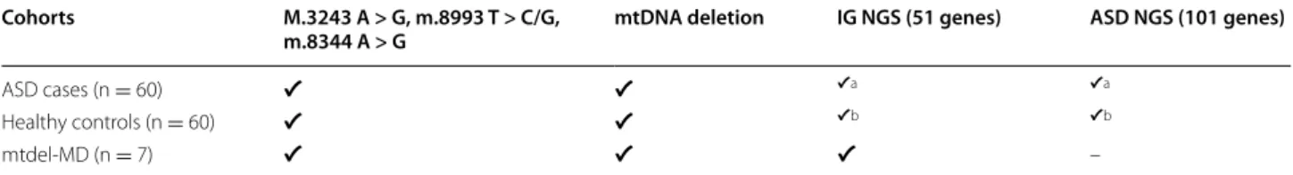
Cohorts M.3243 A > G, m.8993 T > C/G,
m.8344 A > G mtDNA deletion IG NGS (51 genes) ASD NGS (101 genes)
ASD cases (n = 60) ✔ ✔ ✔a ✔a
Healthy controls (n = 60) ✔ ✔ ✔b ✔b
mtdel-MD (n = 7) ✔ ✔ ✔ –
Statistical and bioinformatics analysis
Chi square test with Yates correction/Fisher exact test were used to determine significant differences between patient and control groups [29]. Raw sequences were filtered with Picard tools (version 2.1.0) [30] and quality filtered reads were aligned to the hg19 reference genome with BWA-mem [31] using default parameters. Vari- ant calling was performed using GATK HaplotypeCaller (version 3.3-0) [32] and VCF files were annotated with SnpEff (version 4.1) [33]. We analysed only those vari- ants that were found in the canonical transcript of the gene. To identify potentially causal genetic variations, we used VariantAnalyzer, which is an in-house software developed by András Gézsi from the Budapest Univer- sity of Technology and Economics [34]. This software application annotates SNPs and short indels with several types of annotations, such as their predicted function on genes using SnpEff, observed allele frequencies in several genomic projects including the 1000 Genomes Project and the ESP6500 Project, conservation scores based on PhyloP or PhastCons, predicted function of non-synony- mous SNPs using dbNSFP, and disease associations with HGMD and ClinVar. By creating filter cascades based on these annotations and other information (e.g., genotypes and variant quality annotations), the software can eas- ily be used to filter the variants through a user-friendly graphical interface. Analysis and variant calling of Sure- Select libraries was performed with SureCall software (Agilent, CA, USA). First, we filtered for variants known to be disease-causing, using human gene mutation data- base (HGMD) Professional 2015.1 edition [35]. Second, we filtered for rare variants based on the minor allele fre- quency and frequency of the mutation in our NGS data repository. Since large-scale genomic data of the Hun- garian population is not available, a mutation with a low minor allele frequency may also be population-specific.
We labelled a variant as a rare mutation if it was present in one or two samples within our cohort and the minor allele frequency in Europeans from the 1000 Genomes and ExAC databases was less than 0.5%. It is important to note that a limitation of this method is that it may exclude identification of founder mutations and disease- associated polymorphisms. Finally, mutations were prior- itized based on their predicted effects. Exonic frameshift and stop mutations were considered always damaging, whereas the effects of missense mutations were predicted using Polyphen2, SIFT (Sort Intolerant From Tolerant), or MutationTaster (MT).
Results
Sixty children with ASD (6 females and 54 males, median age = 7 years, IQR = 7.25) were investigated. In our ana- lysed cohort of 60 patients, 29 patients were sporadic
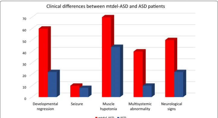
(from simplex families) and their family histories were negative for other neurodevelopmental disorders and major psychiatric or neurological disorders. The dis- tribution of the relevant symptoms for mitochondrial disorders in mtdel-ASD and idiopathic ASD group are shown in Fig. 1. There were some differences between ASD cases with and without mtDNA deletion regarding the clinical phenotype. Developmental regression, mus- cle hypotonia, and additional neurological signs were most common in the mtdel-ASD cases. Multisystemic abnormality appeared also more frequently. Referring to seizures no major differences has been observed between these two groups. Family history was not available in two cases because these children were not living with their biological parents. A positive family history was found in 48% of cases, of which 20 cases (33.3%) had a family his- tory for psychiatric disorders (bipolar disorder, depres- sion, and schizophrenia). In four cases (6.7%), visual and hearing impairments, ataxia, complex endocrine disor- ders, or a combination of these factors was noted. The co-occurrence of these symptoms is an indicator for MD according to mitochondrial disease criteria (MDC) [36].
In five cases (8%), we found a positive family history for psychiatric disorders and some MDC-related symptoms were also noted.
Minor physical anomalies were identified in 44 chil- dren. All children were diagnosed with ASD based on ADI-R, and in most cases, with ADOS as well. We found that serum lactate levels and/or the lactate:pyruvate ratio supported the presence of mitochondrial dysfunction in four patients.
Genetic investigation—mtDNA mutation screening
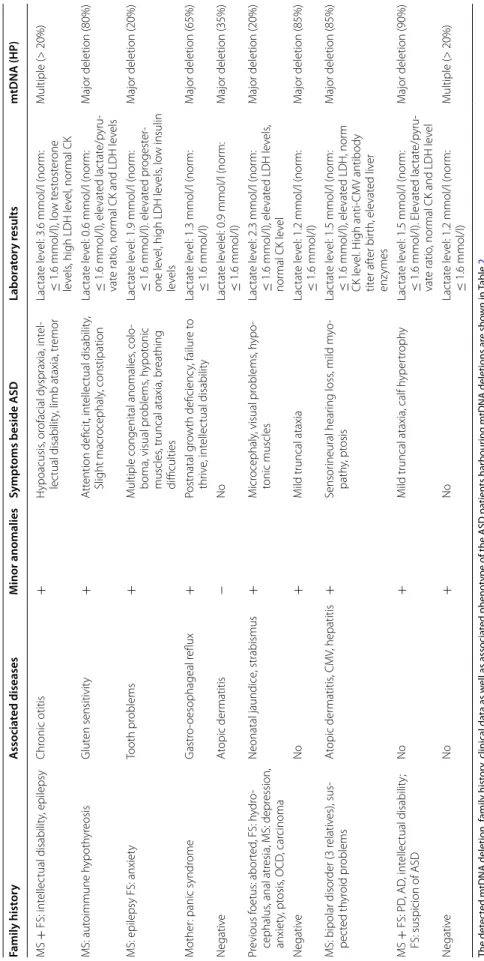
Mitochondrial deletions were identified in 16.6% (10/60) of our patients with ASD. Two children had multiple deletions, whereas a single major deletion was detected in the range of 2.4–7.9 kb in eight children. Detailed clinical and family history data as well as associated phenotype of the patients harbouring mtDNA deletions are shown in Table 2. An evaluation of clinical phenotype, fam- ily history, and laboratory data suggested MD in seven cases with mtDNA deletion. None of the investigated families had a previous diagnosis of primary MD. In all cases, the rate of heteroplasmy (HP) was > 20% in blood samples (Table 2). In the 60 healthy control individuals, a mtDNA deletion was found in two cases. Based on our statistical analysis, there was a significant difference in the frequency of mtDNA deletions between our ASD and control cohorts (χ2 with Yates correction = 4.5; p = 0.03;
odds ratio = 5.8; 95% CI 1.21–27.72). Further analysis of mtDNA mutational “hotspot” regions (m.3243 A > G, m.8993 T > C/G, and m.8344 A > G) did not detect any alterations in our ASD cohort.
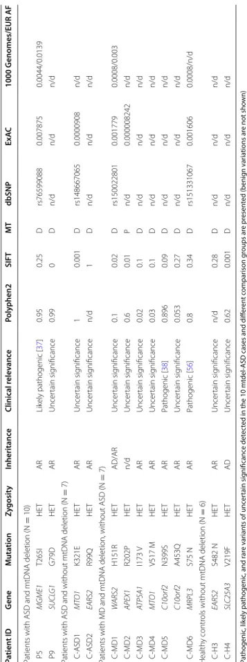
Genetic investigation—nuclear DNA mutation screening of genes responsible for intergenomic communication Next, we focused on those patients with mtDNA dele- tions (mtdel-ASD) and performed nuclear DNA (nDNA) mutation screening to investigate genes involved in intergenomic communication. In this subgroup, we found one rare likely pathogenic variant and one vari- ant with uncertain significance (VUS). The rare variants identified in two patients were both present in only one allele, however the mode of inheritance of these disor- ders is autosomal recessive (Table 3). In Patient 5 (P5), the likely pathogenic heterozygous T265I mutation in the mitochondrial genome maintenance exonuclease 1 (MGME1) is responsible for mtDNA integrity [37]. In Patient 9 (P9), we found a de novo VUS mutation (Clin- Var ID203970) in succinate-CoA ligase alpha subunit (SUCLG1) in a heterozygous form.
In our cohort of seven patients with MD (without ASD) harbouring mtDNA deletions, we found a pathogenic rare variants in two case, and rare VUS in five further cases (Table 2). In one patient the compound heterozy- gous state of one pathogenic and one VUS in C10orf2 gene were detected. In cohorts without MD (patients with ASD lacking mtDNA deletion and healthy individu- als) we found two–two rare VUS in genes responsible for intergenomic communications (Table 3).
Comparing the intergenomic NGS panel results for our different cohorts, we found no significant difference between mtdel-ASD cases and healthy controls with one tailed Fisher exact test (p: 0.4890 odds: 0.5, CI 0.05–
4.97); and mtdel-ASD and ASD without mtDNA dele- tion groups (p:0.55882, Odds: 0625 CI 0.06–5.96). Likely pathogenic variant and VUS were identified in higher number in MD patients with mtDNA deletion and with- out ASD (p = 0.013, Odds: 0.04 CI 0.003–0.5743), in a heterozygous form (Table 3).
Genetic investigation—nDNA mutation screening of ASD‑associated genes
Using the TruSight Autism NGS panel, we detected rare SNVs in 90% (9/10) of our affected children with mtDNA deletion. Syndromic ASD was identified in a single case, Patient 3 (P3), from our mtdel-ASD cohort. A heterozy- gous pathogenic mutation in chromodomain helicase DNA-binding protein 7 (CHD7) was found in this patient as well as a heterozygous mutation of uncertain signifi- cance in tuberin (TSC2). The CHD7 rare variant regarding the ACMG guideline, fulfils the PVS1 and one PS2 crite- ria [38] and based on this we evaluated it as pathogenic.
The patient’s phenotype and the family segregation pat- tern indicated this CHD7 mutation as a de novo mutation resulting in CHARGE syndrome (OMIM 214800) [39].
0 10 20 30 40 50 60 70
Developmental
regression Seizure Muscle
hypotonia Mulsystemic
abnormality Neurological signs
Clinical differences between mtdel-ASD and ASD paents
mtdel-ASD ASD
Fig. 1 The distribution of symptoms which are common in mitochondrial disorders in the patients with mtdel-ASD and in ASD without mtdel
Table 2 Mitochondrial DNA deletion status and clinical data of children with ASD and mtDNA deletion The detected mtDNA deletion, family history, clinical data as well as associated phenotype of the ASD patients harbouring mtDNA deletions are shown in Table2 MS maternal side of the family, FS paternal side of the family, OCD obsessive–compulsive disorder, PD Parkinson’s diseases, AD Alzheimer’s disease, LDH lactate dehydrogenase, CK creatine kinase, CMV cytomegalovirus, mtDNA mitochondrial DNA, HP ratio of heteroplasmy Family historyAssociated diseasesMinor anomaliesSymptoms beside ASDLaboratory resultsmtDNA (HP) MS + FS: intellectual disability, epilepsyChronic otitis+Hypoacusis, orofacial dyspraxia, intel- lectual disability, limb ataxia, tremorLactate level: 3.6 mmol/l (norm: ≤ 1.6 mmol/l), low testosterone levels, high LDH level, normal CK
Multiple (> 20%) MS: autoimmune hypothyreosisGluten sensitivity+Attention deficit, intellectual disability, Slight macrocephaly, constipationLactate level: 0.6 mmol/l (norm: ≤ 1.6 mmol/l), elevated lactate/pyru- vate ratio, normal CK and LDH levels
Major deletion (80%) MS: epilepsy FS: anxietyTooth problems+Multiple congenital anomalies, colo- boma, visual problems, hypotonic muscles, truncal ataxia, breathing difficulties
Lactate level: 1.9 mmol/l (norm: ≤ 1.6 mmol/l). elevated progester- one level, high LDH levels, low insulin levels
Major deletion (20%) Mother: panic syndromeGastro-oesophageal reflux+Postnatal growth deficiency, failure to thrive, intellectual disabilityLactate level: 1.3 mmol/l (norm: ≤ 1.6 mmol/l)Major deletion (65%) NegativeAtopic dermatitis−NoLactate levelel: 0.9 mmol/l (norm: ≤ 1.6 mmol/l)Major deletion (35%) Previous foetus: aborted, FS: hydro- cephalus, anal atresia, MS: depression, anxiety, ptosis, OCD, carcinoma
Neonatal jaundice, strabismus+Microcephaly, visual problems, hypo- tonic musclesLactate level: 2.3 mmol/l (norm: ≤ 1.6 mmol/l), elevated LDH levels, normal CK level
Major deletion (20%) NegativeNo+Mild truncal ataxiaLactate level: 1.2 mmol/l (norm: ≤ 1.6 mmol/l)Major deletion (85%) MS: bipolar disorder (3 relatives), sus- pected thyroid problemsAtopic dermatitis, CMV, hepatitis+Sensorineural hearing loss, mild myo- pathy, ptosisLactate level: 1.5 mmol/l (norm: ≤ 1.6 mmol/l), elevated LDH, norm CK level. High anti-CMV antibody titer after birth, elevated liver enzymes Major deletion (85%) MS + FS: PD, AD, intellectual disability; FS: suspicion of ASDNo+Mild truncal ataxia, calf hypertrophyLactate level: 1.5 mmol/l (norm: ≤ 1.6 mmol/l). Elevated lactate/pyru- vate ratio, normal CK and LDH level
Major deletion (90%) NegativeNo+NoLactate level: 1.2 mmol/l (norm: ≤ 1.6 mmol/l)Multiple (> 20%)
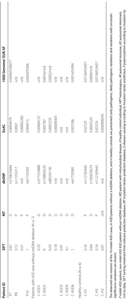
Table 3 Results of the intergenomic NGS panel Pathogenic, likely pathogenic, and rare variants of uncertain significance detected in the 10 mtdel-ASD cases and different comparison groups are presented (benign variations are not shown) P mtdel-ASD patient, non-mtdel-ASD ASD patient without mtDNA deletion, MD patient with mitochondrial disease, H healthy control individual, HET heterozygous, AR autosomal recessive, AD autosomal dominant, n/d no data, SIFT sorting intolerant from tolerant prediction database, MT mutation t@ster prediction database, D disease causing according to mutation t@ster prediction, P polymorphism according to mutation t@ ster prediction, ExAC allele frequency data from exome aggregation consortium, 1000 Genomes allele frequency data from 1000 Genomes project, EUR AF allele frequency in the European Super Population of the 1000 Genomes project Patient IDGeneMutationZygosityInheritanceClinical relevancePolyphen2SIFTMTdbSNPExAC1000 Genomes/EUR AF Patients with ASD and mtDNA deletion (N = 10) P5MGME1T265IHETARLikely pathogenic [37]0.950.25Drs765990880.0078750.0044/0.0139 P9SUCLG1G79DHETARUncertain significance0.990Dn/dn/dn/d Patients with ASD and without mtDNA deletion (N= 7) C-ASD1MTO1K321EHETARUncertain significance10.001Drs1486670650.0000908n/d C-ASD2EARS2R99QHETARUncertain significancen/d1Dn/dn/dn/d Patients with MD and mtDNA deletion, without ASD (N = 7) C-MD1WARS2H151RHETAD/ARUncertain significance0.10.02Drs1500228010.0017790.0008/0.003 C-MD2APEX1R202PHETn/dUncertain significance0.60.01Pn/d0.000008242n/d C-MD3ATP5A1I173 VHETARUncertain significance0.020.1Dn/dn/dn/d C-MD4MTO1V517 MHETARUncertain significance0.030.1Dn/dn/dn/d C-MD5C10orf2N399SHETARPathogenic [38]0.8960.09Dn/dn/dn/d C10orf2A453QHETARUncertain significance0.0530.27Dn/dn/dn/d C-MD6MRPL3S75 NHETARPathogenic [56]0.80.34Drs1513310670.0016060.0008/n/d Healthy controls without mtDNA deletion (N= 6) C-H3EARS2S482 NHETARUncertain significancen/d0.28Dn/dn/dn/d C-H4SLC25A3V219FHETADUncertain significance0.620.001Dn/dn/dn/d
In Patient 8 (P8), we found a pathogenic nonsense mutation in 7-dehydrocholesterol reductase (DHCR7), which was present in only one allele.
In Patient 2 (P2) a rare mutation was detected in autism susceptibility candidate 2 (AUTS2), which previously was associated with syndromic ASD form. The significance of the missense mutation identified in our study is uncer- tain; during segregation analysis the same mutation was present in the healthy mother, however we do not know exactly the penetrance of the genetic defects of AUTS2.
This rare AUTS2 variant coexisted with a rare variant in retinoic acid induced gene 1 (RAI1) (Table 4).
Using in silico analysis, alterations of uncertain sig- nificance were detected in ASD-associated genes in 60%
(6/10) of the mtdel-ASD cases and 71% (5/7) of non- mtdel-ASD cases (Table 4). In the six control individu- als only four rare VUS were detected in genes associated with ASD. The rare variant in zinc finger protein 804A (ZNF804A) was found in two healthy controls, indicating a variant that is likely population-specific (Table 4).
Detailed phenotype of a patient with mtDNA deletion and CHARGE syndrome (CHD7 mutation)
An 11-year-old male patient (P3) had multiple congeni- tal anomalies, such as coloboma of the eyes, oxyceph- aly, epicanthus, convergent strabismus, mild bifid nose/
broad nasal tip, low settled cup ears, mild facial asymme- try, dental dysgenesis, asymmetric chest, macroglossia, cryptorchidism, testicular hypoplasia, and atrial septal defect. He began walking at 3 years of age and suffers from obsessive hand movements, erratic behavior, and sleep disturbance. Aside from his developmental abnor- malities, neurological investigation detected pes varus, severe visual impairment, bilateral ptosis, chewing diffi- culties, mild atrophy and weakness in the distal muscles of the extremities, and truncal ataxia. High lactate lev- els were detected in both serum and cerebrospinal fluid, and decreased levels of serum melatonin, calcium, and vitamin D3 were measured. A brain MRI detected hypo- plastic vermis and transverse sinus on the right side. An EEG found generalized irritative signs, and VEP found an increased P100 on the left side. Brainstem auditory evoked potentials was normal. Family history identified arrhythmia, diabetes mellitus, and colon polypomatosis on the maternal side, and diabetes mellitus, arrhythmia, and dementia on the paternal side.
Detailed phenotype of a patient with congenital cytomegalovirus infection and mtDNA deletion
The 5-year-old female patient (P8) was born at a ges- tational age of 39 weeks by Caesarean section with a birth weight of 3000 g. The pregnancy was complicated with a partial placental abruption at 11 weeks. Evidence
of prolonged neonatal jaundice, highly elevated liver enzymes, low prothrombin level, and high IgM and IgG type anti-cytomegalovirus (CMV) antibodies led to the diagnosis of a congenital CMV infection-induced hepatic lesion. She had congenital sensorineural hearing loss, mild myopathic facies, mild ptosis, and atopic facial der- matitis. A brain MRI identified several T2 hyperintense supratentorial lesions (5–10 mm in size), which were suggested to have an infectious etiology. A heterozygous nonsense mutation in DHCR7 and a major large single deletion in mtDNA were found. The healthy mother also harbours the detected heterozygous mutation. Choles- terol and 7-dehydrocholesterol levels of the child are in the normal range. Homozygous or compound heterozy- gous mutations in DHCR7 result in Smith–Lemli–Opitz (SLOS) syndrome, which is an autosomal recessive dis- ease. However, human CMV infection may lead to altered mitochondrial biogenesis [40]. We believe that this case demonstrates a direct interaction between genetic and environmental risk factors in some forms of ASD.
Discussion
In this study, we provide for the first time a comprehen- sive genetic analysis of patients with ASD that inves- tigates co-occurrence of the most frequent mtDNA alterations, intergenomic communication disturbances (51 genes), and 101 genes previously associated with ASD. We found co-occurrence of mtDNA deletions with ASD-associated genetic alterations, which supports the previous observation that mitochondrial alterations are frequently associated with ASD. In one patient with ASD (P3), we found a mtDNA deletion with CHARGE syn- drome caused by a de novo mutation in CHD7. These genetic alterations in P3 were also accompanied by a TSC2 mutation of uncertain significance. Autistic symp- toms are present in approximately 30% of patients with CHARGE syndrome [39], and lactic acidosis is a rare alteration. Based on the phenotype, we conclude that the driving genetic alteration in this patient is the CHD7 mutation, and the mitochondrial gene defect may not be the true causative factor in the etiology of the dis- ease; however, CHD7 function is strongly ATP-depend- ent [41]. In addition, the associated heterozygous TSC2 mutation is likely a modifying gene. CHD7 is a member of the chromo-domain helicase DNA-binding (CHD) protein family and plays a role in transcription regula- tion through chromatin remodelling. Mutations in TSC2 are known to cause one syndromic form of ASD; how- ever, our patient did not develop the classic symptoms of tuberous sclerosis until recently. TSC2 mutations may induce activation of mTORC1 leading to increased mtDNA expression and mitochondrial density. mTOR is a Ser/Thr kinase that forms complexes with numerous
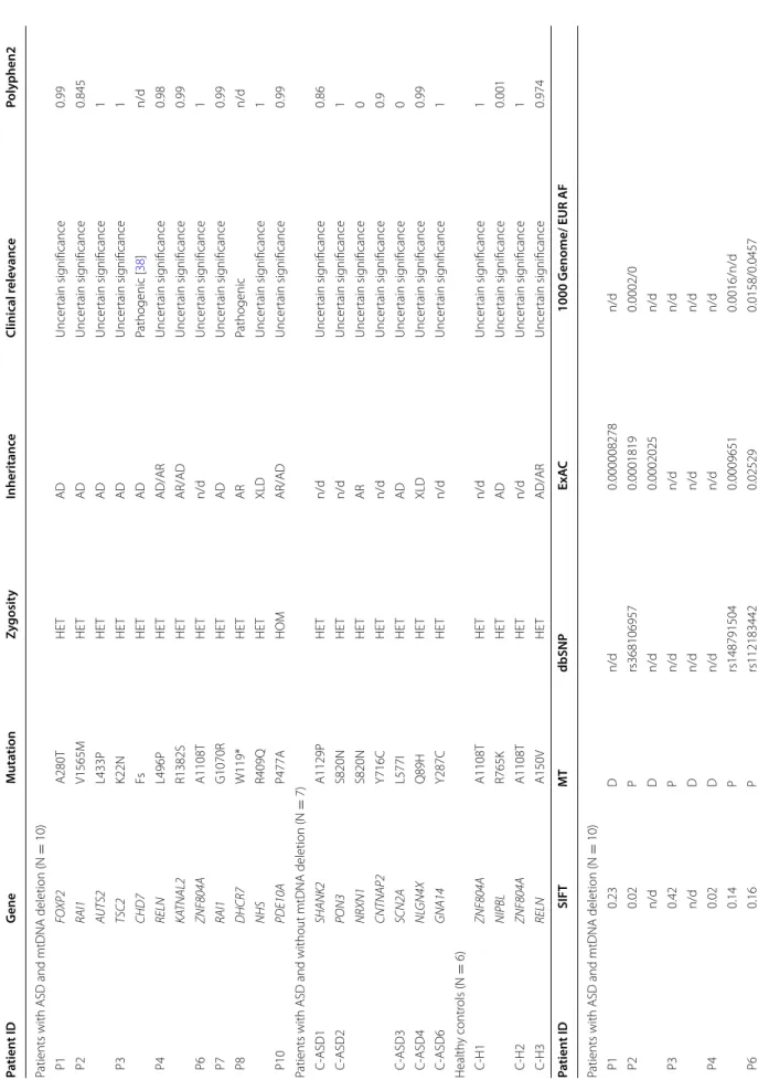
Table 4 Results of the ASD-NGS panel Patient IDGeneMutationZygosityInheritanceClinical relevancePolyphen2 Patients with ASD and mtDNA deletion (N = 10) P1FOXP2A280THETADUncertain significance0.99 P2RAI1V1565MHETADUncertain significance0.845 AUTS2L433PHETADUncertain significance1 P3TSC2K22NHETADUncertain significance1 CHD7FsHETADPathogenic [38]n/d P4RELNL496PHETAD/ARUncertain significance0.98 KATNAL2R1382SHETAR/ADUncertain significance0.99 P6ZNF804AA1108THETn/dUncertain significance1 P7RAI1G1070RHETADUncertain significance0.99 P8DHCR7W119*HETARPathogenicn/d NHSR409QHETXLDUncertain significance1 P10PDE10AP477AHOMAR/ADUncertain significance0.99 Patients with ASD and without mtDNA deletion (N = 7) C-ASD1SHANK2A1129PHETn/dUncertain significance0.86 C-ASD2PON3S820NHETn/dUncertain significance1 NRXN1S820NHETARUncertain significance0 CNTNAP2Y716CHETn/dUncertain significance0.9 C-ASD3SCN2AL577IHETADUncertain significance0 C-ASD4NLGN4XQ89HHETXLDUncertain significance0.99 C-ASD6GNA14Y287CHETn/dUncertain significance1 Healthy controls (N = 6) C-H1ZNF804AA1108THETn/dUncertain significance1 NIPBLR765KHETADUncertain significance0.001 C-H2ZNF804AA1108THETn/dUncertain significance1 C-H3RELNA150VHETAD/ARUncertain significance0.974 Patient IDSIFTMTdbSNPExAC1000 Genome/ EUR AF Patients with ASD and mtDNA deletion (N = 10) P10.23Dn/d0.000008278n/d P20.02Prs3681069570.00018190.0002/0 n/dDn/d0.0002025n/d P30.42Pn/dn/dn/d n/dDn/dn/dn/d P40.02Dn/dn/dn/d 0.14Prs1487915040.00096510.0016/n/d P60.16Prs1121834420.025290.0158/0.0457
The detected rare variants of the 10 mtdel-ASD cases, in ASD patients without a mtDNA deletion, and in healthy controls are presented (only pathogenic, likely pathogenic variations and variations with uncertain significance variations are shown) P mtdel-ASD patient, non-mtdel-ASD ASD patient without mtDNA deletion, MD patient with mitochondrial disease, H healthy control individual, HET heterozygous, AR autosomal recessive, AD autosomal dominant, n/d no data, SIFT sorting intolerant from tolerant prediction database, MT mutation t@ster prediction database, D disease causing according to mutation t@ster prediction, P polymorphism according to mutation t@ ster prediction, ExAC allele frequency data from exome aggregation consortium, 1000 Genomes allele frequency data from 1000 Genomes project, EUR AF allele frequency in the European Super Population of the 1000 Genomes project *The symbol of the non sense mutation in protein level
Table 4 continued Patient IDSIFTMTdbSNPExAC1000 Genome/ EUR AF P70.01Drs3706336840.00046790.0004/0.00077 P80.12Prs115552170.0007n/d 0.31Pn/d0.00002282n/d P101Prs617333920.0045150.0024/0.006 Patients with ASD and without mtDNA deletion (N = 7) C-ASD10.29Drs3772558880.00004137n/d C-ASD20Drs1398565350.0027870.0016/n/d 0.33Drs802931300.00022350.0002/n/d 0.18Dn/d0.00008303n/d C-ASD30.91Dn/dn/dn/d C-ASD40.1Dn/dn/dn/d C-ASD61Drs617550850.0015060.0014/0.004 Healthy controls (N = 6) C-H10.16Prs1121834420.025290.0158/0.0457 0.64Drs1856783740.00055290.0004/n/d C-H20.16Prs1121834420.025290.0158/0.0457 C-H30.01n/dn/d0.000008245n/d
protein partners to regulate cell growth, mitochondrial membrane potential, and ATP synthetic capacity [42].
In Patient 8, we found that the mtDNA deletion was accompanied by a heterozygous pathogenic DHCR7 muta- tion and a rare variant of uncertain significance in NHS actin remodelling activator (NHS). DHCR7 catalyses cho- lesterol production from 7-dehydrocholesterol, and defects in this protein cause SLOS. Furthermore, a high 7-dehydro- cholesterol level results in mitochondrial dysfunction [43].
However, the significance of a heterozygous mutation in this gene is not known. We hypothesize that in the case pre- sented here the co-occurrence of the DHCR7 heterozygous mutation and CMV infection may play a role in changes of mitochondrial biogenesis and in the pathogenesis of autis- tic features. The patient was tested for SLOS; both serum cholesterol and 7-dehydrocholesterol levels were normal, which rules out the presence of typical SLOS.
Evidence of mitochondrial dysfunction in ASD was first described 19 years ago [10]. Currently, it is the most common metabolic abnormality known in ASD with a prevalence of 7.2% [14]. In a subgroup of the CHARGE (Childhood Autism Risk from Genes and Environment) study, decreased NADH activity was found in lympho- cytes in 8 of 10 cases. In this cohort, only 2 of the 10 patients had mtDNA deletions and 5 patients had altered mtDNA copy numbers [12]. However, the genetic back- ground was not clarified in 79% of the patients with ASD-MD [9]. Therefore, the possibility of secondary damage to mitochondria cannot be excluded. A small pilot study examining 12 patients with ASD described 8 mitochondrial deletions [21], which could be the result of intergenomic communication disturbances, environ- mental factors, or other gene–gene interactions. As is the case for many other disorders, it is still not clear whether the detected mitochondrial dysfunction in ASD is a pri- mary or secondary event either having a key role in dis- ease pathogenesis or is simply a downstream effect.
In our study, mtDNA deletions were identified in 16.6%
of evaluated patients with ASD. During mtDNA hotspot screening and NGS analysis, no concomitant primary MD was detected. To examine whether mtDNA deletion is a primary or secondary event in ASD in our cohort, we used different comparison groups to screen the nDNA background of the mtDNA deletion. Pathogenic or likely pathogenic variants were detected in both mtdel-ASD and MD without ASD cases, all in heterozygous form.
A high number of VUS in intergenomic communica- tion genes were detected in the MD without ASD cohort (4/6), and a few rare variants were identified in patients with ASD that lacked mtDNA deletion and in healthy controls (Table 3). Homozygous or compound heterozy- gous mutations in MGME1 and SUCLG1 have been pre- viously correlated with severe early-onset mitochondrial
disorders (OMIM 615084, OMIM 245400) [44]. The importance of the presence of heterozygous mutations is not well understood. It is known in some genes respon- sible for intergenomic communications heterozygous mutations may result in a less severe phenotype than that found with the homozygous form [45].
The question has also been raised whether patients with MD and ASD symptoms have special characteristics. In a study by Rossignol and Frye [9], a cohort of ASD/MD children were compared to two comparison groups: chil- dren with general ASD and children with general MD. In the ASD/MD group, increased lactate and pyruvate levels, seizures, motor delays, and gastrointestinal abnormalities were significantly more prevalent compared to children with general ASD. A more balanced male:female ratio was also detected in the ASD/MD group [9]. Our results con- firm the observations by Rossignol and Frye; however, in our ASD cohort with mtDNA deletion, elevated lactate levels and/or an elevated lactate:pyruvate ratio were found in only four cases, whereas most of our ASD patients with mtDNA deletion had symptoms common to MD, such as hypoacusis, muscle weakness, hypotonia, delayed motor development, and movement disorder (Fig. 1). Signifi- cant difference between mtdel-ASD and non-mtdel-ASD group was found regarding clinical phenotypes (devel- opmental regression, muscle hypotonia, additional neu- rological signs and multisystemic alterations were more common in cases mt-delASD). Interestingly, the phe- notypes of classic mitochondrial deletion syndromes, such as Pearson syndrome, progressive ophthalmoplegia externa, and Kearns–Sayre syndrome, were not detected in any of our patients. The family histories of mtdel-ASD children in our cohort differed from the family histo- ries of the ASD cohort without a mtDNA deletion, since various psychiatric disorders were common among fam- ily members of mtdel-ASD cases both on maternal and paternal side. However none of the parents reached the MDC scoring cut-off value for definitive MD, which could not be independently verified because none of the family members agreed to perform muscle biopsy. MtDNA dis- orders are usually inherited maternally, however single mtDNA deletions are considered sporadic events with low inheritance risk, whereas multiple mtDNA deletions are the result of primary nuclear defects in genes respon- sible for mtDNA maintenance or nucleoside metabolism and follow Mendelian inheritance patterns [46].
Mitochondrial haplogroups were also investigated in association with ASD. Chalkia et al. found that individu- als with European haplogroups designated I, J, K, X, T and U (55% of the European population) had significantly higher risks of ASD compared to the most common European haplogroup, HHV. Asian and Native American haplogroups A and M also were at increased risk of ASD
[47]. In Hungary it is not rare that a person has ancient European haplotype such as T, K, and U haplotype, and rarely Asian haplotype such as B can occur as well. In some Hungarian patients the mtDNA deletion was coex- isting with ancient haplotype [48].
In 90% (9/10) of children from the mtdel-ASD cohort, we found rare SNVs in ASD-associated genes (Table 4).
A rare mutation was detected in AUTS2 in which dele- tions are inherited in an autosomal dominant manner and are associated with neurological symptoms including intellectual disability and developmental delay [49]. In a modest study of 13 cases of ASD associated with AUTS2 alterations, only one patient had a nonsense mutation; all the other patients had a deletion [49]. The significance of the missense mutation identified in our study is uncer- tain (her mother harbours the mutation as well); how- ever, clinical symptoms of the patient correlate with the phenotype of previously published AUTS2 mutations.
This rare AUTS2 variant coexisted with a rare variant in retinoic acid induced gene 1 (RAI1). The gene–gene interaction of these two alterations are hypothesized.
In addition, we found that patients had mutations of uncertain significance in forkhead box P2 (FOXP2), RAI1, phosphodiesterase 10A (PDE10A), katanin catalytic sub- unit A1 like 2 (KATNAL2), and reelin (RELN). Most of these genes play a role in cell regulation, signal transduc- tion, and various signalling pathways, which could influ- ence mitochondrial function. FOXP2 is an evolutionarily conserved transcription factor that regulates the expres- sion of a variety of genes. Mutations in this gene cause speech-language disorder 1 (OMIM 602081), which is also known as autosomal dominant speech and lan- guage disorder with orofacial dyspraxia [50]. RAI1 acts as a transcriptional regulator of chromatin remodel- ling by interacting with basic transcriptional machinery [51]. RAI1 deletion is associated with Smith–Magenis syndrome, whereas duplications are associated with Potocki–Lupski syndrome [52]. Several heterozygous mutations are also associated with Smith–Magenis syn- drome [53]. In our case, we found that the typical symp- toms of Smith–Magenis syndrome were not present.
Mutations in PDE10A can affect cyclic nucleotide con- centrations. This phosphodiesterase selectively catalyzes the hydrolysis of 3′ cyclic phosphate bonds in cAMP and/
or cGMP. The phosphodiesterase family of proteins regu- lates cellular levels, localization, and duration of action of these second messengers by controlling the rate of their degradation. In addition, phosphodiesterases are involved in many signal transduction pathways and are implicated in the pathogenesis of bipolar disorder [54].
Mitochondrial dysfunction may be associated with several forms of syndromic ASD, but is also frequently related to non-syndromic cases [8, 9]. During our
comprehensive analysis, we found examples of both but in most cases we did not find the causative genetic muta- tion that accounts for the mitochondrial dysfunction.
In the examined children from the general ASD cohort (without mtDNA deletion), we found several VUS, most of which were identified in genes without previ- ous correlation to mitochondrial dysfunction. Based on our findings, we conclude that the detected mitochon- drial DNA deletions in patients with ASD in our cohort are a secondary effect. By investigating the most com- mon mtDNA alterations and the most common nuclear genes responsible for intergenomic communications, we did not identify the clear genetic etiology in most of our cases. Therefore, further investigation and characteriza- tion is warranted.
Limitations
We identified certain limitations in our study. We focused our investigation to analyse mutational hotspots and large mtDNA deletions and did not sequence the entire mitochondrial genome. The mtDNA mutations were analysed from blood samples; postmitotic tissue was not available. The used long PCR method detects deletions in the mtDNA with high sensitivity and low specificity.
Deletions under 10% of heteroplasmy could be missed due to technical barrier, overestimation of the HP ratio is not expected. The detection of mtDNA deletion from NGS data will be in the future a new perspective, but today it is not in the everyday praxis. We used targeted NGS panels comprised of the most important genes associated with intergenomic communication and ASD.
However, these panels do not include all currently associ- ated genes and the number of these genes is continuously increasing. Finally, our healthy control group was older than our ASD cohort and had different gender ratios.
However, we felt that ethically it was not appropriate to obtain biomaterial from healthy children for genetic test- ing. Since somatic mtDNA deletion may occur in asso- ciation with ageing, and we detected the mtDNA deletion less frequently in the control group it had no impact on our data. Mitobreak Database [55] supports or presump- tion since deletion were present mostly only aged healthy controls, otherwise they were associated or to sporadic primary mitochondrial disorders (single deletion) or to disorders due to intergenomial gene alterations (multiple deletions).
Conclusions
The aim of our study was to gain a better understand- ing of mitochondrial dysfunction in autism. We found that mtDNA alterations were more common among our cohort of patients with ASD than in control individu- als. In addition, we found that the mtDNA deletion was