1
The role of FDG PET/CT in lymphomas
Ph.D. thesis
Kornélia Kajáry, MD
Doctoral School of Clinical Medicine Semmelweis University
Supervisor: Tamás Györke MD, Ph.D.
Official reviewers: Gabriella Dabasi MD, Ph.D.
László Galuska, MD, Ph.D.
Head of the Final Examination Committee: István Szilvási, MD, Ph.D.
Members of the Final Examination Committee: Andrea Radácsi MD, Ph.D.
Pál Kaposi-Novák MD,Ph.D.
Budapest
2015
2
1. INTRODUCTION
Nowadays, fluorine-18 labelled fluorodeoxyglucose (FDG) positron emission tomography (PET), especially when used as a hybrid modality (positron emission tomography/computed tomography [PET/CT]), is a recommended method for staging (before and after treatment), early response-assessment (commonly referred as interim PET) and detection of recurrence (in case of clinical suspicion) in Hodgkin-lymphoma (HL), as well as, in non-Hodgkin lymphomas (NHL). In my dissertation, I made a report on my scientific results with PET/CT examinations performed before therapy and at the end of first-line treatment in HL and presented a case of neurolymphomatosis (NL) detected by PET/CT.
In Hungary, it is a common practice to perform a computed tomography (CT) of the neck-chest-abdomen-pelvis regions with intravenously contrast material administration for primary staging of HL, before indicating the PET/CT. Based on data analysis in the international literature, CT at full-dose with contrast enhancement (ceCT) of the whole body is not requested as a routine evaluation for that purpose and by skipping it, radiation exposure of these (mostly young) patients may be significantly reduced.
Since 2007, International Harmonization Project (IHP) criteria has been recommended internationally for the evaluation of PET/CT examinations at the end of treatments, but its use in the daily routine is difficult and it also takes into account the size of lesions measured by CT. In addition, though the negative predictive values (NPV) are high in most publications on this system, positive predictive values (PPV) range more widely. Methods used for interpretation of the interim PET(/CT) examinations performed between chemotherapy cycles for evaluation of early therapeutic response are different from those above. These methods are easier to use in clinical practice and do not take into account size of lesions.
During PET/CT investigations of patients suffering from lymphoma, signs of previously unrecognized extranodal involvement are often seen, which usually also modify the treatment plan. NL is defined as an infiltration of cranial or peripheral nerves, nerve roots or nervous plexuses during various haematological malignancies.
Since the rarity of NL, physicians often become not aware that patient’s symptoms may arise from it; therefore, this condition is not recognised in time in many cases. It is a
3
rare entity, which is occasionally difficult to diagnose using conventional imaging modalities.
2. OBJECTIVES
1. To assess in a Hungarian patient sample, whether ceCT is necessary besides the standard FDG PET/CT for primary staging in HL.
Numerous studies have demonstrated so far that for the initial staging of lymphomas, FDG PET and especially PET/CT is more accurate than ceCT used earlier as a routine method. Authors started to analyse relatively soon, whether unenhanced low-dose CT is sufficient or not during hybrid PET/CT assessments, mainly in order to reduce radiation exposure. Results showed that there is no need for intravenously contrast material administration in a number of tumours, such as lymphomas. Based on conclusions of international comparisons made so far, although ceCT may improve detection of abdominal and pelvic manifestations of lymphomas, it can add only very little information, which usually does not modify either disease stage or planned management. In Hungary, it is a common practice to perform a neck-chest-abdomen- pelvis ceCT for primary staging of HL usually before the PET/CT, mostly in another institute, which significantly increases patients’ radiation exposure. One of my objectives was to confirm in a Hungarian patient sample, that unenhanced low-dose CT performed during PET/CT assessment is sufficient for proper primary staging in HL. In this context, I also examined whether breath-hold low-dose chest CT should be recommended in addition to the shallow-breathing low-dose CT performed during the PET/CT investigation for correct assessment of lungs.
2. To compare IHP, London (or Deauville) and Gallamini criteria for the interpretation of FDG PET/CT after the completion of first line therapy in HL.
Based on international literature, PET(/CT) is more accurate than CT alone in post- treatment evaluation of both HL and aggressive NHL, since it can differentiate residual malignant tissue from fibrosis or necrosis. IHP criteria set out in 2007, were generally recommended to be used for interpretation of PET(/CT) scans with the purpose of restaging after completion of therapy. In this system, metabolic response detected by FDG PET became the key factor, but it still takes into account lesion size measured by
4
CT. Post-treatment PET and PET/CT exams have had a high NPV in most of the publications using this system, while PPV ranged widely. In clinical practice, this manifests in a relatively high rate of false positive (FP) cases, which makes decision on need for additional therapy difficult. In my dissertation, I investigated whether the rate of false positivity could be reduced in HL by using semiquantitative or visual interpretation techniques, different from IHP criteria.
3. To investigate the role of FDG PET/CT in NL.
In the daily routine, during PET/CT examinations of patients with various lymphomas, I often encountered signs of extranodal involvement that earlier remained unrecognised, which usually also modified the treatment plan. Besides the relatively more frequently occurring organ involvement, it is a diagnostic challenge to recognise the extremely rare forms of the disease. As the third element of my dissertation, I provided a report on a case of NL detected by FDG PET/CT, which is a haematological malignancy rarely published in the international literature.
3. METHODS 3.1. Primary staging PET/CT and ceCT exams in HL 3.1.1. Patients
In our study, data of 28 newly diagnosed, histologically confirmed HL patients were analysed, who underwent initial staging FDG PET/CT exams between December 2007 and January 2011 at the Pozitron Diagnosztika Központ (Positron Diagnostics Centre) in Budapest and who besides the usual examination protocol, also underwent full-dose, venous phase CT examinations involving regions between the skull base and the inguinal fold. Patient population consisted of 16 female and 12 male patients, with an average age of 34 (18–67) years. Distribution of the disease per histological diagnosis was as follows: nodular sclerosis (NS) – 20 patients, mixed cellularity (MC) – 5 patients, nodular lymphocyte predominant HL (NLPHL) – 2 patients and it could not be exactly determined for 1 patient.
5 3.2.2. Imaging methods, evaluation
Since during PET scans, high blood glucose levels interfere with optimal radiopharmaceutical distribution all patients have fasted for a minimum of 6 hours before investigation. Blood glucose levels measured by fingertip testing before tracer injection were lower than 7.8 (range: 4.1 – 7.8) mmol/L. FDG was administered intravenously in an average amount of 314.5 (292.3 – 336.7) Megabecquerel (MBq), depending on body weights. After an average incorporation time of 81 min (ranging between 58–159 min) spent at rest, whole body (from the skull base to the inguinal fold) unenhanced low-dose CT scans and then PET scans [hereinafter standard PET/CT] were performed. Imaging examination was completed with a full-dose contrast enhanced venous phase whole body CT [hereinafter diagnostic CT]. Finally, breath-hold chest CT scans (during inspiration) were also acquired. Scans were acquired using True Point HD PET/CT scanner and Biograph 16 PET/CT scanner for 27 patients and 1 patient, respectively. Analysis of patient data was carried out in three different ways. First method was based on diagnostic CT scan, in accordance with generally accepted criteria for CT. The second method was based on the standard PET/CT, where also generally accepted criteria for CT were taken in account. During evaluation of PET images, FDG accumulation was considered abnormal if it exceeded surrounding background activity and it was not explainable with physiological causes. With the third method, standard PET/CT and diagnostic CT were evaluated together. Staging was done according to the generally used Cotswolds modified Ann Arbor system. Validation of pre-treatment imaging studies in 27 patients was performed using the post-treatment imaging examination, which was diagnostic CT scan for 1 patient and a second standard PET/CT scan (performed at least 10 days after the end of treatment) for 26 patients. One patient initially refused the offered treatment, for which standard PET/CT was performed again about 4 months after the first PET/CT. Finally, the number and size of lung nodules smaller than 10 mm that could be identified on shallow-breathing and breath-hold CT scans was determined.
6
3.2. Restaging PET/CT exams, after completing the first-line therapy in HL 3.2.1. Patients
In this retrospective study, restaging PET/CT findings and clinical outcomes related to them were analysed in 66 newly diagnosed, histologically confirmed HL patients, who had undergone standard first-line therapy. PET/CT exams were performed at the Pozitron Diagnostic Centre in Budapest between March 2006 and March 2011.
Patients’ data are summarized in Table 1.
Table 1 Characteristics of the patients
Parameters (n=66) Data
Age (years) Mean 37.3
Median 32.5
Range 19-68
Sex (pts) Men 36
Women 30
Clinical stage(pts) I+II 6+39=45
III+IV 13+8=21
Histological type (pts) Nodular sclerosis (NS) 46
Mixed cellularity (MC) 13
Lymphocyta reach (LR) 3
Lymphocyta depletion (LD) 2
Nodular sclerosis/mixed cellularity (NS/MC) 2
Prognostic subgroups (pts)
Stage I-II Favourable 4
Nonfavourable 41
Stage III-IV IPS: 0-3 19
IPS: 4-7 2
Final clinical outcome (pts) Complete remission 60
Progressive/stable disease 3
Death 2
Lost to follow-up 1
Follow-up (n=65!, months) Mean 51.4
Median 54.0
Range 3-78
Chemotherapy (pts) 4 ABVD 24
6 ABVD 42
Chemotherapy + radiotherapy (pts) 46
pts: patients IPS: International Prognostic Score, ABVD: doxorubicin, bleomycin, vinblastine, dacarbazine
7
3.2.2. PET/CT examinations and their interpretation, statistical analysis
For patients treated only with chemotherapy, PET/CT scanning was scheduled at least 24 days after the last day of chemotherapy (between days 24 and 76, a median value of 35 days). For patients who also received irradiation, time interval between the end of radiation therapy and PET/CT was at least 8 weeks for 38 patients and 5 to 8 weeks for 8 patients (overall, PET/CT was performed between weeks 5 and 35 with a median value of 12 weeks). Patients with diabetes fasted for a minimum of 4 hours, whilst the other ones had to fast for at least 6 hours before investigation. Blood glucose levels measured by fingertip testing before tracer injection were lower than 7.3 (ranging between 3.6 and 7.3) mmol/L in all but three patients. Neither did subsequent data analysis reveal why blood levels of these three patients failed to be measured. Since none of them was diabetic, they fasted for at least 6 hours before investigation, and their scans did not show abnormal FDG distribution suggesting high blood glucose levels, thus they were also included in our study. FDG was administered intravenously in an average amount of 341.3 (162.8 – 669.7) MBq, depending on body weights. After an average incorporation time of 84 min (ranging between 52–164 min) spent at rest, whole body PET/CT scans were performed. Forty-three scans were acquired using a Biograph 16 PET/CT scanner and 23 examinations were carried out with a TruePoint HD PET/CT scanner. Patients were allowed to breathe normally during whole-body PET and CT acquisitions. During studies performed using the TruePoint HD scanner, breath-hold CT images of the lung were also acquired.
(1) In accordance with internationally accepted general approach for evaluation of restaging PET/CT exams, analysis was first performed based on recommendations of IHP. According to this system, investigation is considered positive for lymphoma if one or more of the following criteria apply:
- FDG uptake is more intense than mediastinal blood pool (MBP) activity in residual soft tissue lesions ≥2 cm measured by CT
- FDG uptake is more intense than surrounding background uptake in residual soft tissue lesions <2 cm measured by CT
- nodular lung lesion newly revealed on CT, if it is ≥1.5 cm and its FDG uptake exceeds MBP OR if it is <1.5 cm and its FDG uptake is higher than that of the background
8
Exception: if the PET(/CT) scan is otherwise negative and previously there is no history of pulmonary manifestations of lymphoma, the new nodular lesion should be considered benign (first of all an infection or inflammation) regardless of its size and the degree of FDG uptake.
- residual liver or spleen lesion on CT, if it is ≥1.5 cm and its FDG uptake is equal or higher than surrounding liver or spleen activity OR if it is <1.5 cm and FDG uptake is higher than surrounding liver or spleen activity
- uni- or multifocal FDG accumulation in the bone marrow
During further two analyses, the two most important methods from among those usually used for evaluation of interim PET exams were used for interpretation of restaging PET/CT scans.
(2) The second analysis was performed based on the London (or Deauville) criteria, when detected lesions were graded as follows:
1 = no FDG uptake above background within the previously involved lesion;
2 = FDG uptake within the previously involved lesion is equal or lower than MBP activity;
3 = FDG uptake within the previously involved lesion is higher than MBP, but does not exceed liver activity;
4 = FDG uptake within the previously involved lesion is moderately higher than liver activity;
5 = FDG uptake within the previously involved lesion markedly exceeds liver activity, or a new lesion with abnormal FDG uptake has appeared, which has no reasonable physiological or benign cause.
Three different methods were defined depending on what was considered positive and negative as regarding the presence of active lymphoma. When using Method 1, the threshold for positivity was MBP activity, which means that lymphoma was considered to be actively present if the highest score was 3 or higher. For Method 2, liver activity was the threshold, which means that investigation was considered positive for lymphoma, if the highest score was 4 or 5. For Method 3, PET/CTs were considered positive only if lesions with an uptake score of 5 were found. Diffusely increased bone marrow uptake of any degree was not considered a manifestation of lymphoma;
9
however, focal bone marrow accumulations were considered to be a sign of the malignant disease.
(3) Third analysis was based on Gallamini criteria: PET/CT scan was considered to be positive, if FDG uptake within previously involved regions or at new sites (regardless of lesion size) exceeded MBP activity, with a maximum of standard uptake value (SUVmax) greater than 3.5.
A positive FDG PET/CT result in a patient who presented any evidence of disease, progression or relapse (based on progression confirmed by CT and/or cytology or histology and/or on clinical symptoms) or who died of the disease, was considered to be true positive (TP). A positive FDG PET/CT result in a patient who achieved and remained in complete remission (CR) during the follow-up period was considered to be FP. A negative FDG PET/CT result in a patient who presented any evidence of disease, progression or relapse (also based on progression confirmed by CT and/or cytology or histology and/or on clinical symptoms) or who died of the disease was considered to be false negative (FN). A negative FDG PET/CT result in a patient who achieved and remained in CR during the follow-up period was considered to be true negative (TN).
For evaluation of prognostic value of PET/CT examination determined with different analysis methods survival curves were calculated according to the method of Kaplan and Meier for progression-free survival (PFS) and overall survival (OS). PFS was defined as the time elapsed between the end of first-line therapy and the time of any evidence of disease, progression or relapse or HL-related death. Data for patients who did not experience any of the above events were censored at the time of the last follow- up. OS was defined as the time from the end of first-line therapy until death from any cause or until the time of the last control visit. Log-rank test was used for comparison of different survival curves. Statistical analysis for pre-treatment prognostic factors was also performed. Log-rank test result was considered statistically significant if the P- value was less than 0.05.
10 .3. PET/CT in NL
3.3.1. Patient’s remote cancer history
In a 60-year-old male patient surgical removal of the right testis, followed by treatment with rituximab, methotrexate, doxorubicin, bleomycin, cyclophosphamide, dexamethasone and vincristine (R-M-BACOD) was performed due to diffuse large B- cell lymphoma (DLBCL) involving the right testis and supra- and infradiaphragmatic lymph nodes. First-line therapy resulted in CR, and patient had no clinical symptoms for 24 months.
3.3.2. Patient’s neurological history, characteristics of PET/CT study suggesting NL Twenty-nine months after the diagnosis of DLBCL, the patient presented at his attending physician complaining of sharp pains in his right hip and numbness in his right leg. Orthopaedic physical examination and subsequent CT scan did not reveal the cause of his complaints. After that, lumbosacral MRI was performed showing nothing abnormal except a slight protrusion of L5-S1 intervertebral discus. Physiotherapy, non- steroid anti-inflammatory drugs and steroids were prescribed, but they proved to be inefficient. Intensity of pain further increased and then the patient also experienced walking difficulty. Whole body FDG PET/CT was performed 4 months after the onset of symptoms. Being a diabetic, the patient fasted for only 4 h prior scanning instead of the usually recommended 6 hours, and his blood glucose level was 6.4 mmol/L before administration of the radiopharmaceutical substance. After intravenously injection of 436.6 MBq FDG and 70 minutes of waiting time at rest, PET/CT images were acquired with a TruePoint HD PET/CT scanner.
4. RESULTS 4.1. Primary staging PET/CT and ceCT exams in HL
The same disease stage was established with all three methods in 24 patients.
Disease was upstaged by standard PET/CT compared to diagnostic CT in four cases. No difference was found between staging based on standard PET/CT evaluated alone and based on joint evaluation of standard PET/CT and diagnostic CT. Of the four upstaged
11
patients, involvement of subdiaphragmatic lymph nodes sized below abnormal was found in 2 patients besides the supradiaphragmatic manifestation, thus they were reclassified from stage II to stage III. Based on PET/CT scan, the 2 other patients were reclassified from stage III to stage IV, since pathological FDG uptake suggesting bone involvement was detected. Our study results are summarized in Table 2 and Table 3.
Table 2 Primary staging by standard PET/CT vs. diagnostic CT diagnostic CT
standard PET/CT
I (9) II (12) III (4) IV (3)
I (9) 9 - - -
II (10) - 10 - -
III (4) - 2 2 -
IV (5) - - 2 3
Table 3 Primary staging by standard PET/CT vs. diagnostic CT plus standard PET/CT diagnostic CT
+ standard PET/CT standard PET/CT
I (9) II (10) III (4) IV (5)
I (9) 9 - - -
II (10) - 10 - -
III (4) - - 4 -
IV (5) - - - 5
Results of pre-treatment standard PET/CT scans proved to be real for all patients.
Breath-hold low-dose chest CT was not performed in 1 patient, because of his/her poor condition, whilst all methods confirmed multiple lung involvement in 3 patients. For the remaining 24 patients, a total of 10 and 17 nodular lung lesions smaller than 10 mm (2 – 7 mm) were detected on shallow-breathing low-dose CT scans and breath-hold low dose CT scans, respectively.
12
4.2. Restaging PET/CT exams after the first-line therapy in HL
Median follow-up time from the end of first-line therapy was 54 months (between 3 and 78 months). One patient with clinical symptoms and disease progression clearly confirmed by CT refused the recommended second-line therapy, and died 3 months after the end of administered treatment. The other 65 patients were followed-up for a minimum period of 20 months. Ten patients experienced therapy failure (any evidence of disease, progression or relapse) during the follow-up period, which was confirmed by cytology or histology in 4 patients. The other 6 patients showed obvious severe progression on CT, and three of them also had clinical symptoms; therefore, their attending physicians decided to start second-line therapy right away, without histological confirmation of the disease. All of the above patients underwent second- line treatment (chemotherapy and/or bone marrow transplantation). One of them died because of complications of allogeneic bone marrow transplantation 44 months after the end of first-line treatment. One patient was lost to follow-up after the second-line treatment because he did not attend further control visits. Five patients achieved CR, 2 patients had stable disease (SD) and 1 patient had progressive disease (PD) at their last visits (Fig.1). Thus, final clinical outcome was known for 65 patients, and OS curves were calculated for their data. According to IHP criteria and also Method 1, a negative PET/CT result was seen in 50 patients and a positive result in 16 patients. The number of TN cases was 49, and relapse was observed in one patient 9 months after the scan (FN). The number of TP and FP was 10 and 6, respectively. Based on these data, both methods resulted in an NPV of 98.0% and a PPV of 62.5%. Method 2 gave a negative result in 52 cases and a positive result in 14 patients. The number of TN cases was 51, and relapse was detected in the same patient as with the other two methods. The number of TP and FP cases was 10 and 4, respectively. When using Method 3, 57 patients had a negative PET/CT, while result was positive for 9 patients. The number of TN cases was 54. Relapse was observed in three patients: 9 months and 4 months after the scan in one patient and two patients, respectively. The number of TP cases and FP cases was 8 and 1, respectively. Thus, NPV and PPV were 98.1 and 71.4% with Method 2 and 94.7 and 88.9% with Method 3, respectively. Based on Gallamini criteria, a negative PET/CT was found in 53 patients and a positive PET/CT in 13 patients. The number of TP cases and FP cases was 9 and 4, respectively. The number of TN cases was 51, out of which
13
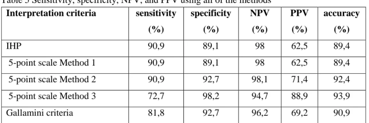
relapse was observed in 2 patients 9 and 4 months after the scan. Based on these data analysis resulted in an NPV of 96.2% and a PPV of 69.2%. PET/CT results were correct (7 TN and one TP) for each of the 8 patients who were examined less than 8 weeks after radiotherapy, regardless of the interpretation method used. The number of true and false results obtained with different analysis methods and sensitivity, specificity, NPV and PPV data are summarized in Table 4 and 5, respectively. It can be noted that NPV was high with all evaluation methods, almost to the same extent. At the same time, PPV values ranged within wider limits (62.5 to 88.9%) when using different analyses.
Fig.1 Final clinical outcome of HL
CR: complete remission, PD: progressive disease, SD: stable disease
However, no statistically significant differences were found between the different interpretation systems. Statistical comparison between groups regarding PFS and OS data is shown in Table 6. In Fig.2 and 3 PFS and OS curves are shown, respectively, comparing different evaluation methods. Significant differences were found between PET negative and PET positive groups as regarding both the PFS and OS and with all analysis methods. However, based on performed analyses, no statistically significant predictive value was found by using pre-treatment prognostic factors commonly used in clinical practice.
14
Table 4 Number of true and false cases using all of the methods
Interpretation criteria TP (pts) TN (pts) FP (pts) FN (pts)
IHP 10 49 6 1
5-point scale Method 1 10 49 6 1
5-point scale Method 2 10 51 4 1
5-point scale Method 3 8 54 1 3
Gallamini criteria 9 51 4 2
IHP: International Harmonization Project, TP: true positive, TN: true negative, FP: false positive, FN:
false negative, pts: patients
Table 5 Sensitivity, specificity, NPV, and PPV using all of the methods Interpretation criteria sensitivity
(%)
specificity (%)
NPV (%)
PPV (%)
accuracy (%)
IHP 90,9 89,1 98 62,5 89,4
5-point scale Method 1 90,9 89,1 98 62,5 89,4
5-point scale Method 2 90,9 92,7 98,1 71,4 92,4
5-point scale Method 3 72,7 98,2 94,7 88,9 93,9
Gallamini criteria 81,8 92,7 96,2 69,2 90,9
IHP: International Harmonization Project, NPV: negative predictive value, PPV: positive predictive value
Table 6 Differences between groups for PFS and OS using the log-rank test
Parameters PFS (n=66) OS (n=65)
Pre-treatment clinical prognosis (good vs. bad *) p= 0.431 p=0.654
Post-treatment PET/CT (positive vs. negative)
IHP p<0.001 p=0.008
5-point scale Method 1 p<0.001 p=0.008
5-point scale Method 2 p<0.001 p=0.003
5-point scale Method 3 p<0.001 p<0.001
Gallamini criteria p<0.001 p=0.003
*Stage I-II: favourable vs. nonfavourable and Stage III-IV: IPS 0-3 vs. IPS 4-7, where IPS: International Prognostic Score; Underline indicates statistically significant values; FDG: 18-fluoro-dezoxi-glukóz, IHP:
International Harmonization Project, PET/CT: positron emission tomography/computer tomography, PFS: progression-free survival, OS: overall survival
15
Fig.2 Kaplan–Meier survival curves showing the PFS according to PET/CT results using the different methods (solid line: PET-negative patients, dashed line: PET-positive patients)
16
Fig.3 Kaplan–Meier survival curves showing the OS according to PET/CT results using the different methods (solid line: PET-negative patients, dashed line: PET-positive patients)
17 4.3. PET/CT in NL
FDG PET/CT performed due to neurological symptoms detailed above (in part 3.3.2) revealed focal increased FDG uptake in the region of the right S1 nerve root and linear FDG uptake along the right sacral plexus with SUVmax of 11.6 and 5.2, respectively (Fig.4).
Fig.4 Transaxial sections of the FDG PET/CT examination demonstrating (red arrows) focal FDG uptake in the region of the right S1 nerve root (a) and linear FDG-uptake along the right sacral plexus (b, c), both suspicious for neurolymphomatosis. The whole-body coronal section PET/CT image is demonstrating the same (d).
18
Based on FDG uptake strong suspicion of NL was described in the investigation report.
After that, a second MRI was performed, showing thickening of the right S1 nerve root and enlargement of the sacral plexus with inhomogeneous contrast enhancement (Fig.5). Tumorous mass was surgically removed, after which patient’s symptoms improved significantly. Histological examination of the removed specimen confirmed the unusual manifestation site of DLBCL.
Fig.5 Coronal T1-weighted (A), coronal (B) and axial fat-suppression (C) contrast–enhanced T1-weighted and axial T2-weighted images of MR examination (D). Red arrows show the thickening of right S1 nerve root (A) with pathological contrast enhancement (B, C), which has heterogeneous signal intensity on the T2-weighted image (D). Blue arrows show the normal left S1 nerve root which has contrast enhancement in the level of ganglion (C), but not elsewhere (B).
Taking into account the uncommon site of the relapse, though diffuse FDG uptake visualised on PET/CT scan was considered to be benign, bone marrow biopsy was performed, based on which bone marrow infiltration was excluded. The patient received four cycles of rituximab, dexamethasone, citarabine and cisplatin (R-DHAP) chemotherapy and then underwent autologous bone marrow transplantation (ASCT).
19
None of PET/CT exams performed following chemotherapy and 6 months after ASCT showed any pathological FDG uptake indicating active lymphoma, therefore, opinion of complete metabolic remission was delivered. Because of the unusual site of remission, though patient had no clinical symptoms, PET/CT examination was repeated 18 and 30 months after ASCT. None of these scans revealed abnormal FDG uptake suggesting lymphoma and thus the opinion of complete metabolic remission was given on both occasions.
5. CONCLUSION
Our results also confirmed in a Hungarian patient population that PET/CT with unenhanced low-dose CT is more accurate than ceCT alone for pre-treatment staging in HL. In line with international analyses, our study confirmed that initially whole body ceCT could be omitted for this indication. However, a low-dose breath-hold chest CT performed concomitantly with the PET/CT exam is still recommended for the correct assessment of lungs. If FDG accumulations with questionable localisations or lesions that cannot be well delimited morphologically from their surrounding were visualized on PET/CT scans and their precise evaluation was essential for further treatment or clinical symptoms with uncertain aetiology developed, ceCT should be still recommended. Ideally, this would mean imaging of the affected anatomical region only and would be performed just after PET/CT scan. Thus, the radiation exposure of these (mostly young) patients could be significantly reduced and this would be probably a more cost-effective solution compared to earlier practices.
In our study, we compared directly for the first time the IHP, Gallamini and London (or Deauville) criteria for interpretation of PET/CT scans performed after first- line treatment in HL. Our results confirmed the high NPV of PET/CT in this indication;
values were similar for all evaluation systems. By comparing different interpretation options, a system resulting in a sufficiently high PPV and easy to use in the daily clinical routine can be chosen. Our summarized data suggest that London (or Deauville) criteria that were originally designed for characterisation of interim scans seem to be the most adequate for this indication because of their high diagnostic accuracy, their simplicity and since they do not depend on SUVmax values. As regarding this 5-point
20
scale, using liver activity for a threshold between negative and positive scans seems to be the most advisable option. Our results are promising, but there are some drawbacks:
retrospective nature of data processing, small study sample, and the heterogeneity of patient population as regarding the disease stage, which naturally resulted in slightly different treatment approaches.
Neurolymphomatosis (NL) is an extremely rare extranodal manifestation of lymphomas. Based on literature review of NL characteristics and on the medical history of our own patient, we can conclude that it is highly recommended to perform FDG PET(/CT) relatively early in all patients with lymphoma (especially in those with NHL), who suffer from neurological symptoms of unclear aetiology. Since FDG PET/CT is highly recommended for the assessment of treatment effectiveness in lymphomas with common manifestations, it seems reasonable to use it in uncommon forms of lymphomas, including NL, too.
6. BIBLIOGRAPHY OF THE CANDIDATE’S PUBLICATIONS 6.1. The publications related to the theme of the PhD thesis
1. Kajáry K, Molnár Z, Deák B. (2012) Az FDG-vel végzett PET/CT vizsgálat prognosztikai éréke Hodgkin-limfómában. Orvostovábbképző szemle, 19: 76-79.
2. Kajáry K, Molnár Z, Mikó I, Barsi P, Lengyel Z, Szakáll S Jr. (2014).
Neurolymphomatosis as a late relapse of non-Hodgkin's lymphoma detected by (18)F- FDG PET/CT: A case report. Rev Esp Med Nucl Imagen Mol, 33: 39-42.
IF: 0.938
3. Kajáry K, Molnár Z, Györke T, Szakáll S Jr, Molnár P, Lengyel Z. (2014) Comparison of the International Harmonization Project, London and Gallamini criteria in the interpretation of 18F-FDG PET/CT examinations after first-line treatment in Hodgkin's lymphoma. Nucl Med Commun, 35: 169-175.
IF: 1.371
4. Bárdi E, Csóka M, Garai I, Szegedi I, Müller J, Györke T, Kajáry K, Nemes K, Kiss C, Kovács G. (2014) Value of FDG-PET/CT Examinations in Different Cancers of Children, Focusing on Lymphomas Pathol Oncol Res, 20: 139-143.
IF: 1.806
21
5. Kajáry K, Molnár Z, Szakáll S Jr, Molnár P, Lengyel Z. (2014) Szükséges-e Hodgkin-limfóma primer stádium-megállapításához FDG-PET/CT mellett iv. ka. CT elvégzése is? Orv Hetil, 155: 226-230.
6.2. The publications related not to the theme of the PhD thesis
1. Kajáry K, Szakáll S Jr., Moravszki M, Molnár P, Lengyel Z. (2007) A PET-CT és szerepe az onkológiában. Orvostudományi Értesítő, 80: 96-105.
2. Lengyel Z, Kajáry K, Moravszki M, Molnár P, Szakáll S Jr. (2007) A PET/CT jelentősége a pulmonológiai onkológiában. Tüdőgyógyászat, 1: 5-13.
3. Kajáry K, Lengyel Z, Molnár P, Szakáll S. (2009) A PET-CT-vizsgálat szerepe nőgyógyászati daganatoknál. Nőgyógyászati onkológia, 1: 8.
4. Kócsák E, Tándor Z, Kajáry K, Gombos J. (2012) Az endometriosis vizsgálata a molekuláris képalkotás eszközeivel a nukleáris medicina szemszögéből. Magyar Radiológia, 86: 119-125.
5. Kajáry K, Temesszentandrási G. (2012) Az FDG-vel végzett PET/CT vizsgálat szerepe az ismeretlen eredetű láz okának kiderítésében-egy arteritiszes beteg bemutatásán keresztül. Orvostovábbképző szemle, 19: 82-86.
6. Tőkés T, Kajáry K, Torgyík L, Lengyel Z, Györke T, Dank M. (2013) PET-CT Imaging in Breast Cancer Patients: New Tracers, Future Directions. J Mol Imaging Dynam, 2: 111.
7. Kajáry K, Molnár P, Lengyel Z, Szakáll S Jr. (2014) Port-Site Metastasis After Laparoscopic Removal of an Isolated Adrenal Metastasis in a Patient With Breast Cancer Detected With FDG PET/CT. Clin Nucl Med, 39: 203-205.
IF: 2,857
8. Kajáry K, Tőkés T, Dank M, Kulka J, Szakáll S Jr, Lengyel Z. (2015) Correlation of the value of 18F-FDG uptake, described by SUVmax, SUVavg, metabolic tumour volume and total lesion glycolysis, to clinicopathological prognostic factors and biological subtypes in breast cancer. Nucl Med Commun, 36: 28-37.
IF: 1,371