Aluminium hydroxide stabilised MnFe 2 O 4 and Fe 3 O 4 nanoparticles as dual-modality contrasts agent for MRI and PET imaging
Xianjin Cui
a, Salome Belo
a, Dirk Krüger
a, Yong Yan
b, Rafael T.M. de Rosales
a, Maite Jauregui-Osoro
a, Haitao Ye
c, Shi Su
c, Domokos Mathe
d, Noémi Kovács
d, Ildikó Horváth
d, Mariann Semjeni
d, Kavitha Sunassee
a, Krisztian Szigeti
f, Mark A. Green
a,e,**, Philip J. Blower
a,g,*aKing’s College London, Division of Imaging Sciences and Biomedical Engineering, 4th Floor Lambeth Wing, St Thomas’Hospital, London SE1 7EH, UK
bSchool of Chemistry, Nottingham University, Nottingham NG7 2RD, UK
cSchool of Engineering and Applied Science, Aston University, Birmingham B4 7ET, UK
dCROmed Ltd., Baross u. 91-95, Budapest H-1047, Hungary
eKing’s College London, Department of Physics, Strand Campus, London WC2R 2LS, UK
fDepartment of Biophysics and Radiation Biology, Nanobiotechnology & In Vivo Imaging Center, Semmelweis University, IX. T}uzoltó u. 37-47, Budapest H-1094, Hungary
gKing’s College London, Division of Chemistry, Britannia House, 7 Trinity St, London SE1 1DB, UK
a r t i c l e i n f o
Article history:
Received 4 February 2014 Accepted 1 April 2014 Available online 24 April 2014
Keywords:
Magnetic nanoparticles PET
MR
Aluminium hydroxide Dual-modal
18F
a b s t r a c t
Magnetic nanoparticles (NPs) MnFe2O4and Fe3O4were stabilised by depositing an Al(OH)3layerviaa hydrolysis process. The particles displayed excellent colloidal stability in water and a high affinity to [18F]-fluoride and bisphosphonate groups. A high radiolabeling efficiency, 97% for18F-fluoride and 100%
for64Cu-bisphosphonate conjugate, was achieved by simply incubating NPs with radioactivity solution at room temperature for 5 min. The properties of particles were strongly dependant on the thickness and hardness of the Al(OH)3layer which could in turn be controlled by the hydrolysis method. The appli- cation of these Al(OH)3coated magnetic NPs in molecular imaging has been further explored. The results demonstrated that these NPs are potential candidates as dual modal probes for MR and PET.In vivoPET imaging showed a slow release of18F from NPs, but no sign of efflux of64Cu.
Ó2014 The Authors. Published by Elsevier Ltd. This is an open access article under the CC BY license (http://creativecommons.org/licenses/by/3.0/).
1. Introduction
Superparamagnetic nanoparticles (NPs) have been intensively investigated due to their potential applications in biosensors
[1e3],targeted drug delivery
[4e7], MRI[8,9]and localised hyperthermia induction
[10,11]. An obstacle to application of these NPs is thatthey tend to aggregate and form larger secondary particles, in order to minimise their surface energy. Moreover, magnetic NPs are most often synthesised in organic solvents and coated with an organic
layer of oleylamine or oleic acid rendering them soluble only in non-polar solvents. On the other hand, medical or bio-applications require colloidal stability and dispersibility in water and biological environments. Many methods have been developed to obtain sta- ble colloids of magnetic NPs, reviewed by Laurent et al.
[12].Amongst them, coating with polyethyleneglycol (PEG)
[8]or Dextran
[13]has been widely used, as these hydrophilic and biocompatible materials not only provide a steric barrier against aggregation, but also make them hardly recognised by the macrophage-monocytic system
[14]. To avoid desorption of thepolymeric coating by heating or dilution, one or more functional groups, such as carbonate or phosphonate, are necessary to bind with the NPs. Such polymers, however, involve a complicated multi-step synthesis approach
[8,15]. Therefore the use of aninorganic shell material that introduces stability, functionality and water-solubility is desirable.
Herein, we report a simple approach to stabilise magnetic NPs by coating them with an Al(OH)
3layer. The aluminium hydroxide
*Corresponding author. King’s College London, Division of Imaging Sciences and Biomedical Engineering, 4th Floor Lambeth Wing, St Thomas’Hospital, London SE1 7EH, UK. Tel.:þ44 20 71889513; fax:þ44 20 71885442.
**Corresponding author. King’s College London, Division of Imaging Sciences and Biomedical Engineering, 4th Floor Lambeth Wing, St Thomas’Hospital, London SE1 7EH, UK. Tel.:þ44 020 78487448.
E-mail addresses:mark.a.green@kcl.ac.uk(M.A. Green),philip.blower@kcl.ac.uk (P.J. Blower).
Contents lists available atScienceDirect
Biomaterials
j o u r n a l h o m e p a g e : w w w . e l s e v i e r. co m/ lo ca t e / b i o m a t e ri a l s
http://dx.doi.org/10.1016/j.biomaterials.2014.04.004
0142-9612/Ó2014 The Authors. Published by Elsevier Ltd. This is an open access article under the CC BY license (http://creativecommons.org/licenses/by/3.0/).
experiment. Transmission electron microscope (TEM) images were taken on a Tecnai FEI T20 at Centre for Ultrastructural Imaging, King’s College London. Attenuated total reflectance infrared (ATR-IR or IR) spectra were recorded on a Perkin Elmer spectrum 100. Dynamic light scattering (DLS) experiments were carried out on Zetasizer Nano ZS from Malvern Instruments with a measure angle 175and a 632.8 nm laser. Zeta potential for all samples was measured in neutral aqueous solution with a pH valuez7.
2.2. Synthesis
2.2.1. Synthesis of MnFe2O4and Fe3O4
Magnetic NPs were obtainedviaa method reported previously[19,20]. Typically, 6 mmol 1,2-hexadecanediol was added to a 100 mlflask containing 20 ml phenyl ether, 5 ml oleylamine and 5 ml oleic acid at 120C, and the resultant solution was kept at this temperature under vacuum for over 30 min to remove water in the solvent. To this light yellow solution, 1 mmol Mn(acac)2and 2 mmol Fe(acac)3(or 2 mmol Fe(acac)3for Fe3O4), was added under N2, and then temperature was increased to 270C at a rate of 10C/min with magnetic stirring. After 30 min, the flask was cooled to room temperature by removal from the hotplate. To precipitate out the NPs, 40 ml ethanol was added. The particles were collected by centrifugation (Jouan CR312, at a speed of 3000 rpm for 30 min) and washed with ethanol/hexane twice.
2.2.2. Synthesis of MnFe2O4@Al(OH)3(1)
MnFe2O4(80 mg, 0.33 mmol) was dissolved in 30 ml diethyl ether by sonication for 20 min to form a dark brown solution, and then 10 ml of a diethyl ether solution containing AlCl3(144 mg, 1 mmol) was added dropwise. The mixture was sonicated for 2 min before the addition of 500ml water (27.8 mmol). The subsequent addition of 10 ml acetone led to a brown suspension. The product was collected by centri- fugation and then dried in a stream of N2to remove ether and acetone, and re- dispersed in water.
2.2.3. Synthesis of Fe3O4@Al(OH)3samples (2e4)
In the case of Fe3O4@Al(OH)3(with a precursor molar ratio of Fe3O4to AlCl3of 1:3) (4), a faster uncontrolled hydrolysis method was used. Fe3O4 (82 mg, 0.33 mmol) was dissolved in 30 ml diethyl ether after sonication for 20 min to form a dark brown solution, and then 10 ml diethyl ether solution containing AlCl3(144 mg, 1 mmol) was added dropwise. The mixture was sonicated for 2 min before the addition of 10 ml acetone leading to a brown suspension. The product was collected by centrifugation and then dried with a stream of N2to remove ether and acetone, and re-dispersed in water. Corresponding amounts of AlCl3were used with the same volume of Et2O to obtain Fe3O4@Al(OH)3(1:1) (2) and Fe3O4@Al(OH)3(1:2) (3) samples with various coreeshell ratios.
2.2.4. Filtration of MFe2O4@Al(OH)3(M¼Mn or Fe)
The Al(OH)3@MFe2O4solution prepared as described in Section2.2.2(200ml) was diluted with water (1 ml) to form a transparent brown solution, and then transferred to a 1 ml centrifuge tube with afilter inside (NanoSep, cut-off-molecular size, 30 K). Brown NPs were obtained on thefilter by centrifugation at 5000 rpm for 20 min.
2.2.5. Preparation of Fe3O4@Al(OH)3-BP-PEG(5K)
Bisphosphonate polyethyleneglycol (prepared as described elsewhere [8]) (5 mg) was added to the aqueous solution of Fe3O4@Al(OH)3(5 ml,ca. 4 mg/ml), followed by a sonication treatment for 10 min.
2.3. Radiolabelling with18F and radiochemical stability in water
18F labelling of MFe2O4@Al(OH)3(M¼Mn, or Fe,1e4) was measured in triplicate at different concentrations. Typically, 50ml aqueous [18F]sodiumfluoride solution
2.4. Radiochemical stability of18F-labelled1,2,3,4in serum
Triplicate samples of labelled NPs were prepared on a NanoSep membrane as described above. The NPs retained in thefiltrate were re-suspended in 25% serum in water (v/v), incubated at 37C for a period of up to 6 h, and then centrifuged at 10,000 rpm (Eppendorf centrifuge 5424) for 30 min. The cumulative binding was calculated using equation(2)as described previously.
2.5. Adsorption of non-radioactive19F
5 mg NP1were dissolved in 5 ml freshly prepared NaF solution with concen- trations of 0.01 mmol/L, 0.1 mmol/L, 1 mmol/L and 10 mmol/L. The suspensions of NPs were sonicated with the laboratory sonicator bath for 1 h, and then left over- night. The samples were centrifuged for 30 min at 3000 rpm (Jouan CR312) and 4 ml of supernatant was then withdrawn from each sample. The concentrations of fluoride anions in supernatant and corresponding particle-free NaF solution were measured with an Orion Star 214 bench-top meter with afluoride combination electrode (from Fisher Scientific). Duplicate samples were prepared for each con- centration. Adsorption percentage was obtained by dividing the concentration dif- ference between the supernatant and the initial particle-free solution by the initial concentration.
2.6. [18F]-fluoride radiolabelling of washed Fe3O4@Al(OH)3samples
500ml of 1.34 mg/ml suspension of2in water (or 2 mg/ml3NPs, or 2.35 mg/ml4 NPs) was placed in a NanoSep tube with omega membrane (molecular weight cutoff, 30 kDa). The tubes were centrifuged at 5000 rpm (Eppendorf centrifuge 5424) for 20 min, and then these NPs were re-dissolved in 450ml water. 50ml [18F]sodium fluoride (ca. 5 MBq) was added to these NPs solutions in the NanoSep tubes. After 10 min incubation by continuous shaking at room temperature, the tubes were centrifuged at 5000 rpm for 20 min. As described before, the activities in thefiltrate and remaining on NPs (on thefilter) were separately measured with a gamma counter, to produce a labelling efficiency for the 1st washed Fe3O4@Al(OH)3samples.
To measure the labelling efficiency for 2nd washed NPs, the washing step was repeat twice before incubation with18F-fluoride radioactivity.
2.7. Radiolabelling of1with64Cu
1 mg bis(dithiocarbamate) bisphosphonate (DTCBP) [15] was dissolved in 100 mMNa2CO3buffer (pH 9). 200ml of the above solution was added to 200ml 64CuCl2radioactivity (ca. 20 MBq) solution that was buffered to pH 5 with sodium acetate. It is essential to maintain the solution at neutral pH, since Al(OH)3is not stable either in acidic or in basic solution. After 5 min, 200 ml 0.5 mg/ml MnFe2O4@Al(OH)3 solution containing 0.2 mg/ml PEG-5K was added and the mixture was incubated at room temperature for another 5 min. The radiolabelled NPs were isolated byfilter centrifugation at speed of 5000 rpm for 15 min, using a Nanosep with a cutoff size of 30 K. There was no radioactivity observed in the filtrate, and all radioactivity remained on NPs in thefilter. The64Cu radiolabelled NPs were re-dissolved in 100ml saline for injection.
2.8. T1, T2and T2* relaxivity measurement
MR imaging of all particles was performed with a standard extremityflex coil on a clinical 3T Philips Achieva MRI scanner (Philips Healthcare, Best, The Netherlands).
T1mapping was obtained by using a 2D sequence that employed two non-selective inversion pulses with inversion times ranging from 20 to 2000 ms, followed by eight segmented readouts for eight individual images [21]. The two imaging trains resulted in a set of 16 images per slice with increasing inversion times (FOV¼200*200 mm, matrix¼200*179 mm, in-plane resolution¼1*1.12 mm, measured slice thickness¼3 mm, slices¼16, TR/TE¼3.2/1.6 ms, FA¼10).T2was
determined with a 2D multi-spin-echo sequence (FOV ¼ 200 200 mm, matrix¼200200, measured slice thickness¼3 mm, ETL¼5, TE¼10 ms, TR¼725 ms, FA¼90). The acquired imaging data were transferred to a computer running Matlab and analysed using an in-house Matlab tool to receive the relaxation timesT1andT2for each NP concentration (in terms of [Fe] or [Fe]þ[Mn]). Excel was used to plot the relaxation rates against concentration and the relaxivity (i.e.
gradient of linearfit) determined from a least squaresfit.
2.9. In vivo PET/MR imaging
A 6e7 weeks old female C57 black mouse with a weight of 20e21 g was used.
Animal experiments were carried out at the Nanobiotechnology & In Vivo Imaging Center, Semmelweis University in Hungary, with permission from the local insti- tutional animal ethics committee and in compliance with the relevant European Union and Hungarian regulations. PET/MRI images were recorded on a nanoScan (r) integrated PET/MRI system (Mediso, Budapest, Hungary), in which the MR is a preclinical 1T MRI scanner (M2, Aspect Imaging) with horizontal bore magnet, so- lenoid coil (diameter of 35 mm) and 450 mT/m gradients. Mice were anaesthetised with isoflurane and placed in prone position on the MRI bed. After the pre-contrast MR scan, 85ml18F-labelled (as described above, Section2.3) NPs3solution in saline containing 0.95 MBq-fluoride radioactivity andca. 60mg Fe was injectedviathe tail vein. PET scanning was started immediately after injection and continued for 120 min. Acquisition took place in 1e5 coincidence mode with 5 ns coincidence window, 400e600 keV energy window, 94.7 mm scan range. A 3D expectation maximisation (3D EM) PET reconstruction algorithm (Mediso Tera-Tomo TM) was applied to produce PET images including corrections for attenuation and scatter, dead time, decay and randoms. After 8 iterations the reconstruction stopped resulting in images with 0.1 mm voxel size and time frames of 815 min. MR scanning was performed immediately after PET. The images of the two modalities were fused automatically.
2.10. In vivo PET/CT imaging
Two normal young C57BL/6 mice were used, at KCL in accordance with UK Research Councils’and Medical Research Charities’guidelines, under a UK Home Office licence. Mice were anaesthetised with isoflurane (Section2.9) and 100ml 0.5 mg/ml solution of1labelled with 6.98 MBq64Cu (as described in Section.) in saline, containing 0.2 mg/ml PEG (5 K), was injectedviatail vein. In the case of PET/
CT imaging with18F radiolabelled MnFe2O4@Al(OH)3-BP-PEG NPs (Sections2.3 and 2.2.5), 105mg NPs in 100ml saline solution containing 4.48 MBq [18F]-fluoride radioactivity was injected. In the case of the control PET/CT imaging with“free64Cu”, 50ml64CuCl2solution buffered with sodium acetate (containing 5 MBq radioactivity) was injected intravenouslyviathe tail vein. PET scanning was commenced imme- diately after injection of NPs using a NanoPET/CT scanner from Mediso, with PET acquisition time 120 min with a coincidence mode 1e5 and energy window 400e 600 keV. CT scans were performed immediately after PET. Adjoint Monte Carlo was used for reconstruction, while the detector model and the number of iterations/
subsets were LORfilter and 5/6, respectively.
3. Results and discussion
Typically, MnFe
2O
4@Al(OH)
3NPs (
1) were obtained by adding a diethyl ether (Et
2O) solution of AlCl
3to a Et
2O solution of MnFe
2O
4NPs, at the selected mole ratios, whilst stirring. After 10 min, the
black mixture was treated with water (500 m l) to induce controlled
hydrolysis and stirred for a further hour. The particles were
precipitated out by the addition of 10 ml acetone, and then isolated
by centrifugal
filtration, washed with ethanol and re-dispersed in
water. Fe
3O
4@Al(OH)
3samples with different Fe:Al ratios (
2e4)
Fig. 1.(a) XRD patterns of MnFe2O4(black line) and MnFe2O4@Al(OH)3(1) NPs (blue line). The red lines show the reference XRD pattern calculated from the published crystal- lographic data of Fe3O4[24]; $ represents the peak of Al(OH)3(nordstrandite phase)[22]; (b) Photographs of MnFe2O4(left) and1(right) NPs in a two-phase mixture of hexane (upper layer) and water (lower layer); (c) TEM images of MnFe2O4NPs isolated from hexane; and (d) TEM image of1NPs isolated from water.were obtained
viaa quick hydrolysis process, where no water was added prior to the addition of acetone and AlCl
3was hydrolysed rapidly when the NPs were dispersed in water, rather than by addition of a small amount of water in Et
2O. Two weak peaks around 21
in the XRD pattern appeared after coating and were associated with the nordstrandite phase of Al(OH)
3(Fig. 1a)
[22].The infrared spectrum of all Al(OH)
3coated samples showed the disappearance of adsorption peaks of C
eH at 2845 cm
1and 2950 cm
1after coating with Al(OH)
3, con
firming that oleylamine had been removed, and the appearance of three absorption peaks at 842 cm
1and 1645 cm
1and a broad band from 3000 to 3500 cm
1, corresponding respectively to the Al
eO stretching
[23],the deformation vibration of water, and O
eH stretching mode (Fig. S1). Nanoparticulate MnFe
2O
4is soluble in hexane but insol- uble in water due to the organic layer (oleylamine and oleic acid) on the surface. Once coated with Al(OH)
3, the NPs become soluble in water but insoluble in hexane (Fig. 1b). All these features suggest a coating of Al(OH)
3replacing the oleylamine on the iron oxide NPs.
Transmission electron microscopy (TEM), however, revealed no obvious difference size or morphology before and after coating with Al(OH)
3(Fig. 1c
ed,
Fig. S2). This could be attributed to thepoorly crystalline and low-density nature of shell, indicated by the weak and broad diffraction peak on XRD pattern in
Fig. 1a and Fig. S3.X-Ray photoelectron spectroscopy (XPS) spectrum and induc- tively coupled plasma mass spectrometry (ICP-MS) both indicated that the content of Al in the shell increased with increasing ratios of
AlCl
3to magnetic NPs (Table S1,
Figs. S4 and 5). NPs with insuffi- cient Al(OH)
3, for example
2tended to aggregate strongly in water, as indicated by TEM images (Fig. S2) and exhibited large hydro- dynamic size (hydrodynamic diameter,
Dh) up to 400 nm as measured by dynamic light scattering (DLS) experiments (Table S2). This suggested the important role of Al(OH)
3in stabil- ising iron oxide NPs in water by converting the hydrophobic surface of oleylamine-coated Fe
3O
4NPs into a hydrophilic surface, as well as offering a highly positive surface potential to protect them from aggregation. DLS experiments con
firmed that NPs
3exhibited a highly positive zeta potential up to
þ70 mV, and a small
Dhof 21 nm, reduced from 43.8 nm for Fe
3O
4in hexane (as measured by DLS). These coated NPs were stable in water with no obvious changes in
Dhfor over 12 months.
Another bene
fit of the Al(OH)
3coating is its high af
finity to
fluoride ions and bisphosphonate groups
[16,17], which allows asimple and easy approach for radiolabelling with [
18F]-
fluoride or
metallic radionuclides conjugated with bisphosphonate. Indeed, a
nearly 100% labelling ef
ficiency (LE) was achieved by simply mixing
a solution of NPs
1with radioactive
64Cu(DTCBP)
2solution (Fig. 4a)
[15]at room temperature for no more than 5 min, and no radio-
activity was observed in the supernatant. Moreover, NPs
1exhibi-
ted a high labelling ef
ficiency (LE) with no-carrier-added [
18F]-
fluoride of up to 97% using as little as 10 m g NPs (Fig. 2). The
adsorption of
fluoride ions by Al(OH)
3-coated NPs was further
con
firmed using a
fluoride selective electrode, with cold NaF
instead of tracer level radioactive
18F (Fig. S6). The binding capacity
Fig. 2.(a) No carrier added [18F]-fluoride radiolabelling of NPs1in water; (b) the amount of radioactivity remaining on18F-fluoride labelled1NPs after washing with water 1, 2 and 3 times respectively; and (c) the amount of radioactivity remaining on NPs after incubation in human serum for different times (0e360 min). In the case of 0 min, the radiolabelled NPs were dissolved in serum in a 1 ml NanoSep with membrane, and then immediately isolated from serum by centrifugation.was measured to be up to 44.45 mg (
fluoride)/g (NPs) (10 times higher than 4
e7 mg/g observed for hydroxyapatite
[16,25]). Thekinetic stability of
18F binding to NPs (0.34 mg and 0.68 mg) was investigated in water and in serum. The results demonstrated that over 99.8%
18F remained on the NPs even after washing with water three times (Fig. 2b). However, the stability appeared to become diminished with a smaller sample of NPs (0.07 mg). This may be simply a result of mechanical losses due to manipulation of the very small sample. Studies on the dynamic stability in human serum indicated that there was a slow release of
18F from radiolabelled NPs
1over a period of 4 h, with
ca. 40%18F remaining on NPs after
4 h incubation and no obvious further release of
18F-
fluoride af- terwards. The release of
18F into serum could be a combination of the dissociation of loosely bonded
18F from the surface, the sub- stitution by other anions in serum, interaction with proteins in serum
viahydrogen bonding or ion pairing, and the dissolution of a labile fraction of the Al(OH)
3layer.
Interestingly, initial results suggested that Fe
3O
4@Al(OH)
3samples (
2e4), prepared by a fast, uncontrolled hydrolysis process, are much less ef
ficient in radiolabelling with
18F than their ana- logues
1prepared by controlled hydrolysis (Fig. S7). Moreover, NPs coated with a thicker Al(OH)
3layer, for example NPs
3and
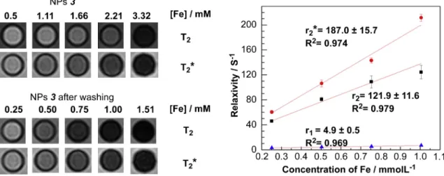
4,
Fig. 3.Left:T2andT2* weighted MR images of aqueous solutions of the NPs3(upper series) or the NPs3after being washed with water using NanoSep (lower series); right: curves of relaxivity against concentration at 3T (red circles,r2*; black squares,r2; blue triangles,r1). The concentration of Fe was measured by ICP-MS.Fig. 4.Structure of64Cu(DTCBP)2, the bisphosphonate derivative used to bind64Cu to NPs (a); andin vivoPET/CT images (maximum intensity projection) of a normal young C57BL/6 mouse after intravenous injection with64Cu radiolabelled1, showing dynamic biodistribution of NPs, 0e15 min (b) and 105e120 min (c).
were detected in the supernatant of
1, which suggested a stable layer of Al(OH)
3consistent with the excellent radiolabelling results above.
As expected, these Al(OH)
3-coated NPs displayed essentially the magnetic properties of the cores and were active as contrast agents in MR imaging, showing a darkening contrast on the
T2or
T2* weighted MR images of solutions of NPs as a result of shortening transverse relaxation time of water molecules (Fig. 3). The trans- verse relaxivity property (r
2) of the NPs strongly depends on the shell thickness, weakening dramatically as the Al(OH)
3shell thickness increases (
3and
4), consistent with previous reports that relaxivity is proportional to the volume fraction of magnetic ma- terials
[26]. Fe3O
4@Al(OH)
3samples
3and
4displayed higher relaxivities (r
1and
r2) after washing off the unstable layer; for example,
r2was improved from 81.6 to 121.9 m
M1s
1for NPs
3, and from 60.5 to 116.6 m
M1s
1for NPs
4at 3T magnetic
field (Fig. 3,
Table S2). For the samples with a stable layer (1), no obvious improvement was observed on the relaxivity properties after washing.
In vivo
PET/CT and PET/MR imaging results showed that both
1and
3labelled with [
18F]-
fluoride exhibited a quick uptake, seen by PET imaging, in the spleen and liver after intravenous injection
viatail vein, despite their small hydrodynamic size of 21 nm in saline solution. Accumulation of NPs in the spleen and liver was evident also by MR, in a signi
ficant darkening contrast in the corresponding areas on MR images in
Fig. S8. The combined im-ages show that the magnetic cores and the radioactivity co- localise in the early period after injection but separate with time. Due to the unstable aluminium hydroxide shell, [
18F]-
fluo- ride radioactivity was gradually released from NPs
3 in vivo,resulting in a signi
ficant bone uptake increasing with time.
Consistent with
in vitrostudies presented above,
1NPs showed a better
in vivostability and slower, but still signi
ficant, release of [
18F]-
fluoride radioactivity (Fig. S9). By contrast, intravenously injected free [
18F]-
fluoride, without NPs, is immediately accu- mulated in bone and not in liver and spleen. PET/CT imaging a normal mouse with
1-
64Cu(DTCBP)
2showed a similar bio- distribution to that of
18F radiolabeled NPs (Fig. 4). All intrave- nously administered NPs were taken up by the spleen and liver within 2 h post-injection, and showed no sign of ef
flux of radio- label from these organs, in contrast to the
18F-labelled particles. By comparison, PET/CT using ionic
64Cu (
64CuCl
2,
Fig. S10) showeduptake dominated by liver and kidneys but not spleen. This con-
firms that
64Cu radioactivity attached to NPs
viabisphosphonate groups co-localises with the magnetic cores and is not rapidly detached from the NPs. The quick clearance of
1NPs by the liver and spleen was not unexpected, as the
in vivobehaviour is determined not only by their hydrodynamic size but also by sur- face properties (surface chemistry and potential)
[27,28].hydrophobic iron oxide-based magnetic NPs into hydrophilic par- ticles stabilised by an Al(OH)
3shell. The features of this system, including high ef
ficiency on
18F or
64Cu labelling, excellent colloidal stability, small hydrodynamic size, good transverse relaxivity and controllable surface potential, suggest that materials based on Fe
3O
4@Al(OH)
3have potential applications as bimodal contrast agents in PET/MRI imaging. A slow release of
18F from NPs was observed
in vivo, whereas PET imaging with64Cu radiolabelled NPs showed no loss of radioactivity from the initially targeted organs (liver, spleen). The ability to derivatise the surface with radiolabels and bisphosphonate groups suggests applications in molecular imaging. Barriers to
in vivouse due to toxicity should be low, because of the established use of Al(OH)
3as adjuvants in vaccines.
The high af
finity to bisphosphonate groups for Al(OH)
3allows us to conjugate these NPs with a range of imaging and therapeutic ra- dionuclides which may be used in conjunction with magnetic im- aging and therapy.
Acknowledgements
Authors thank Drs Alice Warley and Gama Vizcay, at Centre for Ultrastructural Imaging, King
’s College London for TEM, and Mr Andrew Cakebread at King
’s College for ICP-MS measure- ments. RTMR and DM would like to thank EU COST action TD1007 on PET-MRI. The help of László Papp, Sándor Hóbor and Gábor Németh from Mediso is kindly acknowledged. This research was supported by the Centre of Excellence in Medical Engineering funded by the Wellcome Trust and EPSRC under grant number WT088641/Z/09/Z, and the King
’s College London and UCL Comprehensive Cancer Imaging Centre funded by the CRUK (C1519/A10331) and EPSRC in association with the MRC and DoH (WT088641/Z/09/Z) (England), and by the National Institute for Health Research Biomedical Research Centre at Guy
’s and St Thomas
’NHS Foundation Trust and King
’s College London.
PET and SPECT scanning equipment at KCL was funded by an equipment grant from the Wellcome Trust. The views expressed are those of the authors and not necessarily those of the NHS, the NIHR or the Department of Health.
Appendix A. Supplementary data
Supplementary data related to this article can be found at
http://dx.doi.org/10.1016/j.biomaterials.2014.04.004.
References
[1] Safarik I, Horska K, Pospiskova K, Safarikova M. Magnetic techniques for the detection and determination of xenobiotics and cells in water. Anal Bioanal Chem 2012;404(4):1257e73.
[2] Pospiskova K, Safarik I, Sebela M, Kuncova G. Magnetic particles-based biosensor for biogenic amines using an optical oxygen sensor as a trans- ducer. Microchim Acta 2013;180(3e4):311e8.
[3] Safarik I, Pospiskova K, Horska K, Safarikova M. Potential of magnetically responsive (nano)biocomposites. Soft Matter 2012;8(20):5407e13.
[4] Alexiou C. Nanomedicine: innovative applications in medicine. HNO 2013;61(3):197e201.
[5] Kettering M, Winter J, Zeisberger M, Bremer-Streck S, Oehring H, Bergemann C, et al. Magnetic nanoparticles as bimodal tools in magnetically induced labelling and magnetic heating of tumour cells: an in vitro study.
Nanotechnology 2007;18(17).
[6] Tietze R, Lyer S, Duerr S, Alexiou C. Nanoparticles for cancer therapy using magnetic forces. Nanomedicine 2012;7(3):447e57.
[7] Xu C, Xie J, Kohler N, Walsh EG, Chin YE, Sun S. Monodisperse magnetite nanoparticles coupled with nuclear localization signal peptide for cell-nucleus targeting. Chem Asian J 2008;3(3):548e52.
[8] Sandiford L, Phinikaridou A, Protti A, Meszaros LK, Cui X, Yan Y, et al.
Bisphosphonate-anchored PEGylation and radiolabeling of super- paramagnetic iron oxide: long-circulating nanoparticles for in vivo multi- modal (T1 MRI-SPECT) imaging. ACS Nano 2012;7(1):500e12.
[9] Kohler N, Sun C, Fichtenholtz A, Gunn J, Fang C, Zhang MQ. Methotrexate- immobilized poly(ethylene glycol) magnetic nanoparticles for MR imaging and drug delivery. Small 2006;2(6):785e92.
[10] Johannsen M, Thiesen B, Wust P, Jordan A. Magnetic nanoparticle hyper- thermia for prostate cancer. Int J Hyperthermia 2010;26(8):790e5.
[11] Jordan A, Wust P, Fahling H, John W, Hinz A, Felix R. Inductive heating of ferrimagnetic particles and magneticfluids-physical evaluation of their po- tential for hyperthermia. Int J Hyperthermia 1993;9(1):51e68.
[12] Laurent S, Forge D, Port M, Roch A, Robic C, Elst LV, et al. Magnetic iron oxide nanoparticles: synthesis, stabilization, vectorization, physicochemical char- acterizations, and biological applications. Chem Rev 2008;108(6):2064e110.
[13] Berry CC, Wells S, Charles S, Aitchison G, Curtis ASG. Cell response to dextran- derivatised iron oxide nanoparticles post internalisation. Biomaterials 2004;25(23):5405e13.
[14] Jokerst JV, Lobovkina T, Zare RN, Gambhir SS. Nanoparticle PEGylation for imaging and therapy. Nanomedicine 2011;6(4):715e28.
[15] de Rosales RTM, Tavare R, Paul RL, Jauregui-Osoro M, Protti A, Glaria A, et al.
Synthesis of Cu-64(II)-bis(dithiocarbamatebisphosphonate) and its conjuga- tion with superparamagnetic iron oxide nanoparticles: in vivo evaluation as dual-modality PET-MRI agent. Angew Chem Int Ed Engl 2011;50(24):
5509e13.
[16] Jauregui-Osoro M, Williamson PA, Glaria A, Sunassee K, Charoenphun P, Green MA, et al. Biocompatible inorganic nanoparticles for F-18 -fluoride binding with applications in PET imaging. Dalt Trans 2011;40(23):6226e37.
[17] Williamson PA. Strategies for molecular imaging with inorganic nanoparticles.
PhD Thesis. Kings College London; 2011.
[18] Marrack P, McKee AS, Munks MW. Towards an understanding of the adjuvant action of aluminium. Nat Rev Immunol 2009;9:287e93.
[19] Xu Z, Hou Y, Sun S. Magnetic core/shell Fe3O4/Au and Fe3O4/Au/Ag nano- particles with tunable plasmonic properties. J Am Chem Soc 2007;129(28):
8698e9.
[20] Sun SH, Zeng H, Robinson DB, Raoux S, Rice PM, Wang SX, et al. Monodisperse MFe2O4(M¼Fe, Co, Mn) nanoparticles. J Am Chem Soc 2004;126(1):273e9.
[21] Blume U, Orbell J, Waltham M, Smith A, Razavi R, Schaeffter T. 3D T (1)- mapping for the characterization of deep vein thrombosis. MAGMA 2009;22(6):375e83.
[22] Violante A, Huang PM. Formation mechanism of aluminum hydroxide poly- morphs. Clays Clay Miner 1993;41(5):590e7.
[23] Li X, Wang D, Zhou Q, Liu G, Peng Z. Concentration variation of aluminate ions during the seeded precipitation process of gibbsite from sodium aluminate solution. Hydrometallurgy 2011;106(1e2):93e8.
[24] Wechsler BA, Lindsley DH, Prewitt CT. Crystal-structure and cation distribu- tion in titanomagnetites Fe3-xTixO4. Am Miner 1984;69(7e8):754e70.
[25] Miretzky P, Fernandez Cirelli A. Fluoride removal from water by chitosan derivatives and composites: a review. J Fluor Chem 2011;132(4):231e40.
[26] Vuong QL, Berret J-F, Fresnais J, Gossuin Y, Sandre O. A universal scaling law to predict the efficiency of magnetic nanoparticles as MRI T2-contrast agents.
Adv Healthc Mater 2012;1(4):502e12.
[27] Phillips MA, Gran ML, Peppas NA. Targeted nanodelivery of drugs and di- agnostics. Nano Today 2010;5(2):143e59.
[28] Li S-D, Huang L. Pharmacokinetics and biodistribution of nanoparticles. Mol Pharm 2008;5(4):496e504.