Possibilities for protecting cardiac and vascular function against ischaemia-reperfusion injury
Ph.D. Thesis
Enikő Kriebitzsch-Barnucz
Doctoral School of Basic Medicine Semmelweis University
Supervisor: Prof. Gábor Szabó, M.D., med. habil.
Official reviewers:
Zsuzsanna Miklós, M.D., Ph.D.
Tamás Csont, M.D., med. habil.
Head of the Final Examination Committee:
Prof. Emil Monos, M.D., D.Sc.
Members of the Final Examination Committee:
Tamás Ivanics, M.D., med. habil.
Csaba Csonka, M.D., med. habil.
Budapest
2017
2
Contents
1. List of Abbreviations………...5
2. Introduction………..8
2.1. History of operative treatment options for ischaemic heart disease….……..9
2.2. The history of interventional cardiology………...…….…10
2.3. Ischaemia–reperfusion injury of the myocardium and vessels.………....….12
2.4. Myocardial reactive oxygen species generation during ischaemia– reperfusion.……… ………....14
2.4.1. ROS in an ischaemic heart....15
2.4.2. ROS in vessels………...16
2.5. Nitric oxide in cardiovascular physiology and pathophysiology…………..17
2.5.1. Decreased NO production…………..18
2.5.2. NO and vascular integrity in organ transplantation………...…19
2.6. Intracellular antioxidants…………20
2.6.1. Non-enzymatic ROS scavengers………...21
2.6.2. Enzymatic ROS scavengers………...21
2.6.3. Oxidative/nitrosative stress and heme oxygenases…………21 2.7. Myocardial prevention………....23
2.7.1. Myocardial preconditioning………... 23
2.7.2. Cardioprotection through early/classical preconditioning………….23
2.7.3. Cardioprotection via delayed/late preconditioning………. 24
2.7.4. Myocardial postconditioning.….…………........24
2.8. Inhibition of prolyl-hydroxylases and the mechanism of hypoxic adaptation ………... .25
2.9. The role of zinc in cytoprotection during ischaemia–reperfusion injury .... 28
3. Objectives ... .31
4. Methods ... ………32
4.1. Effects of prolyl hydroxylase inhibition on vascular function ... 32
4.1.1. Animals………... 32
4.1.2. Preparation of aortic rings………...32
4.1.3. Experimental groups……….. 32
4.1.4. Model of in vitro cold ischaemic storage – warm reperfusion-induced vascular injury………..……… 33
4.1.5. In vitro assessment of vascular function on aortic rings………. 33
4.1.6. Investigation of cold ischaemic storage – warm reperfusion injury on aortic smooth muscle cells in cell culture……… 34
3
4.1.7. Aortic and vascular smooth muscle cell mRNA expression by
quantitative real-time polymerase chain reaction……… 35
4.1.8. Terminal deoxynucleotidyl transferase-mediated dUTP nick end labelling reaction………... 36
4.1.9. Statistical analysis……… 36
4.1.10.Reagents………... 37
4.2. Q50 in the rat models of ischaemia / reperfusion ... 38
4.2.1. Rat model of myocardial I/R injury: surgical preparation of regional I/R………. ... 38
4.2.1.1. Experimental groups………... 39
4.2.1.2. In vivo hemodynamic parameters……….. 39
4.2.1.3. Determination of area at risk and infarct size……… 39
4.2.1.4. Biochemical estimation………... 40
4.2.2. Rat model of heterotopic heart transplantation ... 40
4.2.2.1. Experimental groups………... 40
4.2.2.2. Hemodynamic measurements………. 41
4.2.2.3. Determination of high-energy phosphate levels……… 41
4.2.2.4. Quantitative real-time polymerase chain reaction…………. 41
4.2.2.5. Western blotting……….. 42
4.2.3. Cardiac myocyte protection studies in vitro ... 43
4.2.3.1. Measurement of human matrix metalloproteinase enzyme activity………...43
4.2.3.2. Statistical analysis………...……… 44
4.2.3.3. Reagents……….. 44
5. Results ... 45
5.1. Effects of prolyl hydroxylase inhibition on vascular function ... 45
5.1.1. Endothelium-dependent and endothelium independent vasorelaxation of aortic rings………. 45
5.1.2. Contractile responses of the aortic rings……….. 46
5.1.3. Results of histopathological staining……… 47
5.1.4. Effects of prolyl hydroxylase inhibition on gene expression……….. 48
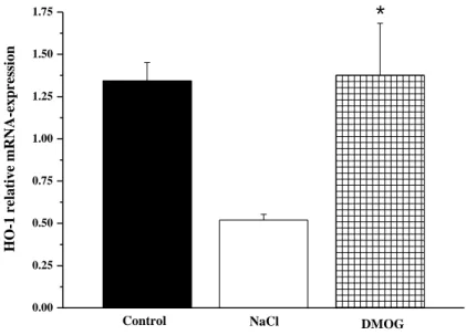
5.1.4.1. Effects of DMOG on HO-1 gene expression in aortic rings.. 48
5.1.4.2. The impact of DMOG on HO-1 gene expression in aortic smooth muscle cell culture………... 49
5.2. The impact of treatment with Q50 on the rodent model of regional and global myocardial ischaemia ... 50
5.2.1. Effects of Q50 post-treatment on regional myocardial ischaemia / reperfusion injury ... 50
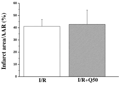
5.2.1.1. Myocardial infarct size………... 50
4
5.2.1.2. Plasma cardiac troponin-T after myocardial infarction……. 51
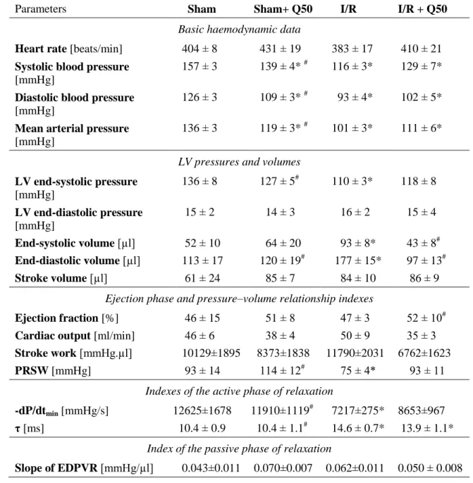
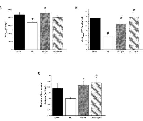
5.2.1.3. Cardiac function after myocardial infarction………. 51
5.2.2. Effect of Q50 pre-treatment on global ischaemia / reperfusion injury ... 54
5.2.2.1. Effect of Q50 on graft function after heart transplantation...54
5.2.2.2. Effect of Q50 on graft myocardial high-energy phosphate contents after heart transplantation……… 56
5.2.2.3. Effect of Q50 on graft gene expression after heart transplantation………. 57
5.2.2.4. Effect of Q50 on graft protein levels after heart transplantation………. 58
5.2.3. H9c2 rat myocardial cells and the post-treatment effect of Q50 after oxidative stress ... 59
5.2.3.1. Cytoprotective effect of Q50 measured using a real-time cell microelectronic sensing technique……….. 59
5.2.3.2. Effect of Q50 on relative HO-1 gene expression in H9c2 rat myocardial cells after H2O2-induced oxidative stress… 60 5.2.3.3. Effect of Q50 on matrix metalloproteinases……….. 61
6. Discussion ... 62
6.1. Mechanism for PHD inhibition by DMOG in the model of cold ischaemia – warm reperfusion injury………... 62
6.2. The effects of Q50 in a rodent models of regional and global myocardial ischaemia………... 66
6.2.1. Effects of Q50 post-treatment on cardiac dysfunction after myocardial infarction……… ... 66
6.2.2. Effects of Q50 pre-treatment on graft dysfunction after heart transplantation ... 67
6.2.3. Mechanism for cardioprotective effects of Q50 against I/R injury ... 67
7. Conclusions ... 70
8. Summary ... 71
9. Összefoglalás ... 72
10. Bibliography ... 73
11. Bibliography of the candidate’s publications ... 84
12. Acknowledgements... 88
5 1. LIST OF ABBREVIATIONS
ACh Acetylcholine
ADP Adenosine diphosphate AMP Adenosine monophosphate ANOVA Analysis of variance
ARNT Aryl hydrocarbon receptor nuclear translocator protein ATP Adenosine triphosphate
Ca2+ Calcium
CA Carbonic anhydrase
cGMP Cyclic guanosine monophosphate
CO Carbon monoxide
CO2 Carbon dioxide
CVD Cardiovascular disease DMOG Dimethyloxalylglycine DNA Deoxyribonucleic acid
dP/dtmax Maximal slope of the systolic pressure increment dP/dtmin Maximal slope of the diastolic pressure decrement ECG Electrocardiography
EDTA Ethylenediaminetetraacetic acid
EDPVR End-diastolic pressure–volume relationship EDV End-diastolic volume
eNOS Endothelial nitric oxide synthase EPO Erythropoietin
FIH Factor inhibiting HIF
GAPDH Glyceraldehyde-3-phosphate dehydrogenase GLUT Glucose transporters
H2O2 Hydrogen peroxide
HO Heme oxygenase
HIF Hypoxia-inducible factor
6 HRE Hypoxia responsible elements iNOS Inducible nitric oxide synthase I/R Ischaemia–reperfusion
KATP ATP-dependent potassium channels KCl Potassium chloride
LDL Low-density lipoprotein LV Left ventricular
LVEDP Left ventricular end-diastolic pressure LVSP Left ventricular systolic pressure MMP Matrix metalloproteinase
MPO Myeloperoxidase
mPTP Mitochondrial permeability transition pore mRNA Messenger RNA
NaCl Sodium chloride NaOCl Sodium hypochlorite
nNOS Neuronal nitric oxide synthase
NO Nitric oxide
NOS Nitric oxide synthase
O2 Oxygen
O2.−
Superoxide anion OCl- Hypochlorite ion
.OH Hydroxyl radical ONOO Peroxynitrite
PCR Polymerase chain reaction
PHD Prolyl hydroxylase domain-containing enzyme PKC Protein kinase C
PKG Protein kinase G PO2 Partial oxygen tension
7 PRSW Preload recruitable stroke work
qRT-PCR Qualitative real-time polymerase chain reaction RNA Ribonucleic acid
RNS Reactive nitrogen species ROS Reactive oxygen species RSS Reactive sulphur species
SDS-PAGE Sodium dodecyl sulphate polyacrylamide gel electrophoresis SEM Standard error of the mean
sGC Soluble guanylyl cyclase SNP Sodium nitroprusside SOD Superoxide dismutase
τ (tau) Time constant of left-ventricular pressure decay TIMP Tissue inhibitor of metalloproteinase
TUNEL Terminal deoxynucleotidyl transferase dUTP nick end labelling VEGF Vascular endothelial growth factor
VHL von Hippel-Lindau tumour suppressor protein VSMC Vascular smooth muscle cells
8 2. INTRODUCTION
Cardiovascular disease (CVD), particularly ischaemic heart disease, is responsible for approximately half of all deaths in the developed world and its treatment places a heavy burden on healthcare systems. The increasing prevalence of CVD can partially be attributed to the ageing society.
The difficulty of reducing ischaemia–reperfusion (I/R) injury and hypoxia is a major problem in cardiology and cardiovascular surgery, as well as being related to the problems that occur during organ transplantation. The aim of cardioprotection during and after ischaemia–reperfusion injury in clinical settings is to reduce infarct size, prevent severe cardiac arrhythmias and increase contractile function of the intact myocardium. Cardioprotection during decreased blood and oxygen supply is an extensively discussed and studied field in basic research: pathophysiology and clinical studies. Therapeutic strategies have improved over recent years; at present, thanks to drug research and operative developments, several strategies that result in a better outcome and prognosis even for patients with complicated disorders are in use.
However, the development and investigation of new cardioprotective drugs is still required because cardiovascular ischaemia-related disorders remain a leading cause of death in the developed world.
It is impossible to point to one pathway in the ischaemia–reperfusion injury.
This thesis therefore focuses on cardiac and vascular protection against ischaemia–
reperfusion injury and attempts to identify potential targets for novel therapeutic options to reduce ischaemia- and reperfusion-related organ damage, loss of function of implanted organs and mortality.
The aim of this study is to describe, using rodent models, the pathophysiological changes to the vascular system and myocardium during and after ischaemia–reperfusion injury. The use of antioxidants and the effect of the hypoxia-inducible factor (Powell et al. 2000) under myocardial or vascular ischaemic conditions were investigated; this may
9
help improve the understanding of the role of these pathways and form the basis for further developments.
2.1. HISTORY OF OPERATIVE TREATMENT OPTIONS FOR ISCHAEMIC HEART DISEASE
In the early development of surgical treatment for ischaemic heart disease, the clinicians made basic observations and performed extracardiac operations for angina. In 1910, Carrel reported the first form of coronary artery bypass (Carrel 1910). Later, operations were performed directly on the heart with the aim of promoting collateral revascularisation of the myocardium from other tissue. One of the pioneers was Beck, who after several years of experimental design created the first cardiopericardiopexy (or pericardial poudrage) (Gage et al. 1958). The first successful experimental arterial coronary artery bypass autograft was performed by Murray in 1954 (Murray et al.
1954). He used an axillary or free autogenous carotid graft using the subclavian artery.
This procedure was the forerunner of the internal mammary artery bypass graft procedure.
Senning reported in 1958 a coronary endarterectomy with direct-vision excision of the plaque followed by patch grafting of the defect in the vessel wall with a split segment of autologous mammary artery (Senning 1958).
From the 1960s, the use of coronary artery bypass grafting was adopted. Favarolo and colleagues used saphenous vein bypass grafts and also used for the first time free interposed saphenous vein autografts (Roncoroni et al. 1973). The first successful heart transplantation was performed in Cape Town by Barnard in 1967 (Barnard et al. 1967).
Along with the development of cardiac surgery, a number of supporting media, solutions and tools were also being developed. One of these was Custodiol® (also called HTK or Histidin-Tryptophan-Ketoglutarat Solution), which was originally developed as a cardioplegia solution but is nowadays used as an organ preservation solution. It is the best alternative to the University of Wisconsin solution (also called UW solution) or other organ transport media. Recently developed solutions such as Custodiol-N® or TiProtec® are more efficient at protecting the myocardium and vessels against ischaemia–reperfusion injury compared with the older generation of preservation
10
solutions or to ordinary physiological salt solution (Garbe et al. 2011). This is particularly important for the situation of cold ischaemia-warm–reperfusion injury;
therefore, it can prolong transport time without severe organ injury (Radovits et al.
2008; Loganathan et al. 2010).
The underlying mechanisms that enable transport media to maintain the physiological function of organs and vessels is the subject of intense investigation. This research has shown that preservation solutions also cause extended tissue injury. This injury is induced by different pathways such as an iron-dependent formation of reactive oxygen species (ROS) or direct cytotoxic effects by histidine (Loganathan et al. 2010). Further research is required to gain further insights into this field.
2.2. THE HISTORY OF INTERVENTIONAL CARDIOLOGY
The first experiments related to interventional cardiology date back several hundred years. One of the first experiments was performed by Harvey, who proved (in 1651) that venous blood flows towards the lungs (Sette et al. 2012). Some years later in 1667, Major became the first to deliver an intravenous injection into a human (Mueller et al.
1995).
The first known cardiac catheterisation was performed by Hales in 1711. During the procedure he inserted a pipe into the vessels of a horse to get into the ventricles, where he measured the pressure with a captured water column. The procedure was named catheterisation by the French physiologist Bernard who also performed other procedures on the heart, such as measurements of left and right ventricular pressure. Chauveau and Marey recorded the pulmonary artery pressure, and they simultaneously registered the aortic and intraventricular pressure (Mueller et al. 1995).
In the last century the field of cardiovascular medicine was characterised by progressive development and refinement of the invasive diagnostic and therapeutic options. New techniques such as cardiac catheterisation, angioplasty and related catheter-based interventions were developed.
11
One of the greatest innovations was Röntgen´s discovery of X-rays, for which he received the Nobel Prize in 1901. This discovery led to experiments in cadavers with intravenous contrast agents enabling Merkel and Jasmin to publish a book of the coronary arteries in 1907 (Mueller et al. 1995).
Forssmann catheterised his own arm with a urethral catheter via the cubital vein and made an X-ray. Furthermore, he documented for the first time a catheter in the right atrium. This led to the rapid development of catheterisation techniques in humans and was completed by documentation with X-ray and contrast agents (Goerig et al. 2008).
Later, diagnostic cardiac catheterisation was introduced by Cournand and Richards and selective coronary angiography by Sones (Cournand et al. 1945).
Further advancements in the field were achieved through the work of Seldinger, who invented a technique for the percutaneous replacement of an access needle with a catheter over guide wires (Seldinger 1957). This technique allowed the insertion of a catheter in different arteries and veins. At present, coronary angiography and percutaneous coronary intervention are mainly based on the insertion of catheters via the femoral or radial artery.
The importance of these discoveries was recognised when the Nobel Prize in Physiology or Medicine was awarded to Cournard, Richards and Forssmann in 1956 for their pioneering work on cardiac catheterisation (Mueller et al. 1995; Sette et al. 2012).
The cardiac catheter has supported the development of basic scientific understanding of cardiac anatomy and physiology and offered a new perspective which cannot be achieved via noninvasive methods.
Work on a range of catheter techniques continued and in the 1970s Gruentzig developed a balloon catheter. He performed the first coronary angioplasty intraoperatively during bypass surgery. Later, balloon dilatation of renal artery stenosis and coronary and coronary angioplasties were performed (Gruntzig et al. 1977; Gruntzig et al. 1978).
The continuous development of cardiological catheter techniques now permits a broad range of functional measurements of the heart. These techniques are used in critical situations such as acute myocardial ischaemic states, and they enable the use of stents and drug-eluting stents or the implantation of heart valves. Because of the economic benefits and relative low complication rates, the number of cardiological interventions is
12
rapidly growing. In contrast, the number of cardiac surgeries is decreasing. This shows that in practice only more complicated cases are being surgically treated.
2.3. ISCHAEMIA–REPERFUSION INJURY OF THE MYOCARDIUM AND VESSELS
A compromised blood flow deprives tissues and organs of oxygen and metabolites, such as glucose. This state of deprivation is called ‘ischaemia’. Ischaemia may require cardiological interventions and cardiac surgical procedures, which are complex and require myocardial reperfusion after finalisation. However after the onset of myocardial reperfusion, many reactions, such as oxidant generation, activation of neutrophil granulocytes and their adhesion to the coronary vascular endothelium or cell damage due to calcium dyshomeostasis, are induced (Vinten-Johansen et al. 2005).
The injury generated by ischaemia and subsequent reperfusion is called ischaemia–
reperfusion injury. The ischaemia–reperfusion-related loss of myocardium due to cell death remains a major issue in cardiovascular protection. In the past decades, various mechanisms for cardioprotection have been discussed and intensively scrutinised.
However, it is impossible to single out just one target for cardioprotection because I/R involves different signalling pathways in the mechanisms of tissue injury.
Organs with high O2-uptake (brain, heart and kidney) need sufficient, continuous blood flow. Blood flow and oxygen supply can be reduced by various pathophysiological conditions, including atherosclerosis, stenosis, reduced blood pressure, shock syndrome and the perioperative period. If the blood flow is insufficient or completely blocked, the organ is subject to hypoxia or anoxia, respectively. The results are tissue injury, cell death and irreversible organ damage when sufficient blood flow is not restored.
The damage caused by reduced oxygen supply can be regional (e.g. because of occluded arteries or myocardial infarction) or global (e.g. via interruption of organ blood flow during organ transplantation or organ transportation). During organ transplantation, two types of ischaemia can be distinguished: warm and cold ischaemia. Warm ischaemia is the period during which a tissue or organ remains at body temperature after its blood supply has been reduced or cut off but before it is cooled or is reconnected to the blood
13
supply. In clinical terminology this means the following: (1) an ischaemic condition during implantation from the removal of the donor organ from ice until the beginning of reperfusion and (2) ischaemia during organ recovery from the beginning of cross- clamping until perfusion (Halazun et al. 2007). Cold ischaemia is the period between the cooling of a tissue or organ after its blood supply has been reduced or cut off and the time it is warmed by having its blood supply restored. This kind of ischaemia can occur while the organ is still in the body or after it has been removed from the body if it is to be used for transplantation.
If the ischaemic period is short (e.g. a brief spasm of a coronary artery, a rapidly lysed thrombus, the rapid ending of exercise or psychic stress or after coronary bypass surgery) the myocardium will completely survive; however, this depends on the duration and degree of ischaemia. The period of dysfunction is called ‘stunning’, and this results in regional wall motion abnormality, decreased systolic pump function and ECG abnormalities. However, a stunned myocardium is able to react to inotropic stimuli such as dopamine, dobutamine or isoproterenol (Kloner et al. 2001). The role of free radicals in stunning has been investigated in several studies, and it has been shown that pretreatment with enzymes that scavenge O2-derived free radicals, such as superoxide dismutase and catalase, can inhibit the process of stunning (Bolli et al.
1989).
Ischaemia–reperfusion injury involves both the myocardium and the coronary endothelium–smooth muscle; therefore, protection of the heart should involve these two aspects (He 2005). Myocardial damage after ischaemia–reperfusion injury is characterised by histomorphological changes such as cell swelling, disrupted ultrastructure, contraction bands, deposition of calcium phosphate granules and inflammatory cell responses (Tsao et al. 1990).
To improve the prognosis for patients suffering from ischaemia-related diseases, further research is warranted to answer the multitude of unanswered questions.
14
2.4. MYOCARDIAL REACTIVE OXYGEN SPECIES GENERATION DURING ISCHAEMIA–REPERFUSION
Atoms, molecules or ions with unpaired electrons are called free radicals. These radicals are most often derived from oxygen (reactive oxygen species, ROS), nitrogen (reactive nitrogen species, RNS) or sulphur (reactive sulphur species, RSS) and are prone to react with other molecules because of their high reactivity.
In cells, energy is generated under physiological conditions in the form of adenosine triphosphate (ATP) by the degradation of glucose during mitochondrial oxidative phosphorylation; this process depends on oxygen. Within the mitochondrial electron transport chain, electrons are transported to molecular oxygen, which is finally reduced to water. As a result, energy is conserved in ATP by mitochondrial synthesis.
If the blood supply of an organ is diminished or completely blocked, this reduces the oxygen supply and subsequently oxidative phosphorylation. When the oxygen supply is insufficient, mitochondria use the available oxygen for the excess generation of mitochondrial ROS. Ongoing hypoxia or ischaemia results in ROS production and causes irreversible oxidative damage to mitochondria followed by cell swelling and cell death (Kiss et al. 2012).
In addition, ROS are generated under normal conditions in cellular metabolism. They not only have detrimental effects but also possess important functions during physiological processes such as cell signalling, apoptosis, gene expression and ion transportation. However, when ROS are generated in excess during pathophysiological conditions (e.g. ischaemia, sepsis or diabetes mellitus), they not only cause mitochondrial damage but also deleterious effects on many molecules including proteins, lipids, RNA and DNA. The process in which ROS damages macromolecules is defined as oxidative stress. Intact cells have the capacity to defend their structures against ROS-induced damage through the use of intracellular enzymes such as superoxide dismutases. The pathophysiological role of oxidative stress is diverse and has frequently been discussed in the literature on inflammatory diseases (e.g. Crohn’s disease), CVD, cancer and ageing (Lu et al. 2010).
15 2.4.1.ROS IN AN ISCHAEMIC HEART
The generation of ROS in an ischaemic heart is a heterogeneous process that depends on the level of tissue oxygenation, which is generally governed by collateral blood flow (Heyman et al.). Even in the case of severe ischaemia, the heart tissue still retains some oxygen, and this small amount of oxygen is sufficient to generate free oxygen radicals (Murphy et al. 2008). ROS generation also has an important role in the protecting signalling pathway of preconditioning (Chen et al. 1995).
During ischaemia, anaerobic glycolysis and high-energy phosphates (tissue ATP, creatine phosphate) provide energy to cardiomyocytes. However, these reserves of high- energy phosphates rapidly decrease, resulting in the accumulation of adenosine diphosphate (ADP). It is known that late in the reversible phase of ischaemia the adenine nucleotide pool radically decreases (Jennings et al. 1985; Kloner et al. 2001).
The ischaemic myocardium switches to anaerobic glycolysis, and the intracellular pH rapidly decreases. To restore intracellular homeostasis, the Na+/H+ exchanger pumps out a high amount of H+, resulting in an excessive Na+ inflow. When the capacity of ATPases (such as Na+/K+ ATPase, ATP-dependent Ca2+ reuptake and active Ca2+
excretion) are reduced, this leads to intracellular Ca2+ overload (Sanada et al. 2004).
Ca2+ overload or ROS are able to open mPTP (mitochondrial permeability transition pores) on the mitochondrial membrane, which leads to H+ influx and loss of mitochondrial membrane potential and results in mitochondrial swelling and the induction of apoptotic pathways (Sanada et al. 2004).
The recovery of arterial blood flow to the ischaemic myocardium re-establishes a sufficient oxygen supply, enabling aerobic cell metabolism, followed by the induction of salvage mechanisms. In the first phase, increased blood flow is observed in the coronary arteries (reactive hyperaemia), which leads to equalisation of the metabolic debt developed during ischaemia. Approximately 20 min after reperfusion, the baseline arterial blood flow is re-established. During reperfusion, the blood flow and oxygen supply of the tissue are restored, which results in a large burst of ROS. One reason for this is the ample amount of mitochondria in the myocardium, which not only generate ROS (by the mitochondrial electron transport chain) but also a major target of the ROS- induced damage.
16
In the past few decades, several studies have confirmed the positive effects of antioxidant treatments during ischaemia–reperfusion injury of the heart. However, the literature also includes controversial reports on the effectiveness of antioxidant strategies after ischaemia–reperfusion injury of the heart (Bolli et al. 1989; Chen et al.
1995; Powell et al. 1997; Tang et al. 1997; Carden et al. 2000; Korichneva 2006; Lu et al. 2010).
2.4.2.ROS IN VESSELS
Arteries play a key role in the regulation of organ blood flow. They have a thin inner lining, a single-layer endothelium, which is known not only to be a physical barrier but also acts as an important regulator of vascular tone. Outside this, the tunica media is composed of smooth muscle cells, which are the effectors in the regulation of blood flow. Fibroblasts, pericytes, dendritic cells, endothelium (from the vasa vasorum), adipocytes and filaments of innervating adrenergic nerves form the outer layer of the vessel, the adventitia. Some authors, such as Stenmark et al., have proposed that the adventitia not only has a barrier function but also plays a complex role in the regulation of vascular physiology (Stenmark et al. 2013).
ROS such as hydrogen peroxide, hypochlorite and superoxide anions are involved in the pathogenesis and progression of various cardiovascular diseases. Under pathophysiological conditions, activated neutrophil granulocytes secrete myeloperoxidase (MPO), an enzyme which in the presence of chloride, converts hydrogen peroxide (H2O2) into hypochlorite. Hypochlorite and H2O2 injure the endothelium and myocardium. This leads to endothelial and myocardial dysfunction, and subsequently contributes to processes such as atherosclerosis and reperfusion injury (Sand et al. 2003).
During pathophysiological conditions, excessive ROS release leads to a reduction in the production of nitric oxide (NO) in the endothelium. As a result, its ability for vasorelaxation is diminished.
17
2.5. NITRIC OXIDE IN CARDIOVASCULAR PHYSIOLOGY AND PATHOPHYSIOLOGY
NO is a polyvalent molecule with a wide range of biological effects. It is a gaseous molecule without charge; it has an important signalling role, and it can easily diffuse between compartments without the need for a transport system. In cardiovascular biology and pathology, the important functions of this molecule are vasodilatation, the regulation of platelet adhesion, involvement in vascular remodelling and mediating cell growth and apoptosis (Furchgott 1983; Postovit et al. 2005).
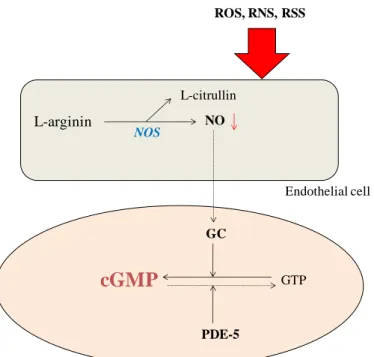
NO is synthesised by nitric oxide synthase (NOS) (Figure 1) and has three isoforms:
endothelial NOS or eNOS is constitutively expressed by the endothelium; neuronal NOS or nNOS is mainly produced by neuronal tissues and inducible NOS or iNOS, which is produced as a response to inflammation in vascular smooth muscle cells and inflammatory cells and results in a large, uncontrolled burst of NO (Zebger-Gong et al.
2010). Both eNOS and nNOS are calcium dependent but iNOS is not.
In the cardiovascular system NO serves as a substrate for protein S-nitrosylation or nitration. In addition, further effects of NO are mediated by the activation of soluble guanylate cyclase (sGC) and the subsequent generation of cyclic guanosine monophosphate (cGMP). Cellular functions are regulated by cGMP binding to cyclic nucleotide phosphodiesterases and by activation of protein kinase G (PKG). PKG is involved in the regulation of the myocardium via regulation of contraction. In the vessel wall, PKG modulates the vascular tone, endothelial permeability and vascular proliferation (Kass et al. 2007).
18
Endothelial cell
Vascular smooth muscle cell
L-arginin NO
L-citrullin
GC
cGMP GTP
PDE-5
ROS, RNS, RSS
NOS
Figure 1. Synthesis of NO. NOS induces synthesis of NO from L-arginine. This leads to an elevated guanylate cyclase level in the vascular smooth muscle cell, which causes endothelium-dependent vasorelaxation through increased intracellular cGMP levels.
During oxidative or nitrosative stess by ROS, RNS or RSS, NO production is reduced followed by a decrease in the cGMP level, which results in reduced vasorelaxation and endothelial dysfunction.
NO reacts with the superoxide anion (O2.−
) under pathophysiologycal conditions; this leads to the formation of peroxynitrite (ONOO−), which causes nitration of proteins, lipid peroxidation and cytotoxicity (Motterlini et al. 2002). Overproduction of NO has been identified as a potent cytotoxic agent against infection, inflammation and cancer (Nathan 1997; Motterlini et al. 2002). The Ca2+-independent isoform of NOS, iNOS, is activated under stress conditions caused by oxygen free radicals, endotoxins or cytokine release (Ca2+-independent isoform), causing excessive NO production (Motterlini et al.
2002).
19 2.5.1.DECREASED NO PRODUCTION
Cardiovascular diseases and pathophysiological conditions associated with ROS excess, such as during hypertension, atherosclerosis, diabetes mellitus, cardiac hypertrophy, heart failure, ischaemia–reperfusion injury or stroke lead to a reduced NO production.
Increased .O2−
levels inactivate NO leading to endothelial dysfunction (Paravicini et al.
2008). Consequently, the effects of decreased NO production are smooth muscle contraction, smooth muscle proliferation, platelet aggregation, increased oxidation of LDL, increased endothelin production, monocyte and platelet adhesion and increased expression of adhesion molecules.
2.5.2.NO AND VASCULAR INTEGRITY IN ORGAN TRANSPLANTATION
NO is important for vascular homeostasis. It helps to maintain the critical balance between endothelium-derived relaxation and contraction factors. When this balance is disrupted, the vasculature is predisposed to vasoconstriction, leukocyte adherence, platelet activation, mitogenesis, pro-oxidation, thrombosis, impaired coagulation, vascular inflammation and in heart transplant patients cardiac allograft vasculopathy (Colvin-Adams et al. 2013). In the 1970s and 80s, there was intensive investigation of NO and its central role in vasorelaxation (Furchgott 1983).
Vascular integrity is a major problem, e.g. during bypass surgery or percutaneous coronary intervention and in general in organ transplantation. The short- and long-term outcomes of organ transplantations (e.g. kidney, liver and heart) are mainly determined by an efficient, continuous blood flow (Woestenburg et al. 2008; Colvin-Adams et al.
2013).
In the initial period after reperfusion, severe endothelial cell dysfunction rapidly occurs and can develop without morphological alteration of cell injury. Those morphological alterations that accompany reperfusion after prolonged ischaemia generally cause cell swelling, a loss of pinocytic vesicles, lifting of the endothelial cells from the underlying basement membranes and the attachment of activated neutrophil granulocytes to the endothelial cell surface. Profound changes in the production of ROS and NO are observed early after reperfusion.
20
After global or regional tissue ischaemia–reperfusion injury, long-term tissue survival is mainly dependent on the extent of the early vascular injury. In general, after organ transplantation, the most important factors responsible for reduced graft survival are occlusive vascular changes. These changes occur because of progressive proliferation of intima, also referred to as allograft arteriosclerosis or transplant vasculopathy. The function of the graft mainly depends on NO provided by intact endothelial cells (Zebger-Gong et al. 2010).
Key factors which determine vascular integrity in the early phase are partly dependent on the organ donor. For live organ donation, the age of the organ donor and pre-existing diseases (diabetes mellitus, hypertension, atherosclerosis) play an important role. Brain death of the organ donor is also problematic because it leads to an excessive cytokine release, which can initiate a diffuse, generalised inflammatory response and severe endothelial injury. In addition, the method of transport and subsequent storage of the transplant need to be considered; transport media cause cold ischaemia. After clamping the blood supply, the subsequent perfusion with a cold preservation solution leads to the iron-dependent formation of ROS or directly to cytotoxic effects, e.g. by histidine (Loganathan et al. 2010). These days, transportation methods and duration have been extended because of the year-on-year decrease in the number of organ donors, which makes it necessary to include matching donors from an extended region.
2.6. INTRACELLULAR ANTIOXIDANTS
Biological intracellular antioxidants are naturally occurring molecules that are able to protect cells and biological structures from uncontrolled oxidant injury. This injury can be provoked by activated oxygen species or free radicals. The reduction of free radicals and ROS is mainly based on the oxidation of endogenous antioxidants by scavenging and reducing molecules (Chaudiere et al. 1999; Squier 2001).
21 2.6.1.NON-ENZYMATIC ROS SCAVENGERS
Non-enzymatic scavengers are mainly compounds of oxidising free radicals, activated oxygen species, or physical quenchers of excited species such as singlet oxygen, hypochlorous acid or hypervalent iron species (Fe2+, Fe3+). The kinetics of scavenging reactions are very fast. The free radical products of scavengers decay through dismutation, recombination or reduction by secondary scavengers (Chaudiere et al.
1999).
2.6.2.ENZYMATIC ROS SCAVENGERS
The elimination of extremely oxidising species, such as O2.− and hydroxyl (OH.) radicals, is mainly performed by superoxide dismutases (SODs) (Chaudiere et al. 1999).
The hydroxyl radical is formed from superoxide by the Haber-Weiss and Fenton reactions and reacts with phenylalanine to form hydroxylated derivates (Kloner et al.
2001).
The most common isoform is Zn2+/Cu2+ (zinc and copper)-SOD (SOD-1) accounting for approximately 50–80% of all SODs in the vascular smooth muscle cells. This is mainly located in the nucleus and cytosol but is absent from mitochondria. In contrast, Mn2+
(manganese)-SOD (also called SOD-2) is abundant in mitochondria. The third form, SOD-3, is anchored to the extracellular matrix (Mendez et al. 2005). SOD-3 has a role in primarily protecting the brain and lungs against oxidative stress.
2.6.3.OXIDATIVE/NITROSATIVE STRESS AND HEME OXYGENASES
Nitrosative stress is similar to oxidative stress. Cells are able to respond to increased ROS and RNS through adaptation or resistance to toxicity, depending on the severity of the nitrosative and/or oxidative stress.
NO plays an important role in cytoprotection against nitrosative and oxidative injury.
NO and NO-related species induce heme oxygenase (HO) expression and activity, particularly in vascular cells. Cells pretreated with different NO-releasing agents increase their resistance to H2O2-mediated cytotoxicity while HO is highly activated.
NO-mediated activation of HO-1 during a stress response has still not been completely explored and needs further investigation.
22
During reperfusion injury, activated neutrophil granulocytes can generate ROS. The production of hypochlorite can cause endothelial dysfunction because it can react with amino acids or proteins. This dysfunction manifests as a reduced response to vasodilative agents such as acethylcoline (Sand et al. 2003; Zhang et al. 2004).
HO has three isoforms. The first, HO-1, is a 32-kDa protein that can be induced by numerous stimuli such as ROS, heavy metals, oxidants, hypoxia. HO-1 plays a pivotal role in vascular function under conditions of increased ROS generation. It is inducible and is involved in the oxidative stress response, and it mediates protective effects. HO catalyses the breakdown of heme to equimolar amounts of biliverdin, iron and carbon monoxide (CO). Biliverdin is reduced to bilirubin by bilirubin reductase, and the free iron is either used in intracellular mechanisms or sequestered in ferritin. Because of its antioxidant property, bilirubin is able to protect cells against peroxynitrite-mediated apoptosis. In addition, it is able to suppress oxidant-induced microvascular leukocyte adhesion and ameliorate post-ischaemic myocardial function. High to normal serum bilirubin levels are inversely related to atherogenic risk, possibly because of inhibitory effects against low-density lipoprotein oxidation and the scavenging of oxygen radicals (Idriss et al. 2008). CO acts as a cellular messenger and has been implicated in vascular tone regulation and neurotransmission. CO acts on vasculature in a manner similar to that of nitric oxide by increasing intracellular cGMP levels (Morita et al. 1995).
Iron released during heme catabolism is reported to have a number of effects. It has been suggested that increased levels of ferritin reduce the cellular oxidant potential by further decreasing the cellular concentration of free iron. On the other hand, ferrous iron has a cytotoxic potential through the generation of ROS. However, these negative effects are negligible in comparison with the cytoprotective potential of bilirubin and CO.
In addition to cellular processes, nutritional deficiencies can also weaken antioxidant protection. However, it should be emphasised that there are many situations of oxidative stress in which an increased intake of elements such as selenium, copper, zinc or manganese will not improve the antioxidant status; this is because these processes are
23
regulated by other factors and cannot be influenced by the nutritional intake of these elements (Chaudiere et al. 1999).
2.7. MYOCARDIAL PREVENTION 2.7.1.MYOCARDIAL PRECONDITIONING
Cardiologists reported the so-called ‘warm up phenomenon’ in the 1980s. They observed that patients who had experienced at least one episode of prodromal angina pectoris before unstable angina or acute myocardial infarction clinically presented with less variation in ST-segments, less cardiac dysfunction and a smaller infarcted area (Jaffe et al. 1980). Following the experiments of Murry, this phenomenon became known as ischaemic preconditioning. In 1986 Murry et al. published results on an experimental model showing the beneficial effect of short ischaemic periods before the onset of a 40-min myocardial coronary occlusion (Murry et al. 1986). Numerous studies confirmed that the cardioprotective effects of ischaemic preconditioning are not present only during myocardial stunning and global or regional acute ischaemia; beneficial effects also appear in chronic cardiac disorders such as hibernating myocardium, contractile dysfunction or myocardial remodelling (Sanada et al. 2004).
Ischaemic preconditioning is a biphasic phenomenon consisting of an early and a late phase. The early phase marks the period starting within minutes following exposure to the stimuli and lasts only 1–2 h. The second window of preconditioning, the late phase, develops more slowly (after 12–24 h) but lasts 3–4 days. The protective effects of acute preconditioning are protein-synthesis independent, while the effects of delayed preconditioning requires protein synthesis (Carden et al. 2000).
2.7.2.CARDIOPROTECTION THROUGH EARLY/CLASSICAL PRECONDITIONING
The potent but short-term protective effect from classic preconditioning is not entirely understood. It has been reported that the early phase developed through rapid posttranslational modification of pre-existing proteins through a series of signalling cascades. In addition, it has been suggested that multiple signal transduction pathways
24
converge on the mitochondria, either preserving ATP synthesis or preventing the onset of mPTP formation after reperfusion or both (Ferdinandy et al. 2007).
2.7.3.CARDIOPROTECTION VIA DELAYED/LATE PRECONDITIONING
The late preconditioning phase is mediated by gene expression and the subsequent synthesis of cardioprotective proteins. This mechanism involves the redox-sensitive activation of transcription factors through the protein kinase C (PKC) and tyrosine kinase signalling pathways that are also involved in the early phase of preconditioning.
Furthermore, the expression of protective mediators such as HO, heat shock proteins, vascular endothelial growth factor (VEGF) and erythropoietin (EPO) is increased after hypoxic preconditioning and plays an important role in protection against tissue injury.
During the late phase of ischaemic preconditioning, these protective mediators are mainly regulated by hypoxia-sensing mechanisms through the stabilisation of HIF (Powell et al. ; Heyman et al. 2011).
2.7.4.MYOCARDIAL POSTCONDITIONING
It has been demonstrated in several studies that brief periods of reperfusion alternating with re-occlusion applied during the very early minutes of reperfusion can protect the myocardium against extended ischaemia reperfusion injury (Zhao et al. 2003).
Generally, postconditioning has a protective effect not only on the cardiomyocytes, but it also reduces infarct size and apoptotic changes and protects the endothelium (Vinten- Johansen et al. 2005; Sanada et al. 2011). In contrast to several smaller trials reported previously, some human studies found no significant effects of ischaemic postconditioning on infarct size or secondary study outcomes (Hahn et al. 2013;
Limalanathan et al. 2014).
25
2.8. INHIBITION OF PROLYL-HYDROXYLASES AND THE MECHANISM OF HYPOXIC ADAPTATION
Hypoxia means an inadequate supply of oxygen in cells (Carden et al. 2000). Mammals have an oxygen sensing mechanism that helps to adapt cells to hypoxia by increasing cell respiration or blood flow (Eltzschig et al. 2011). Organs and areas within an organ have a variable partial pressure of oxygen (PO2). This pressure is mostly in the range 20–45 mmHg. The kidney medulla, bone marrow and the intrauterine foetal compartment normally have a lower PO2 of 10–25 mmHg. In some solid tumours, PO2
can be lower than 1 mmHg. During evolution, certain cell groups differentiated to sense O2 tension; these cells can be found in the carotid body, pulmonary artery, and adrenal chromaffin cells (Aragones et al. 2009).
During hypoxia the transcription factor, hypoxia-inducible factor (HIF-1), becomes activated. HIF-1 is a heterodimeric transcription factor consisting of the consecutively expressed HIF-β (aryl hydrocarbon receptor nuclear translocator protein or ARNT) and a regulatory HIF-α subunit (mainly regulated post-translationally). After dimerisation, the α and β subunits of HIF regulate an overlapping but a distinct set of genes with HIF-2α and HIF-3α. The HIF complex regulates a variety of genes with biological functions including vessel growth (VEGF), vasodilatation (NOS), HO-1, oxygen transport, and metal and energy metabolism to cell fate decisions (e.g. EPO, glucose transporters (GLUT) and carbonic anhydrase (CA)). The target genes mediate adaptive responses to hypoxia/ischaemia at organism, organ and cellular levels (Czibik 2010).
The HIF-α subunit does not sense O2 directly; this activity is performed by the prolyl- hydroxylase domain (PHD) proteins and a single asparaginyl-hydroxyalse, known as factor inhibiting HIF (FIH) (Aragones et al. 2009). These enzymes differ in their affinity for O2. FIH remains active at reduced O2 tensions, when PHDs have already lost their activity. PHD and FIH are 2-oxoglutarate-dependent iron (II)-dioxygenases that use one of the atoms in an O2 molecule to hydroxylate prolyl- or asparagyl-residues, respectively. The second atom of the O2 molecule is used to convert 2-oxoglutarate to carbon dioxide (CO2) and succinate. Iron, maintained in a reduced state by ascorbate, is a necessary cofactor. PHDs hydroxylate N-and C-terminal prolyl residues of the
26
α-subunit. When the prolyl residues are hydroxylated under normal oxygen tension, HIF-α subunits are recognised by the von Hippel-Lindau (VHL) protein in the multiprotein E3 ubiquitin ligase complex and will be ubiquitinated and degraded by the proteasome. When the tissue O2 supply drops, PHD and FIH become progressively inactive, resulting in stabilised (elevated) levels of transcriptionally active HIF complexes (Figure 2).
Three isoenzymes of prolyl 4-hydroxylases specific to HIF-1α have been described in mammals: PHD1, PHD2 and PHD3; these have homology in the C-terminal catalytic domain (Siddiq et al. 2007).
The PHD1 protein is localised in the nucleus and is constitutively expressed and stimulates cell proliferation. Hypoxia does not affect PHD1 gene expression, but it is induced by oestrogen. It is highly expressed in the testis and can be found at low levels in the kidney, liver and heart.
The PHD2 protein is located mainly in the cytoplasm, with lower levels in the nucleus.
Its expression can be regulated by hypoxia and hypoxia mimetics. Basal expression levels are high in the heart and moderate in the brain. A hypoxia-responsible element has been found in the PHD2 gene in humans. In rats, analysis of PHD2 mRNA expression levels in different organs following hypoxia showed reduced levels in the brain but no changes in the heart and kidney (Metzen et al. 2005; Willam et al. 2006).
PHD3 is localised in the nucleus and cytoplasm and its expression can be regulated by hypoxia or hypoxia mimetics.
In the cardiovascular system, PHD2 is the predominant isoform and its expression is induced by hypoxia or hypoxia mimetics such as desferroxamine and cobalt (II) chlorite and by pharmacological PHD inhibitors (such as dimethyloxalylglycine, DMOG) (Czibik 2010).
If oxygen tension is reduced, PHD becomes less active and HIF-α accumulates in the cytosol, and HIF-αβ-heterodimers are formed and translocate into the nucleus. There
27
they bind to the promoter region of genes known as hypoxia-responsive elements (Humphrey et al.).
PHD-HIF-Pathway
HIFα—Pro—OH
Normal O2tension Low O2tension
O2 PHD
Proteasomal degradation
HIFα HIFβ
Hypoxia tolerance regulation of target gene pathways;
e.g. iNOS, GLUT-1, CA-9, VEGF O2 or DMOG treatment
Figure 2. Regulation of HIF via the O2-dependent prolyl-hydoxylase domain containing enzymes under normal oxygen tension and under hypoxic conditions. In normoxia (left side), PHD hydroxylates a specific proline residue that directs the degradation of constitutively synthetised HIF-1α. During hypoxic conditions (right side), inhibition of hydroxylation leads to an increase in HIF-1α protein levels; HIF heterodimers are then formed and this results in hypoxia tolerance via binding to DNA hypoxia-responsive elements.
The importance of HIF in cardiac ischaemic conditions was suggested when it was discovered that HIF-1α in animal infarct models improves myocardial perfusion and left ventricular function. Furthermore, the overexpression of cardiac HIF-1α reduces myocardial infarct size and promotes post-ischaemic function and capillarisation. This suggests that HIF-1α activation/stabilisation is beneficial in cardiac ischaemic syndromes (Czibik 2010). Additionally, it was reported that PHD inhibition (with FG- 2216) did not reduce infarct size, but improved left ventricular function and prevented remodelling (Philipp et al. 2006).
28
The mechanisms that explain how HIF activation can protect cells against ischaemia–
reperfusion injury have not completely been explored. However, it is known that several pathways are involved, such as antioxidant pathways, angiogenesis, cell death and anti- apoptotic pathways.
Drugs that can induce HIF stabilisation under normoxic conditions could provide a new treatment option for myocardial infarction, stroke, renal or liver injury, peripheral vascular disease, or severe anaemia.
In addition, it should be noted that HIF stimulation not only has protective effects for cells and organs, but also can enhance tumour growth and promote fibrosis and may induce pre-eclampsia in pregnant women (Heyman et al. 2011).
In tumours with a high proliferation rate, abnormal vessels and vessel arborisation have been reported. These signs are particularly prevalent in the central regions which became hypoxic or anoxic. This is a consequence of intratumoral hypoxia, which induces overexpression of HIF. This enables tumour cells to adapt to extreme conditions present in solid tumours. Tumour patients with overexpressed HIF have a bad prognosis and higher mortality.
Further research to target HIF pharmacologically and ameliorate the negative effects is therefore warranted.
2.9. THE ROLE OF ZINC IN CYTOPROTECTION DURING ISCHAEMIA–
REPERFUSION INJURY
Zinc plays a vital role in physiological cellular functions. Zinc deprivation results in severe disorders related to growth and maturation and also in stress responses. In the heart, zinc affects the differentiation and regeneration of cardiac muscle, cardiac conductance, acute stress responses and recovery after heart transplantation (Korichneva 2006).
Disruption of zinc homeostasis is associated with severe pathophysiological conditions such as decreased erythrocyte copper-zinc superoxide dismutase, increased low-density lipoprotein cholesterol, decreased high-density lipoprotein cholesterol, decreased
29
glucose clearance, decreased methionine and leucine encephalins and abnormal cardiac function (Sandstead 1995). The central position of zinc in the redox signalling network is based on its chemical nature. Being itself redox inert, zinc creates a redox active environment when it binds to sulphur as a ligand. The most important property of zinc- sulphur ligand interaction is the release of zinc under oxidative circumstances. PKC is one of the redox-sensitive signalling molecules. Under oxidative conditions zinc is released from PKC. Oxidative stress during ischaemia–reperfusion in the myocardium probably triggers changes in the redox status and zinc content of PKC, as well as that of other cellular redox-sensitive proteins, thereby affecting myocardial zinc homeostasis.
The acute protective role of zinc ions for myocardial tissue is mainly due to changes in redox homeostasis: a decrease in generation of .OH from H2O2 due to antagonism of redox-active metals such as iron and copper. The other mechanism is the stabilisation of sulfhydryl, where zinc protects several enzymes (e.g. delta-aminolevulinate dehydratase, dihydroorotase and tubulin) (Powell 2000).
Chevion (1988) discussed the site-specific formation of free radicals. Copper- or iron- binding sites are prevalent in macromolecules such as DNA, peptides or proteins but also exist in nucleotides or glucose. These molecules are the source of the production of hydroxyl radicals via the Fenton reaction. Prevention of site-specific free radical damage can be achieved using selective iron or copper chelating substances.
Furthermore, it is possible displace these metals with other redox-inactive metals such as zinc by introducing high concentrations of hydroxyl-radical scavengers and spin trapping agents and by applying protective enzymes that remove superoxide or hydrogen peroxide (Chevion 1988).
In addition, Powell (2000) suggested that the push-versus-pull reaction can reduce .OH formation. The metal is removed from its binding site by the pull mechanism via a high affinity chelator. This contrasts with the push mechanism where the metal is forced off its binding site via a chemically similar, but redox inactive, agent. As a result, the metal is displaced into the cytosol and undergoes hydrolytic polymerisation, precipitation or possibly redistribution to other less critical sites, thereby shifting the site of .OH- formation. It has been suggested that zinc is able to compete with copper and iron on
30
specific binding sites. This was confirmed in different heme proteins, where zinc is able to compete with Cu2+ for site-specific binding (Powell 2000).
The family of matrix metalloproteinases (MMPs) are zinc-dependent endopeptidases capable of degrading extracellular matrix proteins. Their activity is controlled by limited gene transcription, the synthesis of pro-enzymes, endocytosis and endogenous inhibitors (tissue inhibitors MMP and TIMP). MMP-2 and MMP-9 digest denatured collagens (gelatins). The role of MMPs in chronic heart failure has been confirmed. They contribute to myocardial remodelling through reorganisation of the connective tissues.
Activation of MMPs is observed after elevated levels of ROS or vasoconstrictors such as norepinephrine. Increased MMP activity results in apoptosis. During chronic heart failure, MMPs disrupt the myocardial connective tissue and cause a loss of myocardial integrity, which causes reduced left ventricular function and left ventricular dilatation (Wohlschlaeger et al. 2005). However, after ischaemia–reperfusion injury, an acute release of MMP-2 contributes to cardiac mechanical dysfunction. Pharmacological inhibition of MMP-2 in rats resulted in cardioprotection similar to the effect of ischaemic preconditioning. MMPs are generally inhibited by compounds that contain reactive zinc chelating groups, which opens up potentially new therapeutic options against ischaemia-related cardiac remodelling (Talbot et al. 1996; Giricz et al. 2006;
Cheung et al. 2008; Dorman et al. 2010).
Numerous scientific papers on the protective effects of zinc have been published over the last 10–20 years and several authors have even published data on different zinc complexes, such as chloride salt or zinc complexed with, for example, carnosine, histidinate or aspartate. However, the major facilitator of the effects seen was zinc in all these studies.
In the present project we investigated the potential beneficial effects of Q50, an iron- chelating and zinc-complexing agent belonging to the 8-hydroxyquinoline family.
Therefore, Q50 may be a good candidate as therapeutic agent because of its iron- chelating potential; in addition, it acts on the intracellular source of zinc forming a protective complex.
31 3. OBJECTI VES
The first aim of this work focuses on the role of the hypoxia-inducible factor in vascular cold ischaemic storage and warm reperfusion injury. We therefore treated isolated rat aortic rings with DMOG and simulated reperfusion injury in an organ bath experiment by adding hypochlorite. In addition to the vascular functional measurements, we performed experiments on the cellular and molecular changes.
The second aim of this work was to investigate the activity and the characteristics of the newly developed iron-chelating and zinc-complexing agent, Q50. This work was carried out in rodent models of regional and global myocardial ischaemia–reperfusion.
Regional myocardial ischaemia was induced in the rodent model by ligation of the left anterior descendent coronary artery. Global ischaemia was induced by orthotopic heart transplantation. Cellular and molecular changes of the heart were investigated after the cardiac functional measurements were performed.
32 4. METHODS
4.1. EFFECTS OF PROLYL HYDROXYLASE INHIBITION ON VASCULAR FUNCTION
4.1.1.ANIMALS
Sprague–Dawley rats (male, 250–350 g; Charles River, Sulzfeld, Germany) were used in the experiments. The animals were housed in a room at a constant temperature of 22 ± 2 °C with 12 h light/dark cycles and were fed a standard laboratory rat diet and water ad libitum. The rats were randomly assigned to different groups. The investigation conforms to the Guide for the Care and Use of Laboratory Animals published by the US National Institutes of Health (NIH Publication No. 85-23, revised 1996). All procedures and handling of animals during the investigations were reviewed and approved by the Ethical Committee for Animal Experimentation.
4.1.2.PREPARATION OF AORTIC RINGS
Rats were anaesthetised with an injection (60 mg/kg) of intraperitoneal pentobarbital.
After bilateral thoracotomy, the thoracic aorta was removed and immediately placed in cold (4 °C) Krebs-Henseleit solution (118 mM sodium chloride (NaCl), 4.7 mM potassium chloride (KCl), 1.2 mM KH2PO4, 1.2 mM MgSO4, 1.77 mM CaCl2, 25 mM NaHCO3 and 11.4 mM glucose; pH = 7.4). After dissection of adhering fat and connective tissue, 4 mm length segments were placed in a testing tube in different solutions (NaCl or DMOG supplemented NaCl solution, as described in the following section).
4.1.3.EXPERIMENTAL GROUPS
The aortic segments for the organ bath experiments were randomised into 3 groups:
1) in the control group, the aortic rings were immediately mounted in the organ bath;
2) in the NaOCl group, the aortic rings were preserved in saline at 4 C for 24 h after explantation;
33
3) in the DMOG group, the aortic rings were stored at 4 C for 24 h in saline or in 10-4 M DMOG-supplemented saline.
Vascular smooth muscle cells (VSMC) were divided into 3 groups:
1) the control group: without cold ischaemia and warm reperfusion,
2) the NaCl group: cells were stored at 4 °C in saline for 24 h followed by 6 h warm reperfusion in normal medium at 37 °C,
3) the DMOG (10− 3M) group: cells were stored at 4 °C in DMOG-supplemented saline for 24 h followed by 6 h warm reperfusion in a normal medium at 37 °C.
The DMOG concentrations used were based on previous literature data and our pilot studies on aortic rings and cell culture (VSMC).
4.1.4.MODEL OF IN VITRO COLD ISCHAEMIC STORAGE – WARM REPERFUSION-
INDUCED VASCULAR INJURY
After 24 h of cold storage in different solutions (NaCl or DMOG-supplemented NaCl), we investigated in vitro vascular function in an organ bath experiment. As the major source of free radicals and oxidants produced during ischaemia–reperfusion are activated leukocytes in vivo, which are absent from the present in vitro model, it was necessary to add an external oxidant source to the aortic rings for improved simulation of the clinical situation. The aortic rings were therefore investigated in a similar manner, with additional exposure to hypochlorite (200 µM) for 30 min and rinsing before phenylephrine pre-contraction. Special attention was paid during the preparation to avoid damaging the endothelium. The different preservation solutions were aerated with nitrous oxide to reduce oxygen concentration, simulating hypoxic conditions.
4.1.5.IN VITRO ASSESSMENT OF VASCULAR FUNCTION ON AORTIC RINGS
Isolated aortic rings were mounted on stainless steel hooks in individual organ baths (Radnoti Glass Technology, Monrovia, CA, USA) containing 25 ml of Krebs–Henseleit solution at 37°C and aerated with 95% O2 and 5% CO2. Isometric contractions were recorded using the isometric force transducers of a myograph (159901A, Radnoti Glass
34
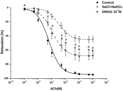
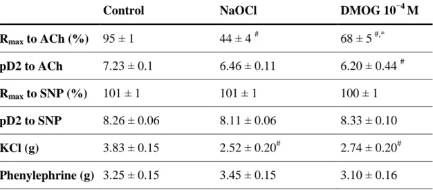
Technology, Monrovia, CA, USA) and digitised, stored and displayed with the IOX Software System (EMKA Technologies, Paris, France). The aortic rings were placed under a resting tension of 2 g and equilibrated for 60 min. During this period, tension was periodically adjusted to the desired level and the Krebs–Henseleit solution was changed every 30 min. At the beginning of each experiment, maximal contraction forces in response to KCl (80 mM) were determined and the aortic rings were washed until the resting tension was again obtained. Aortic preparations were preconstricted with phenylephrine (10−6 M), the α-adrenergic receptor agonist, until a stable plateau was reached, and relaxation responses were examined by adding cumulative concentrations of endothelium-dependent dilator acetylcholine (10−9–10−4 M). For testing the relaxation responses of smooth muscle cells, a direct nitric oxide-donor, sodium nitroprusside (SNP, 10−10–10−5 M), was used. Half-maximal effective concentration (EC50) values were obtained from individual concentration–responses by fitting experimental data with a sigmoidal equation using Origin 7.0 (Microcal Software, Northampton, USA). Contractile responses to phenylephrine are expressed as a percentage of the maximal contraction induced by KCl. The sensitivity to vasorelaxants was assessed using pD2=−log EC50 (M); vasorelaxation (and its maximum, Rmax) are expressed as a percentage of the contraction induced by phenylephrine (10−6 M).
4.1.6.INVESTIGATION OF COLD ISCHAEMIC STORAGE – WARM REPERFUSION INJURY ON AORTIC SMOOTH MUSCLE CELLS IN CELL CULTURE
Vascular smooth muscle cells were isolated from rat aorta with Liberase® following manufacturer instructions, resuspended in base medium and plated and incubated on 6- well plates. The cells were grown over 70% of the plate. To verify the quality of the cells, the α-smooth muscle was immunostained. We performed the experiments with 3–
5 passages of the cells. The medium was changed for saline or DMOG-supplemented saline solution, incubated for 24 h and stored for hypothermic ischaemia at 4 C. After the cold storage, the complete cell culture medium was added and reperfusion was simulated by further incubation at 37 C for 6 h. Samples were harvested in a RLT lysis