Vitamin D deficiency causes inward
hypertrophic remodeling and alters vascular reactivity of rat cerebral arterioles
E´ va Pa´l1*, Leila Hadjadj1, Zolta´n Fonta´nyi2, Anna Monori-Kiss1, Zsuzsanna Mezei3, Norbert Lippai4, Attila Magyar5, Andrea Heinzlmann5, Gelle´rt Karvaly6,7, Emil Monos1, Gyo¨ rgy Na´dasy3, Zolta´n Benyo´1, Szabolcs Va´rbı´ro´2
1 Institute of Clinical Experimental Research, Semmelweis University, Budapest, Hungary, 2 2ndDepartment of Obstetrics and Gynecology, Semmelweis University, Budapest, Hungary, 3 Department of Physiology, Semmelweis University, Budapest, Hungary, 4 Department of Pathology, Ja´sz-Nagykun-Szolnok County Hete´nyi Ge´za Hospital, Szolnok, Hungary, 5 Department of Anatomy, Histology and Embryology,
Semmelweis University, Budapest, Hungary, 6 Department of Laboratory Medicine, Semmelweis University, Budapest, Hungary, 7 Bionics Innovation Center, Budapest, Hungary
*pal.eva@med.semmelweis-univ.hu
Abstract
Background and purpose
Vitamin D deficiency (VDD) is a global health problem, which can lead to several pathophys- iological consequences including cardiovascular diseases. Its impact on the cerebrovascu- lar system is not well understood. The goal of the present work was to examine the effects of VDD on the morphological, biomechanical and functional properties of cerebral arterioles.
Methods
Four-week-old male Wistar rats (n = 11 per group) were either fed with vitamin D deficient diet or received conventional rat chow with per os vitamin D supplementation. Cardiovascu- lar parameters and hormone levels (testosterone, androstenedione, progesterone and 25- hydroxyvitamin D) were measured during the study. After 8 weeks of treatment anterior cerebral artery segments were prepared and their morphological, biomechanical and func- tional properties were examined using pressure microangiometry. Resorcin-fuchsin and smooth muscle actin staining were used to detect elastic fiber density and smooth muscle cell counts in the vessel wall, respectively. Sections were immunostained for eNOS and COX-2 as well.
Results
VDD markedly increased the wall thickness, the wall-to-lumen ratio and the wall cross-sec- tional area of arterioles as well as the number of smooth muscle cells in the tunica media. As a consequence, tangential wall stress was significantly lower in the VDD group. In addition, VDD increased the myogenic as well as the uridine 5’-triphosphate-induced tone and impaired bradykinin-induced relaxation. Decreased eNOS and increased COX-2 expression were also observed in the endothelium of VDD animals.
a1111111111 a1111111111 a1111111111 a1111111111 a1111111111
OPEN ACCESS
Citation: Pa´l E´, Hadjadj L, Fonta´nyi Z, Monori-Kiss A, Mezei Z, Lippai N, et al. (2018) Vitamin D deficiency causes inward hypertrophic remodeling and alters vascular reactivity of rat cerebral arterioles. PLoS ONE 13(2): e0192480.https://doi.
org/10.1371/journal.pone.0192480
Editor: Alain-Pierre Gadeau, "INSERM", FRANCE Received: August 30, 2017
Accepted: January 24, 2018 Published: February 6, 2018
Copyright:©2018 Pa´l et al. This is an open access article distributed under the terms of theCreative Commons Attribution License, which permits unrestricted use, distribution, and reproduction in any medium, provided the original author and source are credited.
Data Availability Statement: All relevant data are within the paper.
Funding: This study has been supported by the Hungarian National Research, Development and Innovation Office (http://nkfih.gov.hu/; OTKA K- 112964, OTKA K-101775 and NVKP_16-1-2016- 0042, to ZB; ED_14-1-2014-0002, to GK), the Hungarian Society of Hypertension (http://www.
hypertension.hu/; to SzV) and by the Dean of Faculty of Medicine, Semmelweis University (http://
semmelweis.hu/english/faculties/medicine/; to SzV and GyN). The funders had no role in study design,
Conclusions
VDD causes inward hypertrophic remodeling due to vascular smooth muscle cell prolifera- tion and enhances the vessel tone probably because of increased vasoconstrictor prosta- noid levels in young adult rats. In addition, the decreased eNOS expression results in endothelial dysfunction. These morphological and functional alterations can potentially com- promise the cerebral circulation and lead to cerebrovascular disorders in VDD.
Introduction
Vitamin D deficiency (VDD) or insufficiency affects 1 billion people from all age groups worldwide. In addition to its well-characterized roles in calcium and phosphate homeostasis as well as in bone metabolism, 1,25-dihydroxyvitamin D—the active metabolite of vitamin D (VitD)—has numerous biological actions [1]. Besides interacting with the intracellular VitD receptor and regulating the expression of up to 200 genes, it mediates non-genomic actions as well [2]. VDD is associated with an increased risk of malignant tumor formation, autoimmune and infectious diseases as well as depression [1]. Diabetes mellitus and metabolic syndrome are also linked to VDD, as 1,25-dihydroxyvitamin D improvesβ-cell function and insulin sensitivity [3]. There is a growing body of evidence linking VDD to cardiovascular diseases including hypertension, atherosclerosis and coronary artery disease. Furthermore, a direct impact of VDD on endothelial dysfunction, arterial stiffness and vascular inflammation was also reported [2,4,5].
The effects of VDD on the cerebrovascular system are as yet less understood, although sev- eral studies highlight the importance of sufficient VitD status in cerebrovascular health. Low concentrations of VitD are associated with an increased risk of cerebrovascular diseases including ischemic stroke [6–9] and with poor poststroke outcome [10]. In addition, VDD is linked to chronic brain injury associated with cerebral small vessel disease [11]. The effect of VitD status on stroke severity was confirmed in rodent models as well: the infarction volume was larger and more severe poststroke behaviour impairment was observed in VitD-deficient rats as compared to VitD-sufficient ones [12].
In the present study, we hypothesized that the aforementioned adverse effects of VDD in stroke are related—at least in part—to VDD-induced alterations in cerebral arterioles. There- fore, we aimed to analyze the changes in the morphological, biomechanical and functional properties of the anterior cerebral artery (ACA) in a rodent model of VDD.
Materials and methods Animals
All procedures conformed to the Guide for the Care and Use of Laboratory Animals published by the US National Institutes of Health (8th edition, 2011) and the EU-conform Hungarian Law on Animal Care (XXVIII/1998). The Institutional Animal Care and Use Committee of Semmelweis University approved the study protocol (IRB: 8/2014, PEI/001/1548-3/2014). All surgery was performed under sodium pentobarbital anesthesia, and all efforts were made to minimize suffering. Twenty-two 4-week-old male Wistar rats were involved in the experi- ments. Eleven of them were fed with VitD-deficient diet (VitD content less than 5 IU/kg) for eight weeks (VDD group). Control animals received conventional rat chow (containing 1500 IU/kg VitD) with per os VitD (Vigantol, 20.000 IU/mL cholecalciferol) supplementation (at
data collection and analysis, decision to publish, or preparation of the manuscript.
Competing interests: The authors have declared that no competing interests exist.
the second week of the study 500 IU/100 g body weight as loading dose, then 140 IU/100 g b.
w. weekly from week 4 to 7 as maintenance dose). Taken together, the daily VitD intake in the Control group was approximately 300 IU/100 g b. w. providing optimal VitD supply. Serum 25-hydroxyvitamin D, testosterone, androstenedione and progesterone levels were measured from blood samples taken at weeks 4 and 8. At week 6 a glucose tolerance test was performed:
fasting as well as 60-minute and 120-minute postload plasma levels were measured before and after oral administration of 2 g/kg b. w. 30% glucose solution.
After 8 weeks, animals were sacrificed as follows. First, blood pressure was measured by cannulation of the carotid artery under general surgical anesthesia (pentobarbital, 45 mg/kg b.
w., i. p.). After perfusionviathe carotid artery with heparinized Krebs-Ringer solution and decapitation under anesthesia, the brain was removed and anterior cerebral artery (ACA) seg- ments were prepared under a stereomicroscope (Wild M3Z, Heerbrugg, Switzerland). Heart and testes were also removed for weight measurement.
Pressure microangiometry
An approximately 2-mm-long segment of the ACA was immersed into an organ chamber (Experimetria Ltd., Budapest, Hungary) filled with normal Krebs-Ringer solution, cannulated at both ends with microcannulas, and extended to itsin vivolength. The chamber was placed on the stage of an inverted microscope (Leica, Wetzlar, Germany). Pressure-servo pumps (Liv- ing Systems, Burlington, VT, USA)–with the belonging pressure transducer (Living Systems, Burlington, VT, USA), which had been calibrated with a mercurial manometer—were con- nected to both cannulas and the arteries were pressurized to maintain 50 mmHg intraluminal pressure. The cerebral arterioles were allowed to equilibrate for 30 min at this pressure in nor- mal Krebs-Ringer solution bubbled with a gas mixture containing 5% CO2, 20% O2and 75%
N2, and the temperature was kept at 37˚C during the entire measurement. After equilibration, 10−4mol/L UTP was added to the chamber and after a 5-min incubation the pressure was increased to 150 mmHg, then decreased to 0 mmHg; this challenge was then repeated once more. Thereafter, the pressure was increased from 0 to 150 mmHg in 10 mmHg steps. All these procedures were performed rapidly—within 5 minutes—to avoid activation of the myo- genic response. After the arterioles were allowed to equilibrate at 50 mmHg intraluminal pres- sure for 10 min, 10−6mol/L bradykinin was added to the vessel chamber and incubated for 5 min at 50 mmHg intraluminal pressure in order to evaluate endothelium-dependent vasore- laxation [13]. Finally, the passive diameter of vessels was measured: the organ chamber was filled with Ca2+-free Krebs solution and after 20 min the pressure-diameter relationship was determined as described above. Pictures were taken during the experiment by a digital camera (Leica DFC 320) connected to the microscope. The outer and inner diameters of the vessels were measured by ImageJ image analysis software (Image J 1.5 NIH, USA). For the calibration, a micrometer etalon (Wild, Heerbrugg, Switzerland) was used.
Calculations
Wall thickness was expressed as the difference between the outer and inner radii:h = Ro-Ri, wherehis wall thickness,Rois the outer radius andRiis the inner radius. The wall cross-sec- tional area (Aw) was computed according to the following equation:Aw=(Ro2-Ri2)π. Tangen- tial wall stress was computed according to the Laplace-equation:σtang=(PRi)/h, whereσtangis the tangential wall stress andPis the intraluminal pressure. The elastic modulus was computed as:E = (ΔP/ΔRo)2Ri2Ro/(Ro2-Ri2), whereEis the incremental elastic modulus,ΔPis the change in intraluminal pressure andΔRois the outer radius change in response toΔP.
Incremental distensibility was computed asD =ΔV/VΔP, whereDis the incremental dis- tensibility andΔVis the change in lumen volume relative to the initial volumeVin response to pressure change (ΔP). Myogenic tone was computed asM% = 100(RiCf-RinKR)/RiCf, where RiCfis the inner radius in Ca2+-free solution andRinKRis the inner radius in normal Krebs- Ringer solution. UTP-induced contraction was expressed as a percentage of complete relaxa- tion:TUTP% = 100(RiCf-RiUTP)/RiCf, whereRiUTPis the inner radius after incubation with UTP. Bradykinin-induced relaxation was expressed as a percentage of UTP-induced precon- traction:TBK% = 100(RiBK-RiUTP)/RiUTP, whereRiBKis the inner radius after bradykinin was added to the chamber.
Histology and immunohistochemistry
ACA segments were freshly fixed with formalin for histological examination and stained for elastic fibers with Weigert’s resorcin-fuchsin. Paraffin-embedded sections of ACA were immu- nostained with anti-COX-2 antibody (1:500, 1 h, 37˚C) or with anti-eNOS antibody (1:200, 1 h, 37˚C) after deparaffinization and antigen retrieval (0.1 mmol/L citrate buffer, pH 6, 30 min, 60˚C). Endogenous peroxidase activity was quenched (with hydrogen-peroxide dissolved in distilled water) and blocked (2.5% normal horse serum). After secondary antibody (biotiny- lated horse anti-mouse antibody for COX-2 or biotinylated horse anti-rabbit antibody for eNOS) staining (30 min, RT) the binding sites of primary antibodies were visualized with DAB Substrate Kit (with 3,3’-diaminobenzidine; 6 min, RT). Sections were counterstained by hema- toxylin. Artery segments were stained for smooth muscle actin (36 min, 37˚C) using Ventana Benchmark Ultra System after deparaffinization and antigen retrieval (8 min, 97˚C). Ultra- View Universal DAB Detection Kit was used for detecting primary antibodies. Data collections were made by microscope (Zeiss AxioImager.A1) coupled with video-camera (Zeiss AxioCAm MRc5 CCD). RGB pictures of resorcin-fuchsin-stained segments were analyzed with the Leica Qwin image analysis software. The green levels (green color was chosen after evaluation of color histograms) were measured starting at the luminal surface and going radially in an out- ward direction. In addition, the thicknesses of the intimal and medial layers of arteries and the intima/media ratio were determined. Pictures of smooth muscle actin, eNOS and COX-2 staining were analyzed with the ImageJ image analysis software using the „Color deconvolu- tion” profile. Optical density of the endothelial layer after eNOS and COX-2 staining and the nucleus count of tunica media after demarcation with smooth muscle actin staining were determined.
Statistical analysis
All data are presented as mean±SEM. For statistical analysis Student’s t test or two-way repeated measures ANOVA followed by Bonferroni post hoc test was used; p<0.05 was con- sidered to be statistically significant. GraphPad Prism version 6.0 was used as statistical software.
Materials
Vigantol 20.000 IU/mL was obtained from Merck Serono (Mumbai, India). Vitamin D defi- cient (EF R/M, E15312-24) and conventional rat chow (SM R/M, S8106-S011) were purchased from ssniff Spezialdia¨ten GmbH (Soest, Germany). Pentobarbital was obtained from Ceva- Phylaxia (Budapest, Hungary). The composition of normal Krebs-Ringer solution (in mmol/
L) was: NaCl 119; KCl 4.7; NaH2PO41.2; MgSO41.17; NaHCO324; CaCl22.5; glucose 5.5 and EDTA 0.034. The composition of Ca2+-free Krebs solution (in mmol/L) was: NaCl 92; KCl 4.7;
NaH2PO41.18; MgCl220; MgSO41.17; NaHCO324; glucose 5.5; EGTA 2 and EDTA 0.025.
Uridine 5’-triphosphate (UTP) and bradykinin were purchased from Sigma-Aldrich (Darm- stadt, Germany). Anti-eNOS [14] and anti-COX-2 [14] antibodies were obtained from Abcam (Cambridge, MA, USA). Anti-smooth muscle actin antibody (1A4) [15] and UltraView Uni- versal DAB Detection Kit were obtained from Ventana Medical Systems, Inc. (Tucson, AZ, USA). Secondary antibodies and DAB Substrate Kit [14] were purchased from Vector Labora- tories (Burlingame, CA, USA).
Results
Physiological parameters and serum levels of hormones, 25-hydroxyvitamin D and glucose
At the end of the experiments the measured physiological parameters of the rats (body weight, heart/body weight ratio, testis weight, mean arterial blood pressure, heart rate, as well as serum testosterone, androstenedione and progesterone levels) did not differ between the two groups (Table 1), indicating that the VitD-deficient diet, at least within 8 weeks, does not affect these parameters. To assess the efficacy of VitD deprivation, serum 25-hydroxyvitamin D levels were measured and found to be significantly lower in VDD as compared to control animals at the 8thweek of treatment (Table 1). In the glucose tolerance test performed at the 6thweek of treatment, blood glucose levels before and after oral glucose administration did not show any difference between the two groups (Table 1). These findings exclude the possibility that any morphological or functional changes of the ACA in the present study would be secondary con- sequences of VDD-induced hypertension, metabolic syndrome or altered androgenic hor- mone status.
Table 1. Physiological parameters and serum levels of hormones, 25-hydroxyvitamin D and glucose in the two experimental groups.
MEASURED PARAMETER CONTROL
n = 11
VDD n = 11
Body weight(g) 435.7±17.7 444.5±10.3
Heart/body weight 0.34±0.01 0.34±0.01
Testis weight(g) 3.74±0.16 3.71±0.33
Mean arterial blood pressure(mmHg) 131±4 134±4
Heart rate (1/min) 357±18 348±11
Serum testosterone(ng/mL) 6.56±0.84 5.94±0.91
Serum androstenedione(ng/mL) 0.57±0.14 0.54±0.12
Serum progesterone(ng/mL) 14.42±2.31 19.86±2.61
Serum 25-hydroxyvitamin D(ng/mL) 19.66±0.81 3.59±0.21
Glucose (OGTT 0 min) (mmol/L) 6.24±0.46 5.49±0.20
Glucose (OGTT 60 min) (mmol/L) 7.61±0.33 7.59±0.37
Glucose (OGTT 120 min) (mmol/L) 5.51±0.39 5.41±0.31
The body and testis weight as well as the heart/body weight ratio of rats did not differ between the groups. VDD did not influence the mean arterial blood pressure, the heart rate or the serum hormone levels. Serum levels of glucose did not show any significant difference between the two groups during the oral glucose tolerance test (OGTT). The VitD-deficient diet induced significantly lower serum 25-hydroxyvitamin D level (p<0.0001). All parameters except blood glucose levels were measured at the 8thweek of treatment. OGGT was performed at week 6. Data are presented as mean±SEM.
p<0.0001
https://doi.org/10.1371/journal.pone.0192480.t001
Arterial geometry
The artery segments were examined in Ca2+-free Krebs solution with pressure myograph to determine vessel geometry under passive conditions, as we hypothesized that a VitD-deficient diet could cause remodeling of cerebral arteries. In Ca2+-free Krebs solution the artery seg- ments showed their fully relaxed inner radii, which decreased tendentiously in the VDD as compared to the Control group, but the difference did not reach the level of statistical signifi- cance (89.6±6.3μm for Control, 78.4±5.6μm for VDD, at 50 mmHg intraluminal pressure).
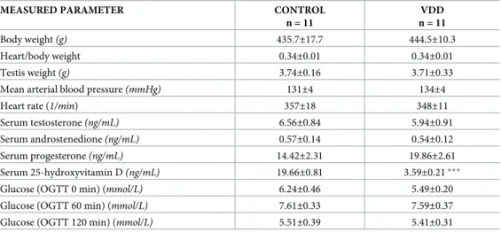
However, wall thickness (Fig 1A) and wall thickness / lumen diameter ratio (Fig 1B) showed a significant increase in the VDD group under passive conditions. In addition, VitD-deficient
Fig 1. Cerebral artery geometry and nucleus count of the smooth muscle layer. (A) VDD significantly increased the wall thickness (p<0.05, n = 10–11) as well as (B) the wall thickness / lumen diameter ratio (p<0.01, n = 10–11). (C) The cross-sectional area of the vessel walls increased significantly in VDD as compared to the Control group (p<0.05, n = 9–9) at 50 mmHg intraluminal pressure. All these parameters were measured under passive conditions. (D) Significantly more nuclei were detected in the smooth muscle layer of anterior cerebral arteries of VDD animals as compared to controls (p<0.0001, n = 4–6). (E) The thickness of the tunica media was increased in the VDD group as compared to the control animals (p<0.05, n = 4–4). (F) In addition, VDD significantly decreased the intima / media ratio of cerebral arteries (p<0.05, n = 4–4).
https://doi.org/10.1371/journal.pone.0192480.g001
feeding increased the wall cross-sectional area as compared to the Control group (Fig 1C), indicating the development of hypertrophic remodeling in the VDD group.
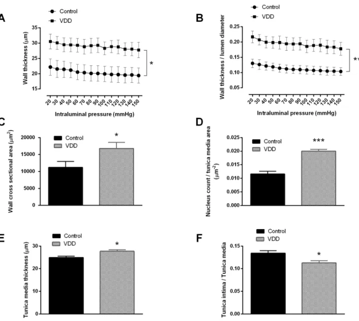
Arterial elasticity
As previously mentioned, the wall thickness / lumen diameter ratio increased in the arterioles of the VDD group. In accordance with this finding, the tangential wall stress was significantly lower in the VDD group under passive conditions (Fig 2), as this biomechanical parameter is inversely proportional to the wall-to-lumen ratio. The incremental elastic modulus and the distensibility did not change significantly between the groups at 50 mmHg intraluminal pres- sure measured under passive conditions (elastic modulus: 2.89±0.27 log(kPa) and 2.65±0.19 log(kPa), distensibility: 1.41±0.12 log(Pa-1) and 1.18±0.23 log(Pa-1) for the Control and VDD groups, respectively).
Smooth muscle tone and endothelial reactivity
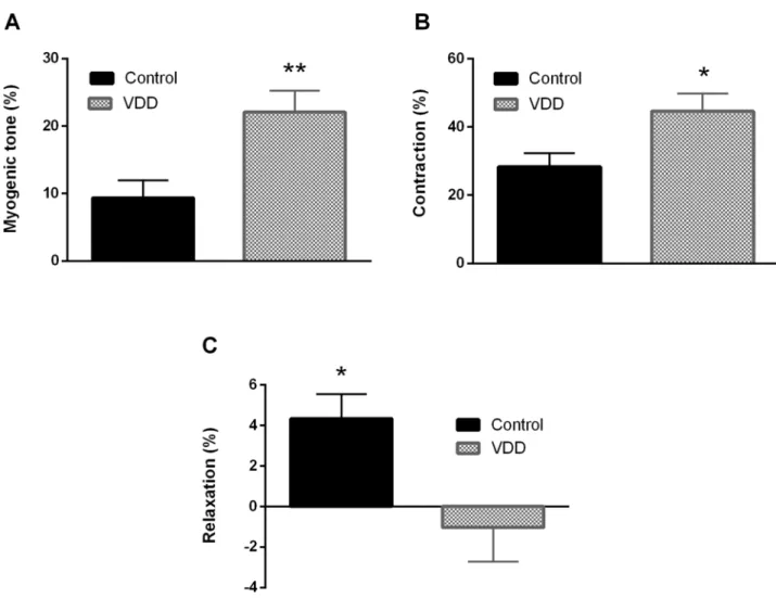
ACA possesses myogenic tone under resting conditions as well [16]. In our present experi- ment, the myogenic tone values of excised ACA segments were determined at 50 mmHg intra- luminal pressure. Segments from VDD animals showed a two-fold increase in myogenic tone as compared to control ones, indicating that the 8-week VitD deprivation doubled the sponta- neous tone of vessels (Fig 3A). Because UTP is a potent vasoconstrictor of cerebral arteries [17], the agonist-induced responsiveness with this agent was also tested at 50 mmHg intralum- inal pressure. UTP caused potent constriction in both groups, but the induced tone was signifi- cantly greater in the VDD group (Fig 3B). To evaluate endothelial function, bradykinin was applied after precontraction with UTP. Whereas bradykinin induced slight vasodilatation in the Control group, it failed to relax the arteries from the VDD group (Fig 3C), indicating endo- thelial dysfunction in VitD-deficient animals.
Fig 2. The tangential wall stress of the cerebral arteries in relaxed condition. VDD caused a decrease in the tangential stress of the vessel wall throughout the entire pressure range under passive conditions (p<0.05, n = 10–11).
https://doi.org/10.1371/journal.pone.0192480.g002
Histology and immunohistochemistry
To gain further insight into the mechanism responsible for the above-mentioned observations, the density of the elastic components of the vessel wall was determined using resorcin-fuchsin staining, which did not show any difference between the groups based on the green color den- sity measurements made in radial direction, outward from the luminal surface (Fig 4A and 4B). Furthermore, after immunohistological staining of smooth muscle actin the amount of nuclei was determined in the tunica media. The significantly increased nucleus count found in the smooth muscle layer of arteries from the VDD group (Fig 1D) indicated the presence of more vascular smooth muscle cells (VSMCs) in the vessel wall of VitD-deficient animals.
Related to this alteration, increased thickness of the tunica media of arteries was observed in the VDD group (Fig 1E), however VDD did not affect the thickness of tunica intima (3.15±0.19μm for Control, 3.15±0.24μm for VDD). Therefore, VDD decreased the intima / media ratio of ACA (Fig 1F). To evaluate the possible role of NO and prostanoids in the effect of VDD, the expression of eNOS and COX-2 was determined by immunostaining. The optical
Fig 3. Alterations of vascular reactivity in VDD. VDD caused alterations in the smooth muscle tone and endothelium-dependent relaxation capacity of arterioles. (A) Anterior cerebral arteries possessed myogenic tone and this tone was greater in the VDD group measured at 50 mmHg intraluminal pressure (p<0.01, n = 8–8). (B) In addition, UTP-induced contraction also increased in the VDD group at 50 mmHg intraluminal pressure (p<0.05, n = 10–9). (C) Bradykinin induced endothelium-dependent relaxation after precontraction in the Control group, but did not relax the arterioles in the VDD group at 50 mmHg intraluminal pressure (p<0.05, n = 9–9).
https://doi.org/10.1371/journal.pone.0192480.g003
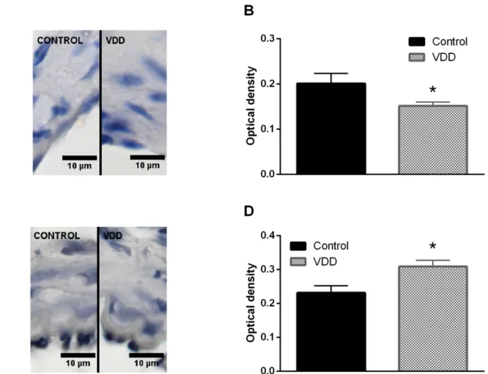
density of endothelial eNOS staining was lower in the VDD group, indicating lower expression of eNOS (Fig 5A and 5B). In contrast, COX-2 expression was enhanced in the endothelial layer of arteries from the VDD group (Fig 5C and 5D).
Discussion
To our knowledge, the present study provides the first report of the deleterious changes in geometry and reactivity of cerebral arteries due to VDD. These alterations were associated with hypertrophic remodeling, increased myogenic tone, endothelial dysfunction, increased COX-2 and decreased eNOS expression. Interestingly, marked changes of morphology and reactivity developed in healthy young adult animals within a relatively short period (8 weeks) of VDD indicating the importance of normal VitD status for the maintenance of cerebrovascu- lar functions.
In humans, the cardiovascular consequences of VDD include hypertension, atherosclerosis, cardiac hypertrophy, cerebrovascular diseases, coronary artery disease, peripheral artery dis- ease, dyslipidemia, insulin resistance and diabetes mellitus [2]; in addition, VDD during preg- nancy leads to cardiomyopathy in infants [2,18]. However, the associations, for instance, between VDD and insulin resistance [19,20] or between VDD and hypertension [21] are not fully confirmed, especially in young healthy subjects [22–24]. The cardiovascular impacts of VitD are attributed to the modulation of immune, inflammatory and endothelial functions, furthermore to the regulation of VSMC proliferation and migration, renin expression, and extracellular matrix homeostasis [2].
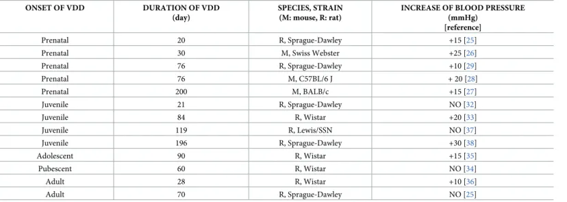
In rodent models of VDD similar disorders of the cardiovascular system have been described as in humans, although their manifestation depends markedly on the experimental protocol applied. For instance, in a number of studies hypertension developed as a conse- quence of VDD, whereas the blood pressure remained unaltered in other studies (Table 2).
According to the literature, VDD during the prenatal period leads to increased blood pressure in rodents’ offsprings [25–29] probably due to the upregulation of the renin-angiotensin system [18,30]. Interestingly, VDDin uterocontributes to the development of hypertension in later life even if the offsprings were fed with VitD sufficient chow after weaning [25,26], which implies that VDD during pregnancy can severely impact the offspring’s long-term health and lead to vulnerability to cardiovascular disease in adult life [18,31]. However, when VDD affects only the postnatal period of life, the observations are controversial both in grow- ing and adult rodents; nevertheless longer exposure to VDD appears to increase the risk of
Fig 4. Elastic components of the vessel wall. (A) Representative images of cerebral arterioles stained with Weigert’s resorcin-fuchsin. (B) Elastic fiber density did not differ between the groups according to measurement of green color intensity as a function of distance from the luminal surface measured on resorcin-fuchsin-stained segments.
https://doi.org/10.1371/journal.pone.0192480.g004
manifestation of hypertension [25,32–38]. Taken together, the development of hypertension appears to depend on the time of onset and the duration of VDD (Table 2). In our study, wean- ling rats were exposed to VDD for only 8 weeks, which explains the unaltered blood pressure levels. Although VDD is a complex disorder linked to several cardiovascular risk factors in the long run, we examined its short-term effects, and found altered cerebrovascular morphology and reactivity without the development of hypertension.
Surprisingly, in spite of the well-preserved systemic cardiovascular and metabolic parame- ters, marked inward hypertrophic remodeling was observed in the cerebral arteries of VDD animals, similar to that observed in secondary hypertension [39]. In rats and humans suffer- ing from secondary hypertension the increase in the wall-to-lumen ratio of small vessels is due to hypertrophic remodeling as a consequence of smooth muscle cell proliferation [39, 40]. In contrast, the hypertrophic remodeling observed in our present study develops at normal blood pressure and therefore is likely to be the direct effect of VDD on VSMCs.
1,25-dihydroxyvitamin D can modulate VSMC proliferation and migration [4]. In our study
Fig 5. Evaluation of immunohistochemical staining of arterioles. (A) Representative immunohistochemical images of cerebral arterioles stained for eNOS. (B) VDD caused a decrease in the expression of eNOS in the endothelium of arterioles (p<0.05, n = 4–6). (C) Representative immunohistochemical images of cerebral arterioles stained for COX-2. (D) The expression of COX-2 in the endothelial layer increased in VDD as compared to Control (p<0.05, n = 4–4).
https://doi.org/10.1371/journal.pone.0192480.g005
the increased cross-sectional area of the vessel wall appears to be the consequence of VSMC proliferation, because we observed increased number of smooth muscle cells in the tunica media of VitD-deficient arteries as compared to controls. On the contrary, we did not observe any changes in the tunica intima of arteries from the VDD group. The literature is controversial regarding the effects of VitD on VSMC proliferation and migration: some stud- ies report enhanced migration and proliferation [41,42], whereas others found VitD-induced inhibition of VSMC growth [43,44]. VitD could inhibit VSMC proliferationviablunting c- myc RNA induction [42], up-regulating the negative modulators of cell proliferation includ- ing TGF-β[45] or decreasing cyclin-dependent kinase 2 activity [46]. The effect of VitD on VSMC definitely depends on the applied dose, and both insufficient and supraphysiological levels of VitD appear to lead to VSMC proliferation. In accordance, U-shaped association has been reported between VitD concentrations and cardiovascular diseases [4] [47]. Therefore, only physiological concentrations of VitD appear to be appropriate for maintaining normal VSMC function.
In hypertension, increased tangential wall stress facilitates wall thickening to compensate increased circumferential stress [48]. In our study, however, mean arterial blood pressure did not differ between the groups, thus the observed wall thickening resulted in decreased tan- gential wall stress according to the Laplace equation. The incremental elastic modulus and dis- tensibility did not differ between the groups, indicating that elastic element density and arrangement were not influenced by VDD. This presumption was also confirmed by the unal- tered elastic fiber density.
Cerebral arteries possess intrinsic myogenic tone [16], which can increase harmfully under pathophysiological conditions. In our study, arteries of VitD-deficient animals developed greater myogenic tone, which is similar to the observation of Tare et al., who reported a two- fold enhancement of the myogenic tone of mesenteric arteries in male VitD-deficient rats as compared to VitD-sufficient ones [29]. In addition, UTP—a potent and partly thromboxane A2-mediated constrictor of cerebral arteries [49]—induced greater tone in the ACA of VDD animals. VitD is an important modulator of the prostanoid system, since it downregulates the expression of COX-2 [50], therefore VDD can lead to enhanced COX-2 expression, as we found in the endothelium of cerebral arteries. In addition, VitD suppresses the expression of
Table 2. Alterations of blood pressure in rodent models of VDD induced by VitD deficient feeding.
ONSET OF VDD DURATION OF VDD
(day)
SPECIES, STRAIN (M: mouse, R: rat)
INCREASE OF BLOOD PRESSURE (mmHg)
[reference]
Prenatal 20 R, Sprague-Dawley +15 [25]
Prenatal 30 M, Swiss Webster +25 [26]
Prenatal 76 R, Sprague-Dawley +10 [29]
Prenatal 76 M, C57BL/6 J + 20 [28]
Prenatal 200 M, BALB/c +15 [27]
Juvenile 21 R, Sprague-Dawley NO [32]
Juvenile 84 R, Wistar +20 [33]
Juvenile 119 R, Lewis/SSN NO [37]
Juvenile 196 R, Sprague-Dawley +30 [38]
Adolescent 90 R, Wistar +15 [35]
Pubescent 60 R, Wistar NO [34]
Adult 28 R, Wistar +10 [36]
Adult 70 R, Sprague-Dawley NO [25]
https://doi.org/10.1371/journal.pone.0192480.t002
TNF-α, NADPH oxidase and its subunits and also increases CuZn-SOD protein expression, therefore prevents inflammatory response and oxidative stress [51]. The increased level of reactive oxygen species (ROS) in VDD can in turn lead to enhanced vasoconstrictor response, as ROS inactivate prostacyclin synthase, shifting the prostanoid balance towards vasoconstric- tion [52]. Bradykinin relaxes cerebral arteries via the B2receptor and NO release [13]; how- ever, in the case of endothelial dysfunction bradykinin can cause endothelium-independent contractions [53]. VitD stimulates NO production through non-genomic eNOS activation via increase in eNOS phosphorylation [54] or due to an increase in eNOS mRNA and promoter activity depending on VitD receptor activation [55]. In our study, we observed decreased eNOS expression in the endothelium of VDD animals, which could contribute to the increased vessel tone and the impaired endothelium-dependent relaxation capacity. In the case of endo- thelial dysfunction, bradykinin-induced contractions might be mediated by COX-2 derived prostanoids and activation of thromboxane-prostanoid receptors [53]. The activation of thromboxane-prostanoid receptors could in turn impair the NO-mediated dilatation of vessels as well [56]. In addition, ROS can also contribute to the diminished endothelium-dependent vasodilatation in VDD due to eNOS uncoupling or inactivation of NO [57]. Enhanced vaso- constrictor prostanoid release and the impairment of the counterbalancing NO pathway could lead to increased myogenic tone and constrictor response as well as endothelial dysfunction in VDD.
VDD influences several pathways relevant to vascular physiology and pathophysiology, therefore it appears to be a significant risk factor for cardiovascular and cerebrovascular dis- ease [2]. Since VDD is associated with dysfunction of endothelial and vascular smooth muscle cells, low VitD levels could predict proinflammatory and prothrombotic alterations, which might lead to atherosclerosis as well as increased thrombosis and arterial stiffness [2,58].
VDD is not only a risk factor for coronary disease, acute myocardial infarction and stroke [2, 5,59], but it also worsens the outcome when myocardial infarction and stroke are already present [10,59]. In addition, low levels of VitD are associated with cerebral small vessel dis- ease related vascular dementia [60,61] and arterial stiffness associated cognitive impairment [62]. The observed alterations in cerebrovascular geometry and reactivity can be considered as prehypertensive changes [39]. Furthermore, the endothelial dysfunction of arteries may lead to the development of atherosclerosis in the long run, as the reduction of biosynthesis and enhanced inactivation of prostacyclin and NO are key components in the initiation of atherogenesis [63]. In addition, the increase in COX-2 expression as well as the proliferation of smooth muscle cells are also predictive of the development of atherosclerosis [63]. There- fore, long-term VDD with or without other disturbing pathophysiological conditions can in turn contribute to hypertension, atherosclerosis and a further increase in constrictor tone as well as to a more deleterious impairment of relaxation capacity, which can aggravate the risk of stroke events [64].
Conclusions
The present study demonstrates the harmful effects of VDD on cerebral artery geometry and function in a rat model. We propose that VDD results in inward hypertrophic remodeling due to VSMC proliferation as well as in enhanced vessel tone due to increased vasoconstrictor prostanoid levels. In addition, impaired NO-mediated vasodilatation leads to endothelial dys- function. Our results imply that a relatively short-term VDD in a relatively young age without any comorbidities can already induce marked morphological and functional alterations in the cerebral vasculature, which underlines the importance of sufficient VitD supply throughout the entire life in order to prevent stroke and other cerebrovascular diseases.
Acknowledgments
The authors thank Attila Pato´cs for determining hormone levels, Eszter M. Horva´th, A´ gnes Nova´k and Rita Benkőfor immunostaining, Ildiko´ Mura´nyi for expert technical assistance and Erzse´bet Fejes for critically reading the manuscript.
Author Contributions
Conceptualization: E´va Pa´l, Leila Hadjadj, Gyo¨rgy Na´dasy, Zolta´n Benyo´, Szabolcs Va´rbı´ro´.
Data curation: E´va Pa´l, Zolta´n Fonta´nyi.
Formal analysis: E´va Pa´l, Anna Monori-Kiss, Zolta´n Benyo´.
Funding acquisition: Gelle´rt Karvaly, Gyo¨rgy Na´dasy, Zolta´n Benyo´, Szabolcs Va´rbı´ro´.
Investigation: E´va Pa´l, Leila Hadjadj, Zolta´n Fonta´nyi, Anna Monori-Kiss, Zsuzsanna Mezei, Norbert Lippai, Attila Magyar, Gelle´rt Karvaly, Gyo¨rgy Na´dasy.
Methodology: E´va Pa´l, Leila Hadjadj, Anna Monori-Kiss, Andrea Heinzlmann, Gyo¨rgy Na´dasy, Szabolcs Va´rbı´ro´.
Project administration: E´va Pa´l, Leila Hadjadj.
Resources: Gyo¨rgy Na´dasy, Zolta´n Benyo´, Szabolcs Va´rbı´ro´.
Supervision: Szabolcs Va´rbı´ro´.
Visualization: E´va Pa´l.
Writing – original draft: E´va Pa´l, Zolta´n Benyo´, Szabolcs Va´rbı´ro´.
Writing – review & editing: E´va Pa´l, Leila Hadjadj, Zolta´n Fonta´nyi, Anna Monori-Kiss, Zsuzsanna Mezei, Norbert Lippai, Attila Magyar, Andrea Heinzlmann, Gelle´rt Karvaly, Emil Monos, Gyo¨rgy Na´dasy, Zolta´n Benyo´, Szabolcs Va´rbı´ro´.
References
1. Holick MF. Vitamin D deficiency. The New England journal of medicine. 2007; 357(3):266–81.https://
doi.org/10.1056/NEJMra070553PMID:17634462
2. Norman PE, Powell JT. Vitamin D and cardiovascular disease. Circulation research. 2014; 114(2):379–
93.https://doi.org/10.1161/CIRCRESAHA.113.301241PMID:24436433
3. Sung CC, Liao MT, Lu KC, Wu CC. Role of Vitamin D in Insulin Resistance. Journal of Biomedicine and Biotechnology. 2012; 2012.
4. Menezes AR, Lamb MC, Lavie CJ, DiNicolantonio JJ. Vitamin D and atherosclerosis. Current opinion in cardiology. 2014; 29(6):571–7.https://doi.org/10.1097/HCO.0000000000000108PMID:25144342 5. Mozos I, Marginean O. Links between Vitamin D Deficiency and Cardiovascular Diseases. BioMed research international. 2015; 2015:109275.https://doi.org/10.1155/2015/109275PMID:26000280 6. Brondum-Jacobsen P, Nordestgaard BG, Schnohr P, Benn M. 25-hydroxyvitamin D and symptomatic
ischemic stroke: an original study and meta-analysis. Annals of neurology. 2013; 73(1):38–47.https://
doi.org/10.1002/ana.23738PMID:23225498
7. Kojima G, Bell C, Abbott RD, Launer L, Chen R, Motonaga H, et al. Low dietary vitamin D predicts 34- year incident stroke: the Honolulu Heart Program. Stroke. 2012; 43(8):2163–7.https://doi.org/10.1161/
STROKEAHA.112.651752PMID:22627988
8. Sun Q, Pan A, Hu FB, Manson JE, Rexrode KM. 25-Hydroxyvitamin D levels and the risk of stroke: a prospective study and meta-analysis. Stroke. 2012; 43(6):1470–7.https://doi.org/10.1161/
STROKEAHA.111.636910PMID:22442173
9. Chowdhury R, Stevens S, Ward H, Chowdhury S, Sajjad A, Franco OH. Circulating vitamin D, calcium and risk of cerebrovascular disease: a systematic review and meta-analysis. European journal of epide- miology. 2012; 27(8):581–91.https://doi.org/10.1007/s10654-012-9729-zPMID:22961293
10. Turetsky A, Goddeau RP Jr., Henninger N. Low Serum Vitamin D Is Independently Associated with Larger Lesion Volumes after Ischemic Stroke. Journal of stroke and cerebrovascular diseases: the offi- cial journal of National Stroke Association. 2015; 24(7):1555–63.
11. Chung PW, Park KY, Kim JM, Shin DW, Park MS, Chung YJ, et al. 25-hydroxyvitamin D status is asso- ciated with chronic cerebral small vessel disease. Stroke; a journal of cerebral circulation. 2015; 46 (1):248–51.
12. Balden R, Selvamani A, Sohrabji F. Vitamin D deficiency exacerbates experimental stroke injury and dysregulates ischemia-induced inflammation in adult rats. Endocrinology. 2012; 153(5):2420–35.
https://doi.org/10.1210/en.2011-1783PMID:22408173
13. Wahl M, Gorlach C, Hortobagyi T, Benyo Z. Effects of bradykinin in the cerebral circulation. Acta physio- logica Hungarica. 1999; 86(2):155–60. PMID:10741874
14. Masszi G, Benko R, Csibi N, Horvath EM, Tokes AM, Novak A, et al. Endothelial relaxation mechanisms and nitrative stress are partly restored by Vitamin D3 therapy in a rat model of polycystic ovary syn- drome. Life sciences. 2013; 93(4):133–8.https://doi.org/10.1016/j.lfs.2013.05.003PMID:23685132 15. Hetthessy JR, Tokes AM, Keresz S, Balla P, Dornyei G, Monos E, et al. High pressure-low flow remod-
eling of the rat saphenous vein wall. Phlebology. 2017:268355516688984.
16. Osol G, Halpern W. Myogenic properties of cerebral blood vessels from normotensive and hypertensive rats. The American journal of physiology. 1985; 249(5 Pt 2):H914–21.https://doi.org/10.1152/ajpheart.
1985.249.5.H914PMID:4061668
17. Hardebo JE, Kahrstrom J, Owman C. P1- and P2-purine receptors in brain circulation. European journal of pharmacology. 1987; 144(3):343–52. PMID:3440480
18. Gezmish O, Black MJ. Vitamin D deficiency in early life and the potential programming of cardiovascular disease in adulthood. Journal of cardiovascular translational research. 2013; 6(4):588–603.https://doi.
org/10.1007/s12265-013-9475-yPMID:23719723
19. Del Gobbo LC, Song Y, Dannenbaum DA, Dewailly E, Egeland GM. Serum 25-hydroxyvitamin D is not associated with insulin resistance or beta cell function in Canadian Cree. The Journal of Nutrition. 2011;
141(2):290–5.https://doi.org/10.3945/jn.110.129619PMID:21178079
20. Poomthavorn P, Saowan S, Mahachoklertwattana P, Chailurkit L, Khlairit P. Vitamin D status and glu- cose homeostasis in obese children and adolescents living in the tropics. International journal of obesity (2005). 2012; 36(4):491–5.
21. Mehta V, Agarwal S. Does Vitamin D Deficiency Lead to Hypertension? Cureus. 2017; 9(2):e1038.
https://doi.org/10.7759/cureus.1038PMID:28357170
22. Black LJ, Burrows S, Lucas RM, Marshall CE, Huang RC, Chan She Ping-Delfos W, et al. Serum 25- hydroxyvitamin D concentrations and cardiometabolic risk factors in adolescents and young adults. The British journal of nutrition. 2016; 115(11):1994–2002.https://doi.org/10.1017/S0007114516001185 PMID:27153206
23. Muhairi SJ, Mehairi AE, Khouri AA, Naqbi MM, Maskari FA, Al Kaabi J, et al. Vitamin D deficiency among healthy adolescents in Al Ain, United Arab Emirates. BMC public health. 2013; 13:33.https://
doi.org/10.1186/1471-2458-13-33PMID:23311702
24. Rafraf M, Hasanabad SK, Jafarabadi MA. Vitamin D status and its relationship with metabolic syndrome risk factors among adolescent girls in Boukan, Iran. Public health nutrition. 2014; 17(4):803–9.https://
doi.org/10.1017/S1368980013003340PMID:24477119
25. Meems LM, Mahmud H, Buikema H, Tost J, Michel S, Takens J, et al. Parental vitamin D deficiency dur- ing pregnancy is associated with increased blood pressure in offspring via Panx1 hypermethylation.
American journal of physiology Heart and circulatory physiology. 2016; 311(6):H1459–h69.https://doi.
org/10.1152/ajpheart.00141.2016PMID:27769995
26. Nascimento FA, Ceciliano TC, Aguila MB, Mandarim-de-Lacerda CA. Maternal vitamin D deficiency delays glomerular maturity in F1 and F2 offspring. PloS one. 2012; 7(8):e41740.https://doi.org/10.
1371/journal.pone.0041740PMID:22927914
27. Pelham CJ, Drews EM, Agrawal DK. Vitamin D controls resistance artery function through regulation of perivascular adipose tissue hypoxia and inflammation. Journal of molecular and cellular cardiology.
2016; 98:1–10.https://doi.org/10.1016/j.yjmcc.2016.06.067PMID:27374117
28. Shi Y, Liu T, Yao L, Xing Y, Zhao X, Fu J, et al. Chronic vitamin D deficiency induces lung fibrosis through activation of the renin-angiotensin system. Scientific reports. 2017; 7(1):3312.https://doi.org/
10.1038/s41598-017-03474-6PMID:28607392
29. Tare M, Emmett SJ, Coleman HA, Skordilis C, Eyles DW, Morley R, et al. Vitamin D insufficiency is associated with impaired vascular endothelial and smooth muscle function and hypertension in young rats. The Journal of physiology. 2011; 589(Pt 19):4777–86.https://doi.org/10.1113/jphysiol.2011.
214726PMID:21807617
30. Li YC, Qiao G, Uskokovic M, Xiang W, Zheng W, Kong J. Vitamin D: a negative endocrine regulator of the renin-angiotensin system and blood pressure. The Journal of steroid biochemistry and molecular biology. 2004; 89-90(1–5):387–92.https://doi.org/10.1016/j.jsbmb.2004.03.004PMID:15225806 31. Hossein-nezhad A, Holick MF. Vitamin D for health: a global perspective. Mayo Clinic proceedings.
2013; 88(7):720–55.https://doi.org/10.1016/j.mayocp.2013.05.011PMID:23790560
32. Andersen LB, Przybyl L, Haase N, von Versen-Hoynck F, Qadri F, Jorgensen JS, et al. Vitamin D deple- tion aggravates hypertension and target-organ damage. Journal of the American Heart Association.
2015; 4(2).
33. Argacha JF, Egrise D, Pochet S, Fontaine D, Lefort A, Libert F, et al. Vitamin D deficiency-induced hypertension is associated with vascular oxidative stress and altered heart gene expression. Journal of cardiovascular pharmacology. 2011; 58(1):65–71.https://doi.org/10.1097/FJC.0b013e31821c832f PMID:21499117
34. Canale D, de Braganca AC, Goncalves JG, Shimizu MH, Sanches TR, Andrade L, et al. Vitamin D defi- ciency aggravates nephrotoxicity, hypertension and dyslipidemia caused by tenofovir: role of oxidative stress and renin-angiotensin system. PloS one. 2014; 9(7):e103055.https://doi.org/10.1371/journal.
pone.0103055PMID:25048368
35. Goncalves JG, de Braganca AC, Canale D, Shimizu MH, Sanches TR, Moyses RM, et al. Vitamin D deficiency aggravates chronic kidney disease progression after ischemic acute kidney injury. PloS one.
2014; 9(9):e107228.https://doi.org/10.1371/journal.pone.0107228PMID:25222475
36. Mirhosseini NZ, Knaus SJ, Bohaychuk K, Singh J, Vatanparast HA, Weber LP. Both high and low plasma levels of 25-hydroxy vitamin D increase blood pressure in a normal rat model. The British journal of nutrition. 2016; 116(11):1889–900.https://doi.org/10.1017/S0007114516004098PMID:27964766 37. Rangan GK, Schwensen KG, Foster SL, Korgaonkar MS, Peduto A, Harris DC. Chronic effects of die-
tary vitamin D deficiency without increased calcium supplementation on the progression of experimental polycystic kidney disease. American journal of physiology Renal physiology. 2013; 305(4):F574–82.
https://doi.org/10.1152/ajprenal.00411.2012PMID:23698116
38. Sundersingh F, Plum LA, DeLuca HF. Vitamin D deficiency independent of hypocalcemia elevates blood pressure in rats. Biochemical and biophysical research communications. 2015; 461(4):589–91.
https://doi.org/10.1016/j.bbrc.2015.04.069PMID:25911319
39. Izzard AS, Rizzoni D, Agabiti-Rosei E, Heagerty AM. Small artery structure and hypertension: adaptive changes and target organ damage. Journal of hypertension. 2005; 23(2):247–50. PMID:15662208 40. Rizzoni D, Porteri E, Castellano M, Bettoni G, Muiesan ML, Muiesan P, et al. Vascular hypertrophy and
remodeling in secondary hypertension. Hypertension. 1996; 28(5):785–90. PMID:8901824
41. Cardus A, Parisi E, Gallego C, Aldea M, Fernandez E, Valdivielso JM. 1,25-Dihydroxyvitamin D3 stimu- lates vascular smooth muscle cell proliferation through a VEGF-mediated pathway. Kidney interna- tional. 2006; 69(8):1377–84.https://doi.org/10.1038/sj.ki.5000304PMID:16557229
42. Mitsuhashi T, Morris RC Jr., Ives HE. 1,25-dihydroxyvitamin D3 modulates growth of vascular smooth muscle cells. The Journal of clinical investigation. 1991; 87(6):1889–95.https://doi.org/10.1172/
JCI115213PMID:1645744
43. Carthy EP, Yamashita W, Hsu A, Ooi BS. 1,25-Dihydroxyvitamin D3 and rat vascular smooth muscle cell growth. Hypertension. 1989; 13(6 Pt 2):954–9. PMID:2786849
44. Somjen D, Weisman Y, Kohen F, Gayer B, Limor R, Sharon O, et al. 25-hydroxyvitamin D3-1alpha- hydroxylase is expressed in human vascular smooth muscle cells and is upregulated by parathyroid hormone and estrogenic compounds. Circulation. 2005; 111(13):1666–71.https://doi.org/10.1161/01.
CIR.0000160353.27927.70PMID:15795327
45. Wu-Wong JR, Nakane M, Ma J, Ruan X, Kroeger PE. Effects of Vitamin D analogs on gene expression profiling in human coronary artery smooth muscle cells. Atherosclerosis. 2006; 186(1):20–8.https://doi.
org/10.1016/j.atherosclerosis.2005.06.046PMID:16095599
46. Chen S, Law CS, Gardner DG. Vitamin D-dependent suppression of endothelin-induced vascular smooth muscle cell proliferation through inhibition of CDK2 activity. The Journal of steroid biochemistry and molecular biology. 2010; 118(3):135–41.https://doi.org/10.1016/j.jsbmb.2009.11.002PMID:
19961935
47. Zittermann A. Vitamin D and cardiovascular disease. Anticancer research. 2014; 34(9):4641–8. PMID:
25202039
48. Lehoux S, Castier Y, Tedgui A. Molecular mechanisms of the vascular responses to haemodynamic forces. Journal of internal medicine. 2006; 259(4):381–92.https://doi.org/10.1111/j.1365-2796.2006.
01624.xPMID:16594906
49. Lacza Z, Kaldi K, Kovecs K, Gorlach C, Nagy Z, Sandor P, et al. Involvement of prostanoid release in the mediation of UTP-induced cerebrovascular contraction in the rat. Brain research. 2001; 896(1–
2):169–74. PMID:11277988
50. Dong J, Wong SL, Lau CW, Liu J, Wang YX, Dan He Z, et al. Calcitriol restores renovascular function in estrogen-deficient rats through downregulation of cyclooxygenase-2 and the thromboxane-prostanoid receptor. Kidney international. 2013; 84(1):54–63.https://doi.org/10.1038/ki.2013.12PMID:23423254 51. Husain K, Suarez E, Isidro A, Ferder L. Effects of paricalcitol and enalapril on atherosclerotic injury in
mouse aortas. American journal of nephrology. 2010; 32(4):296–304.https://doi.org/10.1159/
000319445PMID:20720404
52. Feletou M, Cohen RA, Vanhoutte PM, Verbeuren TJ. TP receptors and oxidative stress hand in hand from endothelial dysfunction to atherosclerosis. Advances in pharmacology (San Diego, Calif). 2010;
60:85–106.
53. More AS, Kim HM, Zhao R, Khang G, Hildebrandt T, Bernlohr C, et al. COX-2 mediated induction of endothelium-independent contraction to bradykinin in endotoxin-treated porcine coronary artery. Jour- nal of cardiovascular pharmacology. 2014; 64(3):209–17.https://doi.org/10.1097/FJC.
0000000000000105PMID:25192543
54. Molinari C, Uberti F, Grossini E, Vacca G, Carda S, Invernizzi M, et al. 1alpha,25-dihydroxycholecalci- ferol induces nitric oxide production in cultured endothelial cells. Cellular physiology and biochemistry:
international journal of experimental cellular physiology, biochemistry, and pharmacology. 2011; 27 (6):661–8.
55. Martinez-Miguel P, Valdivielso JM, Medrano-Andres D, Roman-Garcia P, Cano-Penalver JL, Rodri- guez-Puyol M, et al. The active form of vitamin D, calcitriol, induces a complex dual upregulation of endothelin and nitric oxide in cultured endothelial cells. American journal of physiology Endocrinology and metabolism. 2014; 307(12):E1085–96.https://doi.org/10.1152/ajpendo.00156.2014PMID:
25336523
56. Liu CQ, Leung FP, Wong SL, Wong WT, Lau CW, Lu L, et al. Thromboxane prostanoid receptor activa- tion impairs endothelial nitric oxide-dependent vasorelaxations: the role of Rho kinase. Biochemical pharmacology. 2009; 78(4):374–81.https://doi.org/10.1016/j.bcp.2009.04.022PMID:19409373 57. Laursen JB, Somers M, Kurz S, McCann L, Warnholtz A, Freeman BA, et al. Endothelial regulation of
vasomotion in apoE-deficient mice: implications for interactions between peroxynitrite and tetrahydro- biopterin. Circulation. 2001; 103(9):1282–8. PMID:11238274
58. Wang H, Chen W, Li D, Yin X, Zhang X, Olsen N, et al. Vitamin D and Chronic Diseases. Aging and dis- ease. 2017; 8(3):346–53.https://doi.org/10.14336/AD.2016.1021PMID:28580189
59. Milazzo V, De Metrio M, Cosentino N, Marenzi G, Tremoli E. Vitamin D and acute myocardial infarction.
World journal of cardiology. 2017; 9(1):14–20.https://doi.org/10.4330/wjc.v9.i1.14PMID:28163832 60. Moretti R, Caruso P, Dal Ben M, Conti C, Gazzin S, Tiribelli C. Vitamin D, Homocysteine, and Folate in
Subcortical Vascular Dementia and Alzheimer Dementia. Frontiers in aging neuroscience. 2017; 9:169.
https://doi.org/10.3389/fnagi.2017.00169PMID:28611659
61. Prabhakar P, Chandra SR, Supriya M, Issac TG, Prasad C, Christopher R. Vitamin D status and vascu- lar dementia due to cerebral small vessel disease in the elderly Asian Indian population. Journal of the neurological sciences. 2015; 359(1–2):108–11.https://doi.org/10.1016/j.jns.2015.10.050PMID:
26671097
62. Li X, Lyu P, Ren Y, An J, Dong Y. Arterial stiffness and cognitive impairment. Journal of the neurological sciences. 2017; 380:1–10.https://doi.org/10.1016/j.jns.2017.06.018PMID:28870545
63. Kassi E, Adamopoulos C, Basdra EK, Papavassiliou AG. Role of vitamin D in atherosclerosis. Circula- tion. 2013; 128(23):2517–31.https://doi.org/10.1161/CIRCULATIONAHA.113.002654PMID:
24297817
64. Sierra C, Coca A, Schiffrin EL. Vascular mechanisms in the pathogenesis of stroke. Current hyperten- sion reports. 2011; 13(3):200–7.https://doi.org/10.1007/s11906-011-0195-xPMID:21331606