http://www.j-circ.or.jp
nary bypass surgery, heart transplantation or following acute myocardial infarction (MI). Restoration of blood flow to the ischemic myocardium is necessary to salvage cardiomyocytes from eventual death, but it can itself induce injury, thereby yocardial ischemia/reperfusion (I/R) injury is asso-
ciated with a variety of cardiovascular diseases, but particularly after invasive procedures, such as percutaneous coronary angioplasty, stents application, coro-
M
Received September 17, 2012; revised manuscript received January 28, 2013; accepted February 21, 2013; released online April 11, 2013 Time for primary review: 16 days
Department of Cardiac Surgery, University of Heidelberg, Heidelberg (S.K., E.B., S. Loganathan, S. Li, P.H., A.Z., K.H., A.W., M.K., G.S.), Germany; Heart Center, Semmelweis University, Budapest (E.B., T.R., P.H., B.M.); Avidin Ltd, Szeged (L.G.P., B.O., N.F., I.K., M.G.); Avicor Ltd, Szeged (L.G.P., G.F.); Laboratory for Functional Genomics, Department of Genetics, Biological Research Center, Szeged (L.G.P., N.F.), Hungary; and Department of Anesthesiology, University of Texas Medical Branch, TX (C.S), USA
The first two authors contributed equally to this work (S.K., E.B.).
Mailing address: Sevil Korkmaz, PhD, Laboratory of Cardiac Surgery, Department of Cardiac Surgery, University of Heidelberg, INF 326, 69120 Heidelberg, Germany. E-mail: korkmaz@uni-heidelberg.de
ISSN-1346-9843 doi: 10.1253/circj.CJ-12-1162
All rights are reserved to the Japanese Circulation Society. For permissions, please e-mail: cj@j-circ.or.jp
Q50, an Iron-Chelating and Zinc-Complexing Agent, Improves Cardiac Function in Rat Models of
Ischemia/Reperfusion-Induced Myocardial Injury
Sevil Korkmaz, PhD; Enikő Barnucz, MD; Sivakkanan Loganathan, MD; Shiliang Li, MD;
Tamás Radovits, MD, PhD;Péter Hegedűs, MD; Alina Zubarevich;
Kristóf Hirschberg, MD, PhD; Alexander Weymann, MD; László G. Puskás, MD;
Béla Ózsvári, MD; Nóra Faragó, MD; Iván Kanizsai, MD; Gabriella Fábián, MD;
Márió Gyuris, MD; Béla Merkely, MD, PhD; Matthias Karck, MD;
Csaba Szabó, MD, PhD; Gábor Szabó, MD, PhD
Background: Reperfusion of ischemic myocardium may contribute to substantial cardiac tissue damage, but the addition of iron chelators, zinc or zinc complexes has been shown to prevent heart from reperfusion injury. We in- vestigated the possible beneficial effects of an iron-chelating and zinc-complexing agent, Q50, in rat models of ischemia/reperfusion (I/R)-induced myocardial infarction and on global reversible myocardial I/R injury after heart transplantation.
Methods and Results: Rats underwent 45-min myocardial ischemia by left anterior descending coronary artery ligation followed by 24 h reperfusion. Vehicle or Q50 (10 mg/kg, IV) were given 5 min before reperfusion. In a heart transplantation model, donor rats received vehicle or Q50 (30 mg/kg, IV) 1 h before the onset of ischemia. In myo- cardial infarcted rats, increased left ventricular end-systolic and end-diastolic volumes were significantly decreased by Q50 post treatment as compared with the sham group. Moreover, in I/R rat hearts, the decreased dP/dtmax and load-independent contractility parameters were significantly increased after Q50. However, Q50 treatment did not reduce infarct size or have any effect on increased plasma cardiac troponin-T-levels. In the rat model of heart trans- plantation, 1 h after reperfusion, decreased left ventricular systolic pressure, dP/dtmax, dP/dtmin and myocardial ATP content were significantly increased and myocardial protein expression of superoxide dismutase-1 was upregulated after Q50 treatment.
Conclusions: In 2 experimental models of I/R, administration of Q50 improved myocardial function. Its mechanisms of action implicate in part the restoration of myocardial high-energy phosphates and upregulation of antioxidant enzymes. (Circ J 2013; 77: 1817 – 1826)
Key Words: Antioxidants; Ischemia/reperfusion; Myocardial infarction; Transplantation
Myocardial Disease
humane care in compliance with the ‘Principles of Laboratory Animal Care’ formulated by the National Society for Medical Research and the ‘Guide for the Care and Use of Laboratory Animals’ prepared by the Institute of Laboratory Animal Re- sources and published by the US National Institutes of Health (NIH Publication No. 86-23, revised 1996). This investigation was reviewed and approved by the appropriate institutional review committees.
Rat Model of Myocardial I/R Injury
Surgical Preparation of Regional I/R Rats were anesthe- tized with sodium pentobarbital (60 mg/kg IP). An intratracheal tube was inserted, and the animals were artificially ventilated using a rodent ventilator (Föhr Medical Instruments, Seeheim- Ober Beerbach, Germany). The body temperature was main- tained at 37°C with a controlled heating pad. The chest was opened via a left thoracotomy, followed by a pericardiotomy.
A 6-0 single silk suture was passed around the left anterior descending coronary artery (LAD) and the ends of the tie were pulled through a small pledget to form a snare and then tight- ened. After 45 min of ischemia, reperfusion was achieved by releasing the snare. After surgery, the thorax was closed, the skin was sutured and the rats were allowed to recover on a heating pad. Sham-operated animals were subjected to the same surgical procedures, except that the suture around the LAD coronary artery was not tied.
Experimental Groups Sprague-Dawley rats were random- ized into 4 groups of 6–8 rats: (1) Sham: animals received vehicle but no tightening of the coronary suture, (2) Sham + Q50: rats received Q50 and the ligature placed around the LAD but without occlusion, (3) I/R: rats were treated with vehicle and subjected to I/R, and (4) I/R + Q50: animals were given Q50 and subjected to I/R. Rats underwent 45 min of myocardial ischemia followed by 24 h of reperfusion. Vehicle (10% Solutol® HS15) or Q50 (10 mg/kg) were given as an intravenous bolus 5 min before the onset of reperfusion. The dose of Q50 was chosen on the basis of our pilot studies.
In Vivo Hemodynamic Parameters After 24 h of reper- fusion, the rats were anesthetized with sodium pentobarbital (60 mg/kg IP), tracheotomized, intubated and artificially ven- tilated. LV pressure-volume analysis to assess cardiac func- tion was performed with a 2F microtip pressure-volume cath- eter (SPR-838, Millar Instruments, Houston, TX, USA) as described previously19 (Supplementary File 1).
Determination of Area at Risk and Infarct Size After he- modynamic measurements, the hearts were excised and quick- ly attached to a Langendorff apparatus. Next, 1.5 ml of Evans blue dye (1% w/v) was injected into the aorta and coronary arteries to demarcate the ischemic risk (non-stained) and non- risk (stained) areas of the heart. Heart tissue was excised and transverse slices were incubated with 1% TTC (2,3,5-triphen- yltetrazolium chloride) for 30 min at 37°C (Supplementary File 1).
Biochemical Estimation Blood collected from the rats into EDTA tubes was immediately centrifuged, and the plasma separated. Cardiac troponin-T concentrations were determined by automatic biochemistry analyzer.
Rat Model of Heterotopic Heart Transplantation
Surgical Preparation of Heart Transplantation Transplan- tations were performed in an isogeneic Lewis to Lewis rat strain, so organ rejection was not expected. The experimental model was established according the reported method.7 Brief- ly, the donor rats were anaesthetized intraperitoneally with a mixture of ketamine (100 mg/kg) and xylazine (3 mg/kg) and reducing the beneficial effects of myocardial reperfusion.1 Four
types of reperfusion injury have been described in the litera- ture. In the clinical setting, reperfusion injury is manifested by myocardial stunning,2 which occurs after reperfusion of a glob- ally ischemic myocardium, or in the setting of regional I/R, reperfusion arrhythmias,3 including ventricular arrhythmias such as ventricular tachycardia and fibrillation, myocyte death and necrosis,4 and endothelial- and microvascular dysfunction (including the no-reflow phenomenon).5
Left ventricular (LV) remodeling starts immediately after acute MI and may promote progressive enlargement of the LV to develop the chronic phase of heart failure (HF).6 One pos- sible treatment of end-stage HF is heart transplantation. I/R injury is a common condition during cardiac surgery, which is recognized as a major determinant of primary graft dysfunc- tion.7 Therefore, the prevention of cardiomyocyte loss from I/R insult has particularly important implications in the inter- ventional treatment of coronary ischemia and also in cardiac surgery for cardiopulmonary bypass and heart transplantation.
During reperfusion, the acutely ischemic myocardium is sub- jected to several abrupt biochemical and metabolic changes, including intracellular calcium overload, energy depletion, acidosis and generation of reactive oxygen species (ROS).8,9 All of these changes interact to mediate apoptosis, autophagy and necrosis.10–12 One of the main sources of ROS generation is neutrophil granulocytes, which cause myocardial inflamma- tion, but endothelial cells and cardiomyocytes can also gener- ate ROS at the time of reperfusion.13 Oxidative stress during myocardial reperfusion reduces the bioavailability of the in- tracellular signaling molecule, nitric oxide thereby removing its cardioprotective effects.12 It has been proposed that chela- tion of ferric iron protects against myocardial I/R injury14,15 and in addition, several lines of evidence indicate that anti- oxidant drugs or upregulation of endogenous antioxidant de- fense mechanisms could protect the tissues against reperfusion injury.6 The role of zinc in the antioxidant pathway shows promise as a target for new cardioprotective therapies. Powell et al showed that zinc ions protect isolated rat hearts from I/R injury through inhibition of oxidative stress.16 Additionally, iron in its redox and active form represents the main mediator for the formation of the free radicals that contribute to oxida- tive stress. Its chelation makes it unavailable for this type of generation. Therefore, the use of an agent possessing both iron- chelating and zinc-complexing properties may be a new con- cept in cardioprotection against I/R injury. Substances that belong to the 8-hydroxyquinoline family have been shown to have zinc-complexing properties,17 and 8-hydroxyquinolines are also documented as iron chelators.18 Q50 belongs to the family of 8-hydroxyquinolines and may be a good candidate because it chelates iron and additionally acts on the intracel- lular source of zinc forming a protective complex.
Therefore, in the present study, we investigated the poten- tial beneficial effects of Q50, an iron-chelating and zinc-com- plexing agent, in rat models of MI induced by regional I/R as well as global myocardial I/R injury after heart transplanta- tion.
Methods Animals
Male Lewis and Sprague-Dawley rats (250–350 g; Charles River, Sulzfeld, Germany) were housed in a room at 22±2°C under 12-h light/dark cycles and were fed a standard labora- tory rat diet and water ad libitum. The rats were acclimatized for at least 1 week before experiments. All animals received
group did not receive H2O2 treatment. The H2O2 concentration used here (100 μmol/L) to elicit cell injury was previously op- timized for H9c2 cells according to their sensitivity to oxida- tive stress. Cells were dynamically monitored over 24 h by measuring the electrical impedance every 5 min. The raw plate reads for each titration point were normalized relative to the cell index status immediately before the addition of H2O2. Measurement of Activities of Human Matrix
Metalloproteinase (MMP) Enzymes
The SensoLyte® MMP Assay Kit was used for the continuous spectrophotometric assay of MMP-2 and MMP-9 activities according to the manufacturer’s protocol (Anaspec Inc, San Jose, CA, USA).
Chemical Reagents
Q50 was synthesized at Avidin Ltd (Szeged, Hungary), dis- solved in 10% Solutol® HS15, a polyethylene glycol 660 hy- droxystearate as a nonionic solubilizer for injection solutions.
Custodiol was purchased from Dr Franz Köhler Chemie GmbH (Alsbach-Hähnlein, Germany).
Statistical Analysis
All data are expressed as mean ± SEM. In the case of heart transplantation hemodynamic parameters, Student’s t-test was used to analyze the differences between groups. In all other case, means between groups were compared by 1-way ANOVA with Bonferroni correction for multiple comparisons. P<0.05 was considered statistically significant.
Results
Effect of Q50 Post-Treatment on Regional Myocardial I/R Injury (Post-MI)
Effect of Q50 on Infarct Size In rats subjected to coronary artery occlusion and reperfusion, there was no difference in the area at risk between the vehicle- and Q50-treated rats, in- dicating that a comparable degree of ischemia was induced in both groups. Postischemic treatment with Q50 did not reduce infarct size compared with the I/R group (I/R + Q50: 43±12%
vs. I/R: 41±6%, P>0.05).
Effect of Q50 on Plasma Cardiac Troponin-T Levels After 24 h of reperfusion, the levels of plasma cardiac troponin-T in the I/R group were significantly increased compared with the sham and sham + Q50 groups (I/R: 2,820±584 pg/ml vs. sham:
487±118 pg/ml vs. sham + Q50: 399±114 pg/ml, P<0.05). Post- ischemic treatment with Q50 did not significantly decrease plasma levels of this enzyme (I/R + Q50: 2,210±784 pg/ml).
Effect of Q50 on Cardiac Function Cardiac parameters de- rived from the pressure-volume analysis comparing MI rats with controls are shown in Table 1. There were no significant differences in heart rate, LV end-diastolic pressure, stroke vol- ume, cardiac output, stroke work (SW) or the slope of the end- diastolic pressure-volume relationship (EDPVR) values be- tween the groups (Table 1). However, the increased end- systolic and end-diastolic volumes in the MI rats were signifi- cantly reduced after postischemic treatment with Q50. In the I/R group, decreased LV load-dependent (dP/dtmax) and decreased load-independent (slope of dP/dtmax/end-diastolic volume rela- tionship and maximum time-varying elastance) contractility parameters were significantly increased after postischemic treat- ment with Q50 (Table 1, Figures 1A–C). Moreover, the ejec- tion fraction was significantly increased in the I/R + Q50 group when compared with the I/R group (Table 1).
Systolic and diastolic blood pressures and mean arterial heparinized (400 IU/kg). Cardiac arrest was induced by Cus-
todiol (histidine-tryptophan-ketoglutarate) solution. After 1 h of ischemia, the hearts were implanted intra-abdominally, anas- tomosing the aorta and pulmonary artery of the donor heart with the abdominal aorta and inferior caval vein of the recipi- ent, respectively (Supplementary File 1).
Experimental Groups Rats were randomly divided into 4 groups: (1) control: heart explanted without any treatment, (2) control + Q50: Q50 administered 1 h prior to explantation, (3) I/R: donor rats received vehicle 1 h prior to explantation, then hearts subjected to 1 h ischemia and transplanted, and (4) Q50 + I/R: Q50 treatment of the donor animals 1 h prior to explantation, then hearts subjected to 1 h ischemia and trans- planted. Vehicle (10% Solutol® HS15) or Q50 (30 mg/kg) were given intravenously. There were 6 male Lewis donor and 6 recipient rats in each group and for each measurement.
Hemodynamic Measurements After 1 h of reperfusion, rats were anaesthetized intraperitoneally with a mixture of ketamine (100 mg/kg) and xylazine (3 mg/kg), and a 3F latex balloon catheter (Edwards Lifesciences Corporation, Irvine, CA, USA) was introduced into the LV via the apex to determine LV sys- tolic pressure, LV end-diastolic pressure, maximal slope of the systolic pressure increment, dP/dtmax, and the maximal slope of the diastolic pressure decrement, dP/dtmin, by a Millar mi- cromanometer (Millar Instruments, Houston, TX, USA) at dif- ferent LV volumes. From these data, LV pressure-volume re- lationships were constructed using PVAN 3.6 software (Millar Instruments, Houston, TX, USA).
Determination of High-Energy Phosphate Levels For this analysis, 1 g of heart tissue was homogenized and centrifuged.
Next, 5 ml of supernatant was neutralized with 1 ml of trieth- anolamine-HCl/K2CO3 solution. ATP degradation was assessed with standard photometry. Using an enzyme kinetic assay, the content of each of ATP, ADP and AMP was expressed as micromoles per gram of dry weight. The energy charge poten- tial was calculated as: (ATP + 0.5ADP)/(ATP + ADP + AMP) (Supplementary File 1).
Quantitative Real-Time Polymerase Chain Reaction Total RNA was isolated from each heart with the RNeasy Fibrous Tissue Mini Kit (Qiagen, Hilden, Germany). RNA concentra- tion and purity was determined photometrically (260, 280 and 230 nm) (Supplementary File 1).
Western Blotting Myocardial proteins were extracted into a solution containing 8 mol/L urea, 5 mmol/L EDTA, 0.002%
trasylol, 0.05 mmol/L PMSF, 0.003% triton X-100 containing protease inhibitors (Roche, Mannheim, Germany). Protein con- centration was determined by a commercial kit according to the manufacturer’s protocol (BCA Protein Assay Kit; Thermo Scientific, Rockford, IL, USA) (Supplementary File 1).
Cardiac Myocyte Protection Studies In Vitro
H9c2 rat embryonic cardiac muscle cells (ATCC, Rockville, MD, USA) were cultured in Dulbecco’s Modified Eagle’s Medium and treated with and without Q50 (5 μmol/L) 30 min after exposure of hydrogen peroxide (H2O2; 100 μmol/L). Total RNA was extracted using the RNeasy Mini Kit (Qiagen) and cDNA synthesis was performed with the cDNA Archive kit (Applied Biosystems, Foster City, CA, USA) (Supplementary File 1).
A real-time cell electronic sensing cardioprotection assay was performed as described previously with slight modifica- tions20 (Supplementary File 1). On the following day, H9c2 rat embryonic cardiac muscle cells were post-treated (30 min after H2O2 treatment) with Q50 or solvent (dimethyl sulfoxide;
negative control cells) of the compound. The absolute control
Table 1. Cardiac Hemodynamic Parameters in Rat Model of Myocardial Infarction
Parameter Sham Sham + Q50 I/R I/R + Q50
Basic hemodynamic data
Heart rate (beats/min) 404±8 431±19 383±17 410±21
Systolic blood pressure (mmHg) 157±3 139±4*,# 116±3* 129±7*
Diastolic blood pressure (mmHg) 126±3 109±3*,# 93±4* 102±5*
Mean arterial pressure (mmHg) 136±3 119±3*,# 101±3* 111±6*
LV pressure and volume
LV end-systolic pressure (mmHg) 136±8 127±5# 110±3* 118±8
LV end-diastolic pressure (mmHg) 15±2 14±3 16±2 15±4
End-systolic volume (μl) 52±10 64±20 93±8* 43±8#
End-diastolic volume (μl) 113±17 120±19# 177±15* 97±13#
Stroke volume (μl) 61±24 85±7 84±10 86±9
Ejection phase and pressure-volume relationship indexes
Ejection fraction (%) 46±15 51±8 47±3 52±10#
Cardiac output (ml/min) 46±6 38±4 50±9 35±3
Stroke work (mmHg/μl) 10,129±1,895 8,373±1,838 11,790±2,031 6,762±1,623
PRSW (mmHg) 93±14 114±12# 75±4* 93±11
Indexes of the active phase of relaxation
dP/dtmin (mmHg/s) 12,625±1678 11,910±1,119# 7,217±275* 8,653±967
Tau (ms) 10.4±0.9 10.4±1.1# 14.6±0.7* 13.9±1.1*
Index of the passive phase of relaxation
Slope of EDPVR (mmHg/μl) 0.043±0.011 0.070±0.007 0.062±0.011 0.050±0.008
*P<0.05 vs. sham, #P<0.05 vs. I/R.
I/R, ischemia/reperfusion; LV, left ventricular; PRSW, preload recruitable stroke work; dP/dtmin, maximal slope of the diastolic pressure decrement; Tau, time constant of LV pressure decay; EDPVR, end-diastolic pressure-volume rela- tionship.
Figure 1. Effect of Q50 on left ventricular contractility in a rat model of myocardial infarction. (A) Maximal slope of the systolic pressure increment dP/dtmax, (B) dP/dtmax/end-diastolic volume (EDV) and (C) time-varying elastance in rats subjected to a 45-min occlusion of the left anterior descending coronary artery followed by a 24 h reperfusion. I/R, ischemia/reperfusion. *P<0.05 vs.
sham; #P<0.05 vs. I/R.
charge potential, as an indicator of the myocardial energy level, showed a significant improvement in the Q50-pretreated rats when compared with the I/R group.
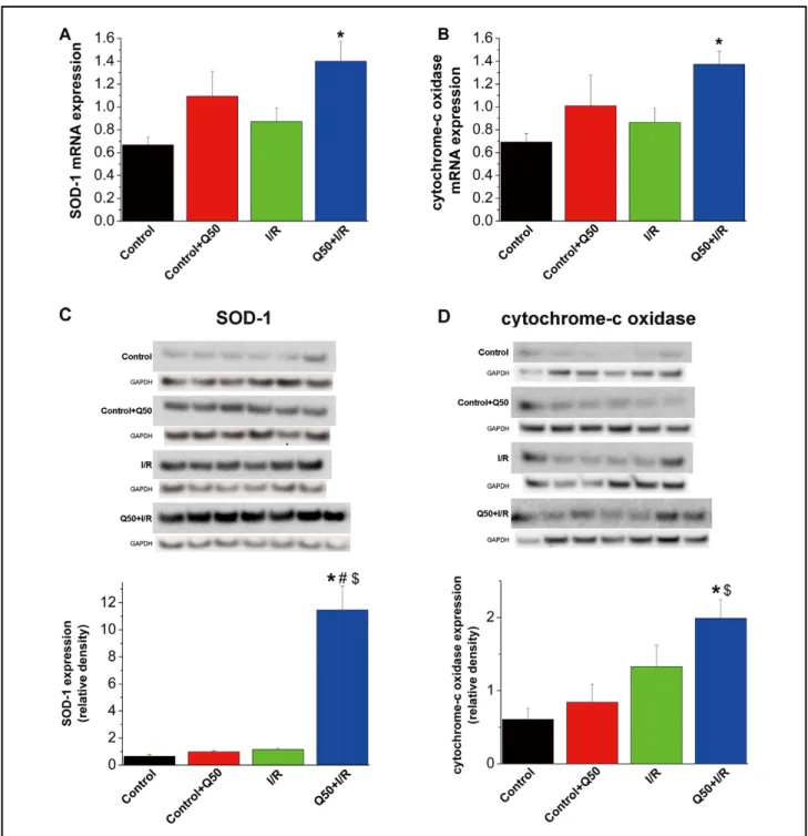
Effect of Q50 on Graft Gene Expression Quantitative real- time polymerase chain reaction of myocardial RNA extracts revealed that relative mRNA expression for superoxide dis- mutase (SOD)-1 and cytochrome-c oxidase remained un- changed in the control, control–Q50 and I/R groups. However, their expressions were significantly upregulated after Q50 treat- pressure were significantly reduced in the I/R, I/R + Q50 and
sham + Q50 groups as compared with sham-operated rats (Table 1). When compared with the sham group, rats with MI showed significantly decreased LV end-systolic pressure, pre- load recruitable SW, dP/dtmin and impaired cardiac relaxation, as reflected by prolonged Tau. Postischemic treatment with Q50 did not significantly restore these parameters (Table 1).
Effect of Q50 Pretreatment on Global I/R Injury (Post-Heart Transplantation)
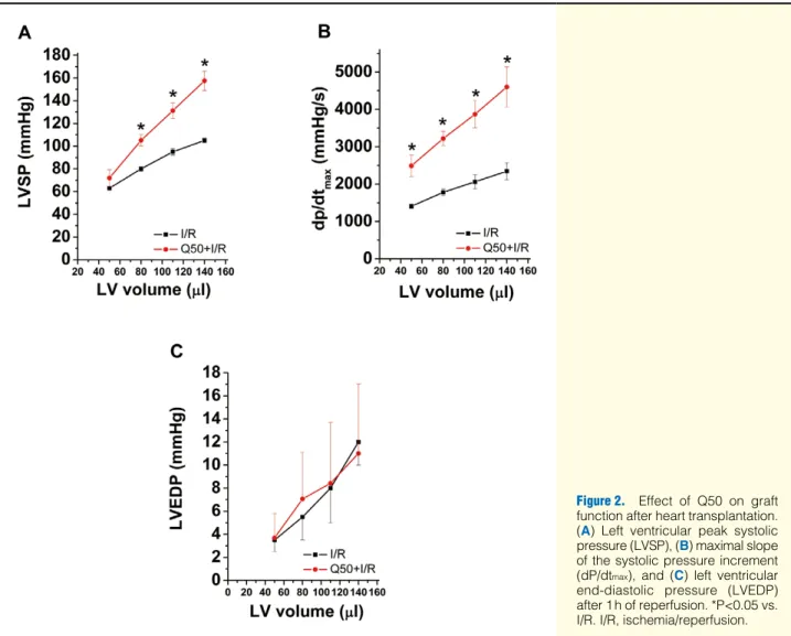
Effect of Q50 on Graft Function After 1 h of reperfu- sion, LV systolic pressure and dP/dtmax were significantly increased in the Q50-treated group compared with the I/R group, indicating improved myocardial contractility (Table 2, Figures 2A,B). Moreover, Q50 treatment resulted in a sig- nificant increase in dP/dtmin values compared with the I/R group, reflecting improved myocardial relaxation (Table 2).
LVEDP, as a marker of the standardized balloon-catheter measurements, did not show any major differences (Table 2, Figure 2C).
Effect of Q50 on Graft Myocardial High-Energy Phosphate Content Myocardial high-energy phosphate content and the ATP and ADP levels were preserved by Q50 preconditioning as compared with the I/R group (Table 3). AMP level did not show any relevant changes between the groups. The energy
Table 2. Effect of Q50 on Graft Function After Heart Transplantation
Parameter I/R Q50 + I/R
LVSP (mmHg) 80±2 105±5*
dP/dtmax (mmHg/s) 1,781±94 3,219±190*
LVEDP (mmHg) 5.5±2.0 7.1±4.1
dP/dtmin (mmHg/s) 989±115 2,477±424*
*P<0.05 vs. I/R.
LVSP, dP/dtmax, LVEDP and dP/dtmin at an intraventricular volume of 80 μl 1 h after reperfusion.
dP/dtmax, maximal slope of the systolic pressure increment;
LVEDP, LV end-diastolic pressure; LVSP, LV peak systolic pres- sure. Other abbreviations as in Table 1.
Figure 2. Effect of Q50 on graft function after heart transplantation.
(A) Left ventricular peak systolic pressure (LVSP), (B) maximal slope of the systolic pressure increment (dP/dtmax), and (C) left ventricular end-diastolic pressure (LVEDP) after 1 h of reperfusion. *P<0.05 vs.
I/R. I/R, ischemia/reperfusion.
Table 3. Effect of Q50 on Myocardial ATP, ADP, AMP Contents in Rat Model of Heart Transplantation
Parameter Control I/R Q50 + I/R
ATP (μmol/g) 6.58±1.12 1.86±0.41* 6.66±0.63
ADP (μmol/g) 3.48±0.16 2.05±0.42* 5.01±0.43
AMP (μmol/g) 1.91±0.22 2.07±0.22 2.59±0.61
Energy charge potential 0.69±0.07 0.49±0.04* 0.85±0.08
*P<0.05 vs. other groups.
I/R, ischemia/reperfusion; ATP, adenosine triphosphate; ADP, adenosine diphosphate; AMP, adenosine monophos- phate.
Figure 3. Effect of Q50 on graft gene and protein expression after heart transplantation. (A) Superoxide dismutase (SOD)-1, (B) cytochrome-c oxidase mRNA expression and immunoblot analysis for (C) SOD-1 and (D) cytochrome-c oxidase protein band densities in the myocardium. *P<0.05 vs. control, $P<0.05 vs. control + Q50, #P<0.05 vs. I/R. I/R, ischemia/reperfusion.
Figure 4. Effects of post-treatment with Q50 on H9c2 rat myocardial cell after hydrogen peroxide (H2O2)-in- duced oxidative stress. (A) Determi- nation of the effects of Q50 on the cell index, measured by real-time cell electronic sensing method in cultured rat cardiomyocytes. H9c2 rat embry- onic cardiac muscle cells were sub- jected to oxidative stress (100 μmol/L H2O2; arrow 1). After 30 min, Q50 was added to the wells (arrow 2). Curves in the Figure are measurements of single wells. Normalized cell index shows the relative viability of cells per well. The black vertical line in the middle of the graph indicates time of normalization of cell index, which is the time of H2O2 application. (B) Ef- fect of Q50 on relative heme oxy- genase (Hmox)-1 gene expression in H9c2 rat myocardial cells after H2O2- induced oxidative stress. Expression ratios were determined at 1, 3 and 24 h post-treatment.
Figure 5. Effect of Q50 on matrix metalloproteinases (MMPs). (A) In vitro effects of Q50 on human MMP-2 and MMP-9 enzyme activities. (B) Effects of Q50 on myocardial MMP-2 protein expression after heart transplantation. *P<0.05 vs. control, $P<0.05 vs.
control+Q50, #P<0.05 vs. I/R. I/R, ischemia/reperfusion.
tion. End-systolic volume, a marker of ventricular contractil- ity, was also increased in the MI group. We found that admin- istration of Q50 before the onset of reperfusion improved LV systolic function. The major indicator of the transition from reversible to irreversible I/R injury is the release of intracel- lular cardiac enzymes or markers, such as troponin-T, lactate dehydrogenase, creatine kinase, and aspartate aminotransfer- ase, into the circulation,21 reflecting major cellular membrane damage and/or death of cardiomyocytes.22 In the present study, the increased plasma levels of cardiac troponin-T were not re- duced by post ischemic administration of Q50. Letienne et al showed that there is a linear relationship between infarct size and plasma levels of biochemical markers.23 Moreover, Q50 failed to result in a reduction of infarct size after temporary occlusion followed by reperfusion as compared with controls.
Even though our study demonstrated improved cardiac func- tion after myocardial ischemia, application of Q50 did not decrease the elevated concentration of cardiac troponin-T or the infarct size, indicating no protective effect on damaged cardiomyocytes. It should be noted that the aforementioned enzyme-biomarker reflects mainly the amount of irreversibly injured myocytes and necrosis, but not the amount of dysfunc- tional myocytes without irreversible injury. Taken together, these observations support the view that the ability of Q50 to improve cardiac performance may be partially related to this iron-chelating and zinc-complexing agent rescuing cardio- myocytes from the border zones and remote regions of the infarcted heart, improving their function, which in turn leads to an improved global cardiac performance.
Effects of Q50 Pretreatment on Graft Dysfunction After Heart Transplantation
Fast recovery of myocardial function is essential for the suc- cess of cardiac transplantation. Therefore, we investigated the effects of Q50 therapy in the early phase (1 h) after heart transplantation. We attempted to simulate clinical conditions encountered during heart transplantation to investigate the potential use of Q50 in enhancing the current protective strat- egy. We previously described that crystalloid cardioplegia as- sociated with cardiac arrest and reperfusion results in a decline of cardiac function.24 In contrast to the MI rats in which only myocardial contractility was improved by post ischemic ad- ministration of Q50, our data showed that treatment of donor rats with Q50 restored both the altered systolic and diastolic LV function after heart transplantation. The different results in these models might be explained by the type of I/R (irre- versible vs. reversible) and timing of application (pre- vs. post- ischemic treatment).
Mechanism of Cardioprotective Effects of Q50 Against I/R Injury
One of the most cited mechanisms of reperfusion injury is the generation of free oxygen radicals at the time of reperfusion.
These free radicals are superoxide anions, hydroxyl radicals and hydrogen peroxide. Therefore, we studied the effects of Q50 on oxidative stress induced by hydrogen peroxide in cul- tured cardiomyocytes. By using a cell-microelectronic sensing technique for the screening of cytoprotective compounds,20 we demonstrated pronounced and concentration-dependent car- dioprotective effects of Q50 on H2O2-treated rat embryonic heart cells in a 30-min post-treatment in vitro model. We found that Q50 induced Hmox-1 expression with similar kinetics as the H2O2 stress in vitro. MMPs are a family of zinc-dependent endopeptidases25 capable of degrading extracellular matrix pro- teins, and zinc is essential for their proteolytic capacity in this ment when compared with the control group (Figures 3A,B).
Effect of Q50 on Graft Protein Expression Densitometric analysis of bands for SOD-1 and cytochrome-c oxidase did not show any differences among the control, control–Q50 and I/R groups. However, after heart transplantation, Q50 treat- ment significantly upregulated the protein expression of SOD-1 when compared with controls and the I/R group (Figure 3C) and increased the cytochrome-c oxidase protein level com- pared with controls (Figure 3D).
Effect of Q50 Post-Treatment on H9c2 Rat Myocardial Cells After H2O2-Induced Oxidative Stress
Cytoprotective Effect of Post-Treatment With Q50 Measured by Real-Time Cell-Microelectronic Sensing Technique Cells were attached and grown overnight and subjected to H2O2- induced oxidative stress (Figure 4A). After 30 min, Q50 was added to the wells (Figure 4A) at given concentrations.
Normalization of cell index was calculated at the time point of starting H2O2 application. Exposure of H9c2 cells to 100 μmol/L H2O2 resulted in a rapid decrease of the cell index, whereas the cell index of the absolute control cells that did not receive H2O2 treatment remained slightly increased. Post- treatment of Q50 exerted a dramatic dose-dependent cyto- protective effect after H2O2 stress: concentrations as low as 0.5μmol/L maintained the cell index near absolute control lev- els after the initial 3 h of the experiments and the cell index remained markedly elevated high during the course of the en- tire experiment.
Effect of Q50 on Relative Heme Oxygenase-1 Gene Expression Hmox-1 expression ratios were determined at 1, 3 and 24 h post-treatment (Figure 4B). We found pronounced induction (on average, 7-fold) after 3 h. This induction decreased to 2-fold, but remained significantly higher in stressed cells com- pared with untreated cells. Q50 alone mimicked the effects of H2O2 on Hmox-1 expression; however, when relative mRNA levels were determined in cells treated with Q50 and exposed to H2O2, no significant differences were recorded compared with Q50 treatment without applying H2O2.
Effect of Q50 on MMPs
Q50 concentrations ranging from 0.3 to 10 μmol/L had no sig- nificant effect on the inhibition of either human MMP-2 or MMP-9 enzyme activities (Figure 5A). However, after heart transplantation, the graft protein expression of MMP-2 was significantly increased compared with controls. Q50-treatment of the donor animals at 1 h prior to explantation significantly downregulated graft MMP-2 expression (Figure 5B).
Discussion
I/R injury frequently occurs in a variety of clinical conditions, including MI and heart transplantation. In the present study using rat models of I/R-induced myocardial injury, we showed that administration of Q50, an iron-chelating and zinc-com- plexing agent, improved cardiac function in vivo and reduced oxidative stress on cardiomyocytes in vitro.
Effects of Q50 Post-Treatment on Cardiac Dysfunction After MIUsing an in vivo experimental model, we studied the cardio- protective effect of Q50 when administered after occlusion before reperfusion to simulate a clinical situation. In the pres- ent work, MI was characterized by significantly decreased systolic performance, impaired ventricular relaxation and an increase in end-diastolic volume, indicative of chamber dila-
Conclusions
This first in vivo study provides experimental evidence that treatment with an iron-chelating and zinc-complexing agent, Q50, improves cardiac functional recovery of the ischemic/
reperfused myocardium in rats. This improvement is indepen- dent of reducing infarct size and cardiac enzyme leakage from the infarcted heart. The mechanisms through which Q50 pro- vides cytoprotection may be restoration of myocardial high- energy phosphate levels, upregulation of the antioxidant en- zyme SOD-1 protein expression and Hmox-1 induction. This study suggests that Q50 may be a promising candidate to treat acute MI, myocardial protection during cardiac transplantation and perhaps more generally, open-heart operations involving a period of global ischemia; however, further experimental studies are required. Additionally, because Q50 is a newly developed agent, its subcellular mechanism of action is un- known at present. Therefore, future studies should aim at clar- ifying possible sites of action of Q50.
Acknowledgments
This study was supported by the Land Baden-Württemberg and by the János Bolyai Research Scholarship of the Hungarian Academy of Sci- ences (to T.R.) and by a grant from the National Development Agency of Hungary (KMR_12-1-2012-0072). S.K. and K.H. are supported by the Medical Faculty of the University of Heidelberg.
The expert technical assistance of Karin Sonnenberg, Patricia Kraft and Lutz Hoffmann is gratefully acknowledged. The authors thank Abigél Farkas and Ferenc Kolonics for their assistance with the manuscript.
Disclosures None.
References
1. Minamino T. Cardioprotection from ischemia/reperfusion injury:
Basic and translational research. Circ J 2012; 76: 1074 – 1082.
2. Ambrosio G, Tritto I. Clinical manifestations of myocardial stun- ning. Coron Artery Dis 2001; 12: 357 – 361.
3. Goldberg S, Greenspon AJ, Urban PL, Muza B, Berger B, Walinsky P, et al. Reperfusion arrhythmia: A marker of restoration of ante- grade flow during intracoronary thrombolysis for acute myocardial infarction. Am Heart J 1983; 105: 26 – 32.
4. Piper HM, Meuter K, Schafer C. Cellular mechanisms of ischemia- reperfusion injury. Ann Thorac Surg 2003; 75: S644 – S648.
5. Kloner RA. Does reperfusion injury exist in humans? J Am Coll Cardiol 1993; 21: 537 – 545.
6. Hori M, Nishida K. Oxidative stress and left ventricular remodelling after myocardial infarction. Cardiovasc Res 2009; 81: 457 – 464.
7. Szabo G, Bahrle S, Stumpf N, Sonnenberg K, Szabo EE, Pacher P, et al. Poly(ADP-Ribose) polymerase inhibition reduces reperfusion injury after heart transplantation. Circ Res 2002; 90: 100 – 106.
8. Ago T, Kuroda J, Kamouchi M, Sadoshima J, Kitazono T. Patho- physiological roles of NADPH oxidase/nox family proteins in the vascular system: Review and perspective. Circ J 2011; 75: 1791 – 1800.
9. Hearse DJ, Bolli R. Reperfusion induced injury: Manifestations, mechanisms, and clinical relevance. Cardiovasc Res 1992; 26: 101 – 108.
10. Gross GJ, Auchampach JA. Reperfusion injury: Does it exist? J Mol Cell Cardiol 2007; 42: 12 – 18.
11. Murphy E, Steenbergen C. Mechanisms underlying acute protection from cardiac ischemia-reperfusion injury. Physiol Rev 2008; 88: 581 – 609.
12. Yellon DM, Hausenloy DJ. Myocardial reperfusion injury. N Engl J Med 2007; 357: 1121 – 1135.
13. Venardos KM, Perkins A, Headrick J, Kaye DM. Myocardial isch- emia-reperfusion injury, antioxidant enzyme systems, and selenium:
A review. Curr Med Chem 2007; 14: 1539 – 1549.
14. Menasche P, Grousset C, Mouas C, Piwnica A. A promising ap- proach for improving the recovery of heart transplants: Prevention of free radical injury through iron chelation by deferoxamine. J Tho- rac Cardiovasc Surg 1990; 100: 13 – 21.
process. However, inappropriate, prolonged or excessive ex- pression of these enzymes has deleterious consequences. It has been shown that acute release of MMP-2 during reperfusion after ischemia contributes to cardiac mechanical dysfunction,26 and pharmacological inhibition of MMP-2 in rats produced cardioprotection similar to the effect of ischemic precondition- ing.27 As a result, MMPs are considered promising drug de- velopment targets28 and pharmacological inhibition of MMPs may be a strategy for the treatment of I/R injury. MMPs are generally inhibited by compounds containing reactive zinc- chelating groups.29 In the present study, regardless of the zinc- binding capacity of Q50, this agent did not show enzymatic inhibition of human MMP-2 and MMP-9 in a biochemical assay. However, Q50-treatment of the donor animals 1 h prior to explantation significantly downregulated increased graft MMP-2 expression after heart transplantation. Taken together, we can speculate that in vivo an indirect inhibitory mechanism (ie, binding zinc, which is essential for the catalytic activity of MMPs) might be possible. During I/R injury, the return of oxygen to ischemic tissues is accompanied by an increased production of ROS.30 Iron plays a role in the formation of the free radicals that contribute to oxidative stress, so its chelation makes it unavailable for this. Chelation of ferric iron with deferoxamine, an iron-chelator, has been shown to reduce the production of the hydroxyl radical, thereby reducing myocar- dial I/R injury.14 In our model of reversible global myocardial I/R, Q50 treatment resulted in a significant increase in SOD-1 protein expression, a first line defense antioxidant enzyme, indicating both a cytoprotective function and free radical scav- enging effect.
In this study, after heart transplantation, comparing with the control group, I/R injury lead to a significant decrease in the high-energy phosphate content. The present data clearly dem- onstrated that pretreatment with Q50 resulted in a better pres- ervation of the high-energy phosphate pool, primarily by in- creased myocardial ATP content, resulting in an improved energy status, as expressed by the significant higher energy charge potential. Importantly, the loss of cellular energetic pools, in turn affects myocardial function.31 Based on the re- sults of the present study, we propose that Q50 may contribute to better recovery of the cellular ATP and therefore improved myocardial contractility. In pathologic conditions, this iron- chelating and zinc-complexing agent may have promising an- tioxidant defense mechanisms.
Study Limitations
The present rat model of heterotopic heart transplantation was selected as a suitable model to evaluate I/R injury. The tech- nique includes both reperfusion with blood in a clinically rel- evant intact animal model, and robust assessment of LV func- tion. This model, however, has certain limitations. In particular, the LV beats in an unloaded condition (ie, the ventricles are perfused via the coronary circulation, but they do not eject), which, on the 1 hand, allows a faster recovery after I/R, but on the other, leads to a time-dependent mechanical deterioration and atrophy. Nevertheless, it has been shown that major dete- rioration does not occur until at least 24 h after implantation.32 A model of irreversible I/R injury (acute MI) gives rise to many unfavorable changes. Therefore, myocardial gene ex- pression and high-energy phosphate content were only assessed in our model of reversible I/R injury (heart transplantation) to examine the mechanism of action of Q50.
25. Hooper NM. Families of zinc metalloproteases. FEBS Lett 1994;
354: 1 – 6.
26. Cheung C, Marchant D, Walker EK, Luo Z, Zhang J, Yanagawa B, et al. Ablation of matrix metalloproteinase-9 increases severity of viral myocarditis in mice. Circulation 2008; 117: 1574 – 1582.
27. Giricz Z, Lalu MM, Csonka C, Bencsik P, Schulz R, Ferdinandy P.
Hyperlipidemia attenuates the infarct size-limiting effect of ischemic preconditioning: Role of matrix metalloproteinase-2 inhibition. J Pharmacol Exp Ther 2006; 316: 154 – 161.
28. Dorman G, Cseh S, Hajdu I, Barna L, Konya D, Kupai K, et al.
Matrix metalloproteinase inhibitors: A critical appraisal of design principles and proposed therapeutic utility. Drugs 2010; 70: 949 – 964.
29. Talbot DC, Brown PD. Experimental and clinical studies on the use of matrix metalloproteinase inhibitors for the treatment of cancer.
Eur J Cancer 1996; 32A: 2528 – 2533.
30. Ferdinandy P, Schulz R, Baxter GF. Interaction of cardiovascular risk factors with myocardial ischemia/reperfusion injury, precondi- tioning, and postconditioning. Pharmacol Rev 2007; 59: 418 – 458.
31. Humphrey SM, Cartner LA, Holliss DG. Critical early metabolic changes associated with myocardial recovery or failure after total ischaemia in the rat heart. Basic Res Cardiol 1987; 82: 304 – 316.
32. Galinanes M, Hearse DJ. Metabolic, functional, and histologic char- acterization of the heterotopically transplanted rat heart when used as a model for the study of long-term recovery from global ischemia.
J Heart Lung Transplant 1991; 10: 79 – 91.
Supplementary Files Supplementary File 1
Methods
Please find supplementary file(s);
http://dx.doi.org/10.1253/circj.CJ-12-1162 15. Ramu E, Korach A, Houminer E, Schneider A, Elami A, Schwalb H.
Dexrazoxane prevents myocardial ischemia/reperfusion-induced oxi- dative stress in the rat heart. Cardiovasc Drugs Ther 2006; 20: 343 – 16. 348.Powell SR, Hall D, Aiuto L, Wapnir RA, Teichberg S, Tortolani AJ.
Zinc improves postischemic recovery of isolated rat hearts through inhibition of oxidative stress. Am J Physiol 1994; 266: H2497 – H2507.
17. Everson RJ, Parker HE. Zinc binding and synthesis eight-hydroxy- quinoline-agarose. Bioinorg Chem 1974; 4: 15 – 20.
18. Pierre JL, Baret P, Serratrice G. Hydroxyquinolines as iron chelators.
Curr Med Chem 2003; 10: 1077 – 1084.
19. Radovits T, Korkmaz S, Loganathan S, Barnucz E, Bomicke T, Arif R, et al. Comparative investigation of the left ventricular pressure- volume relationship in rat models of type 1 and type 2 diabetes mel- litus. Am J Physiol Heart Circ Physiol 2009; 297: H125 – H133.
20. Ozsvari B, Puskas LG, Nagy LI, Kanizsai I, Gyuris M, Madacsi R, et al. A cell-microelectronic sensing technique for the screening of cytoprotective compounds. Int J Mol Med 2010; 25: 525 – 530.
21. Imamura T. Paradigm shift from myocardium-derived to plaque- derived biomarkers for very early diagnosis of acute myocardial in- farction. Circ J 2011; 75: 1322 – 1323.
22. Ravkilde J, Nissen H, Horder M, Thygesen K. Independent prognos- tic value of serum creatine kinase isoenzyme MB mass, cardiac troponin T and myosin light chain levels in suspected acute myocar- dial infarction: Analysis of 28 months of follow-up in 196 patients.
J Am Coll Cardiol 1995; 25: 574 – 581.
23. Letienne R, Bel L, Bessac AM, Denais D, Degryse AD, John GW, et al. Cardioprotection of cariporide evaluated by plasma myoglobin and troponin I in myocardial infarction in pigs. Fundam Clin Phar- macol 2006; 20: 105 – 113.
24. Loganathan S, Radovits T, Hirschberg K, Korkmaz S, Barnucz E, Karck M, et al. Effects of selective phosphodiesterase-5-inhibition on myocardial contractility and reperfusion injury after heart trans- plantation. Transplantation 2008; 86: 1414 – 1418.