Time course analysis of cardiac pacing-induced gene expression changes in the canine heart
Ma´ria Kova´cs•Ma´rton Go¨nczi •Edina Kova´cs• A´ gnes Ve´gh
Received: 1 August 2012 / Accepted: 14 September 2012 / Published online: 27 September 2012 ÓSpringer Science+Business Media New York 2012
Abstract Rapid right ventricular pacing in anesthetized dogs results in marked protection against ischemia and reperfusion-induced ventricular arrhythmias, 24 h later. We have previous evidence that this protection associates with altered expression of genes, encoding proteins involved in the delayed cardioprotection. However, the sequence of transcriptional changes occurring between the pacing stimulus and the test ischemia has not yet been elucidated.
Thus, we designed studies in which the expression of 29 genes was examined by real-time PCR at various time intervals, i.e., immediately (0 h), 6, 12, and 24 h after short periods (4 times 5 min) of rapid (240 beats min-1) right ventricular pacing in the canine. Sham-operated dogs (the pacing electrode was introduced but the dogs were not paced) served as controls. Compared with these dogs, pacing induced an early up-regulation of genes which encode, for example, HSP90, MnSOD, ERK1, PKCe, Bcl2, and sGC; all these somehow relate to the early phase of the protection. These genes remained either up-regulated or, after a transient lower expression (around 6 h), were up- regulated again, suggesting their involvement in the delayed protection. There were also some genes which down-regu- lated soon after the pacing stimulus (e.g., Bax, Casp3, Casp9, MMP9, GSK3b), and showed also low expres- sion 24 h later. Genes encoding eNOS and iNOS, as well as Cx43 were only up-regulated 12 h after pacing. We con- clude that cardiac pacing induces time-dependent changes in gene expression, and the sequence of these changes is important in the development of the delayed protection.
Keywords Cardiac pacingmRNA transcription Gene expression
Introduction
Short periods of rapid cardiac pacing protect the heart against severe ventricular arrhythmias that result from a subsequent ischemia and reperfusion insult in the anes- thetized canine [1]. This pacing-induced protection is short lived; it lasts\1 h [2]. The antiarrhythmic effect, however, re-appears 20–24 h after the initial pacing stimulus [2], and this late or delayed phase of the protection may persist over days, especially if pacing is repeated at a time when the protection from the previous stimulus has already waned [3]. Although the precise mechanisms of this delayed antiarrhythmic effect is still not fully elucidated, there is substantial evidence that endogenous substances that are released in response to the rapid pacing-induced global ischemic changes [4] initiate processes, which ultimately lead to protection several hours later [5,6].
The most likely explanation for the mechanism of the delayed phase of the protection is that these endogenous substances released by a preconditioning stimulus initiate transcriptional changes and modify protein synthesis and enzyme activation (reviewed by, e.g., [7]). One of the first suggestions for this comes from studies which showed that nitric oxide (NO), which is considered to be an important trigger and mediator in preconditioning-induced protection [8–10], releases during the preconditioning procedure (perhaps via eNOS activation) and triggers signaling pathways, leading to transcriptional activation of the iNOS gene, increased iNOS synthesis and activation, and further NO generation during the subsequent prolonged ischemic insult [11].
M. Kova´csM. Go¨ncziE. Kova´csA´ . Ve´gh (&) Department of Pharmacology and Pharmacotherapy, Faculty of Medicine, University of Szeged, Do´m te´r 12,
6720 Szeged, Hungary
e-mail: vegh.agnes@med.u-szeged.hu DOI 10.1007/s11010-012-1467-8
Since then a number of studies have examined the gene expression alterations in both the early [12, 13] and delayed (reviewed by, e.g., [7]) phases of the protection.
These studies showed that several genes are activated after the preconditioning stimulus and induce synthesis of pro- teins which certainly play roles in the development of the late phase of the protection [12–14]. For example, we have just recently shown that cardiac pacing induced changes in connexin43 (Cx43) mRNA and protein expressions, which by modifying gap junctional function influenced arrhyth- mia generation during ischemia and reperfusion [15].
Furthermore, in a previous study, we have reported that several genes, encoding, e.g., NO-producing enzymes, antioxidants, antiapoptotic proteins, etc., were activated in dogs subjected to cardiac pacing and, 24 h later, to a 25-min occlusion and reperfusion insult [16].
Although there is no doubt that gene expression changes occur and play essential role in the development of the delayed phase of the preconditioning-induced protection, the sequence of these changes at the transcript level has not yet been defined. Therefore, we designed studies in which preconditioning was induced by rapid cardiac pacing and changes in mRNA expression of 29 genes were analyzed immediately, 6, 12, and 24 h after cardiac pacing to find a time-dependent relationship in activation of these genes occurring between the pacing stimulus and the appearance of the delayed effect.
Materials and methods
Experimental animals and pacing procedure
Adult mongrel dogs of both sexes were used (mean body weight 20±1 kg). The origin and upkeep of these dogs were in accord with the Hungarian law (XXVIII, Chap. IV, Paragraph 31) regarding large experimental animals, which conforms with the Guide for the Care and Use of Labo- ratory Animals published by the US National Institute of Health (NIH Publication No. 85-23, revised 1996).
The pacing procedure was performed as described pre- viously [2]. In brief, the dogs were lightly anesthetized by the intravenous administration of sodium pentobarbitone (0.5 mg kg-1i.v., EuthanylÒBimeda-MTC Animal Health Inc.) and were allowed to breathe spontaneously. A pacing electrode (CordisF4, Johnson & Johnson Company, USA) was inserted via the right jugular vein into the right ven- tricle such as that it made contact with the ventricular wall.
The correct placing of this electrode was confirmed by recording the endocardial electrocardiogram. Arterial blood pressure was monitored from the left carotid artery using methods previously described in detail [2, 3]. The dogs were then paced, at a rate of 240 beats min-1for four
5-min periods, with 5-min rest intervals between the pacing stimuli.
Experimental groups
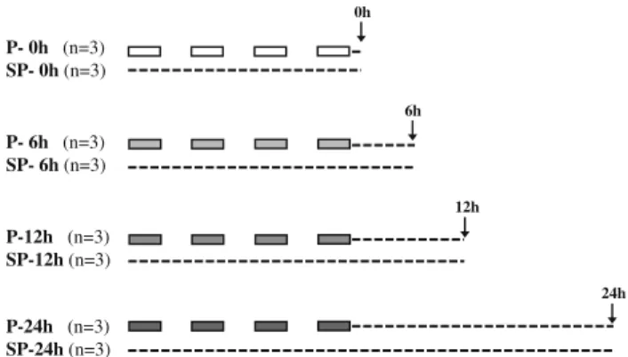
Twenty-four dogs were instrumented by introducing a pacing electrode into the right ventricle. Twelve out of these animals were subjected to rapid cardiac pacing (P), whereas the other 12 dogs served as sham-paced (SP) controls; in these animals the pacing electrode was inserted but the dogs were not paced. Dogs in both the paced and the non-paced groups were divided into four further groups, each containing three animals. From these dogs after euthanasia with an intravenous overdose of the anesthetic the hearts were sectioned immediately (P-0/SP- 0), 6 (P-6/SP-6), 12 (P-12/SP-12), or 24 (P-24/SP-24) h after the pacing or sham-pacing procedures (Fig.1).
Myocardial tissue samples were taken from the wall of the left ventricle, immediately frozen in liquid nitrogen and stored at a temperature of-80°C until further analyses.
Isolation of RNA
The frozen heart tissue (25 mg) was powdered in liquid nitrogen and total RNA was purified with the RNeasy Fibrous Tissue Mini Kit (Qiagen, Germany) according to the manufacturer’s instruction. Total RNA preparations from three dogs in each group were pooled together. The integrity of RNA was assessed by gel electrophoresis, whereas the quantity of RNA was spectrophotometrically measured. The total RNA preparations were used for reverse transcription and quantitative real-time PCR (qRT- PCR) measurements.
P- 0h (n=3) SP- 0h (n=3)
P- 6h (n=3) SP- 6h (n=3)
P-12h (n=3) SP-12h (n=3)
P-24h (n=3) SP-24h (n=3)
0h
6h
12h
24h
Fig. 1 Experimental protocol. Under light pentobarbitone anesthesia a bipolar pacing electrode was introduced into the right ventricle through which the heart was paced (P) four times for 5 min at a rate of 240 beats min-1. Sham-operated dogs (SP; the pacing electrode was introduced but the dogs were not paced) served as controls. The hearts were stopped at various times, i.e., immediately (0 h), 6, 12, and 24 h after pacing with an excess of anesthetic and tissue samples were taken from the wall of the left ventricle
Assessment of mRNA transcription
Quantitative RT-PCR was performed by ABI7000 instru- ment (Applied Biosystems, Carlsbad, USA) with gene- specific primers to monitor gene expression changes. Two micrograms of total RNA from each sample was reverse transcribed using ImPromII reverse transcriptase (Promega, USA) in the presence of poly(dT) primers at 42°C for 1 h.
cDNAs were then diluted in 50-ll water, and 1ll of this reaction mix was used as template for qRT-PCR. This was performed by means of the SybrGreen protocol with SYBR Green PCR Master Mix with passive reference dye (Applied Biosystems, Carlsbad, USA) according to the manufacturer’s instructions at a final primer concentration of 500 nM. The primers were designed using the Primer- Express 2.0 (Applied Biosystems, Carlsbad, USA). Rela- tive expression ratios were calculated as normalized ratios to hypoxanthine guaninephosphoribosyltransferase (HPRT) and to tubulin. A non-template control sample was applied for each run to control the genomic DNA contaminations of the template. Melting temperature analysis has been performed after each reaction to assess the quality of the reaction.
The final relative gene expression changes were calcu- lated as delta–delta Ct values. Genes with expression val- ues lower than 0.5 or higher than 1.5 were considered to be down- or up-regulated (the values correspond to an interval of log2= -1 and log2=0.6) as has been previously described [16]. All the PCRs were performed in triplicate with two parallel running for each reaction. All data were expressed as mean ±SD. To compare the mean score of the relative expression ratios to a given constant (=1) ‘‘one samplettest’’ was performed using the Statistica software.
Differences were considered significant atP\0.05.
Immunoblot analysis
Changes in eNOS protein content and enzyme activation were assessed by western blot. The preparation and the determination of the concentration of total protein were described previously [15]. Protein extracts were resolved using 8 % sodium dodecyl sulfate-polyacrylamide gel electrophoresis and blotted on polyvinylidene fluoride membranes (PVDF). The membranes were incubated with two different primary antibodies; anti-eNOS (BD Trans- duction LaboratoriesTM) was used to measure the protein expression of eNOS and antiphospho-eNOS (peNOS, Ser1177, BD Transduction LaboratoriesTM) to detect the activation of eNOS. Band densities were detected with the ECL Plus kit (GE Healthcare, Buckinghamshire, UK) and developed on Amersham HyperfilmTM (GE Healthcare, Buckinghamshire, UK). Pixel intensities of each band were measured using ImageJ software (NIH) and were
normalized to the highest value on each blot. Three parallel western blots were performed for the statistical analysis using Bonferroni correction. For the verification of equal loading, PVDF membranes were labeled with Coomassie Blue.
Results
Gene expression alterations after rapid cardiac pacing These are illustrated in Table1 and in Figs.2, 3, and 4.
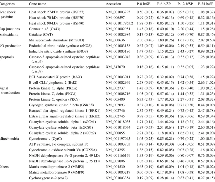
Table1 summarizes the gene expression changes of the total 29 genes, assessed at various time intervals (i.e., immediately 6, 12, and 24 h) after cardiac pacing. The results are expressed as ratios of gene expression data obtained from the time-matched sham-paced control hearts.
There were eight genes which up-regulated immediately after the cessation of pacing (Table1). These early up- regulated genes encode, for example, HSP90, MnSOD, Bcl-2, PKCe, ERK1, and isoforms of the soluble guanylate cyclase (sGCa1, sGCb1, and sGCa2). There were five genes which exhibited an early down-regulation following the pacing stimulus, such as catalase (CAT), caspase3 (casp3), and caspase9 (casp9), GSK3b and MMP9. Of these early up- or down-regulated genes MnSOD, PKCe, casp3 and casp9, GSK3band MMP9 remained unchanged (i.e., up- and down-regulated) at almost each time-point, whereas the others, like ERK1, Bcl-2, and sGC isoforms, showed biphasic transcriptional changes, i.e., after the initial up-regulation these genes were down-regulated (mostly at 6 h) but up-regulated again around 12 and 24 h (Table1). There were also genes, such as those encode NO-producing enzymes (eNOS, iNOS) and the gap junc- tion protein connexin43 (Cx43), which exhibited signifi- cant up-regulation only 12 h after the pacing stimulus (Table1). Surprisingly, the expressions of genes respon- sible for the transcription of mitochondrial proteins, as well as Cox2, were not significantly altered.
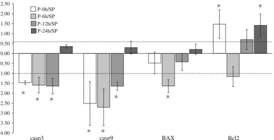
The results are also illustrated in figures by selecting those genes which exhibited significant transcription changes following cardiac pacing, and which may have essential functional roles in the protection afforded by preconditioning. These genes encode proteins that are involved, for example, in apoptosis (both apoptotic and antiapoptotic genes; Fig.2) and signal transduction (Fig.3), or genes, that are responsible for the transcription of NO-producing enzymes (eNOS and iNOS), antioxidants (e.g., MnSOD), metalloproteinases (e.g., MMP9) and Cx43 (Fig.4).
Figure2shows that genes involved in apoptosis such as casp3 and casp9 were significantly down-regulated imme- diately (0 h), 6 and 12 h after cardiac pacing, whereas the
gene, encoding the pro-apoptotic Bax, was significantly down-regulated only at 6 h. In contrast, pacing enhanced at each time point, except 6 h, the expression of gene encoding the antiapoptotic Bcl-2.
The time-course pattern of genes encoding proteins involved in signal transduction showed large variation. Of these genes only PKCeexhibited a significant increase in the transcription at each time point following cardiac pacing. PKCa was only up-regulated at 12 and 24 h, whereas the expression of PKCd was not significantly altered (Fig.3). ERK1 showed an early (0 h) and late (24 h) up-regulation, whereas GSK3b was significantly down-regulated at all time points, except 12 h. Genes encoding the isoforms of sGC were significantly up-
regulated immediately, and 24 h after the cessation of the pacing stimulus (Fig.3).
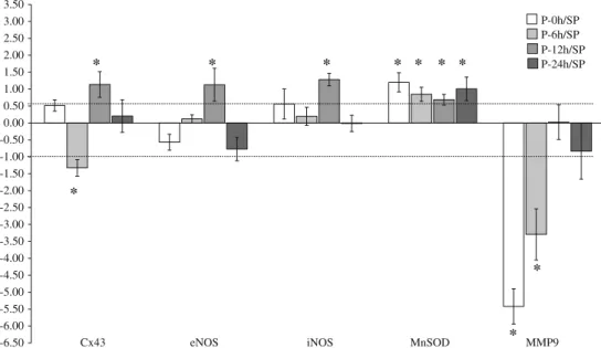
Interestingly, genes responsible for the transcription of eNOS, iNOS were significantly up-regulated only 12 h after pacing; neither before nor after they showed signifi- cant alterations in their expressions (Fig.4). In contrast, the mRNA expression of MnSOD was up-regulated at all time points. The transcription of Cx43 was down-regulated at 6 h, but up-regulated at 12 h after pacing and, as we have described previously, these changes in Cx43 gene expres- sion were in association with arrhythmia generation resulting from ischemia and reperfusion [15]. The gene of the endopeptidase MMP9 was significantly down-regulated immediately and 6 h after pacing (Fig.4).
Table 1 Time-course changes in gene expression following cardiac pacing in dogs
Categories Gene name Accession P-0 h/SP P-6 h/SP P-12 h/SP P-24 h/SP
Heat shock proteins
Heat shock 27-kDa protein (HSP27) NM_001003295 0.50 (0.01) 0.36 (0.07) 0.92 (0.21) 1.08 (0.37) Heat shock 70-kDa protein (HSP70) NM_006597 0.99 (0.72) 0.19 (0.13) 0.69 (0.48) 0.32 (0.16) Heat shock 90-kDa protein (HSP90) NM_001017963.2 1.78 (0.19) 0.85 (0.17) 1.50 (0.25) 1.11 (0.31) Gap junctions Connexin 43 (Cx43) NM_001002951 1.43 (0.18) 0.40 (0.10) 2.20 (0.44) 1.15 (0.28) Antioxidants Catalase (CAT) NM_001002984 0.17 (0.13) 0.25 (0.12) 0.89 (0.70) 0.87 (0.46) Mn superoxide dismutase (MnSOD) NM_000636 2.30 (0.46) 1.80 (0.26) 1.61 (0.15) 2.02 (0.50) NO production Endothelial nitric oxide synthase (eNOS) NM_001003158 0.67 (0.07) 1.09 (0.06) 2.19 (0.53) 0.59 (0.11) Inducible nitric oxide synthase (iNOS) NM_001003186 1.47 (0.45) 1.15 (0.22) 2.43 (0.27) 0.99 (0.21) Apoptosis Caspase-3 apoptosis-related cysteine peptidase
(casp3)
NM_001003042 0.36 (0.09) 0.33 (0.13) 0.32 (0.12) 1.28 (0.08) Caspase-9 apoptosis-related cysteine peptidase
(casp9)
XM_847070 0.18 (0.16) 0.15 (0.11) 0.32 (0.05) 1.23 (0.22) BCL2-associated X protein (BAX) NM_001003011 0.72 (0.28) 0.32 (0.02) 0.74 (0.38) 1.15 (0.22) B cell CLL/lymphoma 2 (Bcl2) NM_001002949 2.78 (0.99) 0.45 (0.15) 1.62 (0.54) 2.66 (1.02) Signal
transduction
Protein kinase C, alpha (PKCa) NM_002737 1.42 (0.39) 0.87 (0.36) 2.15 (0.40) 1.90 (0.23) Protein kinase C, delta (PKCd) NM_001008716 1.05 (0.01) 0.57 (0.14) 1.44 (0.32) 1.31 (0.23) Protein kinase C, epsilon (PKCe) NM_005400 6.73 (2.43) 1.77 (0.32) 2.27 (0.31) 2.08 (0.37) Glycogen synthase kinase 3 beta (GSK3b) NM_002093 0.37 (0.10) 0.34 (0.08) 0.71 (0.30) 0.44 (0.09) Extracellular signal-regulated kinase 1 (ERK1) NM_002746 2.32 (0.37) 0.49 (0.24) 0.72 (0.42) 2.47 (0.78) Extracellular signal-regulated kinase 2 (ERK2) NM_002745 0.98 (0.35) 0.95 (0.36) 1.26 (0.66) 0.59 (0.34) Guanylate cyclase soluble, alpha 1 (sGCa1) NM_001018035 1.71 (0.14) 1.44 (0.26) 1.12 (0.21) 2.44 (0.16) Guanylate cyclase soluble, beta 1(sGCb1) NM_001018034 2.97 (0.53) 2.51 (0.64) 1.27 (0.19) 2.60 (0.51) Guanylate cyclase soluble, alpha 2 (sGCa2) NM_000855 2.21 (0.81) 1.18 (0.07) 1.62 (0.11) 2.41 (0.90) Mitochondria Cytochrome c (CytC) NM_001197045 1.05 (0.25) 0.85 (0.21) 0.79 (0.22) 1.00 (0.54) ATP synthase, Fo complex, subunit F6 NM_001003703 1.48 (0.14) 0.93 (0.30) 0.64 (0.05) 0.51 (0.09) Cytochrome c oxidase subunit Va (COX5A) NM_004255 1.38 (0.15) 0.82 (0.05) 0.02 (0.28) 1.16 (0.07) NADH dehydrogenase Fe–S protein 2, 49 kDa NM_001166159 1.33 (0.19) 0.59 (0.06) 0.80 (0.07) 0.76 (0.09) NADH dehydrogenase Fe–S protein 1, 75 kDa NM_005006 1.05 (0.18) 0.65 (0.16) 0.46 (0.08) 0.52 (0.07) Others Matrix metalloproteinase 2 (MMP2) NM_004530 0.63 (0.19) 0.65 (0.09) 1.04 (0.18) 0.73 (0.02) Matrix metalloproteinase 9 (MMP9) NM_001003219 0.06 (0.08) 0.17 (0.04) 1.08 (0.38) 0.59 (0.31) Cyclooxygenase-2 (cox2) NM_001003354 0.19 (0.09) 0.28 (0.14) 0.87 (0.41) 0.27 (0.15) Relative gene expression ratios were calculated compared the paced with the sham-paced dogs. Genes with expression values lower than 0.5 or higher than 1.5 were considered to be down- or up-regulated. Values are mean±SD (indicated in brackets) of three parallel qRT-PCR reactions
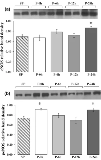
Time-course changes in eNOS protein content and activity following cardiac pacing
The results of the above gene expression studies have prompted us to perform additional experiments in which changes in eNOS protein content and activity were
determined by western blot using the same control and paced heart samples. The results are illustrated in Fig.5.
Compared to the sham-paced controls in dogs subjected to pacing the total eNOS protein content was significantly increased only 24 h after pacing (Fig.5a), whereas the activity of eNOS, as assessed by the measurement of the
-4.00 -3.50 -3.00 -2.50 -2.00 -1.50 -1.00 -0.50 0.00 0.50 1.00 1.50 2.00 2.50
casp3 casp9 BAX Bcl2
P-0h/SP P-6h/SP P-12h/SP P-24h/SP
* * *
* *
* *
* *
Fig. 2 The time-course effect of rapid cardiac pacing on the expression of genes encoding proteins involved in apoptosis.
Pacing-induced biphasic activation (early and late up-regulation) of the gene, encoding the antiapoptotic regulatory protein Bcl-2 and markedly suppressed the expression of genes which encode proteins
involved in the enhancement of apoptotic cell death. Values are mean±SD. Thelinesindicate the interval between 0.5- and 1.5-fold of regulation (the values correspond to an interval of log2= -1 and log2=0.6). *P\0.05 (one samplettest)
-2.50 -2.00 -1.50 -1.00 -0.50 0.00 0.50 1.00 1.50 2.00 2.50 3.00 3.50 4.00
P-0h/SP P-6h/SP P-12h/SP P-24h/SP
PKCα PKCδ PKCε GSK3β ERK1 ERK2 sGCα1 sGCβ1
*
*
* * *
* *
*
*
*
* * *
*
Fig. 3 The time-course changes in transcription of genes encoding proteins involved in intracellular signal transduction. Genes encoding PKCe, ERK1, and subunits of soluble GC were up-regulated either continuously or in a biphasic manner (early and late up-regulation) over the 24-h observation period, indicating a role the activation of these genes in both the early and the late phase of the protection. The
expression of GSKb3 gene was significantly suppressed at almost each time point of the observation. Values are mean±SD. Thelines indicate the interval between 0.5- and 1.5-fold of regulation (the values correspond to an interval of log2= -1 and log2=0.6).
*P\0.05 (one samplettest)
phosphorylated form of this enzyme, was markedly increased twice, i.e., immediately (0 h) and also 24 h after the pacing stimulus (Fig.5b).
Discussion
There is abundant evidence that gene expression changes play an important role in the delayed phase of cardiopro- tection afforded by preconditioning (reviewed by, e.g., 7, 13]). There has not been, however, a systematic evaluation of the sequence of gene expression changes, occurring during that time interval (20–24 h), which elapses between the preconditioning stimulus and the re-appearance of the protection [9,17]. Therefore, we designed experiments to examine the time-course alterations in the expression of 29 genes, encoding proteins that have been proposed to play roles in preconditioning-induced delayed cardioprotection.
We used our established canine model in which precon- ditioning was induced by four 5-min periods of rapid (overdrive) pacing stimulus, interspersed by 5-min reper- fusion intervals [2]. Myocardial tissue samples were har- vested at various time points (0, 6, 12, and 24 h) after pacing to determine transcription changes by qRT-PCR.
The examined genes fall into different categories as regards the function of proteins which they encode, e.g., heat shock proteins, antioxidants, as well as proteins that are involved in signal transduction, mitochondrial respira- tion and ATP synthesis, apoptosis and NO synthesis, etc.
Of these 29 genes 8 genes showed up-regulation and 5 genes exhibited down-regulation immediately after the cessation of the last pacing stimulus. For example, the transcription of MnSOD, which has a particular importance in defenses against oxidative stress, was significantly up- regulated at all time points of the observation, suggesting an essential role of this enzyme in both the early and the delayed phases of the protection [17–20]. We have previ- ous evidence that the expression of MnSOD was signifi- cantly increased in paced dogs subjected to coronary artery occlusion and reperfusion 24 h later, compared with the un-paced controls [16]. This finding supports the proposed parallelism between the induction of MnSOD and the protection [17], although a direct evidence for a cause-and- effect relationship between the up-regulation of MnSOD and the pacing-induced delayed antiarrhythmic effect has not yet been established. Interestingly, the other examined antioxidant enzyme catalase exhibited down-regulation following cardiac pacing. Although it had been found that under in vitro conditions preconditioning increases the mRNA transcription of this enzyme [18,19], these results were not confirmed by the results of the in vivo studies;
i.e., neither catalase in dogs [20] nor the other antioxidant enzymes in conscious pigs [21] were up-regulated in the time of the delayed protection.
Among genes encoding proteins and enzymes involved in signal transduction PKCe, ERK1 and the isoforms of sGC were found to exhibit early activation. This is perhaps not surprising as they are all parts of survival pathways
-6.50 -6.00 -5.50 -5.00 -4.50 -4.00 -3.50 -3.00 -2.50 -2.00 -1.50 -1.00 -0.50 0.00 0.50 1.00 1.50 2.00 2.50 3.00 3.50
eNOS iNOS MnSOD MMP9
Cx43
P-0h/SP P-6h/SP P-12h/SP P-24h/SP
*
* * * * * * *
*
*
Fig. 4 The time-course changes in expression of genes which encode eNOS, iNOS, MnSOD enzymes as well as Cx43 and MMP9. The antioxidant MnSOD was markedly activated at mRNA level at all measured time points, whereas the gene expression of the NOS- producing enzymes showed a significant up-regulation only 12 h after cardiac pacing. The gene of Cx43 down-regulated at 6 h, but up-
regulated at 12 h; the significance of these gene expression changes are explained in detail elsewhere [16]. The transcription of MMP9 was significantly suppressed following cardiac pacing. Values are mean±SD. Thelinesindicate the interval between 0.5- and 1.5-fold of regulation (the values correspond to an interval of log2= -1 and log2=0.6). *P\0.05 (one samplettest)
leading to early cardioprotection [22,23]. For example, the mRNA of PKCe showed a marked up-regulation at all the measured time points, indicating a role of PKCein both the phases of the protection. Indeed, there is strong phar- macological evidence that the translocation of PKCeplays an important role in ‘‘classic’’ preconditioning [22] and that the activation ofeisoform is necessary for the development of the late phase of the protection [24]. In our experiments, PKCa also showed significant up-regulation 12 and 24 h after cardiac pacing, suggesting that in dogs this PKC isoform may also contribute to the delayed effect. Fur- thermore, genes encoding ERK1 and sGC exhibited rather a biphasic pattern in their expression following cardiac pacing. These genes after the initial up-regulation were
either normalized or down-regulated, but a few hours later they were up-regulated again, indicating a role of these pathways in the delayed phase of the preconditioning- induced cardioprotection.
The early and the late activation of genes encoding proteins involved in signal transduction may associate with the subsequent modification of GSK3btranscription. This was markedly down-regulated at almost each time point in the paced dogs. GSK3bis a key enzyme to integrate mul- tiple signaling pathways in the cell and it is considered as an essential part of the mediator pathway leading to early protection [22]. It is usually active under normal physio- logical conditions and inhibits its target proteins through phosphorylation. Stimulation of various kinases involved in cardioprotective signaling (Akt, PKC, PKA, and ERK) inactivates GSK3b via phosphorylation, which in turn reduces the phosphorylation and activation of proteins, such as the pro-apoptotic Bax [25] and inhibits the opening and formation of mPTPs [26]. The fact that cardiac pacing keeps the gene expression of these cardioprotective kinases acti- vated over a longer period and that, simultaneously, down- regulates the expression of GSK3b mRNA may suggest a role for this pathway not only in the early but also in the delayed phase of the protection.
Another important signaling pathway which is certainly involved in cardioprotection associated with precondition- ing is the NO-activated sGC pathway. We have proposed for the first time that NO released during the preconditioning stimulus by activating sGC results in protection against arrhythmias [27]. The evidence for this was that in dogs the administration of methylene blue completely abolished the early antiarrhythmic effect of ischemic preconditioning [28].
More recent studies using ODQ, a more selective inhibitor of sGC, have confirmed this finding and provided further pharmacological evidence for the role of NO-sGC pathway in the early [29] and in the late [30] phase of the protection.
The biphasic transcriptional activation of the various iso- forms of sGC (sGCa1, sCGa2, and sGCb1) at the transcript level following cardiac pacing also supports a role for this pathway in mediating both phases of the protection.
It is well established that NO, released during a pre- conditioning stimulus, plays a trigger and mediator role in both the early and the delayed phase of the protection [8–
10]. We had proposed previously that the antiarrhythmic effects of ischemic preconditioning [31], cardiac pacing [1, 2], and physical exercise [32] can be associated with increased NOS (eNOS and iNOS) expression leading to enhanced NO production with the resultant modulation of the cAMP/cGMP balance in cardiomyocytes [4–6]. We have now provided evidence for this by demonstrating a marked increase in the transcription of both eNOS and iNOS 12 h after cardiac pacing. Furthermore, interestingly, this was the only time point when the expression of these
*
*
0.00 0.20 0.40 0.60 0.80 1.00 0.00 0.20 0.40 0.60 0.80 1.00
eNOS relative band densitypeNOS relative band density
SP P-0h P-6h P-12h P-24h
(a)
(b)
SP P-0h P-6h P-12h P-24h
SP P-0h P-6h P-12h P-24h
SP P-0h P-6h P-12h P-24h
*
Fig. 5 Time-course changes in eNOS protein content (a) and acti- vation (b) determined at various time intervals after rapid cardiac pacing by western blot. Compared to the sham-operated controls, cardiac pacing resulted in significant increase in eNOS protein content only 24 h after pacing (a). However, the enzyme activity showed a significant increase twice, i.e., immediately and 24 h after the cessation of the pacing stimulus (b), indicating a possibility for both early and late enhancements of NO production. Values are mean±SEM. *P\0.05 versus SP group (Bonferroni correction)
genes was significantly up-regulated (Fig.4). This finding might have a particular importance, since previous studies in conscious rabbits have reported that the activity of constitutive calcium-dependent NOS enzymes (eNOS and nNOS) was increased just immediately after precondi- tioning but remained unaffected 24 h later [33], when only iNOS had shown mRNA up-regulation and marked enzyme activation [11].
There is no doubt that iNOS expression and the resultant increase in NO generation is essential for the development of the delayed antiarrhythmic protection. We have phar- macological evidence for this using various, more or less selective inhibitors of iNOS [3, 9, 34, 35] which are all markedly attenuated or even abolished the delayed pro- tection against arrhythmias resulted from cardiac pacing [3, 9, 35] or heavy physical exercise [32, 34]. As these inhibitors are, indeed, not entirely selective for iNOS, for example, dexamethasone inhibits cox-2 as well [9], whereas L-NAME interferes NO formation resulting from activation of any NOS enzymes [34], the role of eNOS- derived NO formation in the delayed phase of the protec- tion cannot be ruled out. To support this hypothesis, we have performed additional experiments in which, using the same control and paced heart samples, the time course of eNOS protein content and activity were determined by western blot analysis (Fig.5). The results clearly show that compared with the non-paced controls, in the paced dogs eNOS protein content was significantly increased only 24 h after the pacing stimulus (Fig.5a), whereas the activity of this enzyme (assessed by measuring the phosphorylated form) was increased twice, i.e., immediately and 24 h after pacing (Fig.5b). Thus, we suggest like the others [11,33]
that the release of NO immediately after the precondi- tioning stimulus most likely results from the increased activation of eNOS. This NO by stimulating the transcript of iNOS and eNOS, that occurs in dogs 12 h after pacing, results in further NO generation and availability at the time of the delayed protection. However, in contrast with others [11, 33], we propose that an increase in eNOS protein expression and activity, besides the increased iNOS acti- vation, may also play a role in NO production which mediates the late phase of the protection.
Another interesting finding of this study is that cardiac pacing protects the heart against apoptotic cell death. The results demonstrate that the transcription of the antiapoptotic protein Bcl-2 was mostly up-regulated (immediately, and then 12 and 24 h after pacing), whereas genes encoding the pro-apoptotic protein Bax and caspases of the apoptotic cascade were rather down-regulated at various times of the observation. Our previous studies have already demon- strated that in paced dogs, subjected to coronary artery occlusion and reperfusion 24 h later, the pro-apoptotic genes were markedly down-regulated [16]. These of our studies
provide further evidence that preconditioning attenuates apoptosis by decreasing the expression of the pro-apoptotic protein, Bax [36], and by increasing the expression of the antiapoptotic Bcl-2 [37], and that the antiapoptotic processes may also be part of the delayed protection [38].
Surprisingly, compared with the controls in the paced dogs we have not found substantial transcriptional changes in genes encoding mitochondrial proteins involved in ATP synthesis and energy production, as well as in cox-2 mRNA expression. However, MMPs, in our case MMP9 exhibited significant down-regulation following cardiac pacing. This family of enzymes is not only responsible for the degradation of the extracellular matrix but also plays an important role in the oxidative stress-induced myocardial injury [39]. The suppression of transcription of this enzyme by cardiac pacing may have a role in the preservation of myocardial structure during a subsequent ischemia and reperfusion. A similar effect of cardiac pacing has been observed previously in our canine model [15]. In this particular study, we have found that cardiac pacing modi- fied the gene and protein expressions of connexin43 (Cx43), the main structural protein of myocardial gap junctions, prevented the structural impairment of the intercalated discs and preserved gap junction permeability and Cx43 phosphorylation during coronary artery occlu- sion, 24 h later [15].
In summary, this study provides evidence that cardiac pacing induces time-course alterations in gene expression which almost certainly contribute to the development of the delayed phase of the antiarrhythmic protection. Of the 29 selected genes, there were genes which showed significant up- or down-regulation immediately after the cessation of the pacing stimulus. These early activated genes primary encode proteins which are known to be involved in the early phase of the preconditioning-induced protection. Some of these genes remained activated at each time point of the 24-h observation period, whereas the others showed a biphasic (early and late) activation pattern, indicating a potential role of these genes in the development of the delayed protection.
Interestingly, genes encoding NOS enzymes (eNOS and iNOS) were activated only 12 h after cardiac pacing;
resulting in marked enzyme protein production and activity at a time (24 h) when the late phase of the protection usually appeared. We are aware that changes in gene transcription does not directly reflect protein production and its associa- tion with the protection, but we think, the present results provide evidence for the sequence of changes which occur at the transcript level between the preconditioning stimulus and the appearance of the delayed protection.
Acknowledgments We gratefully acknowledge Erika Bako´ and Ire´n Biczo´k for expert technical assistance. This study was supported by the Hungarian Scientific Research Foundation (OTKA; Project
number NI61092) and by the National Development Agency (TAMOP-4.2.1/B-09/1/KONV-2010-0005).
References
1. Ve´gh A´ , Szekeres L, Parratt JR (1991) Transient ischaemia induced by rapid cardiac pacing results in myocardial protection.
Cardiovasc Res 25:1051–1053. doi:10.1093/cvr/25.12.1051 2. Kaszala K, Ve´gh A´ , Papp JGY, Parratt JR (1996) Time course of
the protection against ischaemia and reperfusion induced ven- tricular arrhythmias resulting from brief periods of cardiac pac- ing. J Mol Cell Cardiol 28:2085–2095. doi:10.1006/jmcc.
1996.0201
3. Kis A, Ve´gh A´ , Papp JG, Parratt JR (1999) Repeated cardiac pacing extends the time during which canine hearts are protected against ischaemia-induced arrhythmias: role of nitric oxide. J Mol Cell Cardiol 31:1229–1241. doi:10.1006/jmcc.1999.0955 4. Ve´gh A´ , Parratt JR (1996) Delayed ischaemic preconditioning
induced by drugs and cardiac pacing. In: Wainwright CL, Parratt JR (eds) Myocardial preconditioning. Springer, Berlin, pp 251–260 5. Parratt JR, Ve´gh A´ , Kaszala K, Papp JGY (1996) Suppression of
life-threatening ventricular arrhythmias by brief periods of ischaemia and by cardiac pacing with particular reference to delayed myocardial protection. In: Marber M, Yellon DM (eds) Ischaemia, preconditioning and adaptation. BIOS Scientific Publishers, Oxford, pp 85–113
6. Ve´gh A´ , Parratt JR (1998) Delayed preconditioning against ventricular arrhythmias. In: Baxter GF, Yellon DM (eds) Delayed preconditioning and adaptive cardioprotection. Kluwer, Dordr- echt, pp 63–81
7. Bolli R (2000) The late phase of preconditioning. Circ Res 87:972–983. doi:10.1161/01.RES.87.11.972
8. Ve´gh A´ , Szekeres L, Parratt JR (1992) Preconditioning of the ischaemic myocardium; involvement of the L-arginine–nitric oxide pathway. Br J Pharmacol 107:648–652
9. Ve´gh A´ , Papp JGY, Parratt JR (1994) Prevention by dexameth- asone of the marked antiarrhythmic effects of preconditioning induced 20 h after rapid cardiac pacing. Br J Pharmacol 113:
1081–1082
10. Bolli R, Manchikalapudi S, Tang XL, Takano H, Qiu Y, Guo Y, Zhang Q, Jadoon AK (1997) The protective effect of late pre- conditioning against myocardial stunning in conscious rabbits is mediated by nitric oxide synthase. Evidence that nitric oxide acts both as a trigger and as a mediator of the late phase of ischemic preconditioning. Circ Res 81:1094–1107. doi:10.1161/01.RES.
81.6.1094
11. Jones WK, Flaherty MP, Tang X-L, Takano H, Qui Y, Banerjee S, Smith T, Bolli R (1999) Ischemic preconditioning increases iNOS transcript levels in conscious rabbits via a nitric oxide- dependent mechanism. J Mol Cell Cardiol 31:1469–1481. doi:
10.1006/jmcc.1999.0983
12. O´ nody A, Zvara A, Hackler L Jr, Vı´gh L, Ferdinandy P, Puska´s LG (2003) Effect of classic preconditioning on the gene expression pattern of rat hearts: a DNA microarray study. FEBS Lett 536:35–40. doi:10.1016/S0014-5793(03)00006-1
13. Das DK, Maulik N (2006) Cardiac genomic response following preconditioning stimulus. Cardiovasc Res 70:254–263. doi:
10.1016/j.cardiores.2006.02.023
14. Xuan Y-T, Tang X-L, Banerjee S, Takano H, Li RCX, Han H, Qiu Y, Li JJ, Bolli R (1999) Nuclear factor-jB plays an essential role in the late phase of ischemic preconditioning in conscious rabbits. Circ Res 84:1059–1109. doi:10.1161/01.RES.84.9.1095 15. Go¨nczi M, Kova´cs M, Sepre´nyi G, Ve´gh A´ (2012) The
involvement of gap junctions in the delayed phase of the
protection induced by cardiac pacing in dogs. Clin Sci (Lond).
123:39–51. doi:101042/CS20110501
16. Kova´cs M, Papp R, Varga-Orvos Z, Me´nesi D, Puska´s LG, Ve´gh A (2010) Changes in gene expression following cardiac pacing- induced delayed cardioprotection in the canine heart. Acta Biol Hung 61:434–448. doi:10.1556/ABiol.61.2010.4.7
17. Kuzuya T, Hoshida S, Yamashita N, Fuji H, Oe H, Hori M, Kamada T, Tada M (1993) Delayed effects of sublethal ischemia on the acquisition of tolerance to ischemia. Circ Res 72:1293–1299. doi:10.1161/01.RES.72.6.1293
18. Das DK, Engelman RM, Kimura Y (1993) Molecular adaptation of cellular defences following preconditioning of the heart by repeated ischemia. Cardiovasc Res 27:578–584. doi:10.1093/cvr/
27.4.578
19. Sergeev P, da Silva R, Lucchinetti E, Zaugg K, Pasch T, Schaub MC, Zaugg M (2004) Trigger-dependent gene expression profiles in cardiac preconditioning: evidence for distinct genetic programs in ischemic and anesthetic preconditioning. Anesthesiology 100:
474–488
20. Hoshida S, Kuzuya T, Fuji H, Yamashita N, Oe H, Hori M, Suzuki K, Taniguchi N, Tada M (1993) Sublethal ischemia alters myocardial antioxidant activity in canine heart. Am J Physiol 264:H33–H39
21. Tang X-L, Qiu Y, Turrens JF, Sun J-Z, Bolli R (1997) Late preconditioning against stunning is not mediated by increased antioxidant defenses in conscious pigs. Am J Physiol 273:H1631–
H1657
22. Downey JM, Krieg T, Cohen MV (2008) Mapping precondi- tioning’s signalling pathways and engineering approach. Ann NY Acad Sci 1123:187–196. doi:10.1196/annals.1420.02224 23. Murphy E, Steenbergen C (2008) Mechanisms underlying acute
protection from cardiac ischaemia-reperfusion injury. Physiol Rev 88:581–609. doi:10.1152/physrev.00024.2007
24. Qui Y, Ping P, Tang X-L, Manchikalapudi S, Rizvi A, Zung J, Takano H, Wu W-J, Teschner S, Bolli R (1998) Direct evidence that protein kinase C plays an essential role in the development of late preconditioning against myocardial stunning in conscious rabbits and that eis the isoforms involved. J Clin Invest 101:
2182–2198. doi:10.1172/JCI1258
25. Tong H, Imahashi K, Steenbergen C, Murphy E (2002) Phos- phorylation of glycogen synthase kinase-3beta during precondi- tioning through a phosphatidylinositol-3-kinase-dependent pathway is cardioprotective. Circ Res 90:377–379. doi:10.1161/
01.RES.0000012567.95445.55
26. Juhaszova M, Zorov DB, Kim SH, Pepe S, Fu Q, Fishbein KW, Ziman BD, Wang S, Ytrehus K, Antos CL, Olson EN, Sollott SJ (2004) Glycogen synthase kinase-3beta mediates convergence of protection signaling to inhibit the mitochondrial permeability transition pore. J Clin Invest 113:1535–1549. doi:10.1172/JCI 19906
27. Ve´gh A´ , Parratt JR (1996) Ischaemic preconditioning markedly reduces the severity of ischaemia and reperfusion-induced arrhythmias; role of endogenous myocardial protective sub- stances. In: Wainwright CL, Parratt JR (eds) Myocardial pre- conditioning. Springer, Berlin, pp 35–55
28. Ve´gh A´ , Papp J Gy, Parratt JR, Szekeres L (1992) The local intracoronary administration of methylene blue prevents the pronounced antiarrhythmic effect of ischemic preconditioning. Br J Pharmacol 107:910–911
29. Lochner A, Marais E, Du Toit E, Moolman J (2002) Nitric oxide triggers classic ischemic preconditioning. Ann NY Acad Sci 962:404–414. doi:10.1111/j.1749-6632.2002.tb04084.x
30. Kodani E, Xuan YT, Takano H, Shinmura K, Tang XL, Bolli R (2002) Role of cyclic guanosine monophosphate in late precon- ditioning in conscious rabbits. Circulation 105:3046–3052. doi:
10.1161/01.CIR.0000019408.67709.B5
31. Ve´gh A´ , Komori S, Szekeres L, Parratt JR (1992) Antiarrhythmic effects of preconditioning in anaesthetized dogs and rats. Car- diovasc Res 26:486–495. doi:10.1093/cvr/26.5.487
32. Babai L, Szigeti Z, Parratt JR, Ve´gh A´ (2002) Delayed cardio- protective effects of exercise in dogs are aminoguanidine sensi- tive: possible involvement of nitric oxide. Clin Sci 102:435–445 33. Xuan Y-T, Tang X-L, Qiu Y, Banerjee S, Takano H, Han H, Bolli R (2000) Biphasic response of cardiac NO synthase isoforms to ischemic preconditioning in conscious rabbits. Am J Physiol Heart Circ Physiol 279:H2360–H2371
34. Hajnal A´ , Nagy O, Litvai A´, Papp JGY, Parratt JR, Ve´gh A´
(2005) Nitric oxide involvement in the delayed antiarrhythmic effect of treadmill exercise in dogs. Life Sci 77:1960–1971. doi:
10.1016/j.lfs.2005.02.015
35. Kis A, Ve´gh A´ , Papp JGY, Parratt JR (1999) Pacing-induced delayed protection against arrhythmias is attenuated by amino- guanidine, an inhibitor of nitric oxide synthase. Br J Pharmacol 127:1545–1550
36. Nakamura M, Wang NP, Zhao ZQ, Wilcox JN, Thourani V, Guyton RA, Vinten-Johansen J (2000) Preconditioning decreases Bax expression, PMN accumulation and apoptosis in reperfused rat heart. Cardiovasc Res 45:661–670. doi:10.1016/S0008-6363 (99)00393-4
37. Maulik N, Engelman RM, Rousou JA, Flack JE III, Deaton D, Das DK (1999) Ischemic preconditioning reduces apoptosis by upregulating anti-death gene Bcl-2. Circulation 100:11369–
11375. doi:10.1161/01.CIR.100.suppl_2.II-369
38. Baghelai K, Graham LJ, Weschler AS, Jakoi ER (1999) Car- diopulmonary support and physiology. Delayed myocardial pre- conditioning by a1-adrenoceptors involves inhibition of apoptosis. J Thorac Cardiovasc Surg 117:980–986
39. Chow AK, Schulz R (2007) Acute actions and novel targets of matrix metalloproteinases in the heart and vasculature. Br J Pharmacol 152:189–205. doi:10.1038/sjbjp0707344