CYP24A1 inhibition facilitates the anti-tumor effect of vitamin D3 on colorectal cancer cells
János P Kósa, Péter Horváth, János Wölfling, Dóra Kovács, Bernadett Balla, Péter Mátyus, Evelin Horváth, Gábor Speer, István Takács, Zsolt Nagy, Henrik Horváth, Péter Lakatos
János P Kósa, Péter Horváth, Bernadett Balla, Evelin Hor- váth, Gábor Speer, István Takács, Zsolt Nagy, Henrik Hor- váth,Péter Lakatos, 1st Department of Internal Medicine, Sem- melweis University, H-1083 Budapest, Hungary
János Wölfling, Dóra Kovács, Department of Organic Chemis- try, University of Szeged, H-6720 Szeged, Hungary
Péter Mátyus, Department of Organic Chemistry, Semmelweis University, H-1092 Budapest, Hungary
Author contributions: Kósa JP and Lakatos P designed and coordinated the study; Kósa JP, Horváth P, Balla B and Horváth E carried out the cell culture and mRNA experiments; Wölfling J, Kovács D and Mátyus P designed and synthesized the com- pounds used; Speer G, Takács I, Nagy Z and Horvath H contrib- uted to the data analysis; Kósa JP and Horváth P wrote the paper.
Supported by Research Grants ETT 022/2006 and ETT 151/2009 from the Ministry of Health, Hungary; TÁMOP-4.2.1/B-09/1/
KONV-2010-0005 from Creating the Center of Excellence at the University of Szeged, supported by the European Union and co- financed by the European Regional Fund
Correspondence to: János P Kósa, PhD, 1st Department of Internal Medicine, Semmelweis University, Koranyi 2/a, H-1083 Budapest, Hungary. jkosa@bel1.sote.hu
Telephone: +36-1-2100278 Fax: +36-1-2104874 Received: May 23, 2012 Revised: August 21, 2012 Accepted: August 25, 2012
Published online: May 7, 2013
Abstract
AIM: The effects of vitamin D3 have been investigated on various tumors, including colorectal cancer (CRC).
25-hydroxyvitamin-D3-24-hydroxylase (CYP24A1), the enzyme that inactivates the active vitamin D3 metabo- lite 1,25-dihydroxyvitamin D3 (1,25-D3), is considered to be the main enzyme determining the biological half- life of 1,25-D3. During colorectal carcinogenesis, the expression and concentration of CYP24A1 increases significantly, suggesting that this phenomenon could be responsible for the proposed efficacy of 1,25-D3 in the treatment of CRC. The aim of this study was to investi- gate the anti-tumor effects of vitamin D3 on the human
CRC cell line Caco-2 after inhibition of the cytochrome P450 component of CYP24A1 activity.
METHODS: We examined the expression of CYP24A1 mRNA and the effects of 1,25-D3 on the cell line Caco-2 after inhibition of CYP24A1. Cell viability and proliferation were determined by means of sulforhoda- mine-B staining and bromodeoxyuridine incorporation, respectively, while cytotoxicity was estimated via the lactate dehydrogenase content of the cell culture su- pernatant. CYP24A1 expression was measured by real- time reverse transcription polymerase chain reaction.
A number of tetralone compounds were synthesized to investigate their CP24A1 inhibitory activity.
RESULTS: In response to 1,25-D3, CYP24A1 mRNA expression was enhanced significantly, in a time- and dose-dependent manner. Caco-2 cell viability and pro- liferation were not influenced by the administration of 1,25-D3 alone, but were markedly reduced by co- administration of 1,25-D3 and KD-35, a CYP24A1-inhib- iting tetralone. Our data suggest that the mechanism of action of co-administered KD-35 and 1,25-D3 does not involve a direct cytotoxic effect, but rather the inhibi- tion of cell proliferation.
CONCLUSION: These findings demonstrate that the selective inhibition of CYP24A1 by compounds such as KD-35 may be a new approach for enhancement of the anti-tumor effect of 1,25-D3 on CRC.
© 2013 Baishideng. All rights reserved.
Key words: Colorectal cancer; CYP24A1 inhibition; Vita- min D3; Tetralone derivatives; Caco-2 cell culture Kósa JP, Horváth P, Wölfling J, Kovács D, Balla B, Mátyus P, Horváth E, Speer G, Takács I, Nagy Z, Horváth H, Lakatos P.
CYP24A1 inhibition facilitates the anti-tumor effect of vita- min D3 on colorectal cancer cells. World J Gastroenterol 2013;
ORIGINAL ARTICLE
doi:10.3748/wjg.v19.i17.2621 © 2013 Baishideng. All rights reserved.
19(17): 2621-2628 Available from: URL: http://www.wjgnet.
com/1007-9327/full/v19/i17/2621.htm DOI: http://dx.doi.
org/10.3748/wjg.v19.i17.2621
INTRODUCTION
Epidemiologic studies have suggested that maintenance of an adequate level of vitamin D may reduce the inci- dence and development of several types of tumors, in- cluding breast, prostate and colorectal cancers (CRC)[1-4]. The role of vitamin D deficiency in the development of CRC, and the potential use of vitamin D in the treatment of CRC have been the focus of a number of studies, as CRC is one of the most common cancers[5].
There is a vast array of evidence suggesting a protec- tive effect of vitamin D against CRC[6-8]. There is an in- verse association between the serum level of 25-hydroxy vitamin D3 (25-D3) and the risk of CRC[1,9]. In ulcerative colitis, low expression of the vitamin D receptor (VDR) is associated with an elevated risk of the development of CRC[10]. An inadequate dietary intake of vitamin D and a vitamin D deficiency promote the development and growth of CRC in mice[3,11]. In elderly women, higher plasma levels of 25-D3 are accompanied by a lower risk of CRC[12]. Further studies have shown that vitamin D may have a preventive role not only in CRC, but also in other cancers of the alimentary tract[13]. Nevertheless, the exact cellular pathway for the putative anti-tumor effect of vitamin D remains unclear. The action of 1,25-dihy- droxyvitamin-D3 (1,25-D3) through the nuclear VDRs is delayed, but the immediate responses triggered from the cell by cytosolic VDRs acting through Ca2+ influx might also play an important role in this process[14]. However, the application of 1,25-D3 in tumor treatment is restrict- ed due to its tendency to cause hypercalcemia[15].
The anti-tumor efficacy of vitamin D in tumor cell cultures is somewhat contradictory[16-19]. Some cancer cell lines are more susceptible to vitamin D treatment than others[19,20], and the vitamin D-sensitive cell cultures have been shown to resemble early-stage tumors[20]. During the progression of the cancer, this susceptibility is gradu- ally lost, but the underlying pathophysiological process of this loss is not clear. Though numerous clinical stud- ies have been conducted with vitamin D or its analogs, the anti-tumor results were largely disappointing[21]. The current evidence suggests that a relationship does exist between vitamin D and cancer, but the strength of this relationship appears to weaken on progression from the preclinical to the clinical situation[22]. Thus, further exami- nations are needed to identify factors influencing the anti- tumor effect of vitamin D on tumor cells.
The mitochondrial enzyme cytochrome P450 com- ponent of 25-hydroxyvitamin-D3-24-hydroxylase (CY- P24A1), which is the major 1,25-D3-inactivating enzyme, is considered to be an essential factor determining the biological half-life of 1,25-D3. Previous immunohisto-
chemical studies have shown that the level of CYP24A1 rises significantly as the course of colorectal carcinogen- esis progresses[20,23]. This fact might explain why 1,25-D3 cannot exert its anti-tumor effect in many pathological situations. It has also been demonstrated that the higher the level of CYP24A1, the more malignant the CRC[24]. A concomitantly increased expression of the proliferation marker Ki-67 in human CRC samples suggests that the overexpression of CYP24A1 reduces the local availability of 1,25-D3, and hence its antiproliferative effect[24]. Other mechanisms to may be involved in the development of 1,25-D3 insensitivity such as the downregulation of the VDRs[25].
In the present study, we set out to investigate the ef- fects of 1,25-D3 on CRC cells after the inhibition of CYP24A1 activity.
MATERIALS AND METHODS
CYP24A1 inhibitors
The ability of tetralones to inhibit CYP24A1 is less than that of their azole counterparts, but a greater degree of selectivity can be achieved with tetralones through the mechanism of their binding to the active site. Instead of binding to the heme iron, they interact with the active site if the enzyme through hydrogen bonds and van der Waals forces[26]. Thirteen new 2-substituted-benzyl-6- methoxy-1-tetralones synthesized in the Department of Organic Chemistry in Szeged were utilized in the present study.
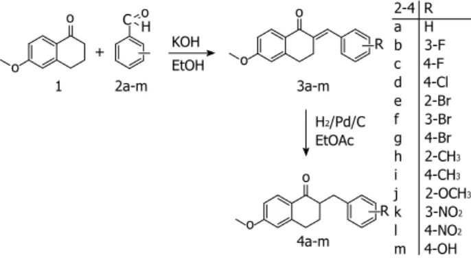
The method employed for the preparation of the tetralones[27] involved the condensation of commercially available 6-methoxy-1-tetralone with benzaldehyde or a substituted benzaldehyde (Figure 1). 6-methoxy-1-te- tralone (1) was dissolved in 4% ethanolic KOH solution, the appropriate benzaldehyde (2a-m) was added, and the reaction mixture was stirred at room temperature for 1-8 h until the starting material had disappeared (thin layer chromatography monitoring), and then allowed to stand overnight. The precipitate that formed was filtered off, washed with water, purified by flash chromatography on silica gel, and recrystallized from ethanol. The synthesis of the hydroxy derivative necessitated initial protection of the hydroxy group in the 4-hydroxybenzaldehyde with a tetrahydropyranyl group, which was stable under the basic ethanolic KOH condensation conditions. The protecting group was removed by heating with aqueous hydrochloric acid in a mixture of ethyl acetate and ethyl methyl ketone. In the next step, the 2-substituted-benzyl- idene-6-methoxy-1-tetralones (3a-m) were dissolved in ethyl acetate, and hydrogenated at 1 atm in the presence of Pd/C as catalyst for 1 h at room temperature. The catalyst was subsequently removed by filtration through a bed of silica gel, the solvent was evaporated in vacuo, and purification by flash chromatography on silica gel fur- nished the 2-substituted-benzyl-6-methoxy-1-tetralones (4a-m).
The resulting tetralones were dissolved individually in dimethyl sulfoxide at a concentration of 10 mmol/L and stored at 4 ℃ until use. In cell culture experiments, com- pounds (4a-m) were dissolved in sterile culture medium (GIBCO’s OPTI-MEM, Life Technologies-Invitrogen, Carlsbad, CA, United States) to the desired concentra- tion. 1,25-D3 at 1 and 10 nmol/L and an untreated con- trol were also applied in these experiments.
Cell culturing
The human epithelial colorectal adenocarcinoma cell line Caco-2 obtained from ECACC was maintained in Dulbecco’s Modified Eagle Medium (D-MEM, Sigma, St.
Louis, MO, United States) supplemented with 10% fetal calf serum (FCS, Sigma) and 1% antibiotic, antimycotic solution (Sigma) at 37 ℃ in a humidified atmosphere containing 5% CO2. Cells were cultured in 6-, 24- and 96-well plates, and all measurements were carried out in triplicate. The cell line was genotyped and identified as Caco-2 in 2011 on the basis of the results of STR analy- sis (DSMZ Profile Database, www.dsmz.de). Twenty- four hours before treatment, the medium was changed to GIBCO’s OPTI-MEM (Life Technologies-Invitrogen, Carlsbad, CA, United States). All experiments were car- ried out with cells from passages 5-25.
Cell viability assay
The protein dye sulforhodamine-B (SRB), was used to test various tetralone derivatives in various concentra- tions for various incubation times in 96-well plates to determine the effects of the compounds alone and in the presence of 1,25-D3 on the Caco-2 cell number. After re- moval of the culture medium, 100 μL of trichloroacetic acid was used to fix the cells during an incubation period of 30 min. The plates were then rinsed 5 times with dis- tilled water. The cells were stained with a 0.4% solution of SRB (Sigma) in acetic acid for 30 min. After removal of the excess dye solution the plates were rinsed 4 times with 1% acetic acid solution and allowed to dry at room temperature. The bound SRB was dissolved in unbuf- fered Trisma-Sol and the plates were shaken for 5 min.
The plates were measured in an Infinite M200 reader (Te- can AG, Männedorf, Switzerland) at 520 nm.
Cytotoxicity measurement
Levels of cytotoxicity were quantified after treatment through measurement of the lactate dehydrogenase (LDH) levels in the wells by using the Cytotoxicity Detec- tion KitPLUS (Roche, Indianapolis, IN, United States). The greater the number of cells that die due to the cytotoxic effect, the higher the amount of LDH in the medium.
The experiments were carried out in accordance with the kit manufacturer’s instructions.
Cell proliferation assays
Cell proliferation was quantified by measurement of the incorporation of 5-bromo-2’-deoxyuridine (BrdU) into the cellular DNA by means of Cell Proliferation enzyme- linked immunosorbent assay, BrdU (colorimetric) (Roche).
The experiments were carried out in accordance with the manufacturer’s instructions.
RNA isolation and Taqman probe-based real-time RT- PCRRNA was isolated through use of the High Pure RNA Isolation Kit (Roche) as prescribed in the manufacturer’
s instructions. The isolated RNA was translated by using Moloney murine leukemia virus reverse transcriptase in accordance with the manufacturer’s instructions (Pro- mega, Madison, WI, United States). Predesigned and validated gene-specific TaqMan Gene Expression Assays from Life Technologies (Life Technologies, Foster City, CA, United States) were used in triplicate for quantitative real-time polymerase chain reaction (PCR) according to the manufacturer’s protocol. Each set contained gene- specific forward and reverse primers and fluorescence- labeled probes. The probes span an exon junction and do not detect genomic DNA [ABI Taqman assay No’s are hs00167999_m1 and hs99999905_m1, for CYP24A1 and glyceraldehyde-3-phosphate dehydrogenase (GAPDH), respectively]. The PCR assays were carried out with the following protocol: denaturation for 10 min at 95 ℃, and 45 cycles of denaturation for 15 s at 95 ℃, anneal- ing and extension for 1 min at 60 ℃. The PCR reaction volume of 20 μL contained 2 μL cDNA, 10 μL of Taq- Man 2x Universal PCR Master Mix NoAmpErase UNG (Life Technologies), 1 μL of gene-specific TaqMan Gene Expression Assay Mix and 7 μL of water. GAPDH was used as a housekeeping gene to normalize for RNA load- ing. Samples were analyzed using the ABI Prism 7500 real-time PCR system (Life Technologies). Relative quan- tification (RQ) studies were carried out on collected data (threshold cycle numbers, referred to as Ct) with the 7500 System SDS software 1.3 (Life Technologies).
Statistical analysis
Data were analyzed by using SPSS for Windows, release 18 (IBM, Armonk, NY, United States). Final data are pre- sented as the means ± SD of at least three independent measurements. Statistical analysis was performed with the unpaired Student t-test; results with P ≤ 0.05 were con-
2-4 R a H b 3-F c 4-F d 4-Cl e 2-Br f 3-Br g 4-Br h 2-CH3
i 4-CH3
j 2-OCH3
k 3-NO2
l 4-NO2
m 4-OH o
o
1 2a-m
C oH KOH
EtOH o
o
R 3a-m
H2/Pd/C EtOAc o
o R
4a-m
Figure 1 Outline of the procedure for the synthesis of the tetralones.
+
sidered statistically significant.
RESULTS
Time and concentration-dependent changes in CYP24A1 mRNA expression after vitamin D3 treatment
An increase in CYP24A1 mRNA level of six orders of magnitude was observed after a brief period of 1,25-D3 treatment. The increase in CYP24A1 mRNA expression was very rapid and it could be observed after 30 min of 1,25-D3 administration, and reached a maximum after 12-16 h of incubation (Figure 2A). After 4 h of incuba- tion in the presence of 1 and 10 nmol/L 1,25-D3, the level of CYP24 mRNA was elevated to 311405-fold and 612801-fold, respectively, relative to the untreated con- trols (Figure 2B).
16 14 12 10 8 6 4 2 0 CYP24A1 mRNA level × 105 (arb. unit)
Figure 2 Time and dose dependent-changes in CYP24A1 mRNA expres- sion in response to 1,25-D3 administration. A: Time course of changes in the cytochrome P450 component of the 25-hydroxyvitamin D3-24-hydroxylase (CYP24A1) mRNA expression in Caco-2 cells after the addition of 100 nmol/L active vitamin D3 metabolite 1,25-dihydroxyvitamin D3 (1,25-D3) to the cell culture supernatant. Glyceraldehyde-3-phosphate dehydrogenase (GAPDH)- normalized CYP24A1 expression levels are shown as a percentage of the CYP24A1 level of the untreated control cells. Points indicate means ± standard deviation (SD) (aP < 0.05 vs untreated control); B: Dose-dependent changes in CYP24A1 mRNA levels in Caco-2 cells after the addition of different amounts of 1,25-D3. GAPDH-normalized CYP24A1 expression levels are shown as a percentage of the CYP24A1 level of the untreated control cells. Points indicate means ± SD (aP < 0.05 vs untreated control).
15 min 30 min 60 min 2 h 3 h 6 h 9 h 12 h 15 h 18 h Incubasion time
a a a a a a
a a a
16 14 12 10 8 6 4 2 0 CYP24A1 mRNA level × 105 (arb. unit)
0.01 0.1 1 10 100 1,25-D3 concentration (nmol/L)
a
a a a a aaaaa a a
a
B
A 1.05
1
0.95
0.9
0.85
0.8 Proportion of viable cells compared to untreated control
Control 0.1 μmol/L 0.3 μmol/L 1 μmol/L 3 μmol/L Concentrations of KD-35
A
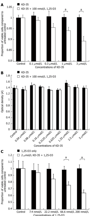
Figure 3 Cell proliferation, lactate dehydrogenase activity and prolifera- tion studies in the presence of KD-35 and 1,25-D3. A: Changes in the num- ber of viable Caco-2 cells (sulforhodamine-B staining) in the presence of differ- ent concentrations of KD-35. Selected wells were treated with 100 nmol/L active 1,25-D3. Data are means ± SD (aP < 0.05 between KD-35 and KD-35 + 1,25-D3 treated cells); B: Changes in the lactate dehydrogenase (LDH) activity of the cell culture supernatant in response to KD-35 with or without 1,25-D3. Data are means ± SD. No significant changes in LDH activity were seen after treat- ment; C: Changes in the proliferation of Caco-2 cells (5-bromo-2’-deoxyuridine incorporation) in response to different concentrations of 1,25-D3. White bars indicate combined treatment with the given 1,25-D3 concentration + 2 μmol/L KD-35. Data are means ± SD. Significance levels were calculated between each sample and the untreated control sample (aP < 0.05 between 1,25-D3 and 1,25-D3 + KD-35 treated cells).
a a
KD-35
KD-35 + 100 nmol/L 1,25-D3
1.8 1.6 1.4 1.2 1 0.8 0.6 0.4 0.2 0
Optical density (A)
KD-35
KD-35 + 100 nmol/L 1,25-D3
Contr ol
Concentrations of KD-35 0.04
μmol/L 0.08
μmol/L 0.16
μmol/L 0.3125
μmol/L 0.625
μmol/L 1.25
μmol/L 2.5 μmol/L
5 μmol/L
1.2 1.1 1 0.9 0.8 0.7 0.6 0.5 0.4 Proportion of viable cells compared to untreated control
Control 7.4 nmol/L 22.2 nmol/L 66.6 nmol/L 200 nmol/L Concentrations of 1,25-D3
1,25-D3 only
2 μmol/L KD-35 + 1,25-D3
a a
C B
Effects of tetralone derivatives on Caco-2 cell line Certain of the tetralones were found to decrease the Caco-2 cell viability but only after 2-4 d of incubation with 1,25-D3. These compounds were tested at various concentrations for various periods to optimize the ef- fect of 1,25-D3 in reducing the total Caco-2 cell count.
Finally, compound KD-35 was selected for further and detailed investigations.
Effects of KD-35 on Caco-2 cell line
When Caco-2 cells were incubated for 4 d in the presence of 100 nmol/L 1,25-D3 with 0.1, 0.3, 1 or 3 μmol/L KD-35, the cell number was reduced by 2.17%, 5.07%, 6.18% and 10.93%, respectively, relative to the controls treated with only 100 nmol/L 1,25-D3 or 3 μmol/L KD-35 (Figure 3).
Results of the cytotoxicity test
To determine the cause of the decrease in viable cell number in the presence of KD-35 and 1,25-D3, we mea- sured LDH concentration in the cell suspension. The concentration of KD-35 ranged between 0.04 μmol/L
and 5 μmol/L. Half of the wells were treated with KD-35 and 100 nmol/L 1,25-D3, the other half were treated with KD-35 only. All experiments were carried out in triplicate. Incubation lasted for 4 d. In all of the experimental setups, the LDH concentrations did not differ significantly in the presence of KD-35 alone or in combination with 1,25-D3 (Figure 3).
Results of the cell proliferation assay
In the presence of 2 μmol/L KD-35, the following con- centrations of 1,25-D3 were used: 7.4, 22.2, 66.6 and 200 nmol/L. Half of the wells were treated only with 2 μmol/L KD-35. Incubation lasted for 4 d. After incuba- tion, the 5-BrdU label was added for an additional 2 h.
The reduction in cell number relative to the control was 3.43%, 14.81%, 22.49% and 35.81%, respectively, com- pared to the wells with 1,25-D3 only (Figure 3).
Changes in CYP24A1 mRNA expression
The amount of CYP24A1 mRNA expressed in the pres- ence of various concentrations of KD-35 did not dif- fer from that of the untreated controls. The CYP24A1 mRNA expression did not depend significantly on the duration of incubation with KD-35 (Figure 4).
DISCUSSION
We have identified a new tetralone compound, KD-35, that effectively and markedly stimulates the anti-prolifera- tive effect of 1,25-D3 in the CRC cell line Caco-2.
CYP24A1, a member of the cytochrome P450 (CYP450) enzyme superfamily is the key enzyme in the metabolism of vitamin D neutralizing the active metabolite 1,25-D3, and thereby controlling its concentration in the tissues.
The CYP450 enzymes all display an iron-containing heme domain at the active site. There are two types of enzyme blockers: azoles and non-azoles[28]. The N-heterocyclic ring of azoles is linked directly to the iron in the heme do- main and, although this inhibition is very potent, it is not selective. Since the other enzymes involved in vitamin D metabolism (CYP27A1 and CYP27B1) are also members of the CYP450 superfamily, this type of nonselective inhi- bition is not specific for CYP24A1.
The enzyme inhibitory effect of non-azoles is medi- ated through hydrogen bonds and hydrophobic interac- tions with the active site of the enzyme. This is a more flexible mechanism which may permit significant selectiv- ity though the inhibitory effect may be less than that of azoles[26]. We investigated 13 tetralones (non-azoles) in a search for a compound that is effective locally in the colon and is not strongly absorbed, so that the risk of adverse systemic effects is minimized.
Most of the 13 tetralones were either toxic or inef- fective, even in the presence of 1,25-D3. Only in the presence of KD-35 did 1,25-D3 markedly inhibit Caco-2 cell proliferation without pronounced cytotoxicity of the tetralone alone. Such inhibition was not observed in the absence of KD-35. Unfortunately, two of the three
8 7 6 5 4 3 2 1 0 CYP24A1 mRNA level × 105 (arb. unit)
0 nmol/L 1,25-D3 1 nmol/L 1,25-D3 10 nmol/L 1,25-D3
B
A KD-35 0 μmol/L
KD-35 2 μmol/L
30
25
20
15
10
5
0 CYP24A1 mRNA level × 105 (arb. unit)
1 d 4 d Incubation time Untreated control
2 μmol/L KD-35 only 1 nmol/L 1,25-D3
1 nmol/L 1,25-D3 + 2 μmol/L KD-35
Figure 4 KD-35 has no effect on CYP24A1 mRNA expression. A: Changes in CYP24A1 mRNA levels in Caco-2 cells incubated with different concentra- tions of 1,25-D3 for 4 h with or without KD-35. Data are means ± SD. No sig- nificant changes in mRNA levels were seen with or without KD-35; B: Effects of KD-35 and KD-35 + 1,25-D3 on CYP24A1 mRNA expression in Caco-2 cells.
Data are means ± SD. No significant change in mRNA levels was seen with or without KD-35 at any time point.
most effective tetralones exhibited much higher cytotox- icity at higher concentrations than KD-35. The question arises as to whether KD-35 exerts its effect via CYP24A1 inhibition. We did not measure CYP24A1 enzyme activ- ity directly since this is technically extremely difficult. It is also complicated to measure the intermediates of the CYP24A1 reaction. Moreover, a simple enzyme kinetic measurement would not reveal whether the compound enters the cell. We therefore chose an indirect approach:
to prove the biological efficacy of the compound. KD-35 was found to exert an effect that allowed 1,25-D3 to re- duce Caco-2 cell proliferation effectively, as reflected by an altered BrdU incorporation. Direct cytotoxicity was excluded by the LDH measurements, and no change in CYP24A1 mRNA expression was detected in response to KD-35, which ruled out alterations in protein synthesis.
Obviously, no direct evidence was obtained to support direct enzyme inhibition, but an alternative mechanism is highly unlikely with this non-azole.
Two major pathways are mediated through the VDRs:
the Wnt-beta-catenin pathway, which is responsible for the loss of adherent cell type, and the E-cadherin path- way, which is responsible for cell-to-cell adhesion and cell differentiation[29,30]. The administration of 1,25-D3 sup- presses the Wnt-beta-catenin pathway and induces the expression of E-cadherin. The Wnt-beta-catenin pathway is constitutionally overregulated in most CRCs, due to the mutation of several members of the pathway (APC, AXIN2, etc.)[29]. There are other participants in colorectal carcinogenesis, such as estrogen receptors, which elevate the number of VDRs in the mucosal cells of the alimen- tary tract, or SNAIL, which inhibits the E-cadherin path- way and expression of VDRs[30-35]. Another important factor in the mucosal cell transition toward adenocarci- noma is an elevated level of CYP24A1, the intracellular concentration of which correlates with the dignity of the tumor[24].
Our results corroborate the earlier finding[36] that the presence of 1,25-D3 dramatically stimulates the expres- sion of CYP24A1 in CRC cells[24,37]. Two vitamin D-re- sponsive elements are present in the promoter region of CYP24A1[38]. Through this pathway, 1,25-D3 stimulates its own destruction through metabolism into inactive forms by enhancing the expression of CYP24A1[39].
Besides the genomic effects, there have also been reports of immediate nongenomic mechanisms. A pos- sible mode of action is activation of the RhoA-ROCK- p38MAPK-MSK signaling pathway. This pathway medi- ates the induction of CST5, which is possibly responsible for tumor suppression and the level of CYP24A1; as a negative feedback mechanism, this eliminates 1,25-D3 from the cell[40]. VDRs found in other tumor cell mem- branes may bind 1,25-D3, and the complex could induce a rapid influx of Ca2+ into the cell[40], which activates RhoA-ROCK and then the p38MAPK-MSK-1 pathway.
Besides the nongenomic activation of this pathway, a vitamin D-responsive element can also be identified in the -1k promoter region of the RhoA gene (http://www.
cbil.upenn.edu/cgi-bin/tess/tess). RhoA plays an impor- tant role in the induction of CDH1/E-cadherin, which is crucial for the acquisition of the polarity and adhesive phenotype of cancer cells[29].
In view of these data, the elevation of CYP24A1 expression might be a self-defense mechanism of tumor cells. By inhibiting the inactivating enzyme, the amount of active vitamin D or its analogs required to elicit their marked anti-tumor effect could be reduced in vivo, thereby preventing elevation of the serum Ca2+ level and avoiding hypercalcemia[36]. The inhibition of CYP24A1 may allow 1,25-D3 to exert its anti-tumor effect, in this way leading to a new approach in the treatment of CRC in the future.
COMMENTS
Background
The effects of vitamin D3 have been investigated on various tumors, including colorectal cancer (CRC). The cytochrome P450 component of 25-hydroxyvi- tamin D3-24-hydroxylase (CYP24A1), the enzyme that inactivates the active vitamin D3 metabolite 1,25-dihydroxyvitamin-D3 (1,25-D3) is considered to be the main enzyme determining the biological half-life of 1,25-D3. During colorec- tal carcinogenesis, the expression and concentration of CYP24A1 increases significantly, suggesting that this phenomenon could be responsible for the controversial efficacy of 1,25-D3 in the treatment of CRC. In the present study, authors set out to investigate the effects of 1,25-D3 on CRC cells after the inhi- bition of CYP24A1 activity.
Research frontiers
The anti-tumor effect of vitamin D3 has been a focus of interest during the last 10-15 years. However, vitamin D3 cannot exert this important effect in a number of tumors. The reasons for this have been investigated intensively. One possible explanation for the reduced anti-tumor efficacy of vitamin D3 is the ac- celerated neutralization of the active vitamin D3 compound in certain cases, e.g., CRC, liver and papillary thyroid cancers.
Innovations and breakthroughs
The authors synthesized a number of compounds potentially able to inhibit the action of CYP24A1, the enzyme neutralizing the effects of vitamin D3. One of these compounds, KD-35, had inhibitory potential without an apparent toxic effect. In the presence of KD-35, vitamin D3 markedly inhibited the growth of CRC cells.
Applications
Selective inhibition of the CYP24A1 by compounds such as KD-35 may permit a new approach to enhancement of the anti-tumor effect of 1,25-D3 on CRC.
Peer review
The authors tackled an interesting topic for investigation. The manuscript is investigating the association between CYP24A1 inhibition and anti-tumor effect of 1a, 25-dihydroxyvitamin-D3 in Caco-2 CRC line. A careful assessment was considered using appropriate cell assays. A major finding of the study was that Caco-2 cell viability and proliferation were markedly reduced in response to 1,25-D3 when the CYP24A1 was inhibited (by KD-35, one of the tetralone com- pounds).
REFERENCES
1 Jenab M, Bueno-de-Mesquita HB, Ferrari P, van Duijnhoven FJ, Norat T, Pischon T, Jansen EH, Slimani N, Byrnes G, Rin- aldi S, Tjønneland A, Olsen A, Overvad K, Boutron-Ruault MC, Clavel-Chapelon F, Morois S, Kaaks R, Linseisen J, Boe- ing H, Bergmann MM, Trichopoulou A, Misirli G, Tricho- poulos D, Berrino F, Vineis P, Panico S, Palli D, Tumino R, Ros MM, van Gils CH, Peeters PH, Brustad M, Lund E, Tormo MJ, Ardanaz E, Rodríguez L, Sánchez MJ, Dorron- soro M, Gonzalez CA, Hallmans G, Palmqvist R, Roddam A, Key TJ, Khaw KT, Autier P, Hainaut P, Riboli E. Association between pre-diagnostic circulating vitamin D concentration
COMMENTS
and risk of colorectal cancer in European populations: a nest- ed case-control study. BMJ 2010; 340: b5500 [PMID: 20093284 DOI: 10.1136/bmj.b5500]
2 Kampman E, Slattery ML, Caan B, Potter JD. Calcium, vita- min D, sunshine exposure, dairy products and colon cancer risk (United States). Cancer Causes Control 2000; 11: 459-466 [PMID: 10877339 DOI: 10.1023/A: 1008914108739]
3 Tangpricha V, Spina C, Yao M, Chen TC, Wolfe MM, Holick MF. Vitamin D deficiency enhances the growth of MC-26 colon cancer xenografts in Balb/c mice. J Nutr 2005; 135:
2350-2354 [PMID: 16177194]
4 Zeeb H, Greinert R. The role of vitamin D in cancer pre- vention: does UV protection conflict with the need to raise low levels of vitamin D? Dtsch Arztebl Int 2010; 107: 638-643 [PMID: 20959891]
5 Ferlay J, Parkin DM, Steliarova-Foucher E. Estimates of cancer incidence and mortality in Europe in 2008. Eur J Cancer 2010; 46: 765-781 [PMID: 20116997 DOI: 10.1016/
j.ejca.2009.12.014]
6 Cross HS, Nittke T, Peterlik M. Modulation of vitamin D synthesis and catabolism in colorectal mucosa: a new target for cancer prevention. Anticancer Res 2009; 29: 3705-3712 [PMID: 19667168]
7 Giovannucci E. Strengths and limitations of current epide- miologic studies: vitamin D as a modifier of colon and pros- tate cancer risk. Nutr Rev 2007; 65: S77-S79 [PMID: 17867375 DOI: 10.1301/nr.2007.aug.S77-S79]
8 Rheem DS, Baylink DJ, Olafsson S, Jackson CS, Walter MH.
Prevention of colorectal cancer with vitamin D. Scand J Gas- troenterol 2010; 45: 775-784 [PMID: 20367197 DOI: 10.3109/00 365521003734125]
9 Gandini S, Boniol M, Haukka J, Byrnes G, Cox B, Sneyd MJ, Mullie P, Autier P. Meta-analysis of observational studies of serum 25-hydroxyvitamin D levels and colorectal, breast and prostate cancer and colorectal adenoma. Int J Cancer 2011;
128: 1414-1424 [PMID: 20473927 DOI: 10.1002/ijc.25439]
10 Wada K, Tanaka H, Maeda K, Inoue T, Noda E, Amano R, Kubo N, Muguruma K, Yamada N, Yashiro M, Sawada T, Nakata B, Ohira M, Hirakawa K. Vitamin D receptor expres- sion is associated with colon cancer in ulcerative colitis. On- col Rep 2009; 22: 1021-1025 [PMID: 19787215]
11 Newmark HL, Yang K, Kurihara N, Fan K, Augenlicht LH, Lipkin M. Western-style diet-induced colonic tumors and their modulation by calcium and vitamin D in C57Bl/6 mice:
a preclinical model for human sporadic colon cancer. Carci- nogenesis 2009; 30: 88-92 [PMID: 19017685 DOI: 10.1093/car- cin/bgn229]
12 Feskanich D, Ma J, Fuchs CS, Kirkner GJ, Hankinson SE, Hollis BW, Giovannucci EL. Plasma vitamin D metabolites and risk of colorectal cancer in women. Cancer Epidemiol Bio- markers Prev 2004; 13: 1502-1508 [PMID: 15342452]
13 Giovannucci E. The epidemiology of vitamin D and colorectal cancer: recent findings. Curr Opin Gastroenterol 2006; 22: 24-29 [PMID: 16319673 DOI: 10.1097/01.mog.0000196150.36701.c2]
14 Ordóñez-Morán P, Muñoz A. Nuclear receptors: genomic and non-genomic effects converge. Cell Cycle 2009; 8:
1675-1680 [PMID: 19448403 DOI: 10.4161/cc.8.11.8579]
15 Mikhail N. Clinical significance of vitamin D deficiency in primary hyperparathyroidism, and safety of vitamin D therapy. South Med J 2011; 104: 29-33 [PMID: 21079532 DOI:
10.1097/SMJ.0b013e3181fcd772]
16 Cho YL, Christensen C, Saunders DE, Lawrence WD, Deppe G, Malviya VK, Malone JM. Combined effects of 1,25-di- hydroxyvitamin D3 and platinum drugs on the growth of MCF-7 cells. Cancer Res 1991; 51: 2848-2853 [PMID: 2032225]
17 Liu G, Hu X, Chakrabarty S. Vitamin D mediates its action in human colon carcinoma cells in a calcium-sensing receptor- dependent manner: downregulates malignant cell behavior and the expression of thymidylate synthase and survivin and promotes cellular sensitivity to 5-FU. Int J Cancer 2010;
126: 631-639 [PMID: 19621386 DOI: 10.1002/ijc.24762]
18 Tanaka H, Abe E, Miyaura C, Kuribayashi T, Konno K, Nishii Y, Suda T. 1 alpha,25-Dihydroxycholecalciferol and a human myeloid leukaemia cell line (HL-60). Biochem J 1982;
204: 713-719 [PMID: 6289803]
19 Lechner D, Kállay E, Cross HS. 1alpha,25-dihydroxyvitamin D3 downregulates CYP27B1 and induces CYP24A1 in colon cells. Mol Cell Endocrinol 2007; 263: 55-64 [PMID: 17029768 DOI: 10.1016/j.mce.2006.08.009]
20 Bareis P, Kállay E, Bischof MG, Bises G, Hofer H, Pötzi C, Manhardt T, Bland R, Cross HS. Clonal differences in ex- pression of 25-hydroxyvitamin D(3)-1alpha-hydroxylase, of 25-hydroxyvitamin D(3)-24-hydroxylase, and of the vi- tamin D receptor in human colon carcinoma cells: effects of epidermal growth factor and 1alpha,25-dihydroxyvitamin D(3). Exp Cell Res 2002; 276: 320-327 [PMID: 12027461 DOI:
10.1006/excr.2002.5528]
21 Ma Y, Trump DL, Johnson CS. Vitamin D in combination cancer treatment. J Cancer 2010; 1: 101-107 [PMID: 20842231 DOI: 10.7150/jca.1.101]
22 Mocellin S. Vitamin D and cancer: deciphering the truth.
Biochim Biophys Acta 2011; 1816: 172-178 [PMID: 21767609]
23 Edlich R, Mason SS, Chase ME, Fisher AL, Gubler K, Long WB, Giesy JD, Foley ML. Scientific documentation of the relationship of vitamin D deficiency and the development of cancer. J Environ Pathol Toxicol Oncol 2009; 28: 133-141 [PMID:
19817700 DOI: 10.1615/JEnvironPatholToxicolOncol.v28.
i2.50]
24 Horváth HC, Lakatos P, Kósa JP, Bácsi K, Borka K, Bises G, Nittke T, Hershberger PA, Speer G, Kállay E. The candidate oncogene CYP24A1: A potential biomarker for colorectal tumorigenesis. J Histochem Cytochem 2010; 58: 277-285 [PMID:
19901270 DOI: 10.1369/jhc.2009.954339]
25 Larriba MJ, Martín-Villar E, García JM, Pereira F, Peña C, de Herreros AG, Bonilla F, Muñoz A. Snail2 cooperates with Snail1 in the repression of vitamin D receptor in colon can- cer. Carcinogenesis 2009; 30: 1459-1468 [PMID: 19502595 DOI:
10.1093/carcin/bgp140]
26 Aboraia AS, Makowski B, Bahja A, Prosser D, Brancale A, Jones G, Simons C. Synthesis and CYP24A1 inhibitory activ- ity of (E)-2-(2-substituted benzylidene)- and 2-(2-substituted benzyl)-6-methoxy-tetralones. Eur J Med Chem 2010; 45:
4427-4434 [PMID: 20655626 DOI: 10.1016/j.ejmech.2010.07.001]
27 Yee SW, Jarno L, Gomaa MS, Elford C, Ooi LL, Coogan MP, McClelland R, Nicholson RI, Evans BA, Brancale A, Simons C. Novel tetralone-derived retinoic acid metabolism block- ing agents: synthesis and in vitro evaluation with liver mi- crosomal and MCF-7 CYP26A1 cell assays. J Med Chem 2005;
48: 7123-7131 [PMID: 16279770 DOI: 10.1021/jm0501681]
28 Schuster I, Egger H, Nussbaumer P, Kroemer RT. Inhibi- tors of vitamin D hydroxylases: structure-activity relation- ships. J Cell Biochem 2003; 88: 372-380 [PMID: 12520539 DOI:
10.1002/jcb.10365]
29 Ordóñez-Morán P, Larriba MJ, Pálmer HG, Valero RA, Barbáchano A, Duñach M, de Herreros AG, Villalobos C, Berciano MT, Lafarga M, Muñoz A. RhoA-ROCK and p38MAPK-MSK1 mediate vitamin D effects on gene expres- sion, phenotype, and Wnt pathway in colon cancer cells. J Cell Biol 2008; 183: 697-710 [PMID: 19015318 DOI: 10.1083/
jcb.200803020]
30 Peña C, García JM, Silva J, García V, Rodríguez R, Alonso I, Millán I, Salas C, de Herreros AG, Muñoz A, Bonilla F.
E-cadherin and vitamin D receptor regulation by SNAIL and ZEB1 in colon cancer: clinicopathological correlations.
Hum Mol Genet 2005; 14: 3361-3370 [PMID: 16203744 DOI:
10.1093/hmg/ddi366]
31 Gilad LA, Bresler T, Gnainsky J, Smirnoff P, Schwartz B.
Regulation of vitamin D receptor expression via estrogen-in- duced activation of the ERK 1/2 signaling pathway in colon and breast cancer cells. J Endocrinol 2005; 185: 577-592 [PMID:
15930183 DOI: 10.1677/joe.1.05770]
32 Gilad LA, Tirosh O, Schwartz B. Phytoestrogens regulate transcription and translation of vitamin D receptor in colon cancer cells. J Endocrinol 2006; 191: 387-398 [PMID: 17088408 DOI: 10.1677/joe.1.06930]
33 Lechner D, Bajna E, Adlercreutz H, Cross HS. Genistein and 17beta-estradiol, but not equol, regulate vitamin D synthesis in human colon and breast cancer cells. Anticancer Res 2006;
26: 2597-2603 [PMID: 16886669]
34 Larriba MJ, Muñoz A. SNAIL vs vitamin D receptor ex- pression in colon cancer: therapeutics implications. Br J Cancer 2005; 92: 985-989 [PMID: 15770204 DOI: 10.1038/
sj.bjc.6602484]
35 Pálmer HG, Larriba MJ, García JM, Ordóñez-Morán P, Peña C, Peiró S, Puig I, Rodríguez R, de la Fuente R, Bernad A, Pollán M, Bonilla F, Gamallo C, de Herreros AG, Muñoz A.
The transcription factor SNAIL represses vitamin D recep- tor expression and responsiveness in human colon cancer.
Nat Med 2004; 10: 917-919 [PMID: 15322538 DOI: 10.1038/
nm1095]
36 Tashiro K, Abe T, Oue N, Yasui W, Ryoji M. Character-
ization of vitamin D-mediated induction of the CYP 24 transcription. Mol Cell Endocrinol 2004; 226: 27-32 [PMID:
15489002 DOI: 10.1016/j.mce.2004.07.012]
37 Meyer MB, Goetsch PD, Pike JW. A downstream intergenic cluster of regulatory enhancers contributes to the induction of CYP24A1 expression by 1alpha,25-dihydroxyvitamin D3.
J Biol Chem 2010; 285: 15599-15610 [PMID: 20236932 DOI:
10.1074/jbc.M110.119958]
38 Chen KS, DeLuca HF. Cloning of the human 1 alpha,25- dihydroxyvitamin D-3 24-hydroxylase gene promoter and identification of two vitamin D-responsive elements. Biochim Biophys Acta 1995; 1263: 1-9 [PMID: 7632726 DOI: 10.1016/01 67-4781(95)00060-T]
39 Adams JS, Hewison M. Update in vitamin D. J Clin Endocri- nol Metab 2010; 95: 471-478 [PMID: 20133466 DOI: 10.1210/
jc.2009-1773]
40 Ordóñez-Morán P, Alvarez-Díaz S, Valle N, Larriba MJ, Bo- nilla F, Muñoz A. The effects of 1,25-dihydroxyvitamin D3 on colon cancer cells depend on RhoA-ROCK-p38MAPK- MSK signaling. J Steroid Biochem Mol Biol 2010; 121: 355-361 [PMID: 20223287 DOI: 10.1016/j.jsbmb.2010.02.031]
P- Reviewers Braet F, Lakatos PL S- Editor Gou SX L- Editor Cant MR E- Editor Zhang DN