The apoptotic effects of progesterone on breast cancer (MCF-7) and human osteosarcoma
(MG-636) cells
H.R. MOTAMED
1, M. SHARIATI
1, R. AHMADI
2,3p, S. KHATAMSAZ
1and M. MOKHTARI
11Department of Biology, Kazerun Branch, Islamic Azad University, Kazerun, Islamic Republic of Iran
2Department of Biology, Hamedan Branch, Islamic Azad University, Hamedan, Islamic Republic of Iran
3Avicenna International College, Budapest, Hungary
Received: January 7, 2020 • Accepted: June 13, 2020 Published online: October 9, 2020
© 2020 Akademiai Kiado, Budapest
ABSTRACT
Purpose:Progesterone has been reported to inhibit the proliferation of breast cancer and osteosar- coma cells; however, its inhibitory mechanism has not yet been clarified. The aim of the present study was to clarify the effects of progesterone on apoptosis in breast cancer (MCF-7) and human osteosarcoma (MG-63) cells.Materials and methods:In this experimental study the cytotoxic effect of progesterone was measured in MCF-7 and MG-63 cells exposed to different concentrations of proges- terone using MTT assay, and effective concentrations were identified. The expression levels of theBax, P53 and Bcl-2 genes were evaluated by real-time PCR, and caspase-3, 8 and 9 activity levels were determined using a colorimetric method. Hoechst staining andflow cytometry were used to confirm apoptosis. The data were statistically analyzed using one-way analysis of variance (ANOVA) and in- dependent-samples t-test.Results:Compared to the control group, we observed a significant increase in the expression levels of theBaxandP53genes and the activity levels of caspase-3 and 9, and a significant decrease in the expression level of theBcl-2 gene in MCF-7 and MG-63 treated with effective concen- tration of progesterone. The caspase-8 activity level did not change significantly in treated MG-63 but increased in treated MCF-7 cells. Hoechst staining andflow cytometry results confirmed apoptosis in the cells exposed to effective concentration of progesterone.Conclusions:The cytotoxic effect of progesterone on breast cancer and osteosarcoma cells was mediated by apoptotic pathways. In this context,
*Corresponding author. Department of Biology, Hamedan Branch, Islamic Azad University, Prof. Mousivand Blvd, Hamedan, Islamic Republic of Iran. Tel.:þ98 8134494002; fax:þ98 8134494001. E-mail: drrahahmadi@yahoo.com
progesterone triggers the extrinsic and intrinsic apoptotic pathways in MCF-7 cells and induces the intrinsic apoptotic pathway in MG-63 cells.
KEYWORDS
apoptosis, progesterone, MCF-7, MG-63, caspase
INTRODUCTION
Breast cancer is the most frequently diagnosed cancer and the second leading cause of cancer deaths in women worldwide. Its prevalence is influenced by different factors including gender, genetics, age, geography and ethnic background [1]. Among human neoplasms, primary ma- lignant bone tumors are fairly uncommon. The most common primary bone malignancies account for only 0.2% of all malignancies in the UK and USA [2]. Years of scientific research have shown that female sex hormones are linked to the etiopathogenesis of breast [3] and bone cancer [4]. The relationship between progestogens and the increased risk of breast and bone cancers has been reported in recent studies [5].
The findings suggest that sex steroid hormones may influence the growth and prolifera- tion of breast cancer cells either directly or indirectly [6]. A large body of experimental and clinical data have indicated that progestogens induce the proliferation of breast cancer cells and play a significant role in the development of breast cancer [7]. Studies have also shown that progesterone is capable of inhibiting apoptosis in breast cancer cells by suppressing expression of the proapoptotic proteins and upregulating expression of the antiapoptotic proteins, which leads to the enhanced survival ability of breast cancer cells [8]. However, there have been reports indicating that various progestogens have diverging effects on pro- gesterone receptor expression followed by different effects on the growth of breast cancer cells. Indeed, although several studies have demonstrated that progestogen administration in vivoor in vitro enhances the cell growth and viability of breast cancer cells [9], several investigations clearly show that progestogens can inhibit growth and induce apoptosis in breast cancer cells [10, 11]. The effect of progesterone on the basal levels of apoptosis suggests that this mechanism may also be important for normal labor at term [12]. The research data indicate that progesterone can trigger apoptosis by upregulating proapoptotic (p-53andBAX) and decreasing antiapoptotic (BCL-2) gene expression in cancer cells [13]. Progesterone also has the capability to induce the caspase cascade through extrinsic or intrinsic apoptosis in many cancer cells [14].
Sex steroids have an important impact on bone cell growth. The importance of progesterone in bone cell development is widely accepted [15]. Research findings suggest a crucial role for progesterone signaling in bone acquisition to augment bone mass, which may have the potential to reduce the burden of osteoporosis [16]. Bone loss associated with the reduction of certain sex steroid hormones in breast cancer survivors may also represent the effects of sex hormones on bone cells [17]. Experimental data suggest that sex steroid hormones including progesterone induce apoptosis in rat osteosarcoma cells, which is accompanied by caspase-3 activation [18, 19]. However, progesterone may protect certain bone cells against apoptosis through its receptor and the downstream mitochondrial pathway [20]. It has also been shown that the effects of
progesterone on bone cell apoptosis may contribute to the mechanisms, by which progesterone exerts its action on bone formation [15]. Although various studies have shown that progesterone induces apoptosis in osteosarcoma cells [21], the role of the progesterone in the apoptosis of bone cancer cells has been poorly clarified.
Regarding conflicting reports on the effect of progesterone on breast cancer cells and few reports concerning the effects of progesterone on osteosarcoma cells, the present in vitro study was carried out to clarify the cytotoxic and apoptotic effects of progesterone on human breast cancer (MCF-7) and human osteosarcoma (MG-63) cells through evaluation of the expression levels of theBAX,P53andBCL-2genes and the activity levels of caspase-3, 8 and 9 as well as detecting cell nuclear morphology alterations and discriminating between apoptosis and necrosis in MCF-7 and MG-63 cell lines exposed to cytotoxic doses of pro- gesterone.
MATERIALS AND METHODS
Progesterone
In this experimental study progesterone was obtained from the Abu Reyhan Pharmaceutical Company (Tehran-Iran) and dissolved in DMSO, Dulbecco’s modified Eagle’s medium (DMEM) or phosphate buffered saline (PBS) to produce different concentrations (0.001, 0.01, 0.1, 1 and 10 mg/mL).
Cell culture
MCF-7 and MG-63 cells were obtained from the National Cell Bank of Iran (Pasteur Institute, Tehran, Iran). The cells were cultured in DMEM supplemented with 10% Fetal Bovine Serum (FBS) and 1% antibiotics (gentamicin). Cells were then preserved in a humidified atmosphere with 5% CO2in a 378C incubator. Cultured cells at 70–80% confluency were washed with PBS and detached from theflask using trypsin-EDTA with incubation at 378C for 3–4 min, followed by addition of culture medium containing 10% FBS to neutralize the excess trypsin-EDTA activity. The cell suspension was eventually centrifuged and the cell pellet was re-suspended in fresh culture medium to be used in the experiments.
MTT assay
The effect of progesterone on cell viability was determined with 3-(4,5-dimethylthiazol-2yl)-2,5- biphenyl tetrazolium bromide (MTT) assay. Cells were seeded in 96-well culture plates. After syncytialization, cells were treated with progesterone (0.001, 0.01, 0.1, 1 and 10 mg/mL) for 24 h.
MTT (DOBIO Biotech, Shanghai, China) solution (5 mg/mL) diluted in PBS was then added to cells (100
m
L) and incubated with the cells for 3 h at 378C in darkness. During this incubation period, water-insoluble formazan crystals were formed and dissolved by the addition of 100m
L/well DMSO (Sigma) (DMSO is usually well tolerated with no observable toxic effects to cells at 0.1%final concentration). The optical density (OD) of each culture well was measured at 570 nm using a microplate reader. Wells containing cells and MTT solution without progesterone were taken as blank samples. The percentage of cell viability was calculated as [Optical density (OD) of the sample/OD of the control]3100 [22].
Real time RT-PCR assay
Cancerous cells were seeded into 6-well plates (5 3 105cells/well) and incubated for 24 h. The cells were then exposed to effective concentration of progesterone (2.5 mg/mL) and incubated for an additional 24 h. Total RNA was extracted with the high purity RNA extraction kit (Takara, Japan) according to the manufacturer’s instructions and reverse-transcribed into cDNA. Then, real-time quantitative PCR was conducted to analyzeBax,P53,Bcl-2and GAPDH expression levels. The primer sequences are shown inTable 1. Each amplification reaction was performed in a 20-
m
L reaction mixture containing 10m
L Power SYBR Green PCR Master Mix (2X), 1m
L of each primer (2m
M), 1m
L cDNA, and 7m
L double-distilled water. The expression levels of genes was calculated by the 2ΔΔCT method and were normalized to the loading control,GAPDH[23].Caspase assay
The activity levels of caspase-3, 8 and 9 were detected using the ApoTarget colorimetric protease assay kit (Abnova, Taiwan) according to the manufacturer’s instructions. MCF-7 and MG-63 cells were treated with progesterone, while concurrently incubating a control culture without treatment. 33106 cells per sample were counted. The cells were resuspended in 50
m
L of chilled cell lysis buffer, incubated on ice for 10 min, and then centrifuged for 1 min in a microcentrifuge (10,0003g). The supernatant (cytosol extract) was transferred to a fresh tube and put on ice.50
m
L of 23reaction buffer (containing 0.5m
L DTT) was added to each sample followed by adding 5m
L of 4 mM DEVD-pNA, 4 mM IETD-pNA, and 4 mM LEHD-pNA substrate to assay the activity level of caspase-3, 8, and 9, respectively. The samples were incubated in the dark at 378C for 2 h. Samples were read in a microplate reader set at 405 nm. Fold increase in caspase-3, 8 and 9 activity was determined compared to that in untreated controls [24].Analysis of MCF-7 and MG-63 cell nuclear morphology using Hoechst staining
Nuclear morphology was detected using Hoechst staining [25]. Progesterone-treated MCF-7 and MG-63 cells werefixed in 80% acetone for 30 min followed by staining with Hoechst 33,342 (5
m
g/mL) for 5 min at room temperature. The cells were then washed twice with PBS, examined and immediately photographed under afluorescence microscope (Nikon Corporation, Chiyoda- ku, Tokyo, Japan) with excitation wavelength of 330–380 nm. Apoptotic cells were defined on the basis of changes in nuclear morphology such as chromatin condensation and fragmentation [26].Flow cytometric analyses of cell death
MCF-7 and MG-63 cells (13105cells/well) were treated with effective concentration (2.5 mg/
mL) of progesterone in a 24-well plate and untreated cells were considered as negative control.
The Annexin-V-Fluos Staining Kit (Biolegend, USA) was used to discriminate between apoptosis and necrosis in the given culture system. After treating the cells with 2.5 mg/mL of progesterone and 42 h incubation, cells were trypsinized and washed by PBS. To resuspend cells 100
m
L binding buffer was added. Upon adding binding buffer, 10m
L PI and 5m
L Annexin-VFITC were also added to the microtube. The samples were then incubated at 258C for 15 min
in a dark room. 400
m
L Annexin-V was added to each microtube andfinally cellular analysis was carried out byflow cytometry, which involves simultaneous staining with both annexin-V and the DNA stain propidium iodide (PI). Four subpopulations of cells were discriminated: (a) PI- negative and (FITC)-negative viable cells (PI–/FITC–) that maintain the typical asymmetry of plasma membrane lipids; (b) PI-negative and FITC-positive early apoptotic cells (PI–/FITCþ) capable of transporting PI outside the cell; (c) PI-positive and (FITC)-positive late apoptotic or necrotic cells (PIþ/FITCþ) with loss of plasma membrane integrity; and (d) PI-positive and FITC-negative necrotic cells (PIþ/FITC-). Cells were prepared following the manufacturer’s protocol. Fluorescence intensity was measured by flow cytometry (FACSCalibur, BD Bio- sciences, Franklin Lakes, NJ, USA) [27].Statistical analysis
Statistical analysis was performed by SPSS software (version 21.0; SPSS, Chicago, IL, USA).
Differences between cell viabilities in the groups were tested using one-way analysis of vari- ance (ANOVA) followed by Tukey’s multiple comparisonpost-hoc test. Independent samples t-test was used to detect differences in gene expression and caspase activity between experi- mental and control groups. All experiments were performed at least three times. All data are expressed as the mean ± standard deviation (S.D.). Value differences were considered sig- nificant if <0.05.
RESULTS
Effect of progesterone on the viability of MCF-7 and MG-63 cells. Viability of MCF-7 cells did not significantly change when exposed to 0.001, 0.01, 0.1 and 1 mg/mL of progesterone compared with the control group; however, exposure of MCF-7 cells to 10 mg/mL of proges- terone resulted in a significant decrease in cell viability compared to the control and other experimental groups. The viability of MG-63 cells significantly decreased in groups exposed to 0.1, 1 and 10 mg/mL of progesterone compared to the control group (Fig. 1). There was also a significant difference in cell viability between the group exposed to 10 mg/mL and the groups
Table 1.Primer sequences used in real-time RT-PCR
Gene Primer Sequences
Product Size (bp)
Bax Forward: 59-TTGCTTCAGGGTTTCATCCAG-39 101
Reverse: 59- AGCTTCTTGGTGGACGCATC-39
P53 Forward: 59- CATCTACAAGCAGTCACAGCACAT-39 194
Reverse: 59- CAACCTCAGGCGGCTCATAG-39
Bcl-2 Forward: 59- TGTGGATGACTGAGTACCTGAACC-39 122
Reverse: 59- CAGCCAGGAGAAATCAAACAGAG-39
GAPDH Forward: 59- CCCACTCCTCCACCTTTGAC-39 75
Reverse: 59- CATACCAGGAAATGAGCTTGACAA-39
exposed to 0.1 and 1 mg/mL of progesterone. The effective concentration of progesterone was calculated by regression analysis.
Effect of progesterone on expression level of Bax, P53 and Bcl-2 genes in MCF-7 and MG-63 cells. Compared to the control group, the partial expression levels of the P53 and Bax genes significantly increased in MCF-7 and MG-63 cells exposed to effective concentration (2.5 mg/
mL) of progesterone (P< 0.05,P< 0.001, respectively); however, the partial expression level of theBcl-2gene significantly decreased (P< 0.001) (Fig. 2).
Effect of progesterone on caspase-3,8 and 9 activity level in MCF-7 and MG-63 cells. Com- pared to the control group, caspase-3, 8 and 9 activity levels significantly increased in MCF-7 cells exposed to effective concentration (2.5 mg/mL) of progesterone (P< 0.001,P< 0.01 andP<
0.001, respectively). A significant increase in caspase-9 and 3 activity levels was also observed in MG-63 cells exposed to effective concentration (2.5 mg/mL) of progesterone compared with the control group (P< 0.001 andP< 0.01, respectively); however, the caspase 8 activity level did not change significantly (Fig. 3).
Morphological characteristics of MCF-7 and MG-63 cells nuclei with Hoechst staining. To further confirm apoptosis and to determine the nuclear morphology characteristics of MCF-7 and MG-63 cells, the Hoechst staining method was used. Nuclear morphology was evaluated with membrane-permeable blue Hoechst 33,342. Nuclei of the cells appeared with regular contours and were round and large in size in untreated (control) MCF-7 and MG-63 cells. They were rarely seen with smaller nuclei and condensed chromatin. In contrast, most nuclei of Fig. 1.Viability of MCF-7 and MG-63 cells exposed to 0.001, 0.01, 0.1, 1 and 10 mg/mL of progesterone.
*and # indicate significant differences compared to control group and groups exposed to 0.001, 0.01, 0.1 and 1 mg/mL of progesterone, respectively (*:P< 0.05, **:P< 0.01, ***:P< 0.001, ###:P< 0.001)
Fig. 2.Partial expression levels (RQ) of P53, Bax and Bcl-2 genes in MCF-7 and MG-63 cells exposed to effective concentration (2.5 mg/mL) of progesterone compared with control group. * represents significant
difference compared to control group (*:P< 0.05, ***:P< 0.001)
Fig. 3.Activity levels of caspase-3, -8 and -9 in MCF-7 and MG-63 cells exposed to effective concentration (2.5 mg/mL) of progesterone compared with control group. * represents significant difference compared to
control group (**:P< 0.01, ***:P< 0.001)
progesterone-treated MCF-7 and MG-63 cells appeared hypercondensed (brightly fluorescent) due to chromatin being dense caused by apoptosis.
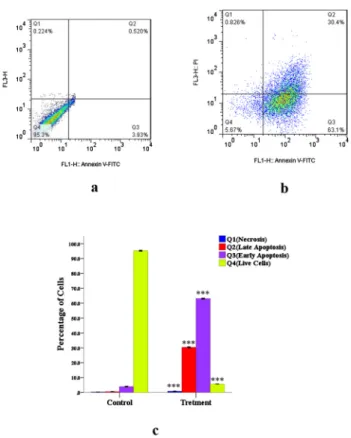
Determination of apoptosis in MCF-7 and MG-63 cells by flow cytometry.Flow cytometry was used in our study to discriminate early apoptotic cells from late apoptotic and[1–40]necrotic ones. In the early stages of apoptosis, phosphatidylserine (PS) is translocated from the inner side of the plasma membrane to the outer layer. Annexin V, a calcium-dependent phospholipid- binding protein with a high affinity for PS can therefore be used as a sensitive probe for the exposure of PS on the cell membrane and hence as a marker of apoptosis.Figs. 4a and 5aare representative of control MCF-7 and MG-63 cells, in which almost no apoptotic cells were detected. However, in progesterone-treated MCF-7 and MG-63 cells (Figs. 4b and 5b, respec- tively), a significant increase in early and late apoptotic cells and a significant decrease in live cells were shown. As shown inFigs. 4b and 5b, analysis of the cell population showed distinct
Fig. 4.Necrosis, early and late apoptosis in MCF-7 cell line induced by progesterone (2.5 mg/mL): Q1:
Necrosis; Q2: Late Apoptosis; Q3: Early Apoptosis; Q4: Viable cells. (4.a) Control MCF-7 cells; (4.b) MCF- 7 cells treated with progesterone; (4.c) Q1, Q2, Q3 and Q4 phases in control and treated MCF-7 cells. The analysis was done by FACSDiva Version 6.1.3. *** represent significant difference compared to control
group (P< 0.001)
sets in the population. The proportion of Annexin Vþand propidium iodide-negative cells was significantly increased by the treatment of MCF-7 and MG-63 cells with effective dose (2.5 mg/
mL) of progesterone compared to the control, indicating the translocation of phosphatidyl serine, an early event of the apoptotic process. The percentage of necrotic cell death was almost negligible in the control and treated groups (Figs. 4c and 5c).
DISCUSSION
In vitro cytotoxicity in MCF-7 cells was evaluated by MTT assay to determine the cytotoxic effect of progesterone. Progesterone (10 mg/mL) exposure was found to have significant cyto- toxic effect on MCF-7 cells. In line with this finding, there are several studies showing the cytotoxic effects of progesterone on cancer cellsin vivoandin vitro. Thein vitroinvestigation on the antiproliferative effects of progesterone on ovarian cancer cells has revealed that the pro- liferation of epithelial ovarian cancer cells is significantly reduced by progesterone treatment Fig. 5.Necrosis, early and late apoptosis in MG-63 cell line induced by progesterone (2.5 mg/mL):
Q1: Necrosis; Q2: Late Apoptosis; Q3: Early Apoptosis; Q4: Viable cells. (5.a) Control MG-63 cells, (5.b) MG-63 cells treated with progesterone; (5.c) Q1, Q2, Q3 and Q4 phases in control and treated MG-63 cells.
The analysis was done by FACSDiva Version 6.1.3. * and *** represent significant difference compared to control group (*:P< 0.05 and ***:P< 0.001)
[28]. The inhibitory effects of progesterone on breast cancer cells have also been reported in previous [11, 29] and recent studies [30]. A recent research indicates that progesterone inhibits breast cancer cell invasion and migration potential [31]. By contrast, there are reports indicating that progesterone may promote breast cancer cell proliferation. A recent study has revealed that progesterone promotes the proliferation of breast cancer cells by inducing the expression of cyclin G1 [9]. Progesterone enhances the proliferation and migration of tumor cells in sporadic breast cancer by decreasing BRCA1 expression level [1]. However, the proliferative effects of progesterone on breast cancer cells might be associated with doses lower than used in this research.
Our findings revealed that treatment of MG-63 cells with progesterone (0.1, 1, 10 mg/mL) reduced cell viabilityin vitro. Previous studies showed that bone-forming osteoblast cells are direct targets for progesterone action [32]. It was also observed that the proliferation, migration, invasion, colony formation and apoptosis of osteosarcoma cells are remarkably affected by sex steroid hormones [33]. Sex steroids, particularly estrogen and progesterone have been shown to play an important role in the regulation of cell proliferation in human osteosarcoma [21].
Progesterone promotes osteocalcin gene transcription by stimulating the expression of c-fos and c-jun, resulting in osteoblast proliferation and differentiation [20]. Depending on their osteo- blastic commitment, osteosarcoma cell lines respond to sex steroids [34]. In contrast to our findings and the results of studies supporting the inhibitory effects of progesterone on the proliferation and development of bone cells, there are reports suggesting that progesterone signaling is not essential for bone growth and turnover [19]. It has also been shown that hor- monal expression is practically negative in osteosarcoma of craniofacial bones [35].
We carried out real-time PCR assay for detection of the expression of theBax, Bcl-2 and P53 genes in MCF-7 and MG-63 cells exposed to effective concentration (2.5 mg/mL) of proges- terone. The results revealed a decrease in the expression level of the anti-apoptoticBcl-2and an increase in the tumor suppressor P53 and pro-apoptotic Bax genes in both cell lines. These results indicated that the cytotoxic effects of progesterone on MCF-7 and MG-63 cells were mediated through Bax- and P53-dependent apoptosis. The role played by progesterone in inducingBax-dependent apoptosis has been reported in previous studies. Although progester- one induces apoptosis in cervical, ovarian and breast cancer cells through Bax- or P53- dependent pathways [19, 28], exposure of T47D human breast cancer cells to progesterone may result in reduced P53 expression level [11]. There have also been few literature reports about the apoptotic effects of progesterone on osteosarcomain vivoandin vitro. In one study, researchers found that progesterone protects osteoblasts against apoptosis through the downstream mito- chondrial pathway [15].
To discriminate the extrinsic and intrinsic apoptotic pathway, the activity levels of the initiator caspases-8 and -9 and the executioner caspase-3 were measured calorimetrically in MCF-7 and MG-63 cells exposed to effective concentration (2.5 mg/mL) of progesterone. Our findings indicated a significant increase in activity levels of caspases-8, -9 and -3 in MCF-7 cells and of caspase-9 and -3–but not caspase-8 –in MG-63 cells. Regarding the involvement of caspase-9 in the intrinsic pathway and of caspase-8 in the extrinsic apoptotic pathway, pro- gesterone induces both the intrinsic and the extrinsic pathway in MCF-7 cells and only the intrinsic pathway in MG-63 cells. Apoptosis in progesterone-treated MCF-7 and MG-63 cells was also confirmed by Hoechst staining and flow cytometry. Consistent with our findings there are reports indicating that effective concentration (2.5 mg/mL) of progesterone induces
apoptosis in cancer cells through triggering of caspase cascade. The results of a study have shown that progesterone induces apoptosis in ovarian cancer cells via activation of a caspase-8 pathway [36]. Caspase-8 has also been shown to be associated with breast cancer risks and the effect may be modified by progesterone receptor status [37]. Caspase-3 expression is also associated with adverse breast cancer-specific survival in breast cancer patients [38]. A high expression of caspase-3 has been observed in patients with cancer surviving symptoms [39]. The effects of progesterone on the caspase cascade in osteosarcoma cells have not been reported in previous studies as far as we know.
CONCLUSION
In conclusion, the results of the present study suggest that progesterone has a potential to impose cytotoxic effects on breast and osteosarcoma cells and the effect is mediated by apoptotic pathways. In this context, progesterone triggers extrinsic and intrinsic apoptotic pathways via increasing the expression level of theBaxandP53genes and activating caspase-3, -8 and -9 in MCF-7 cells; however, the hormone induces the intrinsic apoptotic pathway through enhancing the expression level of theBaxandP53genes and activating caspase-9 and -3 in MG-63 cells. In a broader context, our study may provide a basis for an association between progesterone treatment and recurrence reduction in breast and osteosarcoma cancer patients, thereby providing a lead for modeling a randomizedin vitrostudy.
Conflict of interest:The authors state that there are no conflicts of interest regarding the pub- lication of this article.
ACKNOWLEDGEMENTS
This research was financially supported by Kazerun branch, Islamic Azad University, Kazerun, Iran. We appreciate all who assisted us to carry out this research.
REFERENCES
1. Nagini S. Breast cancer: Current molecular therapeutic targets and new players. Anticancer Agents Med Chem 2017; 17: 152–63.
2. Kindblom L. Bone tumors: epidemiology, classification, pathology. In: Davies A, Sundaram M, James S, editors. Imaging of bone tumors and tumor-like lesions. Medical radiology. Berlin, Heidelberg: Springer;
2009. p. 1–15.
3. Gadducci A, Biglia N, Sismondi P, Genazzani AR. Breast cancer and sex steroids: critical review of epide- miological, experimental and clinical investigations on etiopathogenesis, chemoprevention and endocrine treatment of breast cancer. Gynecol Endocrinol 2005; 20: 343–60.
4. Farach-Carson MC, Lin S-H, Nalty T, Satcher RL. Sex differences and bone metastases of breast, lung, and prostate cancers: do bone homing cancers favor feminized bone marrow?. Front Oncol 2017; 7: 163.
5. Gonzalez Ricarte M, Castro Perez Ad, Tarın JJ, Cano A. Progestogens and risk of breast cancer: a link between bone and breast?. Gynecol Endocrinol 2016; 32: 6–8.
6. Chaiwongwatanakul S, Yanatatsaneejit P, Tongsima S, Mutirangura A, Boonyaratanakornkit V. Sex steroids regulate expression of genes containing long interspersed elements-1s in breast cancer cells. Asian Pac J Cancer Prev 2016; 17: 4003–7.
7. Ruan X, Neubauer H, Yang Y, Schneck H, Schultz S, Fehm T, et al. Progestogens and membrane-initiated effects on the proliferation of human breast cancer cells. Climacteric 2012; 15: 467–72.
8. Vares G, Ory K, Lectard B, Levalois C, Altmeyer-Morel S, Chevillard S, et al. Progesterone prevents radiation- induced apoptosis in breast cancer cells. Oncogene 2004; 23: 4603.
9. Tian JM, Ran B, Zhang CL, Yan DM, Li XH. Estrogen and progesterone promote breast cancer cell prolif- eration by inducing cyclin G1 expression. Braz J Med Biol Res 2018; 51: 1–7.
10. Chen F-P, Chien M-H, Chen H-Y, Huang T-S, Ng Y-T. Effects of estradiol and progestogens on human breast cells: Regulation of sex steroid receptors. Taiwan J Obstet Gynecol 2013; 52: 365–73.
11. Im JY, Kim TH, Lee YJ, Kim IY, Kwack SJ, Byung Mu Lee CSM, et al. Molecular mechanism of progesterone- induced apoptosis in human breast cancer T47D cells. Cancer prevention research 2008; 13: 177–83.
12. Luo G, Abrahams VM, Tadesse S, Funai EF, Hodgson EJ, Gao J, et al. Progesterone inhibits basal and tnf-a- induced apoptosis in fetal membranes: a novel mechanism to explain progesterone-mediated prevention of preterm birth. Reprod Sci 2010; 17: 532–9.
13. Nguyen H, Syed V. Progesterone inhibits growth and induces apoptosis in cancer cells through modulation of reactive oxygen species. Gynecol Endocrinol 2011; 27: 830–6.
14. Kon A, Yuan B, Hanazawa T, Kikuchi H, Sato M, Furutani R, et al. Contribution of membrane progesterone receptorato the induction of progesterone-mediated apoptosis associated with mitochondrial membrane disruption and caspase cascade activation in Jurkat cell lines. Oncol Rep 2013; 30: 1965–70.
15. Wang Q-P, Xie H, Yuan L-Q, Luo X-H, Li H, Wang D, et al. Effect of progesterone on apoptosis of murine MC3T3-E1 osteoblastic cells. Amino Acids 2009; 36: 57–63.
16. Yao W, Dai W, Shahnazari M, Pham A, Chen Z, Chen H, et al. Inhibition of the progesterone nuclear re- ceptor during the bone linear growth phase increases peak bone mass in female mice. PLoS One 2010; 5:
e11410.
17. Chlebowski RT. Bone health in women with early-stage breast cancer. Clin Breast Cancer 2005; 5: S35–40.
18. Bravo D, Shogren KL, Zuo D, Wagner ER, Sarkar G, Yaszemski MJ, et al. 2-methoxyestradiol-mediated induction of frzb contributes to cell death and autophagy in MG-63 osteosarcoma cells. J Cell Biochem 2017;
118: 1497–504.
19. Rickard DJ, Iwaniec UT, Evans G, Hefferan TE, Hunter JC, Waters KM, et al. Bone growth and turnover in progesterone receptor knockout mice. Endocrinology 2008; 149: 2383–90.
20. Liang M, Liao Ey, Xu X, Luo Xh, Xiao Xh. Effects of progesterone and 18-methyl levonorgestrel on osteo- blastic cells. Endocr Res 2003; 29: 483–501.
21. Dohi O, Hatori M, Suzuki T, Ono K, Hosaka M, Akahira JI, et al. Sex steroid receptors expression and hormone-induced cell proliferation in human osteosarcoma. Cancer Sci 2008; 99: 518–23.
22. Zhang N, Wang W, Li W, Liu C, Wang Y, Sun K. Reduction of progesterone, estradiol and hCG secretion by perfluorooctane sulfonate via induction of apoptosis in human placental syncytiotrophoblasts. Placenta 2015;
36: 575–80.
23. Li Y, Wang H, Zhou D, Shuang T, Zhao H, Chen B. Up-regulation of long noncoding rna sra promotes cell growth, inhibits cell apoptosis, and induces secretion of estradiol and progesterone in ovarian granular cells of mice. Med Sci Monit 2018; 24: 2384.
24. Chang W-T, Cheng H-L, Hsieh B-S, Chiu C-C, Lee K-T, Chang K-L. Progesterone increases apoptosis and inversely decreases autophagy in human hepatoma HA22T/VGH cells treated with epirubicin. Sci World J 2014: 2014.
25. Araki T, Yamamoto A, Yamada M. Accurate determination of DNA content in single cell nuclei stained with Hoechst 33258fluorochrome at high salt concentration. Histochemistry 1987; 87: 331–8.
26. Yao G, Ling L, Luan J, Ye D, Zhu P. Nonylphenol induces apoptosis of Jurkat cells by a caspase-8 dependent mechanism. Int Immunopharmacol 2007; 7: 444–53.
27. Grott M, Karakaya S, Mayer F, Baertling F, Beyer C, Kipp M, et al. Progesterone and estrogen prevent cisplatin-induced apoptosis of lung cancer cells. Anticancer Res 2013; 33: 791–800.
28. Hu Z, Deng X. The effect of progesterone on proliferation and apoptosis in ovarian cancer cell. Zhonghua Fu Chan Ke Za Zhi 2000; 35: 423–6.
29. Chavez-Riveros A, Garrido M, Apan MTR, Zambrano A, Dıaz M, Bratoeff E. Synthesis and cytotoxic effect on cancer cell lines and macrophages of novel progesterone derivatives having an ester or a carbamate function at C-3 and C-17. Eur J Med Chem 2014; 82: 498–505.
30. Zhou L, Zhou W, Zhang H, Hu Y, Yu L, Zhang Y, et al. Progesterone suppresses triple-negative breast cancer growth and metastasis to the brain via membrane progesterone receptora. Int J Mol Med 2017; 40: 755–61.
31. Godbole M, Tiwary K, Badwe R, Gupta S, Dutt A. Progesterone suppresses the invasion and migration of breast cancer cells irrespective of their progesterone receptor status-a short report. Cell Oncol (Dordr) 2017;
40: 411–7.
32. MacNamara P, O'Shaughnessy C, Manduca P, Loughrey H. Progesterone receptors are expressed in human osteoblast-like cell lines and in primary human osteoblast cultures. Calcif Tissue Int 1995; 57: 436–41.
33. Fang D, Yang H, Lin J, Teng Y, Jiang Y, Chen J, et al. 17beta-estradiol regulates cell proliferation, colony formation, migration, invasion and promotes apoptosis by upregulating miR-9 and thus degrades MALAT-1 in osteosarcoma cell MG-63 in an estrogen receptor-independent manner. Biochem Biophys Res Commun 2015; 457: 500–6.
34. Quinkler M, Kaur K, Hewison M, Stewart P, Cooper M. Progesterone is extensively metabolized in osteo- blasts: implications for progesterone action on bone. Hormone Metab Res 2008; 40: 679–84.
35. Domınguez-Malagon HR, Gonzalez-Conde E, Cano-Valdez AM, Luna-Ortiz K, Mosqueda-Taylor A.
Expression of hormonal receptors in osteosarcomas of the jaw bones: Clinico-pathological analysis of 21 case.
Medicina oral, patologia oral y cirugia bucal 2014; 19: e44.
36. Syed V, Ho S-M. Progesterone-induced apoptosis in immortalized normal and malignant human ovarian surface epithelial cells involves enhanced expression of FasL. Oncogene 2003; 22: 6883.
37. Han S, Lee K-M, Choi J-Y, Park SK, Lee J-Y, Lee JE, et al. CASP8 polymorphisms, estrogen and progesterone receptor status, and breast cancer risk. Breast Cancer Res Treat 2008; 110: 387–93.
38. Pu X, Storr SJ, Zhang Y, Rakha EA, Green AR, Ellis IO, et al. Caspase-3 and caspase-8 expression in breast cancer: caspase-3 is associated with survival. Apoptosis 2017; 22: 357–68.
39. Himuro T, Horimoto Y, Arakawa A, Matsuoka J, Tokuda E, Tanabe M, et al. Activated caspase 3 expression in remnant disease after neoadjuvant chemotherapy may predict outcomes of breast cancer patients. Ann Surg Oncol 2016; 23: 2235–41.
40. Xiong J, Zhao J, Peng L, Wang H, Liang W. BRCA1 inhibits progesterone-induced proliferation and migration of breast cancer cells. Nan Fang Yi Ke Da Xue Xue Bao 2012; 32: 1105–10.