Contents lists available atScienceDirect
Fitoterapia
journal homepage:www.elsevier.com/locate/fitote
Flavonoid, stilbene and diarylheptanoid constituents of Persicaria maculosa Gray and cytotoxic activity of the isolated compounds
Andrea Vasas
a, Ildikó Lajter
a, Norbert Kúsz
a, Péter Forgó
a, Gusztáv Jakab
b, Csilla Fazakas
c, Imola Wilhelm
c,d, István A. Krizbai
c,d, Judit Hohmann
a,e,⁎aDepartment of Pharmacognosy, University of Szeged, H-6720 Szeged, Hungary
bInstitute of Environmental Sciences, Faculty of Agricultural Water and Environmental Management, Tessedik Samuel College, H-5540 Szarvas, Hungary
cInstitute of Biophysics, Biological Research Centre, H-6726 Szeged, Hungary
dInstitute of Life Sciences, Vasile Goldiş Western University of Arad, RO-310414 Arad, Romania
eInterdisciplinary Centre of Natural Products, University of Szeged, H-6720 Szeged, Hungary
A R T I C L E I N F O Keywords:
Persicaria maculosa Flavanones Chalcones Stilbene Diarylheptanoids Cytotoxicity
A B S T R A C T
Persicaria maculosa(Polygonaceae) has been used as edible and as medicinal plant since ancient times. As a result of multistep chromatographic purifications, chalcones [2′-hydroxy-3′,4′,6′-trimethoxychalcone (1), pashanone (2), pinostrobin chalcone (3)], flavanones [6-hydroxy-5,7-dimethoxyflavanone (4), pinostrobin (5), onysilin (6), 5-hydroxy-7,8-dimethoxyflavanone (7)], flavonol [3-O-methylgalangin (8)], stilbene [persilben (9)], dia- rylheptanoids [1,7-diphenylhept-4-en-3-one (10), dihydroyashabushiketol (12), yashabushidiol B (13)] and 3- oxo-α-ionol-glucoside (11) were isolated fromP. maculosa. The present paper reports for the first time the occurrence of diarylheptanoid-type constituents in the family Polygonaceae. Cytotoxicity of1–5,7and9–11on 4 T1 mouse triple negative breast cancer cells was assayed by MTT test. None of the tested compounds reduced the cell viability to less than 80% of the control. On non-tumorigenic D3 human brain endothelial cells the decrease of cell viability was observed in case of1and2. Further impedance measurements on 4 T1 and D3 cells a concentration-dependent decrease in the cell index of both cell types was demonstrated for1, while2proved to be toxic only on endothelial cells.
1. Introduction
The genusPersicaria (smartweed) (family Polygonaceae) includes about 100 species nearly worldwide [1]. The plants are perennials or annuals [2–3].Persicaria maculosaGray (syn.Polygonum persicariaL., lady's thumb) is an annual plant, native to Europe and widely dis- tributed as a weed throughout temperate and tropical North and South America, Asia, North Africa and Australia [4].The Cherokee, Chippewa, and Iroquois native Americans prepared simple or complex decoctions of P. maculosa, which they used as dermatological, urinary, gastro- intestinal, and veterinary aids, for cardiac diseases, and as an analgesic [5]. The plant has also been used to treate.g.diarrhoea and infectious diseases, and the leaves and young shoots can be eaten in salads [6].
Previous phytochemical studies revealed the presence of stilbenes, fla- vonoids, phenolic acids, sesquiterpenes and diterpenes in this species [2,7–8].
In vitro pharmacological studies demonstrated the antibacterial, antifungal and insecticidal activities of the plant [9,10], while inin vivo
studies the hydroalcoholic extract of the herb exhibited anti-in- flammatory effect and decreased locomotion after intraperitoneal ad- ministration to rats; therefore, it possessed spasmolytic activity [11]. As concerns the chemical constituents responsible for the observed activ- ities, persilben, a unique naturally occurringE-stilbene attracted great interest because of its antimicrobial, antifungal and antioxidant activ- ities and its good penetration through biological membranes in con- sequence of its high lipophilicity [12,13]. Moreover, flavonoids of the plant have anti-inflammatory and antioxidant activities. Different ex- tracts prepared by our group fromP. maculosawere investigated on G protein-activated inwardly rectifying K+channel (GIRK) using patch clamp method. The CHCl3 extract of the plant exhibited high GIRK channel inhibitory activity at 0.1 mg/mL concentration [9].
In continuation of our work onP. maculosa, thirteen compounds (1−13), among them chalcones, flavanones, flavonol, diarylhepta- noids, a stilbene derivative and an α-ionol-glycoside were identified.
Cytotoxic activity of the isolated compounds 1–5, 7 and9–11 was evaluated against 4T1 and D3 cell linesin vitro.
https://doi.org/10.1016/j.fitote.2020.104610
Received 18 March 2020; Received in revised form 16 April 2020
⁎Corresponding author at: Department of Pharmacognosy, University of Szeged, H-6720 Szeged, Hungary.
E-mail address:hohmann@pharm.u-szeged.hu(J. Hohmann).
Available online 17 May 2020
0367-326X/ © 2020 The Authors. Published by Elsevier B.V. This is an open access article under the CC BY-NC-ND license (http://creativecommons.org/licenses/BY-NC-ND/4.0/).
T
2. Experimental section
2.1. General experimental procedures
Vacuum liquid chromatography (VLC) was carried out on silica gel G (15 μm, Merck); preparative thin-layer chromatography (preparative TLC) was performed on silica gel 60 F254 plates (Merck). Medium- pressure liquid chromatography (MPLC) was performed by a Biotage SP1™ Purification System using a KP-C18HS 40 + M column. HPLC was performed on a LiChrospher RP-18 (5 μm, 250 × 4 mm, Merck) column using mixture of acetonitrile–H2O as mobile phase on a Waters 600 instrument. NMR spectra were recorded on a Bruker Avance DRX 500 spectrometer at 500 MHz (1H) and 125 MHz (13C). The peaks of the residual solvents were taken as reference. Two-dimensional data were acquired and processed with standard Bruker software. In the1He1H COSY, HSQC and HMBC experiments, gradient-enhanced versions were used. ESI and APCI mass spectra were recorded on an API 2000 triple quadrupole mass spectrometer equipped with an electrospray and APCI interfaces.
2.2. Plant material
Persicaria maculosa Gray was collected in the flowering period in Homoródalmás (Hungary) in July 2012. Botanical identification was performed by G. J. (Institute of Water Management and Irrigation, Szent István University, H-5540 Szarvas, Hungary). A voucher spe- cimen (No. 811) has been deposited at the Herbarium of the Department of Pharmacognosy, University of Szeged, Szeged, Hungary.
2.3. Extraction and isolation
The air-dried and ground whole plants ofP. maculosa(3.15 kg) were extracted with MeOH (20 L) at room temperature. The crude extract was concentratedin vacuo and subjected to solvent–solvent partition first with 3 × 500 mLn-hexane, then with 3 × 500 mL of CHCl3. After evaporation, the CHCl3phase (36.8 g) was fractionated by MPLC on reversed-phase silica gel, using a gradient system of MeOH–H2O (from 3:7 to 8:2). The fractions were combined into twelve subfractions (I–XII) according to the TLC monitoring. Subfraction IV (229.4 mg) was separated by VLC on silica gel, using a gradient system of CH2Cl2–acetone (from 99:1 to 8:2) to yield 13 main fractions (IV/1–13).
Fraction IV/5 was further purified by preparative TLC with CH2Cl2–MeOH (4:1) to yield compound11 (3.2 mg). Subfraction IX (282.8 mg) was chromatographed by VLC on silica gel with the gradient system of cyclohexane–CH2Cl2–acetone (from 5:5:0 to 0:95:5). After TLC monitoring, 12 main fractions (IX/1–12) were obtained. Fraction IX/1 was subjected to preparative TLC on silica gel using cyclohexane–CH2Cl2–acetone (5:5:1) as developing system, to yield compounds10(2.3 mg) and12(1.8 mg). Fraction IX/3 was also pur- ified by prep. TLC with cyclohexane–CH2Cl2–MeOH 40:20:1, and compounds9(2.8 mg) and7 (4.5 mg) were afforded. Fraction IX/4 (30.3 mg) was chromatographed by Sephadex LH-20 gel using MeOH as eluent, and thereafter by prep. TLC with cyclohexane–CH2Cl2–MeOH 20:30:1 to obtain compounds6(5.2 mg) and13(4.0 mg). Fraction XI (229.4 mg) was separated by normal phase VLC, which was eluted with the gradient system of CH2Cl2–MeOH (from 99:1 to 9:1) to yield 13 main fractions (XI/1–13). Compound 5(4.8 mg) was obtained from fraction XI/2 (8 mg) by RP-HPLC, using acetonitrile–H2O (9:1) (iso- cratic elution, flow: 0.5 mL/min). Fraction XI/5 (76.5 mg) was purified by Sephadex LH-20 gel chromatography, using MeOH as eluent to yield 6 subfractions (XI/5/1–6). From subfraction XI/5/2 compounds 2 (4.5 mg) and 8(1.1 mg) were separated by prep. TLC, using tolue- ne–ethyl acetate–MeOH (5:4:1). Compound 1(7.0 mg) was obtained from subfraction XI/5/3 by prep. TLC using CH2Cl2–acetone (19:1).
Compound 3 (3.1 mg) was crystallized from fraction XI/7, and 4 (2.2 mg) from fraction XI/10.
2.3.1. Characterization of pinostrobin chalcone (3)
Orange crystals, m.p. 149–150 °C;1H NMR (500 MHz, CDCl3)δppm 14.1 (1H, s, 2’OH), 7.86 (1H, d, J = 15.6 Hz, H-β), 7.76 (1H, d, J= 15.6 Hz, H-α), 7.59 (2H, m, H-2, H-6), 7.38 (3H, m, H-3–H-5), 6.01 and 5.94 (2 × 1H, 2 × d,J= 1.0 Hz, H-3′, H-5′) 3.91 (3H, s, OCH3);
ESI-MS positivem/z293 [M + Na]+, 271 [M + H]+, 167 [C8H7O4]+. 2.3.2. Characterization of 6-hydroxy-5,7-dimethoxyflavanone (4)
White crystals, m.p. 148–149 °C;1H NMR (500 MHz, CD3OD)δppm 7.53 (2H, d,J= 7.3 Hz, H-2′, H-6′), 7.42 (2H, t,J= 7.2 Hz, H-3′, H-5′), 7.37 (1H, t,J= 7.3 Hz, H-4′), 6.17 (1H, s, H-8), 5.49 (1H, dd,J= 12.7, 2.9, H-2), 3.80 (3H, s, 5-OCH3), 3.76 (3H, s, 7-OCH3), 3.01 (1H, dd, J= 12.7, 16.7 Hz, H-3a), 2.76 (1H, dd,J= 16.7, 3.0, H-3b);13C NMR (125 MHz, CD3OD):δppm 192.0 (C-4), 159.7 (C-5), 159.4 (C-6), 140.5 (C-1′), 131.1 (C-7), 129.7 (C-3′, C-5′), 129.6 (C-4′), 127.2 (C-2′, C-6′), 106.3 (C-10), 94.2 (C-8), 80.6 (C-2), 61.4 (7-OCH3), 56.2 (5-OCH3), 46.4 (C-3).
2.3.3. Characterization of pinostrobin (5)
Yellow solid;1H NMR (500 MHz, CD3OD)δppm 12.0 (1H, s, 5-OH), 7.50 (2H, d,J= 7.4 Hz, H-2′, H-6′), 7.42 (2H, t,J= 7.2 Hz, H-3′, H-5′), 7.37 (1H, t,J= 7.1 Hz, H-4′), 6.10 (1H, d,J= 1.8 Hz, H-8), 6.06 ((1H, d,J= 1.8 Hz, H-6), 5.59 (1H, dd,J= 12.5, 2.6 Hz, H-2), 3.82 (3H, s, OCH3), 3.13 (1H, dd,J= 12.8, 17.1 Hz, H-3a), 2.82 (1H, dd,J= 17.1, 2.9 Hz, H-3b); APCI-MSm/z271 [M + H]+, 167 [M–C8H8]+, 131, 103.
2.3.4. 3-Methylgalangin (8)
Yellow powder;1H NMR (500 MHz, CD3OD)δppm 8.06 (2H, m, H- 2, H-6′), 7.55 (3H, m, H-3’–H-5′), 6.43 (1H, d,J= 2.0 Hz, H-8), 6.23 (1H, d,J= 2.0 Hz, H-6), 3.80 (3H, s, OCH3); ESI-MS positivem/z285 [M + H]+, 270 [M + H-CH3]+.
2.3.5. Persilben (9)
Yellowish powder;1H NMR (500 MHz, CDCl3) 13.9 (1H, s, COOH), 8.15 (1H, d,J= 15.6, Hα), 7.86 (1H, d,J= 15.6 Hz, Hβ), 7.65 (2H, m, H-2, H-6), 7.40 (3H, m, H-3–H-5), 6.88 (1H, s, H-6), 6.10 (1H, s, H-4), 3.91, 3.86 (2 × 3H, 2 × s, 2 × OCH3).
2.4. Cell culture and toxicity tests
4T1 (mouse triple negative breast cancer cells) were cultured in RPMI 1640 medium supplemented with 5% foetal bovine serum (FBS) (both from Thermo Fischer Scientific, Waltham, MA, USA). D3 (hCMEC/D3 human cerebral microvascular endothelial cells) were kept in rat tail collagen coated dishes in EBM-2 medium complemented with 2% FBS and EGM-2MV kit (all of them purchased from Lonza, Basel, Switzerland).
For MTT (3-[4,5-dimethylthiazole-2-yl]-2,5-diphenyltetrazolium bromide) assay, cells were plated in 96-well plates (Corning, Corning, NY, USA) in a density of 5000 4 T1 cells/well or 25,000 D3 cells/well.
After 24 h, half of the medium was replaced with serum-free medium, containing the compounds in a final concentration of 10, 20 or 50 μmol/L. Control wells received solvent (DMSO) in max. 0.2% con- centration. After 48 h, MTT reagent (Sigma-Aldrich, St. Louis, MO, USA) was added to the cells in a final concentration of 2.5 mg/mL. After incubation at 37 °C for one hour, acidified isopropanol solution was added to each well. Absorbance was measured at 595 nm with a FLUOstar OPTIMA microplate reader (BMG LABTECH, Offenburg, Germany).
For impedance measurements, cells were plated in 96-well E-plates having micro-electrodes integrated on the bottom (ACEA Biosciences, San Diego, CA, USA), and allowed to attach onto the electrode surface.
After 24 h, cells were treated with the test compounds as described above. Electrical impedance was recorded in real-time using an xCELLigence® Real-Time Cell Analysis (RTCA) instrument (ACEA Biosciences). Cell impedance (which depends on cell number, degree of
adhesion, spreading and viability), expressed in arbitrary units (cell index) was automatically calculated by the software of the instrument.
3. Results and discussion
3.1. Isolation and structure elucidation of the compounds
MeOH extract was prepared from dried whole plant ofP. maculosa and subjected to solvent-solvent partition, yielding n-hexane, CHCl3
and remaining aqueous extracts. The CHCl3extract was fractionated by medium pressure liquid chromatography on reversed phase silica gel resulting twelve fractions. Further purification of the fractions with combination of different chromatographic techniques (VLC, prep TLC, Sephadex LH-20 gel chromatography and HPLC) resulted in the isola- tion of thirteen compounds (1–13).
Eight compounds (1–8) are belonging to the group of flavonoids, interestingly all of them have an unsubstituted ring B. Three chalcones were identified as 2′-hydroxy-3′,4′,6′-trimethoxychalcone (1), 2′,6′-di- hydroxy-3′,4′-dimethoxychalcone (pashanone = polygochalcone, 2),
and 2′,6′-dihydroxy-4′-methoxychalcone (pinostrobin chalcone, 3) by comparison with reference data [14,15]. Pinostrobin (5) [16,17], and three isomeric rare flavanones, 6-hydroxy-5,7-dimethoxyflavanone (4) [18], 5-hydroxy-6,7-dimethoxyflavanone (onysilin,6) [19], and 5-hy- droxy-7,8-dimethoxyflavanone (7) [22] were identified by analysis of their 1D and 2D NMR spectra and comparison with the data published in the literature [22]. In previous studies, onysilin (6) and 5-hydroxy- 7,8-dimethoxyflavanone (7) were differentiated on the basis of minor difference in13C NMR data, UV spectra with shift reagents, and melting points [22,20]; however, in our HMBC investigations clear arguments were found for structural assignment of6and7(Fig. 1).
In the present experiment, the only isolated flavonol is 3-O-me- thylgalangin (8) [21]. Compound9was identified as the known per- silben [15], and compound 11 as (6R,9S)-3-oxo-α-ionol-glucoside based on the NMR and optical rotation data [αD28 + 43° (c 0.19, MeOH)] [22]. Compound10revealed to be 1,7-diphenyl-4-en-3-hep- tanone based on 1D and 2D NMR and MS data, which were in agree- ment with published data [23].
Close analogues of 10, (5S)-1,7-diphenylhept-5-ol-3-one (=dihy- droyashabushiketol) (12) [measured [α]D= +14 (c0.1, CHCl3)] [24], and (3S,5S)-1,7-diphenylhept-3,5-diol (=yashabushidiol B) (13) [measured [α]D= −5 (c0.1, CHCl3)] [25] were also identified fromP.
maculosa. Our NMR measurements allowed previously unpublished1H and13C NMR assignments for compounds3,4,5,8, and9, these data are listed in Materials and Methods section.
3.2. Chemotaxonomic significance
A variety of diarylheptanoids have been isolated previously from plant families Aceraceae, Actinidiaceae, Betulaceae, Burseraceae, Casuarinaceae, Juglandaceae, Leguminosae, Myricaceae, and Zingiberaceae, but the present paper reports for the first time the oc- currence of diarylheptanoid-type constituents in the family Polygonaceae [26]. The isolation of compounds10, 12, and13pro- vided new chemotaxonomic information, the presence of diarylhepta- noids might serve as a chemotaxonomic marker forPersicariaspecies.
1,7-Diphenylhept-4-en-3-one (10) was reported previously only as the metabolite ofAlpinia officinarum(Zingiberaceae) with potent PAF re- ceptor binding inhibitory and inducible NO synthase protein and mRNA expression suppressing activities [27,28]. Dihydroyashabushiketol (12), and yashabushidiol B (13) were isolated formerly fromAlpinia, Acorus, AmmomumandAlnus species, with cytotoxic activity against IMR-32 human neuroblastoma cells [29]. These compounds were pre- viously obtained by chemical synthesis, too [30,31].
The present experiment afforded the first isolation of chalcones1,3 and flavanone4from the family Polygonaceae. 2’-Hydroxy-3′,4′,6′-tri- methoxychalcone (1) was obtained formerly from Annonaceae, Piperaceae and Rosaceae species [32], while pinostrobin chalcone (3) fromAlpiniaspecies [18]. 6-Hydroxy-5,7-dimethoxyflavanone (4) was isolated previously only fromPiper hispidum[21]. Onysilin (6), 5-hy- droxy-7,8-dimethoxyflavanone (7), 3-O-methylgalangin (8), 3-oxo-α- ionol-glucoside (11) were isolated previously from different species of family Polygonaceae [33].
Fig. 1.Diagnostic HMBC correlations (H→C) of compounds6and7between 5- OH and C-5, C-6 and C-10 (chemical shifts shown inδppm).
Flavonoids have generally been used as chemotaxonomic marker in genus Polygonum. The most common feature of Polygonum and Persicaria genus was hold the flavonoid spectrum with glycosylated and/or methoxylated derivatives of kaempferol, quercetin, myricetin, apigenin and luteolin, glycosylated at C-3 [34]. Interestingly in our study, with except of 3-O-methylgalangin (8), chalcones and flavanones were isolated, among them biogenetically related chalcone–flavanone pairs, such as pinostrobin chalcone (3) and pinostrobin (5), and pa- shanone (2) and 5-hydroxy-7,8-dimethoxyflavanone (7). This finding serves as a chemotaxonomic marker; the common occurrence of chal- cone–flavanone pairs is regarded as taxonomic characteristic only to PolygonumandPersicariagenus in Polygonaceae family.
3.3. Evaluation of biological effects of compounds isolated from P.
maculosa
In order to test the possible toxic effects of the isolated compounds on tumor cells, at first an MTT assay was performed on proliferating mouse breast cancer cells. At 10 μM concentration, none of the tested compounds (1–5,7and9–11) reduced viability of 4 T1 cells to less than 80% of the control after 48 h of treatment (not shown). As a model of non-tumorigenic cells, D3 human brain endothelial cells were used.
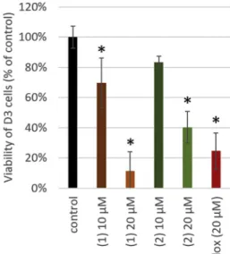
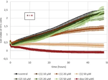
Confluent D3 monolayers were treated with the test compounds at concentrations of 10 and 20 μM, and real-time impedance measure- ments were performed, which revealed that only 2′-hydroxy-3′,4′,6′- trimethoxy chalcone (1) and pashanone (2) decreased cell index (Fig. 2). MTT assay data were in line with this result, showing a con- centration-dependent decrease in the viability of D3 cells in response to 2′-hydroxy-3′,4′,6′-trimethoxy chalcone (1) and pashanone (2) (Fig. 3).
As a next step, we aimed to characterize in detail the effects of compounds1and2on tumor and normal cells. Impedance measure- ments did not show tumor cell selectivity; a concentration-dependent decrease in the cell index of both cell types was found in response to 2′- hydroxy-3′,4′,6′-trimethoxy chalcone (1), while pashanone (2) proved to be toxic only on endothelial cells (Figs. 4 and 5).
MTT data confirmed the results obtained with impedance mea- surements (Fig. 6).
In the course of our studies, chalcone (1–3), flavanone (4–7), fla- vonol (8), diarylheptanoid (10,12,13), stilbene (9), and ionol (11) derivatives were isolated fromP. maculosa, most of them having che- motaxonomic significance. The cytotoxicity assays of selected com- pounds (1–5,7and9–11) on tumorigenic 4 T1 mouse triple negative breast cancer and non-tumorigenic D3 human brain endothelial cells by impedance measurements and MTT assay revealed moderate activity of two chalcones, 2′-hydroxy-3′,4′,6′-trimethoxy chalcone (1) and
pashanone (2), the other compounds did not show any potency. A concentration-dependent decrease in the cell index of both cell types was demonstrated for 2′-hydroxy-3′,4′,6′-trimethoxy chalcone (1), while pashanone (2) proved to be toxic only on endothelial cells.
Previous studies have been proved that chalcones are promising antitumor lead compounds due to their antioxidant, cytotoxic, and apoptosis inducing activities. The cytotoxicity of chalcones against tumor cell lines may be the result of disruption of the cell cycle, in- hibition of angiogenesis, mitochondrial uncoupling, apoptosis induc- tion, antiproliferation, and antimetastasis. For antimitotic activity, the α,β-unsaturated carbonyl part, the planar structure geometry, and presence of methoxy and 2′ oxygenated substituents are favourable features [35,36]. 2’-Hydroxy-3′,4′,6′-trimethoxychalcone (1) and pa- shanone (2) fulfil these requirements.
Although the tumor specificity of chalcones has been reported several times,e.g.in comparing sensitivity of HepG2 cells to normal liver cells; osteosarcoma to bone marrow and small intestinal epithelial cells; murine leukemia cells to normal human lymphocytes; and human prostate cancer cells to normal human prostate epithelial cells [37], the Fig. 2.Impedance of D3 human brain endothelial cells treated for 48 h with
compounds1–5,7, and9–11isolated fromPersicaria maculosa. Doxorubicin was used as a positive control of toxicity. *P < .01 compared to control (ANOVA and Bonferroni's post-hoc test).
Fig. 3.Viability of D3 cells treated for 48 h with 2′-hydroxy-3′,4′,6′-trimethoxy chalcone (1) and pashanone (2), as assessed by MTT assay. *P < .01 com- pared to control (ANOVA and Bonferroni's post-hoc test).
Fig. 4.Impedance changes of D3 cells treated with 2′-hydroxy-3′,4′,6′-tri- methoxy chalcone (1) and pashanone (2) in concentrations of 10, 20 or 50 μmol/L. *P< .01 [1and2in 20 and 50 μmol/L concentrations] compared to control, as assessed by comparing areas under curves with ANOVA and Bonferroni's post-hoc test.
tumor specificity could not be presented in our experiment when chalcones1and2on 4T1 mouse triple negative breast cancer and non- tumorigenic D3 human brain endothelial cells were tested.
Declaration of Competing Interest
The authors declare no competing financial interest.
Acknowledgments
Financial support for this research was provided by the Economic Development and Innovation Operative Program GINOP-2.3.2-15- 2016-00012 and GINOP-2.3.2-15-2016-00020. Grant 20391-3/2018/
FEKUSTRAT awarded by the Ministry of Human Capacities, Hungary, is acknowledged. C.F. is recipient of BO/00213/19/8 and UNKP-19-4- SZTE-18 fellowships and support from NKFIH (PD-121130). Work of I.W. is supported by the NKFIH FK-124114 and the UEFISCDI PN-III-P1- 1.1-TE-2016-1352 projects.
References
[1] http://www.efloras.org/florataxon.aspx?flora_id=1&taxon_id=124629.
[2] M. Derita, S. Zacchino, Chemotaxonomic importance of sesquiterpenes and flavo- noids in five Argentinian species ofPolygonumgenus, J. Essent. Oil Res. 23 (2011) 11–14.
[3] M.G. Derita, S.J. Gattuso, S.A. Zacchino, Occurrence of polygodial in species of Polygonumgenus belonging toPersicariasection, Biochem. Syst. Ecol. 36 (2008) 55–58.
[4] http://www.efloras.org/florataxon.aspx?flora_id=1&taxon_id=220010181.
[5] D.F. Austin, Baboquivari Mountain Plants: Identification, Ecology, and
Ethnobotany, The University of Arizona Press, Tucson, Arizona, 2010, pp. 222–223.
[6] https://bie.ala.org.au/species/http://id.biodiversity.org.au/node/apni/2894047.
[7] H.D. Smolarz, Chromatographical analysis of phenolic acids in some species of PolygonumL genus. Part 2 – Quantitative determination of the major components by high performance liquid chromatography (HPLC), Acta Soc. Bot. Pol. 69 (2000) 21–23.
[8] N. Prota, R. Mumma, H.J. Bouwmeester, M.A. Jongsma, Comparison of the che- mical composition of three species of smartweed (genusPersicaria) with a focus on drimane sesquiterpenoids, Phytochemistry 108 (2014) 129–136.
[9] F. Hussain, B. Ahmad, I. Hameed, D. Dastagir, P. Sanaullah, S. Azam, Antibacterial, antifungal and insecticidal activities of some selected medicinal plants of Polygonaceae, Afr. J. Biotechnol. 9 (2010) 5032–5036.
[10] M. Derita, S. Zacchino, Validation of the ethnopharmacological use ofPolygonum persicariafor its antifungal properties, Nat. Prod. Commun. 6 (2011) 931–933.
[11] H.M. Yano, E.M. Bacchi, L.S.S. Hayashi, R. De Lucia, Anti-inflammatory activity and effect on locomotion ofPolygonum persicariaL. (Polygonaceae) extract in rats, Lat.
Am. J. Pharm. 30 (2011) 1635–1638.
[12] H.D. Smolarz, M.J. Potrzebowski, Persilben, a new carboxystilbene fromPolygonum persicaria, J. Mol. Struct. 605 (2002) 151–156.
[13] H.D. Smolarz, U. Kosikowska, B. Baraniak, A. Malm, A. Persona, Lipophilicity, antifungal and antioxidant properties of persilben, Acta Pol. Pharm. 62 (2005) 457–460.
[14] K. Ichino, H. Tanaka, K. Ito, Synthesis of helilandin B, pashanone, and their isomers, J. Nat. Prod. 51 (1988) 906–914.
[15] S.N. Malek, C.W. Phang, H. Ibrahim, A.W. Norhanom, K.S. Sim, Phytochemical and cytotoxic investigations ofAlpinia muticarhizomes, Molecules 16 (2011) 583.
[16] V.I. Yamovoi, E.A. Kulmagambetova, A.T. Kulyyasov, K.M. Turdybekov, S.M. Adekenov, Molecular structure of a novel polymorphic modification of pi- nostrobin, Chem. Nat. Compd. 37 (2001) 424–427.
[17] M. Tanjung, T.S. Tjahjandarie, M.H. Sentosa, Antioxidant and cytotoxic agent from the rhizomes ofKaempferia pandurata, Asian Pac. J. Trop. Dis. 3 (2013) 401–404.
[18] P.C. Vieira, M.A. De Alvarenga, O.R. Gottlieb, H.E. Gottlieb, 4-Hexadecenylphenol and flavonoids fromPiper hispidum, Planta Med. 39 (1980) 153–156.
[19] C. Kamperdick, N.H. Van, T.V. Sung, Constituents fromMiliusa balansae (Annonaceae), Phytochemistry 61 (2002) 991–994.
[20] E. Wollenweber, The occurrence of flavanones in the farinose exudate of the fern Onychium siliculosum, Phytochemistry 21 (1982) 1462–1464.
[21] M. Xin, S. Guo, W. Zhang, Z. Geng, J. Liang, S. Du, Z. Deng, Y. Wang, Chemical constituents of supercritical extracts fromAlpinia officinarumand the feeding de- terrent activity againstTribolium castaneum, Molecules 22 (2017) 647.
[22] A. Pabst, D. Barron, E. Sémon, P. Schreier, Two diastereomeric 3-oxo-α-ionol β-D- glucosides from raspberry fruit, Phytochemistry 31 (1992) 1649–1652.
[23] Z. Liu, S. Sang, T.G. Hartman, C.T. Ho, R.T. Rosen, Determination of diarylhepta- noids fromAlpinia officinarum(Lesser Galangal) by HPLC with photodiode array and electrochemical detection, Phytochem. Anal. 16 (2005) 252–256.
[24] C.H. Park, K.H. Kim, I.K. Lee, S.Y. Lee, S.U. Choi, J.H. Lee, K.R. Lee, Phenolic constituents ofAcorus gramineus, Arch. Pharm. Res. 34 (2011) 1289–1296.
[25] V.U. Pawar, V.S. Shinde, Chiron approach to the synthesis of yashabushidiol B, (3S,5S)-1-(4′-hydroxyphenyl)-7-phenylheptane-3,5-diol, and its 4′-methoxy ana- logue, Tetrahedron-Asymmetry 22 (2011) 8–11.
[26] Y. Jahng, J.G. Park, Recent studies on cyclic 1,7-diarylheptanoids: their isolation, structures, biological activities, and chemical synthesis, Molecules 23 (2018) 3107/
1–3107/42.
[27] G.J. Fan, Y.H. Kang, Y.N. Han, B.H. Han, Platelet-activating factor (PAF) receptor binding antagonists fromAlpinia officinarum, Bioorg. Med. Chem. Lett. 17 (2007) 6720–6722.
[28] H.J. Lee, J.S. Kim, J.H. Ryu, Suppression of inducible nitric oxide synthase ex- pression by diarylheptanoids fromAlpinia officinarum, Planta Med. 72 (2006) 68–71.
[29] Y. Sun, K. Tabata, H. Matsubara, S. Kitanaka, T. Suzuki, K. Yasukawa, New cyto- toxic diarylheptanoids from the rhizomes ofAlpinia officinarum, Planta Med. 74 (2008) 427–431.
[30] J. Romanski, P. Nowak, C. Chapuis, J. Jurczak, Total synthesis of (5S)-dihydroya- shabushiketol, Tetrahedron-Asymmetry 22 (2011) 787–790.
[31] M. Narasimhulu, T. Srikanth Reddy, K. Chinni Mahesh, A. Sai Krishna, J. Venkateswara Rao, Y. Venkateswarlu, Synthesis of yashabushidiol and its ana- logues and their cytotoxic activity against cancer cell lines, Bioorg. Med. Chem.
Lett. 19 (2009) 3125–3127.
Fig. 5.Impedance changes of 4 T1 mouse triple negative breast cancer cells treated with 2′-hydroxy-3′,4′,6′-trimethoxy chalcone (1) and pashanone (2) in concentrations of 10, 20 or 50 μmol/L. *P < .01 [1in 20 and 50 μmol/L concentrations and doxorubicin] compared to control, as assessed by com- paring areas under curves with ANOVA and Bonferroni's post-hoc test.
Fig. 6.Viability 4 T1 cells treated for 48 h with different concentrations 2′- hydroxy-3′,4′,6′-trimethoxy chalcone (1) and pashanone (2), as assessed with MTT assay. * P < .01 compared to control (ANOVA and Bonferroni's post-hoc test).
[32] T.P. Lien, A. Porzel, J. Schmidt, T.V. Sung, G. Adam, Chalconoids fromFissistigma bracteolatum, Phytochemistry 53 (2000) 991–995.
[33] B.K. Datta, S.K. Datta, M.A. Rashid, S.D. Sarker, Flavonoids fromPolygonum stag- ninum, Biochem. Syst. Ecol. 30 (2002) 693–969.
[34] C.W. Park, Flavonoid chemistry ofPolygonumsect.Echinocaulon: a systematic survey, Syst. Bot. 12 (1987) 167–179.
[35] M.L. Go, X. Wu, X.L. Liu, Chalcones: an update on cytotoxic and chemoprotective properties, Curr. Med. Chem. 12 (2005) 481–499.
[36] S. Marquina, M. Maldonado-Santiago, J.N. Sánchez-Carranza, M. Antúnez-Mojica, L. González-Maya, R.S. Razo-Hernández, L. Alvarez, Design, synthesis and QSAR study of 2′-hydroxy-4′-alkoxy chalcone derivatives that exert cytotoxic activity by the mitochondrial apoptotic pathway, Bioorg. Med. Chem. 27 (2019) 43–54.
[37] H. Sakagami, Y. Masuda, M. Tomomura, S. Yokose, Y. Uesawa, N. Ikezoe, D. Asahara, K. Takao, T. Kanamoto, S. Terakubo, H. Kagaya, H. Nakashima, Y. Sugita, Quantitative structure–cytotoxicity relationship of chalcones, Anticancer Res. 37 (2017) 1091–1098.