Article
Coumarin-Based Triapine Derivatives and Their Copper(II) Complexes: Synthesis, Cytotoxicity and mR2 RNR
Inhibition Activity
Iryna Stepanenko1,*, Maria V. Babak2 , Gabriella Spengler3 , Marta Hammerstad4, Ana Popovic-Bijelic5 , Sergiu Shova6 , Gabriel E. Büchel7 , Denisa Darvasiova8, Peter Rapta8 and Vladimir B. Arion1,*
Citation: Stepanenko, I.; Babak, M.V.;
Spengler, G.; Hammerstad, M.;
Popovic-Bijelic, A.; Shova, S.; Büchel, G.E.; Darvasiova, D.; Rapta, P.; Arion, V.B. Coumarin-Based Triapine Derivatives and Their Copper(II) Complexes: Synthesis, Cytotoxicity and mR2 RNR Inhibition Activity.
Biomolecules2021,11, 862.
https://doi.org/10.3390/
biom11060862
Academic Editor: S. Lorenzo
Received: 3 May 2021 Accepted: 27 May 2021 Published: 9 June 2021
Publisher’s Note:MDPI stays neutral with regard to jurisdictional claims in published maps and institutional affil- iations.
Copyright: © 2021 by the authors.
Licensee MDPI, Basel, Switzerland.
This article is an open access article distributed under the terms and conditions of the Creative Commons Attribution (CC BY) license (https://
creativecommons.org/licenses/by/
4.0/).
1 Institute of Inorganic Chemistry, University of Vienna, Währinger Strasse 42, A-1090 Vienna, Austria
2 Drug Discovery Lab, Department of Chemistry, City University of Hong Kong, 83 Tat Chee Avenue, Hong Kong SAR 518057, China; mbabak@cityu.edu.hk
3 Department of Medical Microbiology, Albert Szent-Györgyi Health Center and Faculty of Medicine, University of Szeged, Semmelweis utca 6, H-6725 Szeged, Hungary; spengler.gabriella@med.u-szeged.hu
4 Section for Biochemistry and Molecular Biology, Department of Biosciences, University of Oslo, P.O. Box 1066, Blindern, NO-0316 Oslo, Norway; marta.hammerstad@ibv.uio.no
5 Faculty of Physical Chemistry, University of Belgrade, Studentski trg 12–16, 11158 Belgrade, Serbia;
ana@ffh.bg.ac.rs
6 “Petru Poni” Institute of Macromolecular Chemistry, Aleea Gr. Ghica Voda 41A, 700487 Iasi, Romania;
shova@icmpp.ro
7 ChemConsult GmbH, P.O. Box 43, 9485 Nendeln, Liechtenstein; gabriel.buechel@gmail.com
8 Faculty of Chemical and Food Technology, Institute of Physical Chemistry and Chemical Physics, Slovak University of Technology in Bratislava, Radlinského 9, SK-812 37 Bratislava, Slovakia;
denisa.darvasiova@stuba.sk (D.D.); peter.rapta@stuba.sk (P.R.)
* Correspondence: iryna.stepanenko@univie.ac.at (I.S.); vladimir.arion@univie.ac.at (V.B.A.)
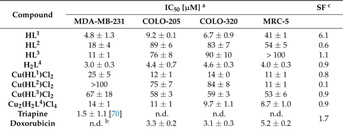
Abstract:A series of thiosemicarbazone-coumarin hybrids (HL1-HL3andH2L4) has been synthe- sised in 12 steps and used for the preparation of mono- and dinuclear copper(II) complexes, namely Cu(HL1)Cl2 (1),Cu(HL2)Cl2 (2),Cu(HL3)Cl2 (3) andCu2(H2L4)Cl4 (4), isolated in hydrated or solvated forms. Both the organic hybrids and their copper(II) and dicopper(II) complexes were comprehensively characterised by analytical and spectroscopic techniques, i.e., elemental analysis, ESI mass spectrometry, 1D and 2D NMR, IR and UV–vis spectroscopies, cyclic voltammetry (CV) and spectroelectrochemistry (SEC). Re-crystallisation of1from methanol afforded single crystals of copper(II) complex with monoanionic ligandCu(L1)Cl, which could be studied by single crystal X-ray diffraction (SC-XRD). The prepared copper(II) complexes and their metal-free ligands revealed antiproliferative activity against highly resistant cancer cell lines, including triple negative breast cancer cells MDA-MB-231, sensitive COLO-205 and multidrug resistant COLO-320 colorectal adeno- carcinoma cell lines, as well as in healthy human lung fibroblasts MRC-5 and compared to those for triapine and doxorubicin. In addition, their ability to reduce the tyrosyl radical in mouse R2 protein of ribonucleotide reductase has been ascertained by EPR spectroscopy and the results were compared with those for triapine.
Keywords:triapine; coumarin; thiosemicarbazones; copper(II); electrochemistry; antiproliferative activity; tyrosyl radical reduction
1. Introduction
Synthetic nucleoside analogues and Schiff bases are used as suitable models for in- vestigation of nucleic acids and as chelating agents for application in different fields of research [1,2]. Schiff bases are easily prepared and their electronic and steric properties can be fine-tuned for biomedical applications.α-N-Heterocyclic thiosemicarbazones (TSCs) are excellent metal chelators, which act as mono- or polydentate ligands in metal complexes.
They are well known for their broad spectrum of biological effects, including antitumour,
Biomolecules2021,11, 862. https://doi.org/10.3390/biom11060862 https://www.mdpi.com/journal/biomolecules
antiviral, antifungal, antibacterial and antimalarial activity [3,4]. The most well-known representative of this class of compounds is 3-aminopyridine-2-carboxaldehyde thiosemi- carbazone or triapine, (3-AP) (Chart1, left), which entered more than 30 phase I and II clinical trials [5]. 3-AP demonstrated excellent anticancer potential and was also shown to enhance the anticancer effects of other anticancer drugs, such as cisplatin, gemcitabine, dox- orubicin, irinotecan, as well as the effect of radiotherapy [6–10]. The mechanism of action of 3-AP was linked to iron sequestration from the catalytic centre of ribonucleotide reductase (RNR) and transferrin, resulting in the formation of the inhibitory iron(II)-(3-AP)2complex.
It has been shown that this complex can generate reactive oxygen species (ROS), and that only catalytic amounts are needed for the complete reduction of the tyrosyl radical in mouse and human R2 RNR subunit [11–15]. TSCs were also shown to inhibit topoisomerase IIα(Topo IIα), which controls DNA topology upon cell division [16–18]. Quite recently, two other TSCs, namely di-2-pyridylketone 4-cyclohehyl-4-methyl-3-thiosemicarbazone (COTI-2) and (E)-N0-(6,7-dihydroquinolin-8(5H)-ylidene)-4-(pyridine-2-yl)piperazine-1- carbothiohydrazide (DpC), entered phase I clinical trials, reinforcing the interest to this class of compounds [19–22].
Chart 1.Line drawings of triapine (left) and coumarin (right).
Despite the advances in preclinical development of TSCs, these compounds are still fac- ing some fundamental problems, including low aqueous solubility and high toxicity [23–25].
The insightful application of high-impact molecular design elements might significantly shorten the optimisation process required to obtain highly efficacious drug candidates. For example, the insertion of a biologically active morpholine fragment into TSC backbones improved their aqueous solubility and anticancer activity in various cancer cell lines [23,26].
Herein, a biologically active coumarin (or 2H-chromen-2-one) fragment (Chart1, right) was selected to be incorporated, due to its well-documented wide spectrum of activities, including antibacterial, antifungal, neuroprotective, antiamoebic, anti-inflammatory, and cytotoxic properties [27–38]. In addition, another biologically active piperazine fragment was included, since heteroaromatic ring systems are considered highly impactful design elements for the optimisation of key pharmacological parameters [23,39].
A series of potentially bidentate (NS), tridentate (ONS or CNS, in particular, for Pd(II) and Ru(II)) Schiff bases is well documented in the literature [40–50]. These have been obtained by condensation reactions of acetyl-, formyl- or trifluoroacetyl-coumarins with substituted thiosemicarbazides. In the case of tridentate ONS Schiff bases, the start- ing coumarins contain a second carbonyl or hydroxyl function in a position suitable for chelation to a transition metal. Here, we propose a new synthetic pathway to coumarin- thiosemicarbazone hybrids with NNS binding site, as is the case for triapine, which is considered the most favourable for the design of potent metal-based anticancer drugs [11].
A series of novel antitumor TSC-coumarin hybrids and their Cu(II) complexes were pre- pared, by attachment of 3-AP-related moiety to a coumarin fragment at position 3 via a piperazine spacer. Copper(II) complexes with potentially tetradentate piperazine ligands bearing pendant pyridyl groups were reported to effectively cleave DNA and to be cyto- toxic [51]. An additional 3-AP-based moiety was attached at position 7 (for both type of structures see Chart2).
Chart 2.TSC-coumarin hybrids (HL1-HL3andH2L4) and their copper(II) complexes studied in this work,10was studied by X-ray crystallography.
It is well known that these two frameworks separately exhibit anticancer activity, therefore the aim was to determine if they can work in synergy. It was hypothesised that the novel hybrid molecules could display multiple biological activities with an improved selectivity profile, and reduced side effects. The novel compounds were characterised by spectroscopic and spectroelectrochemical techniques, and their antiproliferative activity was screened by a colorimetric MTT assay in various cancer cell lines (human breast adenocarcinoma (MDA-MB-231), human colorectal adenocarcinoma cell lines (COLO-205 and COLO-320)) and human healthy lung fibroblasts (MRC-5). The inhibition of mouse R2 RNR protein, a likely target for some TSCs, by the metal-free ligandHL1and copper(II) complexes1and3(see Chart2) was also investigated, and compared with that of triapine.
2. Experimental Part 2.1. Chemicals
2,4-Pyridinedicarboxylic acid, 2,4-dihydroxybenzaldehyde, diethyl malonate,t-butyl piperazine-1-carboxylate, 1-ethyl-3-(3-dimethylaminopropyl)carbodiimide (EDCI), 1- hydroxybenzotriazole (HOBt), diisopropylethylamine (DIEA), 4,4-dimethyl-3- thiosemicarbazide, 4-phenylthiosemicarbazide were purchased from Acros Organics (Fis- cher Scientific UK; Geel, Belgium), Alfa Aesar (Karlsruhe, Germany), Sigma-Aldrich (Schnelldorf, Germany) and/or Iris-Biotech (Marktredwitz, Germany). N-(4-Hydroxy- 3,5-dimethylphenyl)hydrazinecarbothioamide was synthesised in five steps according to published protocols [52].
2.2. Synthesis of TSCs
The syntheses of 4-chloromethyl-2-dimethoxymethylpyridine (E) starting from 2,4- pyridinedicarboxylic acid in five steps (Scheme S1) and 7-hydroxy-3-(piperazine-1-carbonyl)- 2H-chromen-2-one (H) starting from 2,4-dihydroxybenzaldehyde in four steps (Scheme S2) are given in details in ESI † (see also Schemes S3 and S4, Figures S1 and S2).
3-(4-((2-(Dimethoxymethyl)pyridin-4-yl)methyl)piperazine-1-carbonyl)-7-hydroxy-2H-chromen -2-one (I1 in Scheme 1). To a solution of 4-chloromethyl-2-dimethoxymethylpyridine
E (0.92 g, 4.6 mmol) in a 1:1 mixture of dry CH2Cl2 and THF (50 mL), 7-hydroxy-3-
(piperazine-1-carbonyl)-2H-chromen-2-oneH(asH·TFA, 3.5 g, 9.01 mmol) and 1,1,3,3- tetramethylguanidine (TMG, 1.7 mL, 13.5 mmol) were added. The reaction mixture was stirred at 50 ◦C for 24 h. The solvent mixture was removed under reduced pressure and the brown oily residue was purified by column chromatography on silica by using CH2Cl2/MeOH 10:1 as eluent to give the side productI2(Rf= 0.64) as a cream-coloured solid (ca. 0.06 g) andI1(Rf= 0.63) as a bright-yellow solid (1.34 g, 67.3%). Other eluents can be also used: MeOH/EtOAc 2:1: fr1isI1(Rf= 0.78), fr2is a I2(Rf= 0.67); acetone (used for TLC plates): fr1isI1(Rf= 0.54), fr2isI2(Rf= 0.34). Positive ion ESI-MS forI1(ACN/MeOH + 1% H2O):m/z440.18 [M + H]+, 462.16 [M + Na]+; negative: m/z437.98 [M–H]−. 1H NMR (I1, 600 MHz, DMSO-d6)δ, ppm: 8.48 (d,J= 5.5 Hz, 1H, H19), 8.05 (s, 1H, H4), 7.57 (d,J= 8.6 Hz, 1H, H5), 7.43 (s, 1H, H21), 7.33 (dd,J= 5.0, 1.5 Hz, 1H, H18), 6.81 (dd,J= 8.5, 2.2 Hz, 1H, H6), 6.73 (d,J= 2.1 Hz, 1H, H8), 5.27 (s, 1H, H22), 3.60 (s, 2H, H12or H15), 3.57 (s, 2H, H16), 3.34 (s, 2H, H12or H15(overlapped by H2O signal, from1H,1H-COSY)), 3.29 (s, 6H, H23and H230), 2.41 (s, 2H, H13or H14), 2.35 (s, 2H, H13or H14).13C NMR (I1, 151 MHz, DMSO-d6)δ, ppm: 163.34 (C2or C11), 162.41 (C7), 157.97 (C2or C11), 157.04 (C20), 155.66 (C9), 148.76 (C19), 147.70 (C17), 143.04 (C4), 130.38 (C5), 123.71 (C18), 120.62 (C21), 119.59 (C3), 113.73 (C6), 110.63 (C10), 103.95 (C22), 102.03 (C8), 60.43 (C16), 53.50 (C23), 52.71 (C13
or C14, {2.35 ppm}), 52.15 (C13or C14, {2.41 ppm}), 46.46 (C12or C15, {3.34 ppm}), 41.32 (C12
or C15, {3.60 ppm}). For atom labelling used in the NMR resonances assignment ofI1, see Scheme S5, ESI †.
Scheme 1. Synthesis of TSCs HL1-HL3 and H2L4. Reagents and conditions: (x) E, H·TFA (or H), 1,1,3,3- tetramethylguanidine (TMG), dry CH2Cl2/THF 1:1, 50◦C, 24 h, purification by column chromatography; (xi1)I1, water, 12 M HCl, 60◦C, 3 h, Et3N, purification by column chromatography; (xi2)I2, water, 12 M HCl, 60◦C, 4 h, Et3N, purification by column chromatography; (xii) thiosemicarbazide, C2H5OH, 80◦C, 2–3 h.
7-((2-(Dimethoxymethyl)pyridin-4-yl)methoxy)-3-(4-((2-(dimethoxymethyl)pyridin-4-yl)methyl) piperazine-1-carbonyl)-2H-chromen-2-one(I2in Scheme1). To a solution of 4-chloromethyl-2-
dimethoxymethylpyridineE(1.0 g, 4.9 mmol) in a 1:1 mixture of dry CH2Cl2/THF (50 mL), 7-hydroxy-3-(piperazine-1-carbonyl)-2H-chromen-2-oneH(asH·TFA, 2.5 g, 6.4 mmol) and 1,1,3,3-tetramethylguanidine (TMG, 1.85 mL, 14.7 mmol) were added. The reaction mixture was stirred at 50◦C for 24 h. The solvents were removed under reduced pressure and the brown oily residue was purified on silica by using eluents specified previously forI1, to produceI2(0.27 g, 18.0%) andI1(1.14 g, 52.2%). Positive ion ESI-MS for I2(ACN/MeOH + 1% H2O):m/z605.28 [M + H]+, 627.28 [M + Na]+; negative:m/z603.28 [M–H]−.1H NMR (I2, 600 MHz, DMSO-d6)δ, ppm: 8.57 (d,J= 5.0 Hz, 1H, H27), 8.48 (d,J= 5.5 Hz, 1H, H19), 8.11 (s, 1H, H4), 7.70 (d,J= 8.7 Hz, 1H, H5), 7.56 (s, 1H, H29), 7.44 (dd, 1H, H26), 7.43 (s, 1H, H21), 7.33 (dd,J= 5.0, 1.5 Hz, 1H, H18), 7.13 (d,J= 2.4 Hz, 1H, H8), 7.10 (dd,J= 8.6, 2.4 Hz, 1H, H6), 5.36 (s, 2H, H24), 5.30 (s, 1H, H30), 5.27 (s, 1H, H22), 3.61 (s, 2H, H12or H15), 3.58 (s, 2H, H16), 3.37 (s, 2H, H12or H15), 3.31 (s, 6H, H31and H310), 3.29 (s, 6H, H23and H230), 2.42 (s, 2H, H13or H14), 2.35 (s, 2H, H13or H14).13C NMR (I2, 151 MHz, DMSO-d6) δ, ppm: 163.04 (C2or C11), 161.56 (C7), 157.76 (C2or C11), 157.38 (C28), 157.04 (C20), 155.34 (C9), 149.03 (C27), 148.76 (C19), 147.69 (C17), 146.03 (C25), 142.50 (C4), 130.23 (C5), 123.71 (C18), 121.71 (C26), 121.31 (C3), 120.61 (C21), 118.67 (C29), 113.43 (C6), 112.32 (C10), 103.95 (C22), 103.89 (C30), 101.56 (C8), 68.22 (C24), 60.42 (C16), 53.54 (C23or C31), 53.43 (C23or C31), 52.70 (C13or C14, {2.34 ppm}), 52.15 (C13or C14, {2.42 ppm}), 46.40 (C12or C15, {3.36 ppm}), 41.32 (C12or C15, {3.60 ppm}). For atom labelling used in the NMR resonances assignment ofI2, see Scheme S5, ESI †.
4-((4-(7-Hydroxy-2-oxo-2H-chromene-3-carbonyl)piperazin-1-yl)methyl)picolinaldehyde(J1 in Scheme 1), accompanied by 7-hydroxy-3-(4-((2-(hydroxy(methoxy)methyl)pyridin-4 -yl)methyl)piperazine-1-carbonyl)-2H-chromen-2-one(J1h). To a suspension of acetalI1(0.41 g, 0.93 mmol) in water (20 mL), 12 M HCl (0.23 mL, 2.76 mmol) was added. The yellow solution was heated at 60◦C for 3 h. Then, Et3N (0.4 mL, 2.87 mmol) was added. The solvent was removed under reduced pressure. The residue was purified on silica by using CH2Cl2/MeOH 10:1 as eluent. The first fraction as a mixture of hemiacetalJ1hand alde- hydeJ1(Rf= 0.63) was collected as a cream-coloured solid. Yield: 0.29 g, 78.0%. The molar ratio of hemiacetalJ1h/aldehydeJ1is 1:6.5. Positive ion ESI-MS forJ1h(ACN/MeOH + 1%
H2O):m/z394.13 [M + H]+, 416.12 [M + Na]+; negative:m/z392.11 [M–H]−.1H NMR (J1h, 600 MHz, DMSO-d6)δ, ppm: 10.75 (s, 1H, OH), 9.99 (s, 1H, H22), 8.76 (d,J= 5.0 Hz, 1H, H19), 8.07 (s, 1H, H4), 7.89 (s, 1H, H21), 7.67 (dd,J= 4.9, 1.5 Hz, 1H, H18), 7.58 (d,J= 8.6 Hz, 1H, H5), 6.82 (dd,J= 8.5, 2.3 Hz, 1H, H6), 6.74 (d,J= 2.2 Hz, 1H, H8), 3.67 (s, 2H, H16), 3.61 (s, 2H, H12or H15), 3.37 (s, 2H, H12or H15), 2.44 (s, 2H, H13or H14), 2.39 (s, 2H, H13or H14).
13C NMR (J1, 151 MHz, DMSO-d6)δ, ppm: 193.78 (C22), 163.27 (C2or C11), 162.08 (C7), 157.93 (C2or C11), 155.58 (C9), 152.48 (C20), 150.25 (C19), 149.07 (C17), 143.02 (C4), 130.39 (C5), 128.13 (C18), 121.35 (C21), 119.78 (C3), 113.60 (C6), 110.75 (C10), 102.01 (C8), 59.90 (C16), 52.73 (C13or C14, {2.39 ppm}), 52.08 (C13or C14, {2.44 ppm}), 46.45 (C12or C15, {3.36 ppm}), 41.30 (C12or C15, {3.61 ppm}). Positive ion ESI-MS forJ1(ACN/MeOH + 1% H2O):m/z 426.16 [M + H]+, 448.14 [M + Na]+; negative:m/z424.15 [M–H]−.1H NMR (J1, 600 MHz, DMSO-d6)δ, ppm: 10.75 (s, 1H, OH), 8.43 (d,J= 5.6 Hz, 1H, H19), 8.07 (s, 1H, H4), 7.58 (d,J= 8.6 Hz, 1H, H5), 7.46 (s, 1H, H21), 7.28 (dd,J= 5.0, 1.5 Hz, 1H, H18), 6.82 (dd,J= 8.5, 2.3 Hz, 1H, H6), 6.74 (d,J= 2.2 Hz, 1H, H8), 6.71 (d,J= 7.3 Hz, 1H, OH (H23)), 5.38 (d,J= 7.2 Hz, 1H, H22), 3.61 (s, 2H, H12or H15), 3.56 (s, 2H, H16), 3.37 (s, 2H, H12or H15), 3.33 (s, 3H, H24), 2.43 (s, 2 H, H13or H14), 2.36 (s, 2 H, H13or H14).13C NMR (J1, 151 MHz, DMSO-d6)δ, ppm: 163.28 (C2or C11), 162.07 (C7), 159.78 (C20), 157.93 (C2or C11), 155.59 (C9), 148.21 (C19), 147.61 (C17), 143.01 (C4), 130.39 (C5), 123.31 (C18), 119.98 (C21), 119.81 (C3), 113.61 (C6), 110.76 (C10), 102.02 (C8), 97.88 (C22), 60.55 (C16), 53.65 (C24), 52.73 (C13or C14, {2.36 ppm}), 52.15 (C13or C14, {2.43 ppm}), 46.45 (C12or C15, {3.37 ppm}), 41.31 (C12or C15, {3.61 ppm}). For atom labelling used in the NMR resonances assignment ofJ1andJ1h, see Scheme S6, ESI †. The mixture of hemiacetalJ1hand aldehydeJ1was successfully used in the next step (xii1).
4-((4-(7-((2-Formylpyridin-4-yl)methoxy)-2-oxo-2H-chromene-3-carbonyl)piperazin-1-yl)methyl) picolinaldehyde(J2in Scheme1). To a suspension of diacetalI2(0.35 g, 0.58 mmol) in water
(25 mL), 12 M HCl (0.25 mL, 3 mmol) was added. The yellow solution was heated at 60◦C for 4 h. Et3N (0.45 mL, 3.23 mmol) was added and then water was removed under reduced pressure. The residue was purified on silica by using a mixture of CH2Cl2/MeOH 8:1 as eluent. The first fraction with Rfca. 0.73 as a mixture ofJ2(main product) and “hemiacetal and aldehyde” was collected as a cream-coloured solid. Yield: 0.163 g, 55.0% (calculated for J2). Positive ion ESI-MS for “hemiacetal-hemiacetal” (ACN/MeOH + 1% H2O):m/z577.26 [M + H]+, 599.20 [M + Na]+. Positive ion ESI-MS for “hemiacetal-aldehyde” (ACN/MeOH + 1% H2O):m/z545.21 [M + H]+, 567.19 [M + Na]+; negative:m/z543.19 [M–H]−. Positive ion ESI-MS for “aldehyde-aldehyde”,J2:m/z513.2 [M + H]+, 535.19 [M + Na]+; negative:
m/z511.18 [M–H]−.1H NMR (J2, 600 MHz, DMSO-d6)δ, ppm: 10.01 (s, 1H, H30), 9.99 (s, 1H, H22), 8.84 (d,J= 4.9 Hz, 1H, H27), 8.77 (d,J= 4.8 Hz, 1H, H19), 8.12 (s, 1H, H4), 7.99 (s, 1H, H29), 7.89 (s, 1H, H21), 7.77 (dd,J= 5.0, 1.7 Hz, 1H, H26), 7.72 (d,J= 8.7 Hz, 1H, H5), 7.67 (dd,J= 4.9, 1.4 Hz, 1H, H18), 7.15 (d,J= 2.4 Hz, 1H, H8), 7.13 (dd,J= 8.6, 2.4 Hz, 1H, H6), 5.46 (s, 2H, H24), 3.67 (s, 2H, H16), 3.62 (s, 2H, H12or H15), 3.39 (s, 2H, H12 or H15), 2.44 (s, 2H, H13or H14), 2.39 (s, 2H, H13or H14).13C NMR (J2, 151 MHz, DMSO-d6)δ, ppm:
193.78 (C22), 193.56 (C30), 163.01 (C2or C11), 161.36 (C7), 157.74 (C2or C11), 155.33 (C9), 152.56 (C20), 152.48 (C28), 150.49 (C27), 150.26 (C19), 149.06 (C17), 147.23 (C25), 142.49 (C4), 130.28 (C5), 128.13 (C18), 126.08 (C26), 121.37 (C3), 121.35 (C21), 119.32 (C29), 113.37 (C6), 112.42 (C10), 101.60 (C8), 67.75 (C24), 59.89 (C16), 52.73 (C13or C14, {2.39 ppm}), 52.08 (C13
or C14, {2.44 ppm}), 46.40 (C12or C15, {3.39 ppm}), 41.32 (C12or C15, {3.62 ppm}). Atom labelling used for the NMR resonances assignment ofJ2is shown in Scheme S6, ESI †. The mixture of “hemiacetal and aldehyde” and aldehydeJ2(Scheme S7, ESI †) was successfully used for the next (xii2) step.
2-((4-((4-(7-Hydroxy-2-oxo-2H-chromene-3-carbonyl)piperazin-1-yl) methyl)pyridin-2-yl)methylene) -N,N-dimethylhydrazine-1-carbothioamide(HL1·0.5H2O). A suspension of aldehydeJ1(260 mg, 0.66 mmol) and 4,4-dimethyl-3-thiosemicarbazide (79 mg, 0.66 mmol) in ethanol (10 mL) was heated at 80◦C for 2 h, then concentrated to ca. 12of the original volume and stored overnight at 4◦C. The yellow product was filtered off and dried in vacuo (220 mg). The filtrate was evaporated and the residue was suspended in a small amount of water and an additional amount of the product was collected by filtration (39 mg). Total yield: 259 mg, 78.0%. Anal. Calcd for C24H26N6O4S·0.5H2O (Mr= 503.57), %: C, 57.24; H, 5.40; N, 16.69;
S, 6.37. Found, %: C, 57.01; H, 5.38; N, 16.76; S, 6.74. Positive ion ESI-MS (ACN/MeOH + 1% H2O):m/z495.19 [M + H]+, 517.17 [M + Na]+, 533.14 [M + K]+; negative:m/z493.16 [M–H]−. IR (ATR, selected bands,νmax, cm−1): 2924.98, 1715.91, 1604.23, 1571.96, 1460.70, 1314.99, 1220.21, 1143.61, 1001.04, 863.53. UV–vis (MeOH),λmax, nm (ε, M−1cm−1): 396 (2881), 331 (24112), 278 sh, 261 sh.1H NMR (600 MHz, DMSO-d6,Z-isomer)δ, ppm: 15.12 (s, 1H, H23), 10.73 (s, 1H, OH), 8.71 (d,J= 5.1 Hz, 1H, H19), 8.07 (s, 1H, H4), 7.73 (s, 1H, H21), 7.63–7.55 (m, 2H, H5+ H22), 7.50 (d,J= 4.9 Hz, 1H, H18), 6.82 (dd,J= 8.5, 2.1 Hz, 1H, H6), 6.74 (d,J= 1.9 Hz, 1H, H8), 3.65 (s, 2H, H16), 3.61 (s, 2H, H12 or H15), 3.37 (s, 2H, H12 or H15), 3.36 (s, 6H, H25), 2.43 (s, 2H, H13 or H14), 2.39 (s, 2H, H13or H14). 13C NMR (151 MHz, DMSO-d6,Z-isomer)δ, ppm: 180.08 (C24), 163.30 (C2or C11), 162.08 (C7), 157.93 (C2or C11), 155.59 (C9), 151.79 (C20), 149.98 (C17), 148.02 (C19), 143.05 (C4), 136.22 (C22), 130.39 (C5), 125.42 (C21), 123.99 (C18), 119.80 (C3), 113.60 (C6), 110.76 (C10), 102.01 (C8), 60.11 (C16), 52.76 (C13or C14, {2.39 ppm}), 52.16 (C13or C14, {2.44 ppm}), 46.44 (C12or C15, {3.37 ppm}), 41.30 (C12or C15, {3.61 ppm}), 40.69 (C25).1H NMR (600 MHz, DMSO-d6, E-isomer)δ, ppm: 11.16 (s, 1H, H23), 10.73 (s, 1H, OH), 8.51 (d,J= 5.0 Hz, 1H, H19), 8.24 (s, 1H, H22), 8.07 (s, 1H, H4), 7.84 (s, 1H, H21), 7.63–7.55 (m, 1H, H5), 7.33 (d,J= 4.8 Hz, 1H, H18), 6.82 (dd,J= 8.5, 2.1 Hz, 1H, H6), 6.74 (d,J= 1.9 Hz, 1H, H8), 3.61 (s, 2H, H12or H15), 3.59 (s, 2H, H16), 3.37 (s, 2H, H12or H15), 3.32 (s, 6H, H25), 2.43 (s, 2H, H13or H14), 2.39 (s, 2H, H13or H14). 13C NMR (151 MHz, DMSO-d6,E-isomer)δ, ppm: 180.47 (C24), 163.27 (C2or C11), 162.07 (C7), 157.93 (C2or C11), 155.59 (C9), 153.66 (C20), 149.39 (C19), 147.72 (C17), 144.08 (C22), 143.01 (C4), 130.39 (C5), 123.95 (C18), 119.80 (C3), 119.09 (C21), 113.60 (C6), 110.76 (C10), 102.01 (C8), 60.35 (C16), 52.80 (C13or C14, {2.39 ppm}), 52.16 (C13
or C14, {2.44 ppm}), 46.44 (C12or C15, {3.37 ppm}), 42.16 (C25), 41.30 (C12or C15, {3.61 ppm}).
The molar ratio ofZ-isomer/E-isomer in DMSO-d6is 1:3.4. An overview of condensation reactions with thiosemicarbazides is shown in Scheme S8, ESI †, while atom labelling used for the NMR resonances assignment is shown in Scheme S9, ESI †. The line drawings forZ- andE-isomers ofHL1are shown in Chart3.
Chart 3.The structure ofZ- andE-isomer ofHL1.
2-((4-((4-(7-Hydroxy-2-oxo-2H-chromene-3-carbonyl)piperazin-1-yl)methyl)pyridin-2-yl)methylene) -N-phenylhydrazine-1-carbothioamide(HL2·0.5C2H5OH·H2O). A suspension of hemiacetal J1h/aldehydeJ1as 1:4 mixture (237 mg, 0.6 mmol) and 4-phenylthiosemicarbazide (100.8 mg, 0.6 mmol) in ethanol (15 mL) was heated at 80◦C for 3 h and allowed to stand at 4◦C overnight. The yellow product was filtered off and dried in vacuo. Yield: 294 mg, 84%.
Anal. Calcd for C28H26N6O4S·0.5C2H5OH·H2O (Mr= 583.66), %: C, 59.68; H, 5.35; N, 14.39;
S, 5.49. Found, %: C, 59.58; H, 5.34; N, 14.16; S, 5.38. Positive ion ESI-MS (ACN/MeOH + 1% H2O):m/z543.19 [M + H]+, 565.18 [M + Na]+; negative:m/z541.20 [M–H]−. IR (ATR, selected bands,νmax, cm−1): 3144.24, 1710.66, 1613.92, 1538.17, 1225.83, 1203.21, 1149.00, 1001.08, 926.26, 839.56. UV–vis (MeOH),λmax, nm (ε, M−1cm−1): 390 sh, 339 (38378), 260 sh.1H NMR (600 MHz, DMSO-d6,E-isomer)δ, ppm: 12.01 (s, 1H, H23), 10.77 (s, 1H, OH), 10.20 (s, 1H, H25), 8.54 (d,J= 5.0 Hz, 1H, H19), 8.28 (s, 1H, H21), 8.20 (s, 1H, H22), 8.04 (s,1H, H4), 7.56 (d,J= 8.9 Hz, 1H, H5), 7.54 (d,J= 7.5 Hz, 2H, H27+ H31), 7.40 (m, 3H, H28+ H30
+ H18), 7.24 (t,J= 7.4 Hz, 1H, H29), 6.80 (d,J= 8.4 Hz, 1H, H6), 6.71 (s, 1H, H8), 3.61 (s, 2H, H12or H15), 3.59 (s, 2H, H16), 3.34 (s, 2H, H12or H15), 2.43 (s, 2H, H13or H14), 2.39 (s, 2H, H13or H14). 13C NMR (151 MHz, DMSO-d6,E-isomer)δ, ppm: 176.48 (C24), 163.33 (C2or C11), 162.82 (C7), 157.97 (C2or C11), 155.68 (C9), 153.13 (C20), 149.37 (C19), 147.60 (C17), 143.20 (C22), 143.06 (C4), 138.95 (C26), 130.36 (C5), 128.16 (C18), 126.36 (C27 + C31), 125.67 (C29), 124.20 (C28+ C30), 120.53 (C21), 119.39 (C3), 113.78 (C6), 110.38 (C10), 102.03 (C8), 60.57 (C16), 52.82 (C13or C14, {2.38 ppm}), 52.26 (C13or C14, {2.43 ppm}), 46.43 (C12or C15, {3.34 ppm}), 41.29 (C12or C15, {3.61 ppm}).15N NMR (61 MHz, DMSO-d6,E-isomer)δ, ppm: 174.27 (N23), 128.95 (N25). Atom labelling used for the NMR resonances assignment ofHL2is shown in Scheme S9, ESI †.
7-Hydroxy-3-(4-((2-((2-(((4-hydroxy-3,5-dimethylphenyl)amino)methyl)hydrazineylidene)-methyl) pyridin-4-yl)methyl)piperazine-1-carbonyl)-2H-chromen-2-one(HL3·0.25C2H5OH·0.5H2O). A sus- pension of hemiacetal J1h/aldehydeJ1 as 1:4 mixture (224 mg, 0.56 mmol) and N-(4- hydroxy-3,5-dimethylphenyl)hydrazinecarbothioamide (120.3 mg, 0.57 mmol) in ethanol (15 mL) was heated at 80◦C for 3 h, then concentrated to ca. 1/3 of original volume and allowed to stand at 4◦C overnight. The yellow precipitate was filtered off and dried in vacuo. Yield: 220 mg, 65.0%. Anal. Calcd for C30H30N6O5S·0.25C2H5OH·0.5H2O (Mr= 607.19), %: C, 60.33; H, 5.39; N, 13.84; S, 5.28. Found, %: C, 60.17; H, 5.31; N, 14.03; S, 5.63.
Positive ion ESI-MS (ACN/MeOH + 1% H2O):m/z587.22 [M + H]+, 609.20 [M + Na]+; negative: m/z585.20 [M–H]−. IR (ATR, selected bands,νmax, cm−1): 3280.19, 1695.04, 1615.80, 1542.25, 1484.85, 1468.79, 1221.47, 1197.21, 1004.82, 846.37. UV–vis (MeOH),λmax, nm (ε, M−1cm−1): 400 sh, 337 (36839), 260 sh.1H NMR (600 MHz, DMSO-d6,E-isomer)δ, ppm: 11.85 (s, 1H, H23), 10.74 (s, 1H, OH), 9.94 (s, 1H, H25), 8.52 (d,J= 5.0 Hz, 1H, H19), 8.28 (s, 1H, H21), 8.23 (s, 1H, H32), 8.16 (s, 1H, H22), 8.05 (s, 1H, H4), 7.57 (d,J= 8.6 Hz, 1H, H5), 7.38 (d,J= 6.0 Hz, 1H, H18), 6.99 (s, 2H, H27+ H31), 6.82 (dd,J= 8.5, 2.3 Hz, 1H, H6), 6.74 (d,J= 2.2 Hz, 1H, H8), 3.60 (s, 2H, H12 or H15), 3.58 (s, 2H, H16), 3.36 (s, 2H, H12or H15), 2.43 (s, 2H, H13or H14), 2.37 (s, 2H, H13or H14), 2.17 (s, 6H, CH3).13C NMR (151 MHz, DMSO-d6,E-isomer)δ, ppm: 176.71 (C24), 163.26 (C2or C11), 162.08 (C7), 157.93 (C2or C11), 155.58 (C9), 153.27 (C20), 151.23 (C29), 149.30 (C19), 147.53 (C17), 143.00 (C4), 142.62 (C22), 130.39 (C5), 130.15 (C26), 126.60 (C27+ C31), 124.06 (C18), 123.89 (C28+ C30), 120.50 (C21), 119.79 (C3), 113.61 (C6), 110.74 (C10), 102.01 (C8), 60.56 (C16), 52.80 (C13or C14, {2.37 ppm}), 52.26 (C13or C14, {2.43 ppm}), 46.42 (C12or C15, {3.35 ppm}), 41.29 (C12or C15, {3.60 ppm}), 16.59 (CCH3).15N NMR (61 MHz, DMSO-d6,E-isomer)δ, ppm: 173.22 (N23), 127.65 (N25). Atom labelling used for the NMR resonances assignment ofHL3is shown in Scheme S9, ESI †.
2-((4-((4-(7-((2-((2-(dimethylcarbamothioyl)hydrazineylidene)methyl)pyridin-4-yl)methoxy)-2-oxo- 2H-chromene-3-carbonyl)piperazin-1-yl)methyl)pyridin-2-yl)methylene)-N,N-dimethylhydrazine-1- carbothioamide (H2L4·0.5C2H5OH·0.75H2O). A suspension of dialdehyde J2 (150 mg, 0.29 mmol) and 4,4-dimethyl-3-thiosemicarbazide (70 mg, 0.59 mmol) in ethanol (15 mL) was heated at 80◦C for 2 h. The solvent was removed under reduced pressure and the yellow residue was suspended in water (5 mL), filtered off and dried in vacuo. Yield:
150 mg, 69.0%. Anal. Calcd for C34H38N10O4S2·0.5C2H5OH·0.75H2O (Mr= 751.41), %: C, 55.94; H, 5.70; N, 18.64; S, 8.53. Found, %: C, 56.05; H, 5.37; N, 18.31; S, 8.68. Positive ion ESI-MS (ACN/MeOH + 1% H2O):m/z715.25 [M + H]+, 737.24 [M + Na]+; negative:m/z 713.26 [M–H]−. IR (ATR, selected bands,νmax, cm−1): 2924.29, 1718.17, 1605.17, 1363.66, 1311.27, 1287.79, 1220.09, 1146.74, 1003.07, 864.65. UV–vis (MeOH),λmax, nm (ε, M−1cm−1):
330 (15870), 270 sh.1H NMR (600 MHz, DMSO-d6, Z,Z-isomer)δ, ppm: 15.12 (s, 1H, H31), 15.06 (s, 1H, H34), 8.79 (d,J= 5.1 Hz, 1H, H27), 8.71 (d,J= 5.0 Hz, 1H, H19), 8.13 (s, 1H, H4), 7.84 (s, 1H, H29), 7.75–7.68 (m, 2H, H5+ H21), 7.63 (s, 1H, H30), 7.60 (s, 1H, H22), 7.59 (dd,J
= 5.1, 1.4 Hz, 1H, H26), 7.50 (dd,J= 5.2, 1.1 Hz, 1H, H18), 7.15 (d,J= 2.1 Hz, 1H, H8), 7.11 (dd,J= 8.7, 2.4 Hz, 1H, H6), 5.43 (s, 2H, H24), 3.65 (s, 2H, H16), 3.62 (s, 2H, H12or H15), 3.38 (s, 2H, H12or H15), 3.39 (s, 6H, H33or H36), 3.36 (s, 6H, H33or H36), 2.44 (s, 2H, H13or H14), 2.39 (s, 2H, H13or H14).13C NMR (151 MHz, DMSO-d6,Z,Z-isomer)δ, ppm: 180.08 (C35
or C32), 180.07 (C35or C32), 163.05 (C2or C11), 161.53 (C7), 157.75 (C2or C11), 155.32 (C9), 151.89 (C20or C28), 151.79 (C20or C28), 149.98 (C17), 148.32 (C27), 148.03 (C19), 147.69 (C25), 142.55 (C4), 136.22 (C22), 135.88 (C30), 130.25 (C5), 125.42 (C21), 123.99 (C18), 123.38 (C29), 121.92 (C26), 121.30 (C3), 113.41 (C6), 112.34 (C10), 101.66 (C8), 67.91 (C24), 60.11 (C16), 52.80 (C13or C14, {2.39 ppm}), 52.16 (C13or C14, {2.44 ppm}), 46.40 (C12or C15, {3.38 ppm}), 41.31 (C12or C15, {3.62 ppm}), 40.86 (C30+ C36).1H NMR (600 MHz, DMSO-d6,E,E-isomer)δ, ppm: 11.19 (s, 1H, H34), 11.16 (s, 1H, H31), 8.59 (d,J= 5.1 Hz, 1H, H27), 8.51 (d,J= 5.0 Hz, 1H, H19), 8.25 (s, 1H, H30), 8.24 (s, 1H, H22), 8.12 (s, 1H, H4), 7.94 (s, 1H, H29), 7.84 (s, 1H, H21), 7.75–7.68 (m, 1H, H5), 7.43 (dd,J= 5.1, 1.4 Hz, 1H, H26), 7.33 (dd,J= 5.0, 1.2 Hz, 1H, H18), 7.15 (d,J= 2.1 Hz, 1H, H8), 7.11 (dd,J= 8.7, 2.4 Hz, 1H, H6), 5.39 (s, 2H, H24), 3.62 (s, 2H, H12or H15), 3.59 (s, 2H, H16), 3.38 (s, 2H, H12or H15), 3.31 (s, 6H, H33or H36), 3.30 (s, 6H, H33or H36), 2.44 (s, 2H, H13or H14), 2.39 (s, 2H, H13or H14).13C NMR (151 MHz, DMSO-d6,E,E-isomer)δ, ppm: 180.51 (C35), 180.47 (C32), 163.03 (C2or C11), 161.53 (C7), 157.75 (C2or C11), 155.32 (C9), 153.86 (C20or C28), 153.67 (C20or C28), 149.68 (C27), 149.40 (C19), 147.72 (C17), 145.97 (C25), 144.09 (C22or C30), 143.67 (C22or C30), 142.51 (C4), 130.25 (C5), 123.95 (C18), 121.84 (C26), 121.30 (C3), 119.10 (C21), 117.17 (C29), 113.41 (C6), 112.34 (C10), 101.60 (C8), 68.11 (C24), 60.34 (C16), 52.80 (C13or C14, {2.39 ppm}), 52.16 (C13or C14, {2.44 ppm}), 46.40 (C12or C15, {3.38 ppm}), 42.18 (C30+ C36), 41.31 (C12or C15, {3.62 ppm}).
15N NMR (61 MHz, DMSO-d6,E,E-isomer)δ, ppm: 172.37 (N31+ N34). The molar ratio of
Z,Z-isomer/E,E-isomer ofH2L4in DMSO-d6is 1:4.3. Atom labelling used for the NMR resonances assignment ofH2L4is shown in Scheme S10, ESI †.
2.3. Synthesis of the Copper(II) Complexes
Cu(HL1)Cl2·2.25H2O (1·2.25H2O).A solution of CuCl2·2H2O (34.5 mg, 0.2 mmol) in methanol (2 mL) was added to a warm solution (40–50◦C) ofHL1(100 mg, 0.2 mmol) in methanol (20 mL). The resulting suspension was stirred at room temperature for 3 h and then left to stand at 4◦C overnight. The greenish precipitate of 1 was filtered off, washed with a small amount of methanol and dried in vacuo at 40◦C. Yield: 100 mg, 75.0%. Anal.
Calcd for C24H26Cl2CuN6O4S·2.25H2O (Mr= 669.55), %: C, 43.05; H, 4.59; N, 12.55; S, 4.79.
Found, %: C, 42.95; H, 4.36; N, 12.49; S, 5.03. Positive ion ESI-MS (ACN/MeOH + 1% H2O):
m/z556.13 [Cu(HL1)]+; negative: m/z590.12 [Cu(L1)Cl–H]−. IR (ATR, selected bands, νmax, cm−1): 3335.23, 1707.99, 1606.25, 1570.08, 1510.03, 1366.67, 1223.68, 1120.81, 958.56, 914.56. UV–vis (MeOH),λmax, nm (ε, M−1cm−1): 420 (16097), 342 (29742), 315 sh, 249 sh. The light brown single crystals ofCu(L1)Cl·1.775H2O (10·1.775H2O)were obtained by re-crystallisation of1·2.25H2Ofrom methanol.
Cu(HL2)Cl2·2.25H2O (2·2.25H2O).A solution of CuCl2·2H2O (31.4 mg, 0.18 mmol) in ethanol (2 mL) was added to a warm solution ofHL2(100 mg, 0.18 mmol) in ethanol (15 mL). (HL2 was originally dissolved in ethanol (80 mL) at 70◦C, then this solution was concentrated to ca. 15 mL at 40◦C.) The resulting suspension was stirred at room temperature for 2 h and allowed to stand overnight at 4◦C. The brown precipitate was filtered off, washed with ethanol (2 mL) and dried in vacuo. Yield: 100 mg, 77.0%. Anal.
Calcd for C28H26Cl2CuN6O4S·2.25H2O (Mr = 717.59), %: C, 46.86; H, 4.28; N, 11.71; S, 4.47; Cl, 9.88. Found, %: C, 46.54; H, 3.91; N, 11.41; S, 4.57; Cl, 9.71. Positive ion ESI-MS (ACN/MeOH + 1% H2O):m/z604.12 [Cu(L2)]+; negative: m/z638.11 [Cu(L2)Cl–H]−. IR (ATR, selected bands,νmax, cm−1): 3043.82, 1708.93, 1603.13, 1501.19, 1419.14, 1343.40, 1308.50, 1220.69, 1120.12, 958.32. UV–vis (MeOH),λmax, nm (ε, M−1cm−1): 415 (19464), 345 (32857), 250 sh.
Cu(HL3)Cl2·0.5C2H5OH·2.5H2O (3·0.5C2H5OH·2.5H2O).A solution of CuCl2·2H2O (29.1 mg, 0.17 mmol) in ethanol (2 mL) was added to a solution ofHL3(100 mg, 0.17 mmol) in warm ethanol (20 mL). (HL3was first dissolved in ethanol (75 mL) at 70◦C, then this solution was concentrated to ca. 20 mL at 40 ◦C.) The resulting brown so- lution was stirred at room temperature overnight, then concentrated to 12 of original volume and allowed to stand at 4 ◦C for 72 h. The brown precipitate was filtered off, washed with ethanol (2 mL) and dried in vacuo. Yield: 110 mg, 82.0%. Anal. Calcd for C30H30Cl2CuN6O5S·0.5C2H5OH·2.5H2O (Mr= 789.19), %: C, 47.18; H, 4.85; N, 10.65; S, 4.06. Found, %: C, 46.89; H, 4.45; N, 10.29; S, 4.17. Positive ion ESI-MS (ACN/MeOH + 1% H2O):m/z648.16 [Cu(L3)]+; negative:m/z682.13 [Cu(L3)Cl–H]−. IR (ATR, selected bands,νmax, cm−1): 3287.21, 2968.62, 1704.98, 1605.13, 1568.45, 1416.15, 1219.17, 1119.15, 1040.61, 849.77. UV–vis (MeOH),λmax, nm (ε, M−1cm−1): 422 (12125), 343 (18473), 288 (13458), 258 sh.
Cu2(H2L4)Cl4·1.5CH3OH·2.5H2O (4·1.5CH3OH·2.5H2O). CuCl2·2H2O (28.6 mg, 0.17 mmol) was added to a suspension of H2L4 (60 mg, 0.08 mmol) in DMF (15 mL) at 50◦C. The resulting brown solution was stirred at room temperature for 2 h and the solvent was removed under reduced pressure. The green-isch residue was suspended in a small amount of methanol (5 mL), filtered off and dried in vacuo. Yield: 68 mg, 79.0%.
Anal. Calcd for C34H38Cl4Cu2N10O4S2·1.5CH3OH·2.5H2O (Mr= 1076.87), %: C, 39.59; H, 4.59; N, 13.01; S, 5.96. Found, %: C, 39.89; H, 4.52; N, 13.05; S, 5.69. Positive ion ESI-MS (ACN/MeOH + 1% H2O):m/z419.02 [Cu2(L4)]2+, 875.06 [Cu2(L4)Cl]+; negative: m/z 944.96 [Cu2(L4)Cl3]−. IR (ATR, selected bands,νmax, cm−1): 3519.95, 3451.46, 3030.62, 2927.48, 1704.81, 1606.91, 1506.32, 1371.97, 1243.96, 1126.58, 1046.47, 933.37, 850.79. UV–vis (MeOH),λmax, nm (ε, M−1cm−1): 426 (19893), 340 sh, 318 (25679), 254 sh.
2.4. Physical Measurements
Elemental analyses for1–4were carried out in a Carlo Erba microanalyser at the Micro- analytical Laboratory of the University of Vienna. TFA content was measured by using free zone capillary electrophoresis (Agilent Scientific Instruments, Santa Clara, CA 95051,United States). Separation of ions was achieved at−30 kV along a 45 cm silica capillary with 50 µm bore width. The electrolyte was a Good’s buffer solution prepared from 50 mM cyclo- hexylaminosulfonic acid and 20 mM arginine (pH = 9.1). Trimethyl(tetradecyl)ammonium hydroxide was added as EOF modifier also influencing the separation of TFA from acetate.
Electrospray ionisation mass spectrometry (ESI-MS) was carried out with an amaZon speed ETD Bruker (Bruker Daltonik GmbH, Bremen, Germany,m/zrange 0–900, ion posi- tive/negative mode, 180◦C, heating gas N2 (5 L/min), Capillary 4500 V, End Plate Offset 500 V) instrument for all compounds. Expected and experimental isotopic distributions were compared. UV–vis spectra ofHL1-HL3andH2L4,1–4were measured on a Perkin Elmer UV–Vis spectrophotometer Lambda 35 in the 240 to 700 nm window using samples dissolved in methanol. IR spectra of1–4were recorded on a Bucker Vertex 70 Fourier transform IR spectrometer (4000–600 cm−1) using the ATR technique. 1D (1H,13C) and 2D (1H-1H COSY,1H-1H TOCSY,1H-1H NOESY,1H-13C HSQC,1H-13C HMBC,1H-15N HSQC,1H-15N HMBC) NMR spectra of intermediates andHL1-HL3andH2L4were mea- sured on a Bruker AV (Bruker BioSpin GmbH, Rheinstetten, Germany) NEO 500 or AV III 600 spectrometers in DMSO-d6at 25◦C. Fluorescence excitation and emission spectra ofHL1-HL3andH2L4were recorded in H2O, 1% DMSO/H2O solutions with a Horiba FluoroMax-4 (HORIBA Jobin Yvon GmbH, Unterhaching, Germany) spectrofluorimeter and processed using the FluorEssence v3.5 software package.
2.5. Crystallographic Structure Determination
X-ray diffraction measurement ofCu(L1)Cl·1.775H2O (10·1.775H2O)was performed on a Bruker D8 (Karlsruhe, Germany) Venture diffractometer. A single crystal was posi- tioned at 45 mm from the detector, and 1591 frames were measured, each for 60 s over 0.7◦scan width. Crystal data, data collection parameters, and structure refinement details are given in Table1. The structure was solved by direct methods and refined by full- matrix least-squares techniques. Non-H atoms were refined with anisotropic displacement parameters except those belonging to disordered fragments. H atoms were inserted in cal- culated positions and refined with a riding model. The following computer programs and hardware were used: structure solution,SHELXS-2014and refinement,SHELXL-2014[53]
molecular diagrams, ORTEP [54] computer, Intel CoreDuo. CCDC 2047825.
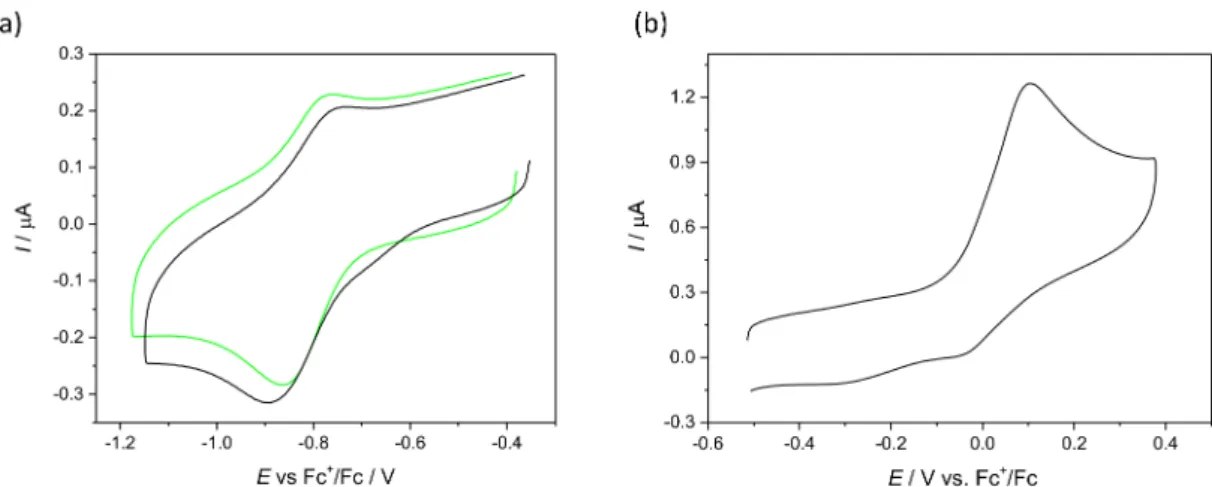
2.6. Electrochemistry and Spectroelectrochemistry
Cyclic voltammetry experiments with 0.5 mM solutions of Cu(II) complexes and ligands in 0.1 MnBu4NPF6DMSO were performed under argon atmosphere using a three- electrode arrangement with glassy carbon 1 mm disc working electrode (from Ionode, Aus- tralia), platinum wire as counter electrode, and silver wire as pseudo-reference electrode.
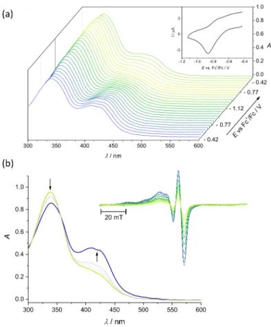
Ferrocene (Fc) served as the internal potential standard. A Heka PG310USB (Lambrecht, Germany) potentiostat with a PotMaster 2.73 software package served as the potential control in voltammetry studies. In situ ultraviolet-visible-near-infrared (UV-vis-NIR) spec- troelectrochemical measurements were performed on a spectrometer Avantes (Apeldoorn, The Netherlands) (Model AvaSpec-2048x14-USB2) in the spectroelectrochemical cell kit (AKSTCKIT3) with the Pt-microstructured honeycomb working electrode (1.7 mm optical path length), purchased from Pine Research Instrumentation (Lyon, France). The cell was positioned in the CUV-UV Cuvette Holder (Ocean Optics, Ostfildern, Germany) connected to the diode-array UV-vis-NIR spectrometer by optical fibres. UV-vis-NIR spectra were processed using the AvaSoft 7.7 software package. Halogen and deuterium lamps were used as light sources (Avantes, Model AvaLight-DH-S-BAL). The in situ EPR spectroelectro- chemical experiments were carried out under an argon atmosphere in the EPR flat cell (0.5 mm thickness of the inner space) equipped with a large platinum mesh working electrode.
The freshly prepared solutions were carefully purged with argon and the electrolytic cell was polarised in the galvanostatic mode directly in the cylindrical EPR cavity TM-110 (ER 4103 TM) and the electron paramagnetic resonance (EPR) spectra were measured in situ. The EPR spectra were recorded at room temperature or at 77 K with the EMX Bruker spectrometer (Rheinstetten, Germany).
Table 1.Crystal data and details of data collection forCu(L1)Cl·1.775H2O.
Compound Cu(L1)Cl·1.775H2O
empirical formula C24H28.6ClCuN6O5.78S
fw 624.58
space group triclinicP1
a, Å 8.2906(6)
b,Å 15.1634(13)
c, Å 43.244(4)
β,◦ 94.026(3)
V[Å3] 5422.9(8)
Z 8
λ[Å] 0.71073
ρcalcd, g cm−3 1.530
cryst size, mm3 0.12×0.08×0.03
T [K] 110(2)
µ, mm−1 1.031
R1a 0.0968
wR2b 0.2099
GOFc 1.084
aR1=∑||Fo|−|Fc||/∑|Fo|.bwR2= {∑[w(Fo2−Fc2)2]/∑[w(Fo2)2]}1/2.cGOF = {∑[w(Fo2−Fc2)2]/(n− p)}1/2, wherenis the number of reflections andpis the total number of parameters refined.
2.7. Cell Lines and Culture Conditions
Human breast adenocarcinoma cells MDA-MB-231, sensitive COLO-205 and mul- tidrug resistant COLO-320 colorectal adenocarcinoma cells and normal human lung fibrob- lasts MRC-5 were obtained from ATCC. COLO-205 and COLO-320 cells were cultured in RPMI-1640 medium containing 10% foetal bovine serum, MRC-5 cells were cultured in EMEM medium containing 10% foetal bovine serum. MDA-MB-231 cells were cultured in DMEM/high glucose medium containing 10% FBS. Adherent MDA-MB-231 cells were grown in Falcon tissue culture 75 cm2flasks and all other cells were grown in tissue culture 25 cm2flasks (BD Biosciences, Singapore). All cell lines were grown at 37◦C in a humidified atmosphere of 95% air and 5% CO2. The stock solutions of copper(II) complexes were prepared in DMSO.
2.8. Inhibition of Cell Viability Assay
The cytotoxicity of the compounds was determined by colorimetric MTT assay. The cells were harvested from culture flasks by trypsinisation and seeded into Cellstar 96-well microculture plates at the seeding density of 6000 cells per well (6×10⁴cells/mL, MDA- MB-231) or 10,000 cells per well (1×10⁵cells/mL cells/mL, COLO-205, COLO-320 and MRC-5). The cells were allowed to resume exponential growth for 24 h, and subsequently were exposed to drugs at different concentrations in media for 72 h. The drugs were diluted in complete medium at the desired concentration and added to each well (100µL) and serially diluted to other wells. After exposure for 72 h, the media were replaced with MTT in media (5 mg/mL, 100µL) and incubated for additional 50 min. Subsequently, the media were aspirated and the purple formazan crystals formed in viable cells were dissolved in DMSO (100µL). Optical densities were measured at 570 nm using the BioTek H1 Synergy (BioTek, Singapore) microplate reader. The quantity of viable cells was expressed in terms of treated/control (T/C) values in comparison to untreated control cells, and 50% inhibitory concentrations (IC50) were calculated from concentration-effect curves by interpolation.
Evaluation was based on at least three independent experiments, each comprising six replicates per concentration level.
2.9. Tyrosyl Radical Reduction in Mouse R2 RNR Protein
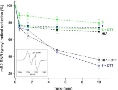
The 9.4 GHz EPR spectra were recorded at 30 K on a Bruker Elexsys II E540 EPR spectrometer with an Oxford Instruments ER 4112HV helium cryostat, essentially as de- scribed previously [13]. The concentration of the tyrosyl radical in mouse R2 ribonucleotide reductase protein (mR2) was determined by double integration of EPR spectra recorded at non-saturating microwave power levels (3.2 mW) and compared with the copper stan- dard [55] mR2 protein was expressed, purified, and iron-reconstituted as described previ- ously [56] and passed through a 5 mL HiTrap desalting column (GE Healthcare) to remove excess iron. The purified, iron-reconstituted mR2 protein resulted in the formation of 0.76 tyrosyl radical/polypeptide. Samples containing 20µM mR2 in 50 mM Hepes buffer, pH 7.50/100 mM NaCl, and 20µMHL1, complex1, or complex3in 1% (v/v) DMSO/H2O, and 2 mM dithiothreitol (DTT) were incubated for indicated times and quickly frozen in cold isopentane. The same samples were used for repeated incubations at room temperature.
The experiments were performed in duplicates.
2.10. Redox Activity of[FeII(L1)2]
The generation of paramagnetic intermediates was monitored by cw-EPR spectroscopy using the EMX spectrometer (Bruker). Deionised water was used for the preparation of ethanol/water solutions in whichHL1was dissolved (ethanol was used to increase the solubility ofHL1). The spin trapping agent (DMPO; Sigma-Aldrich) was distilled prior to use.
3. Results and Discussion
3.1. Synthesis and Characterisation ofHL1-HL3andH2L4, Their Copper(II) Complexes1–4and10 The synthesis of ligandsHL1-HL3andH2L4was realised in 12 steps. Two building blocks were prepared first, namely 4-chloromethyl-2-dimethoxymethylpyridine (E) starting from 2,4-pyridinedicarboxylic acid in five steps, as shown in Scheme S1, and 7-hydroxy-3- (piperazine-1-carbonyl)-2H-chromen-2-one (H) starting from 2,4-dihydroxybenzaldehyde in four steps, as shown in Scheme S2 and discussed in detail in ESI †. The atom labelling of the precursors for NMR resonances assignment is given in Schemes S3 and S4. Then these building blocksEandHwere combined to give aldehydes or aldehyde precursors, which entered condensation reactions with thiosemicarbazide derivatives with formation ofHL1-HL3andH2L4in 3 steps as shown in Scheme1.
3.1.1. Synthesis of 3-(4-((2-(Dimethoxymethyl)pyridin-4-yl)methyl)piperazine-1-carbonyl)- 7-hydroxy-2H-chromen-2-one (I1) and 7-((2-(Dimethoxymethyl)pyridin-4-yl)methoxy)-3-(4- ((2-(dimethoxymethyl)pyridin-4-yl)methyl)piperazine-1-carbonyl)-2H-chromen-2-one (I2)
The condensation of two building blocksHandE(Scheme1) was first attempted in the presence of Et3N (pKa= 10.75) to yield the desired productI1in low yield (12–17%). By using a much stronger base, namely 1,1,3,3-tetramethylguanidine (TMG, pKa= 13), and optimising the molar ratio of reactants theI1was produced in good to very good yield (50–80%) (Table S1, ESI †). We also noticed that the synthesis ofI1was accompanied by the formation of another product of condensation, i.e.,I2, with two pyridine rings and one coumarin-piperazine moiety even if a double excess ofHin comparison toEwas used.
Interestingly, the same reaction performed in the presence of K2CO3as a base resulted only inI2. The two productsI1andI2were separated by column chromatography with MeOH/EtOAc 2:1 as eluent (for details see Experimental part). The identity ofI1andI2
was confirmed by positive ESI mass spectra, which showed peaks atm/z440.18, 462.16 forI1and 605.28, 627.28 forI2, attributed to [M + H]+and [M + Na]+, respectively.1H and
13C NMR spectra were also in agreement with the structures proposed. Assignment of proton resonances was carried out based on 2D NMR experiments. The presence of the

![Figure 1. ORTEP view of one of the two crystallographically independent molecules of [Cu(L 1 )Cl] (1 0 ) with thermal ellipsoids at 50% probability level for anisotropically refined atoms](https://thumb-eu.123doks.com/thumbv2/9dokorg/962921.56944/15.892.186.710.458.758/figure-crystallographically-independent-molecules-thermal-ellipsoids-probability-anisotropically.webp)
![Figure 4. (a) UV–vis spectrum of [Fe II (L 1 ) 2 ] (blue trace) in H 2 O/EtOH (4:1, v/v) compared to that of HL 1 (black trace)](https://thumb-eu.123doks.com/thumbv2/9dokorg/962921.56944/19.892.260.587.126.719/figure-spectrum-blue-trace-etoh-compared-black-trace.webp)