1
Comparative solution equilibrium and structural studies of half-sandwich ruthenium(II)(
6-toluene) complexes of picolinate derivatives
Jelena M. Poljarević,a,b G. Tamás Gál,c Nóra V. May,c Gabriella Spengler,d Orsolya Dömötör,a Aleksandar R. Savić,b Sanja Grgurić-Šipka,b Éva A. Enyedya*
a Department of Inorganic and Analytical Chemistry, University of Szeged, Dóm tér 7. H-6720 Szeged, Hungary
b Faculty of Chemistry, University of Belgrade, Studentski trg 12-16, 11000 Belgrade, Serbia
c Research Centre for Natural Sciences Hungarian Academy of Sciences, Magyar tudósok körútja 2, H-1117 Budapest, Hungary
d Department of Medical Microbiology and Immunobiology, University of Szeged, Dóm tér 10, H-6720 Szeged, Hungary
Keywords: Stability constants; X-ray crystal structures; Half-sandwich complexes; Speciation;
Antiproliferative activity
* Corresponding author.
E-mail address: enyedy@chem.u-szeged.hu (É.A. Enyedy).
ABSTRACT
Five Ru(II)(6-toluene) complexes formed with 2-picolinic acid and its various derivatives have been synthesized and characterized. X-ray structures of four complexes are also reported. Complex formation processes of [Ru(II)(6-toluene)(H2O)3]2+ organometallic cation with the metal-free ligands were studied in aqueous solution in the presence of chloride ions by the combined use of 1H NMR spectroscopy, UV-visible spectrophotometry and pH- potentiometry. Solution stability, chloride ion affinity and lipophilicity of the complexes were characterized together with in vitro cytotoxic and antiproliferative activity in cancer cell lines being sensitive and resistant to classic chemotherapy and in normal cells as well. Formation of mono complexes such as [Ru(6-toluene)(L)(Z)]+/o (L: completely deprotonated ligand; Z = H2O/Cl‒) with high stability and [Ru(6-toluene)(L)(OH)] was found in solution. The pKa
values (8.3-8.7) reflect the formation of low amount of mixed hydroxido species at pH 7.4 at 0.2 M KCl ionic strength. The complexes are fairly hydrophilic and show moderate chloride ion affinity and fast chloride-water exchange processes. The studied complexes exhibit no cytotoxic activity in human cancer cells (IC50 > 100 M), only complexes formed with 2-
2
picolinic acid (1) and its 3-methyl derivative (2) represented a moderate antiproliferative effect (IC50 = 84.8 (1), 79.2 μM (2)) on a multidrug resistant colon adenocarcinoma cell line revealing considerable multidrug resistant selectivity. Complexes 1 and 2 bind to human serum albumin covalently and relatively slowly with moderate strength at multiple binding sites without ligand cleavage.
3
1. Introduction
Ruthenium complexes have emerged as attractive alternatives to platinum based compounds such as cisplatin, carboplatin and oxaliplatin which are undoubtedly successful anticancer drugs but have several drawbacks such as serious side-effects and lack of activity against certain types of cancer. Ruthenium compounds have different physico-chemical and pharmacokinetic properties compared to the platinum drugs, and they have different mechanism of action as well, that is the reason why they are the subject of extensive drug discovery efforts [1-3]. Imidazolium trans-[tetrachlorido(DMSO)(imidazole)ruthenate(III)]
(NAMI-A) was the first Ru(III) complex reached clinical trials [4], while sodium trans- [tetrachloridobis(1H-indazole)ruthenate(III)] (NKP-1339, IT-139) is one of the most promising investigational non-Pt drugs in current clinical development. NKP-1339 is active against solid malignancies such as non-small cell lung cancer, colorectal carcinoma and the treatment is accompanied by minor side effects [5,6]. While cisplatin induces DNA damage via adduct formation [7], endoplasmic reticulum stress and reactive oxygen species-related effects were found to be involved in the mechanism of action of NKP-1339 [5,8]. Ru(III) complexes are considered as prodrugs that are activated by reduction and it provides the impetus for the development of various Ru(II) anticancer compounds [5]. It is noteworthy that a novel Ru(II) compound [Ru(4,4’-dimethyl-2,2’-bipyridine)2-(2-(2’,2’’:5’’,2’’’- terthiophene)-imidazo[4,5-f][1,10]phenanthroline)]Cl2 (TLD-1433) has entered a human clinical trial recently as nontoxic photosensitizing agent [9]. Ru(II) is often stabilized in the +2 oxidation state by the coordination of η6-arene type ligands and there are two main prototypes of Ru(II)-arene complexes [3]: i) 1,3,5-triaza-7-phosphatricyclo-[3.3.1.1]decane (PTA) containing Ru(II)-arene (RAPTA) compounds such as [Ru(6-p-cymene)(PTA)Cl2] (RAPTA-C) possessing significant antimetastatic property and is ready for translation into clinical evaluation [10,11]; ii) the bidentate 1,2-ethylenediamine (en) containing Ru(II)-arene (RAED) complexes such as [Ru(6-biphenyl)(en)Cl]PF6 (RM175) that possesses a similar cytotoxic activity to cisplatin [12,13]. In most of the half-sandwich organoruthenium(II) compounds a bidentate ligand with an (O,O), (O,S), (O,N), (N,N) or (N,S) binding mode is coordinated and a chloride ion acts as the leaving group [3,14-16]. Aquation (replacement of the chlorido ligand by a water molecule) facilitates the reaction with biological macromolecules such as proteins or DNA, therefore the strength of the Ru-Cl bond and the rate of its cleavage have a strong impact on the bioactivity of the Ru(II)-arene complexes
4
[17]. Notably, the chemical and pharmacological properties of the Ru(II)-arene half-sandwich compounds can be fine-tuned by variation of the coordinated ligand, the arene ring and the leaving group [1,3,10]. Although a large number of Ru(II)-arene compounds has been developed and extensively investigated, information about their solution speciation and stability constants is still limited in the literature. Most of the solution equilibrium studies are focused on [Ru(6-p-cymene)(X,Y)Cl] type complexes [18-24]. For the better understanding of the pharmacokinetic properties and mechanisms of action of these metal complexes, the knowledge of the aqueous chemistry and the most plausible chemical forms in water, especially at physiological pH, is a mandatory prerequisite.
In our previous works we have studied the biological activity of Ru(II)(6-p-cymene) complexes of various pyridine derivatives [25-28] and moderate-to-low cytotoxicity was found in six tumor cell lines; although the complex of pyridine-2-carboxylic acid (2-picolinic acid, picH) represents an enhanced antiproliferative activity (e.g. IC50 = 82 M in HeLa cells, 36 M in FemX cells [27]) and antimetastatic effect based on wound migration assay [25].
The solution speciation of Ru(II)(6-p-cymene) picolinate complexes was also studied by some of us revealing the formation of mono-ligand complexes with high stabilities [23].
Notably, the Os(II) congener of the picolinate complex showed very high in vitro cytotoxic activity [29].
As the physico-chemical and biological properties can be modified by the exchange of the arene ring, in this work we have prepared and structurally characterized Ru(II)(6-toluene) complexes formed with picH and its 3-methyl (3-Me-picH), 5-bromo (5-Br-picH), 4- carboxylic (2,4-dipicH2) and 5-carboxylic (2,5-dipicH2) derivatives (Chart 1). In addition to the determination of the solid phase structures of four complexes by X-ray crystallography, solution speciation of these Ru(II)(6-toluene) complexes in water was revealed by UV- visible (UV-vis) spectrophotometry, 1H NMR spectroscopy and pH-potentiometry involving studies on their stability and chloride ion affinity. The antiproliferative and cytotoxic effectiveness of these complexes in multidrug resistant/non-resistant human cancer lines was also tested. Interactions between human serum albumin and the complexes showing antiproliferative effect were monitored using fluorometry and ultrafiltration.
5
Chart 1. Chemical structures of the ligands in their completely deprotonated forms (a) and the general formula of the prepared [Ru(6-toluene)(L)(Cl)] complexes.
2. Experimental
2.1. Chemicals
All solvents were of analytical grade and used without further purification. 2-Picolinic acid (picH), 3-methylpyridine-2-carboxylic acid (3-Me-picH), 5-bromo-2-pyridinecarboxylic acid (5-Br-picH), 2,4-pyridinedicarboxylic acid monohydrate (2,4-dipicH2·H2O), 2,5- pyridinedicarboxylic acid (2,5-dipicH2), RuCl3·3H2O, KCl, HCl, KOH, 4,4-dimethyl-4- silapentane-1-sulfonic acid (DSS), 1-methylimidazole (N-MeIm), human serum albumin (HSA, as lyophilized powder with fatty acids, A1653), NaClO4, KH2PO4, NaH2PO4·2H2O, Na2HPO4·2H2Owere purchased from Sigma-Aldrich in puriss quality. Doubly distilled Milli- Q water was used for preparation of samples. The purity of the ligands and the exact concentration of their stock solutions were determined by pH-potentiometric titrations and by the computer program Hyperquad2013 [30]. [Ru(η6-toluene)Cl2]2 was prepared according to a well known procedure [31]. A stock solution of [Ru(η6-toluene)(Z)3]2+/+/o/‒, where Z is H2O or Cl‒, was obtained by dissolving [Ru(η6-toluene)Cl2]2 in water and the exact concentration of this stock was determined with pH-potentiometric titrations. The modified phosphate-buffered saline (PBS’) contains 12 mM Na2HPO4, 3 mM KH2PO4, 1.5 mM KCl and 100.5 mM NaCl;
and the concentration of the K+, Na+ and Cl‒ ions corresponds approximately to that of the human blood serum (c(K+) = 3.5-5.1 mM, c(Na+) = 135-145 mM, c(Cl‒) = 96-106 mM [32]).
HSA solution was freshly prepared before the experiments and its concentration was estimated from its UV absorption: 280 nm(HSA) = 36850 M−1cm−1 [33]. Stock solution of N- MeIm was prepared on a weight-in-volume basis in PBS’ solution.
2.2. Synthesis of the complex [(η6-toluene)RuCl(μ-Cl)]2 with different picolinic acids
N
O O-
N
O O- Br
N
O O- O O-
N
O O- O
-O
N
O O-
pic 3-Me-pic 5-Br-pic 2,4-dipic 2,5-dipic [Ru(6-toluene)(L)(Cl)]
(a) (b)
N Ru Cl O
6
For the characterization of the prepared complexes 1H and 13C NMR spectroscopy, elemental analysis and electrospray ionization mass spectrometry (ESI-MS, Fig. S1) were used in addition to X-ray crystallography (vide infra). NMR spectra were recorded on a Bruker Avance III 500 spectrometer or a Bruker Ultrashield 500 Plus instrument, and DMSO-d6 was used as solvent. ESI-MS measurements were performed using a Micromass Q-TOF Premier (Waters MS Technologies) mass spectrometer equipped with electrospray ion source.
Elemental analysis of all compounds was performed with a Perkin–Elmer 2400 CHN Elemental Analyser (Perkin–Elmer, Waltham, MA) at the Microanalytical Laboratory of the University of Vienna.
2.2.1. Synthesis of the precursor [Ru(η6-toluene)Cl(μ-Cl)]2
[Ru(η6-toluene)Cl(μ-Cl)]2 was prepared according the literature procedure used for the analogous [Ru(η6-benzene)Cl(μ-Cl)]2 [31] by adding 5 mL of 1-methyl-1,4-cyclohexadiene to a solution of 0.5 g RuCl3·3H2O (1.9 mmol) in 40 mL of absolute ethanol. This mixture was refluxed for 8 h. The reddish brown precipitate formed during the synthesis was filtered off, washed with diethyl ether and left to dry in exsiccator. Yield: 85%, 0.450 g; 1H NMR (500.26 MHz, DMSO-d6, , ppm): 2.12 (3H, s, CH3), 5.68 (3H, m, C2, C4, C6 toluene), 5.97 (2H, m, C3, C5 toluene); 13C NMR (125.79 MHz MHz, DMSO-d6) 18.73 (CH3), 82.22 (C4 toluene), 84.83 (C5, C3 toluene), 89.28 (C6, C2 toluene), 105.82 (C1 toluene).
2.2.2. Synthesis of chlorido[(pyridine-κN-2-carboxylato-κO)(η6-toluene)ruthenium(II)] (1):
A solution of picH (0.015 g, 0.13 mmol) in 2 mL of 2-propanol was added to a warm solution of [Ru(η6-toluene)Cl2]2 (0.030 g, 0.057 mmol) in 25 mL of 2-propanol. The reaction mixture was stirred at room temperature for 7 days and the yellow-range precipitate was formed.
Solution was filtered off and product was dried in exsiccator. Yield: 58%, 0.023 g; 1H NMR (500.26 MHz, DMSO-d6, , ppm): 2.15 (3H, s, CH3), 5.60 (2H, m, C2, C6 toluene), 5.70 (1H, t, C4 toluene), 5.99 (2H, m, C3, C5 toluene), 7.71 (1H, dd, C5 ligand), 7.75 (1H, d, C3, ligand), 8.06 (1H, t, C4 ligand), 9.29 (1H, d, C6 ligand); 13C NMR (125.79 MHz, DMSO-d6) 18.57 (CH3), 77.07 (C4 toluene), 78.44 (C5 toluene), 79.71 (C3 toluene), 86.15 (C6 toluene), 88.06 (C2 toluene), 101.01 (C1 toluene), 125.31 (C3 ligand), 128.09 (C5 ligand), 139.64 (C4 ligand), 150.89 (C2 ligand), 153.88 (C6 ligand). (Numbering of the ligand protons is shown in Chart S1.) ESI/MS (m/z) (Fig. S1): [M‒Cl]+ (C13H12NO2Ru, calculated: 315.9912) = 315.9947 and [M‒Cl‒COO]+ (C12H12NRu, calculated: 272.0013) = 272.0025. Elemental
7
analysis: calculated for: C13H12O2NClRu (%): C, 44.51; H, 3.45; N, 3.99. Found (%): C, 44.41; H, 3.37; N, 4.01.
2.2.3. Synthesis of complexes of chlorido[(3-methylpyridine-κN-2-carboxylato-κO)(η6- toluene)ruthenium(II)] (2), chlorido[(5-bromopyridine-κN-2-carboxylato-κO)(η6- toluene)ruthenium(II)] (3), chlorido[(4-carboxylate-pyridine-κN-2-carboxylato-κO)(η6- toluene)ruthenium(II)] (4), chlorido[(5-carboxylate-pyridine-κN-2-carboxylato-κO)(η6- toluene)ruthenium(II)] (5):
Methanolic solution of the ligand (3-Me-picH (10.4 mg, 0.076 mmol) or 5-Br-picH (15.4 mg, 0.076 mmol) or 2,4-dipicH2·H2O (14.1 mg, 0.076 mmol) or 2,5-dipicH2 (12.7 mg, 0.076 mmol)) was slowly added in the methanolic (5 mL) solution of [Ru(η6-p-toluene)Cl2]2
(20.0 mg, 0.038 mmol) and reaction mixture was stirred for 3 h, at 40°C. Then, reaction volume was reduced to half and desired orange complex was precipitated. Solution was filtered off and product was dried in exsiccator.
2: Yield: 57%, 0.016 g; 1H NMR (500.26 MHz, DMSO-d6, , ppm): 2.16 (3H, s, toluene CH3), 2.54 (3H, s, ligand CH3), 5.57 (1H, d,C2, toluene), 5.60 (1H, d, C6, toluene), 5.68 (1H, t, C4 toluene), 5.97 (2H, dd, C3, C5 toluene), 7.59 (1H, dd, C5 ligand), 7.89 (1H, d, C4 ligand), 9.22 (1H, d, C6 ligand); 13C NMR (125.79 MHz, DMSO-d6) 18.37 (CH3, ligand), 18.65 (CH3, toluene), 77.11 (C4 toluene), 78.88 (C5 toluene), 79.21 (C3 toluene), 86.62 (C6 toluene), 88.43 (C2 toluene), 101.18 (C1 toluene), 126.88 (C5 ligand), 137.92 (C4 ligand), 142.70 (C6 ligand), 147.29 (C3 ligand), 152.48 (C2 ligand), 170.89 (COO-Ru). ESI/MS (m/z) (Fig. S1): [M‒Cl]+ (C14H14NO2Ru, calculated: 330.0068) = 330.0079 and [M‒Cl‒COO]+ (C13H14NRu, calculated: 286.0170) = 286.0176. Elemental analysis: calculated for:
C14H14O2NClRu∙1.25H2O (%): C, 43.42; H, 4.29; N, 3.62. Found (%): C, 43.37; H, 4.08; N, 3.68.
3: Yield: 52%, 0.017 g; 1H NMR (500.26 MHz, DMSO-d6, , ppm): 2.17 (3H, s, CH3), 5.63 and 5.67 (2H,dd ,C2, C6 toluene), 5.77 (1H, t,C4 toluene), 6.06 (2H, m, C3, C5 toluene), 7.68 (1H, d, C3 ligand), 8.34 (1H, d, C4 ligand), 9.52 (1H, s, C6 ligand); 13C NMR (125.79 MHz, DMSO-d6) 18.41 (CH3), 76.98 (C4 toluene), 78.54 (C5 toluene), 79.21 (C3 toluene), 86.58 (C6 toluene), 88.16 (C2 toluene), 101.60 (C1 toluene), 122.95 (C5 ligand), 126.30 (C3 ligand), 142.28 (C4 ligand), 149.76 (C2 ligand), 154.04 (C6 ligand), 169.69 (COO-Ru).
ESI/MS (m/z) (Fig. S1): [M‒Cl]+ (C13H11BrNO2Ru, calculated: 395.8996) = 395.9078 and [M‒Cl‒COO]+ (C12H11BrNRu, calculated: 351.9098) = 351.9121. Elemental analysis:
8
calculated for C13H11O2NBrClRu∙0.3H2O (%): C, 35.89; H, 2.69; N, 3.22. Found (%): C, 35.90; H, 2.79; N, 2.96.
4: Yield: 56%, 0.017 g; 1H NMR (500.26 MHz, DMSO-d6, , ppm): 2.18 (3H, s, CH3), 5.66 (2H,dd ,C2, C6 toluene),5.75 (1H, t,C4 toluene), 6.06 (2H, m, C3, C5 toluene), 8.06 (2H, m, C3, C5 ligand), 9.51 (1H, d, C6 ligand), 14.22 (1H, s, free COOH ligand); 13C NMR (125.79 MHz, DMSO-d6) 18.41 (CH3), 77.27 (C4 toluene), 78.94 (C5 toluene), 79.68 (C3 toluene), 86.61 (C6 toluene), 88.17 (C2 toluene), 101.54 (C1 toluene), 123.82 (C3 ligand), 126.63 (C5 ligand), 140.93 (C4 ligand), 151.88 (C6 ligand), 155.17 (C2 ligand), 164.66 (COO-Ru), 169.73 (COOH). ESI/MS (m/z) (Fig. S1): [M‒Cl]+ (C14H12NO4Ru, calculated: 359.9810) = 359.9876 and [M‒Cl‒COO]+ (C13H12NO2Ru, calculated: 315.9912) = 315.9947. Elemental analysis: calculated for C14H12O4NClRu∙0.5H2O (%): C, 41.64; H, 3.25; N, 3.47. Found (%):
C, 41.64; H, 3.04; N, 3.52.
5: Yield: 50%, 0.015 g; 1H NMR (500.26 MHz, DMSO-d6, , ppm): 2.18 (3H, s, CH3), 5.66(1H, d ,C2 toluene), 5.70(1H, d, C6 toluene),5.80(1H, t, C4 toluene), 6.08 (2H, m, C3, C5 toluene), 7.89 (1H, d, C3 ligand), 8.51 (1H, d, C4 ligand), 9.56 (1H, s, C6 ligand), 14.20 (1H, s, free COOH ligand); 13C NMR (125.79 MHz, DMSO-d6) 18.43 (CH3), 77.11 (C4 toluene), 78.72 (C5 toluene), 79.32 (C3 toluene), 86.53 (C6 toluene), 87.99 (C2 toluene), 101.46 (C1 toluene), 125.29 (C3 ligand), 130.63 (C4 ligand), 140.24 (C5 ligand), 153.28 (C6 ligand), 154.32 (C2 ligand), 164.42 (COO-Ru), 169.56 (COOH). ESI/MS (m/z) (Fig. S1): [M‒Cl]+ (C14H12NO4Ru, calculated: 359.9810) = 359.9876 and [M‒Cl‒COO]+ (C13H12NO2Ru, calculated: 315.9912) = 315.9947. Elemental analysis: calculated for C14H12O4NClRu∙0.4H2O (%): C, 41.83; H, 3.21; N, 3.48. Found (%): C, 41.92; H, 3.05; N, 3.45.
2.3. Crystallographic structure determination
Single crystals, suitable for X-ray diffraction experiment, of compounds [Ru(6- toluene)(pic)Cl] (1), [Ru(6-toluene)(3-Me-pic)Cl]∙H2O (2∙H2O), [Ru(6-toluene)(5-Br- pic)Cl] (3) and [Ru(6-toluene)(2,5-dipic)Cl] (5) were grown from methanol solution of the solid complexes.
Orange (1) and yellow (2∙H2O, 3, 5) single crystals were mounted on loops and transferred to the goniometer. X-ray diffraction data were collected at ‒170 °C (for 1, 2∙H2O) or 20 °C (for 3, 5) on a Rigaku RAXIS-RAPID II diffractometer using Mo-K radiation. A numerical absorption correction [34] was carried out using the program CrystalClear [35].
Sir2014 [36] and SHELXL [37] under WinGX [38] software were used for structure solution
9
and refinement, respectively. The structures were solved by direct methods. The models were refined by full-matrix least squares on F2. Refinement of non-hydrogen atoms was carried out with anisotropic temperature factors. Hydrogen atoms were placed into geometric positions (except for water hydrogen atoms which were constrained). They were included in structure factor calculations but they were not refined. The isotropic displacement parameters of the hydrogen atoms were approximated from the U(eq) value of the atom they were bonded to.
The summary of data collection and refinement parameters are collected in Table S1. Selected bond lengths and angles of compounds were calculated by PLATON software [39]. The graphical representation and the edition of CIF files were done by Mercury [40] and PublCif [41] softwares, respectively. The crystallographic data files for the complexes have been deposited with the Cambridge Crystallographic Database as CCDC 1574021-1574024.
2.4. pH-potentiometric measurements and data evaluation
The pH-potentiometric measurements determining the proton dissociation and formation constants were carried out at 25 ± 0.1°C and an ionic strength I = 0.20 M (KCl) in order to keep the activity coefficients constant. The titrations were performed with a carbonate-free KOH solution (0.20 M). The exact concentrations of HCl and KOH were determined by pH- potentiometric titrations. An Orion 710A pH-meter equipped with a Metrohm combined electrode (type 6.0234.100) and Methrom 665 Dosimat burette were used for the pH- potentiometric measurements. The volume resolution of the burette is 0.001 mL and its precision is 0.002 mL. The electrode system was calibrated to the pH = ‒ log[H+] scale by means of blank titrations (strong acid HCl vs. strong base KOH), as suggested by Irving et al.
[42]. The average water ionization constant, pKw, was determined as 13.76 ± 0.01, which corresponds well to the literature data [43]. The reproducibility of the titration points included in the calculations was within 0.005 pH. The pH-potentiometric titrations were performed in the pH range 2.0 to 11.5. The initial volume of the samples was 5 mL. The ligand concentration was 2 mM and metal to ligand ratios of 1:1 and 1:2 were used. The samples were degassed by bubbling purified argon through them for 10 min prior the measurements and the argon was also passed over the solutions during the titrations.
The computer program Hyperquad2013 [30] was utilized to establish the stoichiometry of the complexes and to calculate the overall stability constants. β(MpLqHr) is defined for the general equilibrium:
pM + qL + rH ⇌ MpLqHr as β(MpLqHr)
=
[MpLqHr]/[M]p[L]q[H]r (1)10
where M denotes the metal moiety [Ru(η6-toluene)(Z)3] (Z = H2O/Cl‒) and L the completely deprotonated ligand. In all calculations exclusively titration data were used from experiments in which no precipitate was visible in the reaction mixture. The goodness-of-fit measured in Hyperquad2013 by sigma () represents the overall goodness-of-fit derived from the sum of squared residuals (calculated-experimental titration data). The model was accepted when was close to one (< 1.5). The standard deviation of the log values of species included into the model was always lower than 0.1. As equilibrium constants were determined in the presence of 0.20 M chloride ion, they are considered as conditional constants. log values for the various hydroxido complexes[(Ru(6-toluene))2(2-OH)i](4-i)+ (i=2,3) were calculated based on the pH-potentiometric titration data in the presence of chloride ions and were found to be in fairly good agreement with previously published data [44].
2.5. UV-vis spectrophotometric and 1H NMR spectroscopic titrations, and determination of the distribution coefficients
A Hewlett Packard 8452A diode array spectrophotometer was used to record the UV-vis spectra in the interval 200 – 800 nm. The path length was 1 cm. Equilibrium constants (proton dissociation, stability constants and H2O/Cl− exchange constants) and the individual spectra of the species were calculated with the computer program PSEQUAD [45]. The spectrophotometric titrations were performed in aqueous solution on samples containing the ligands with or without the organometallic cation, and the concentration of the ligands was 100-120 μM. The organometallic cation was also titrated (120 M) separately. The metal-to- ligand ratios were 1:1 in the pH range from 3 to 11.5 at 25.0±0.1 °C at an ionic strength of 0.20 M (KCl). Measurements for 1:1 metal-to-ligand systems were also carried out by preparing individual samples in which KCl was partially or completely replaced by HCl; pH values, varying in the range ca.0.8–2.5, were calculated from the strong acid content. The absorbance data were recorded after various waiting time (1-48 h). UV-vis spectra recorded as a function of chloride concentrations (0–252 mM) were used to investigate the H2O/Cl− exchange processes of complexes [Ru(6-toluene)(L)(H2O)] at pH 5.5-7.0 (at a constant pH).
In order to check the effect of the variable ionic strength on the logK’ (H2O/Cl−) constant in this titration experiment individual samples of complex 2 were also prepared in which a constant ionic strength was applied, namely the sum of concentrations of NaClO4 and NaCl was 0.30 M.
11
1H NMR titrations were carried out on a Bruker Ultrashield 500 Plus instrument using WATERGATE water suppression pulse scheme. DSS was used as an internal NMR standard.
1H NMR spectra of samples containing [Ru(II)(η6-toluene)(H2O)3]2+ (1 mM) and ligand picH (1 mM) in D2O at various pH values were recorded after 4 h of incubation (25 °C, I = 0.20 M (KCl)). Titration of 2 mM solution of [Ru(η6-toluene)(Z)3] was also performed separately. To study the interaction with HSA and N-MeIm 1H NMR spectra were recorded for samples containing precursor [Ru(η6-toluene)Cl(μ-Cl)]2 or complex 1 (1 mM), with or without half equivalent of HSA or N-MeIm. Samples were prepared in PBSʹ buffer and incubated for 24 h at 25 °C.
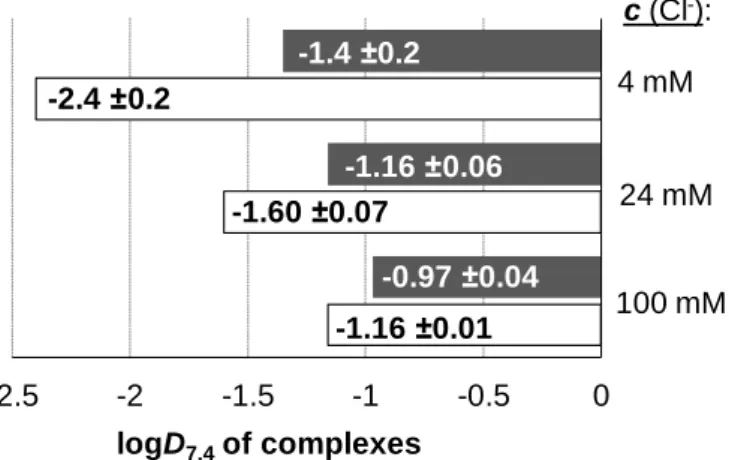
Distribution coefficients at physiological pH (D7.4) of the complexes 1–5 and the ligands as well as the Ru precursor were determined by the traditional shake-flask method in n- octanol/buffered aqueous solution at pH 7.40 at various chloride concentrations using UV-vis detection as described in our former work [24].
2.6. Fluorescence and membrane ultrafiltration/UV-vis studies with HSA
Fluorescence spectra were recorded on a Hitachi-F4500 fluorometer in 1 cm quartz cell at 25.0 ± 0.1 °C. All solutions were prepared in PBS’ (pH 7.4) and were incubated for 24 h following a time-dependence experiment. Samples contained 1 M HSA, and various HSA- to- Ru(6-toluene) or 1 or 2 ratios (from 1:0 to 1:10) were used. The excitation wavelength was 295 nm and the emission was read in the range of 310-500 nm. The quenching (KQ’) constants were calculated with the computer program PSEQUAD [45] using the same approach applied in our previous works [46,47].
Samples (0.50 mL) used for the ultrafiltration studies contained 40 M HSA and Ru(6-toluene) or 1 or 2 (up to 1:10 protein-to-complex ratio) in PBS’ buffer (pH 7.4) at 25.0 ± 0.1 °C and were incubated for 24 h. Samples were separated by ultrafiltration through 10 kDa membrane filters (Millipore Amicon Ultra-0.5 centrifugal filter unit) in low (LMM) and high molecular mass (HMM) fractions with the help of a temperature controlled centrifuge (Sanyo, 10000 rpm, 10 min). The LMM fraction containing the non-bound metal complex was separated from the protein and its adducts in the HMM fraction. The concentration of the non-bound compounds in the LMM fractions was determined by UV-vis spectrophotometry by comparing the recorded spectra to those of reference samples without the protein.
12 2.7. Cell lines
Human colonic adenocarcinoma cell lines Colo 205 doxorubicin-sensitive (ATCC-CCL-222) and Colo 320/MDR-LRP multidrug resistant overexpressing ABCB1 (MDR1)-LRP (ATCC- CCL-220.1) were purchased from LGC Promochem, Teddington, UK. The cells were cultured in RPMI 1640 medium supplemented with 10% heat-inactivated fetal bovine serum, 2 mM L- glutamine, 1 mM sodium pyruvate and 100 mM 4-(2-hydroxyethyl)-1- piperazineethanesulfonic acid (HEPES). The cell lines were incubated at 37 °C, in a 5% CO2, 95% air atmosphere. The semi-adherent human colon cancer cells were detached with Trypsin-Versene (EDTA) solution for 5 min at 37 C.
MRC-5 human embryonal lung fibroblast cell line (ATCC CCL-171) was purchased from LGC Promochem, Teddington, UK. The cell line was cultured in Eagle’s Minimal Essential Medium (EMEM, containing 4.5 g/L glucose) supplemented with a non-essential amino acid mixture, a selection of vitamins and 10% heat-inactivated fetal bovine serum. The cells were incubated at 37 °C, in a 5% CO2, 95% air atmosphere.
2.8. Assay for cytotoxic effect
In the study MRC-5 non-cancerous human embryonic lung fibroblast and human colonic adenocarcinoma cell lines (doxorubicin-sensitive Colo 205 and multidrug resistant Colo 320 colonic adenocarcinoma cells) were used to determine the effect of compounds on cell growth. The effects of increasing concentrations of compounds (complexes 1-5, the metal-free ligands, the precursor [Ru(η6-toluene)Cl(μ-Cl)]2, and the positive control cis-[Pt(NH3)2(Cl)2] (cisplatin, Teva) on cell growth were tested in 96-well flat-bottomed microtiter plates. The compounds were diluted in a volume of 100 μL of medium.
The adherent human embryonal lung fibroblast cells were cultured in 96-well flat- bottomed microtiter plates, using EMEM supplemented with 10% heat-inactivated fetal bovine serum. The density of the cells was adjusted to 2×10⁴ cells in 100 μL per well, the cells were seeded for 24 h at 37 C, 5% CO2, then the medium was removed from the plates containing the cells, and the dilutions of compounds previously made in a separate plate were added to the cells in 200 μL.
In case of the colonic adenocarcinoma cells, the two-fold serial dilutions of compounds were prepared in 100 μL of RPMI 1640, horizontally. The semi-adherent colonic adenocarcinoma cells were treated with Trypsin-Versene (EDTA) solution. They were adjusted to a density of 2×10⁴ cells in 100 μL of RPMI 1640 medium, and were added to each
13
well, with the exception of the medium control wells. The final volume of the wells containing compounds and cells was 200 μL.
The culture plates were incubated at 37 °C for 24 h; at the end of the incubation period, 20 μL of MTT (thiazolyl blue tetrazolium bromide, Sigma-Aldrich) solution (from a stock solution of 5 mg/mL) were added to each well. After incubation at 37 °C for 4 h, 100 μL of sodium dodecyl sulphate (SDS) (Sigma-Aldrich) solution (10% in 0.01 M HCI) were added to each well and the plates were further incubated at 37 °C overnight. Cell growth was determined by measuring the optical density (OD) at 540/630 nm with Multiscan EX ELISA reader (Thermo Labsystems, Cheshire, WA, USA). Inhibition of the cell growth was determined according to the formula below:
100
100
control medium
OD control cell
OD
control medium
OD sample OD
Results are expressed in terms of IC50, defined as the inhibitory dose that reduces the growth of the cells exposed to the tested compounds by 50%.
2.9. Assay for antiproliferative effect
The method is similar to the one described in the assay described in Section 2.8 and antiproliferative effect of complexes 1-5, the metal-free ligands, the precursor [Ru(η6- toluene)Cl(μ-Cl)]2 and cisplatin was determined. In the assay testing the inhibition of cell proliferation, 6×10³ colon adenocarcinoma cells were distributed in 100 μL of medium with the exception of the medium control wells. The culture plates were incubated at 37 °C for 72 h and after the incubation time the plates were stained with MTT according to the experimental protocol applied for the cytotoxicity assay vide supra.
3. Results and discussion
3.1. Synthesis, characterization and X-ray diffraction analysis of organometallic Ru(II) complexes
The Ru(II) precursor [Ru(η6-toluene)Cl(μ-Cl)]2 and the complexes of picH, 3-Me-picH, 5-Br- picH, 2,4-dipicH2 and 2,5-dipicH2 (Chart 1) were obtained according to the literature procedure used for the analogous Ru(η6-p-cymene) complexes [25-28]. Pure compounds (1-5) were isolated from methanol or 2-propanol with moderate yields 50-58%. The organometallic Ru(II) complexes were characterized by means of standard analytical methods (1H , 13C
14
NMR, elemental analysis and ESI-MS). The 1H NMR spectra of complexes confirm the coordination of the ligands manifesting itself in downfield or upfield shifts of the pyridine protons (e.g. in the case of 1 the C3 proton of the ligand is upfield while C4, C5 and C6 are downfield shifted upon coordination as shown in Fig. S2). The coordination of the pyridine nitrogen via its non-bonding electron pair results in a decrease of the electron density especially in the neighboring C6 proton. On the other hand the effect of the coordination of the carboxylate oxygen is reverse as the electron density is increased locally due to the negative charge decreasing the chemical shift of the nearest C3 proton compared to the case of the free ligand with the protonated COOH moiety. Similar observations were made for the analogous Ru(II)(6-p-cymene) complex of picH [27]. In general, signals representing protons next to the pyridine nitrogen were shifted distinctly upon coordination.
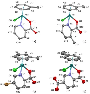
Single crystals of complexes 1, 2∙H2O, 3 and 5 were obtained by the slow evaporation from methanol and their structures were determined by single crystal X-ray diffraction. The ORTEP representations of these complexes are depicted in Fig. 1. The complexes 1 and 2∙H2O crystallized in monoclinic crystal systems in space group P21/n and P21, respectively.
Figure 1. Molecular structures of ruthenium complexes in crystal 1 (a) in crystal 2(b) in crystal 3 and (c) in crystal 5 (d). Displacement parameters are drawn at 50% probability level; hydrogen atoms and water molecule for 2 are omitted for clarity.
The crystals 3 and 5 crystallized in triclinic crystal systems in space group P-1. All of the complexes adopt the so-called “piano stool” configuration, whereby toluene forms the seat and
15
the chelating picolinate ligand as well as the chlorido leaving group constitute the chair legs. In these half-sandwich complexes the ligand is coordinated through the pyridine nitrogen and the carboxylate oxygen. In these structures Ru(II) is a chiral centre. In crystals 1, 3 and 5 both enantiomers were crystallized in non-chiral space groups. On the other hand complex 2 crystallized together with a solvate water molecule and only one enantiomer could be found in the chiral space group P21. The absolute configuration RRu could be determined according to CIP convention [48], the Flack parameter is 0.01(5). The molecular structures of the studied complexes were directly compared to that of the benzene derivative [Ru(6-C6H6)(pic)(Cl)]
defined previously (Ref. code OHUFUT [49]) which crystallized without solvate inclusion in triclinic P-1 space group (Fig. 2.) Selected bond distances and angles are collected in Table 1 for comparison. Distances between the toluene ring and the Ru ion are within the range observed for other ruthenium arene half-sandwich complexes (2.079(11)-2.392(7) Å) [50].
Bond lengths and angles do not show significant differences compared to each other (data are within the experimental errors, Table 1). However, slight differences can be observed, namely the Ru-O bond length according to the influence of picolinate substituents. This bond length is increasing in the order of 2 < 1 ~ OHUFUT < 3 < 5 which is in agreement with the increasing electron-withdrawing effect the substituents in the order of -CH3 < -H < -Br < -COO- group.
Figure 2. Comparison of molecular structures of Ru(II)(η6-toluene) picolinate complexes 1 (colored by element), 2 (orange), 3 (yellow), 5 (violet) together with [Ru(6-C6H6)(pic)(Cl)] (CSD Ref. code OHUFUT) (cyan) [49].Atoms Ru1, Cl1, N1 and O1 are superimposed.
However, the angles between planes of CgA and CgB (where Cg is the centre of gravity
16
calculated for rings A and B, respectively) show slight differences (Table 1 and Fig. 2). The methyl groups of the toluene molecule are almost in the same position for crystals 1, 2∙H2O and 3 (the torsion angle O1-Ru1-Cg(A)-C7 is 5.5 o, 13.3o and -7.7o degree for 1, 2∙H2O and 3, respectively). However, there is a significant difference in crystal 5 where the methyl group turns to the side of the chloride ion and this torsion angle is 116.2o).
Table 1. Selected bond distances (Å) and angles (o) of the studied Ru(II)(η6-toluene) picolinate complexes in crystals 1-3, 5 and [Ru(6-C6H6)(pic)(Cl)] (OHUFUT [49])
1 2∙H2O 3 5 OHUFUT
Bond length (Å)
Ru1-Cl1 2.4133(5) 2.415(2) 2.405(4) 2.396(3) 2.4133(6) Ru1-O1 2.074(1) 2.063(6) 2.085(9) 2.093(6) 2.075(2) Ru1-N1 2.089(2) 2.092(8) 2.11(1) 2.095(7) 2.087(2) Ru1-C1 2.183(2) 2.179(9) 2.17(1) 2.21(1) 2.178(3) Ru1-C2 2.194(2) 2.18(1) 2.23(1) 2.17(1) 2.179(3) Ru1-C3 2.185(2) 2.18(1) 2.16(1) 2.14(1) 2.191(3) Ru1-C4 2.187(2) 2.17(1) 2.16(1) 2.16(1) 2.190(3) Ru1-C5 2.148(2) 2.159(8) 2.21(1) 2.14(1) 2.168(3) Ru1-C6 2.174(2) 2.172(9) 2.16(1) 2.16(1) 2.160(3) Ru1-Cg(A) a 1.6564(9) 1.656(4) 1.662(6) 1.659(5) 1.662 Bond angles (o)
O1-Ru1-N1 77.04(6) 76.8(3) 77.0(4) 77.4(3) 77.54(9) O1-Ru1-Cl1 87.44(4) 84.9(2) 85.9(3) 87.0(2) 87.04(6) N1-Ru1-Cl1 85.73(4) 83.7(2) 83.6(3) 84.2(2) 84.20(7) Cg(A)-Ru1-O1 a 127.69(5) 129.3(2) 129.6(4) 128.4(3) 128.77 Cg(A)-Ru1-N1 a 132.73(5) 133.6(2) 134.2(4) 132.8(3) 132.55 Cg(A)-Ru1-Cl1 a 128.74(4) 129.66(17) 128.2(2) 129.1(2) 128.97 Cg(A)-Cg(B) b 52.94(9) 64.1(5) 61.9(7) 58.9(6) 55.30
O1-Ru1-Cg(A)-Cl1 5.5 13.3 -7.8 116.2 -
a Cg is the centre of gravity calculated for ring A. b Angles between planes calculated for rings A and B.
The positions of the picolinate ligands are slightly different in the studied complexes due to secondary interactions with adjacent molecules as different molecular arrangements and solvate inclusion (for crystal 2∙H2O) are encountered in these crystal structures. The packing
17
arrangements are shown in Figs. S3-S5 viewing along selected crystallographic axes. The main secondary interactions between molecules are C-H…O hydrogen bonds between the toluene hydrogens and the carboxylate oxygen (O1) of the picolinate ligand. Beside the hydrogen bonds considerable secondary interactions are formed between neighboring complexes by C- H…Cl interactions (e.g. C12-H12…Cl1 in 2∙H2O and C5-H5…Cl1 in 4, Table S2 and Figs. S4 and S6).
3.2. Proton dissociation processes of the studied ligands and hydrolysis of the [Ru(6- toluene)(H2O)3]2+ organometallic cation
Proton dissociation constants of the ligands picH, 3-Me-picH, 5-Br-picH, 2,4-dipicH2
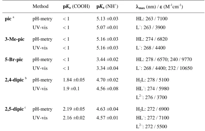
and 2,5-dipicH2 (Chart 1) were determined by pH-potentiometric and UV-vis spectrophotometric titrations performed in the pH range from 2 up to 11.5 (Table 2). Molar absorbance spectra of the ligand species in the different protonation states were calculated via the deconvolution of the spectra recorded at various pH values as it is shown in Fig. S7 for 5- Br-picH. The pKa value of picH and the calculated molar absorbance spectra of the HL and L‒ forms are in reasonably good agreement with data reported previously [23,51,52]. The protonated compounds picH, 3-Me-picH, 5-Br-picH possess two, while 2,4-dipicH2 and 2,5- dipicH2 have three dissociable protons. It was found in all cases that the first deprotonation step assigned to the carboxylic group at position 2 takes place in a fairly acidic range and no pKa values could be determined for this process. Therefore this carboxylate remains deprotonated in the whole studied pH range. pKa determined for picH, 3-Me-picH, 5-Br-picH can be attributed to the deprotonation of the pyridinium (NH+) group as well as the higher pKa
of 2,4-dipicH2 and 2,5-dipicH2. The lower pKa of the latter two ligands belongs to the carboxylic group at position 4 and 5, respectively. Comparing the pKa values to that of Hpic, it is worth mentioning that the methyl substituent has no measurable effect at position 3, while the bromo and the carboxylic groups decrease the pKa (NH+) significantly due to the electron withdrawing power of the halogen substituent and the mesomeric effect of the COO‒ moiety.
The pKa values of picH, 2,4-dipicH2 and 2,5-dipicH2 are in good agreement with data reported in the literature [53,54] (Table 2).
Based on the determined pKa values it can be declared that all the studied ligands are present in their completely deprotonated forms (L‒: pic, 3-Me-pic, 5-Br-pic; L2‒: 2,4-dipic, 2,5-dipic) at pH 7.4 resulting in their strongly hydrophilic character (logD7.4 < ‒2.5).
18
Table 2. Proton dissociation constants (pKa) of the studied ligands determined by pH-potentiometric and UV-vis spectrophotometric titrations; max and molar absorptivity () values for the ligand species in the different protonation states. {T = 25.0˚C, I = 0.20 M (KCl)}
Method pKa (COOH) pKa (NH+) max (nm) / (M-1cm-1) pic a pH-metry
UV-vis
< 1
< 1
5.13 ±0.03 5.07 ±0.01
HL: 263 / 7100 L‒: 263 / 3900 3-Me-pic pH-metry
UV-vis
< 1
< 1
5.16 ±0.03 5.16 ±0.03
HL: 274 / 6820 L‒: 268 / 4400 5-Br-pic pH-metry
UV-vis
< 1
< 1
3.44 ±0.02 3.34 ±0.04
HL: 278 / 6570; 240 / 9770 L‒: 268 / 4400; 232 / 10650 2,4-dipic b pH-metry
UV-vis
1.84 ±0.05 1.9 ±0.1
4.70 ±0.02 4.56 ±0.08
H2L: 278 / 5100 HL‒: 274 / 5980 L2‒: 276 / 3700 2,5-dipic c pH-metry
UV-vis
2.19 ±0.05 2.16 ±0.02
4.63 ±0.04 4.57 ±0.01
H2L: 272 / 6900 HL‒: 272 / 7100 L2‒: 272 / 5500
a Reported pKa values for picH: 5.17 (NH+), ~1 (COOH) at I = 0.20 M (KCl), T = 25.0˚C in ref. [23]; 5.19 (NH+), ~1 (COOH) at I = 0.20 M (KCl), T = 25.0˚C in ref. [52]. b Reported pKa values for 2,4-dipic: 4.79 (NH+), 2.23 (COOH) at I = 0.10 M (KNO3), T = 25.0˚C in ref. [53]. c Reported pKa values for 2,5-dipic:
4.58 (NH+), 2.17 (COOH) at I = 0.50 M (NaClO4), T = 25.0˚C in ref. [54].
Hydrolytic behavior of the organometallic cation [Ru(6-toluene)(H2O)3]2+ has been already studied by Buglyó et al. in the presence and in the absence of chloride ions [44]. In the latter case the fast hydrolysis of the aquated organoruthenium cation yields the species [(Ru(6-toluene))2(μ2-OH)3]+ that becomes predominant at pH > 5. When 0.2 M KCl was used as the background electrolyte, as in our studies, formation of various chlorido and mixed chlorido/hydroxido species as intermediates was found in addition to the major hydrolysis product [(Ru(6-toluene))2(μ2-OH)3]+. In a good accordance with their findings based on the combined use of 1H NMR spectroscopy and ESI-MS, we have also detected three different species based on the 1H NMR spectra recorded at various pH values (Fig. S8). Namely, the identified species are [Ru(6-toluene)(H2O)2Cl]+ (= M), [(Ru(6-toluene))2(-OH)2Cl]+ (=
[M2(OH)2]) and [(Ru(6-toluene))2(-OH)3]+ (= [M2(OH)3]). Overall stability constants for the dinuclear hydrolysis products [(Ru(6-toluene))2(2-OH)i](4-i)+ (i = 2,3) were determined by pH-potentiometric and UV-vis spectrophotometric titrations at 0.20 M chloride ion
19
concentration (Table 3) and are in good agreement with data obtained by Buglyó et al. using pH-potentiometry [44]. Notably these are conditional stability constants being valid only at 0.20 M KCl ionic strength. Concentration distribution curves were computed on the basis of the stability constants determined by pH-potentiometry showing that the hydrolysis is suppressed somewhat due to the presence of chloride ions, since [M2(OH)3] dominates only at pH > 6 (Fig. S9). The 1H NMR signals of the three kinds of species (M, [M2(OH)2],
[M2(OH)3]) could be integrated and distribution of the organometallic fragment was calculated showing an acceptable match between the two kinds of methods.
3.3. Complex formation equilibria of [Ru(6-toluene)(H2O)3]2+ with the picolinate ligands:
stability, deprotonation, chloride ion affinity and lipophilicity
Complexation processes were studied by the combined use of pH-potentiometric, UV-vis spectrophotometric titrations and 1H NMR spectroscopy in a 0.20 M chloride-containing medium. Therefore the formation (logK [ML]) and deprotonation (pKa [ML]) constants determined herein are considered as conditional stability constants. The complex formation between [Ru(6-toluene)(H2O)3]2+ and the studied bidentate picolinate ligands follows a fairly simple scheme (Chart S2). Namely a mono complex [Ru(6-toluene)(L)(Z)] (=[ML]) is formed, and a mixed hydroxido species [ML(OH)] appears by the deprotonation of the coordinated H2O molecule and/or by the displacement of the chlorido co-ligand by OH‒ in the basic pH range, similarly to the behavior of analogous half-sandwich Ru(6-p-cymene) complexes [22,23]. The complex formation of the organometallic cation with the picolinate ligands was found to be a rather slow process especially in the strongly acidic pH range. The steady state could be reached after ~4 h waiting time in the [Ru(6-toluene)(H2O)3]2+ ‒ 3-Me- picH (1:1) system at pH 1.92 as the time-dependence of the UV-vis spectra indicates (Fig. 3).
However, the complexation becomes faster at higher pH values and e.g. in the case of the picH the reaction was finished within 1 h at pH 2.79 (Fig. S10). Based on the recorded spectra it could be concluded that the complex formation proceeds in a great extent already at pH 2 in all cases. As a consequence logK [ML] constants were attempted to be determined from the UV-vis spectral changes of the metal-to-ligand charge-transfer (Ru 4d6→π*) and ligand (π
→π*) transition bands in the pH range from 0.8 to 2.5 (Table 2). Although in this pH range the complex formation is even slower, and the waiting time has a strong impact on the fraction of the complex formed (Fig. S11). It was found that at pH 0.86 in the [Ru(6- toluene)(H2O)3]2+ ‒ 3-Me-picH system more than 15 h is needed to reach the constant
20
absorbance value (Fig. S12). Therefore 24 h waiting time was applied, however the measurements revealed only minor spectral changes (<5%) in this pH range in all the studied [Ru(6-toluene)(H2O)3]2+ – ligand systems. It indicates negligible decomposition of the complexes under such strongly acidic conditions. Thus for the logK [ML] constants only a lower limit could be estimated (Table 3). Based on these findings the complexation of picH with [Ru(6-p-cymene)(H2O)3]2+ was reinvestigated using longer incubation times (24 h) needed to reach steady state in the presence of chloride ions (0.2 M KCl) and a higher logK [ML] value (>10.7) was obtained than previously published [23].
Figure 3. Time-dependence of UV-vis absorption spectra recorded for the [Ru(6-toluene)(H2O)3]2+ ‒ 3-Me-picH (1:1) system at pH = 1.92 in the presence of chloride ions. The inset shows the absorbance changes at 308 nm with fitted curve. {cRu = cL =123 M; T = 25 ˚C; I = 0.20 M (KCl); ℓ = 1.0 cm}.
Table 3. Stability constants logK [ML], pKa [ML] values of the [Ru(6-toluene)(H2O)3]2+ complexes formed with picolinate ligands in 0.2 M chloride-containing aqueous solutions determined by various methods and estimated H2O/Cl− exchange constants (logK’) for the [Ru(6-toluene)(L)(H2O)]+ complexes. {T = 25.0 ˚C, I = 0.20 M (KCl)} a
logK [ML] pKa [ML] pKa [ML] logK’ (H2O/Cl−)
ligand complex UV-vis UV-vis pH-metry UV-vis
pic 1 >10.8 b 8.53 ±0.01 c 8.47 ±0.01 c 1.3 ±0.1 3-Me-pic 2 >10.7 b 8.71 ±0.01 8.68 ±0.05 1.3 ±0.1 d 5-Br-pic 3 > 9.1 b 8.47 ±0.01 8.41 ±0.03 1.5 ±0.1 2,4-dipic 4 > 11.6 b 8.44 ±0.01 8.37 ±0.06 1.2 ±0.1 2,5-dipic 5 > 11.9 b 8.58 ±0.01 8.38 ±0.07 1.1 ±0.1
0.0 0.2 0.4 0.6 0.8
220 280 340 400 460 520
Absorbance
/ nm 0.3 min
1420 min 0.0 0.1 0.2 0.3 0.4
0 300 600 900 1200
Abs.at 308 nm
t / min
21
a M denotes the organometallic fragment Ru(η6-toluene) that appears as [Ru(η6-toluene)(Z)3] (Z = H2O/Cl‒) in water in the presence of chloride ions. Hydrolysis products of the organometallic cation obtained in our work: log [M2(OH)2] = ‒6.32±0.05, log [M2(OH)3] = ‒10.58±0.02; and log [M2(OH)2] = ‒6.50, log [M2(OH)3] = ‒10.56 reported in ref. [44]. b Estimated values based on UV-vis spectrum recorded at pH 0.8; c pKa [ML] values based on 1H NMR titrations: 8.52 ±0.09 (0.20 M KCl) and 7.87 ±0.09 (0 M KCl). d logK’
(H2O/Cl‒) = 1.19 ±0.06 determined at constant ionic strength (I = 0.30 M (NaClO4/NaCl)).
Increasing the pH values the studied [ML] complexes may undergo deprotonation and/or decomposition. Deprotonation of the coordinated water molecule (and/or Cl‒→ OH‒ exchange) results in the formation of mixed hydroxido [ML(OH)] complexes, while decomposition can yield unbound ligand and metal ion in hydrolyzed forms depending on the actual pH. The recorded UV-vis spectra were the same in a wide pH range (e.g. in the [Ru(6- toluene)(H2O)3]2+ ‒ 3-Me-picH system at pH between 3.1 and 7.6 shown in Fig. 4), while significant spectral changes are observed at pH > 8 due to the formation of [ML(OH)]. The appearance of isosbestic points suggests that the metal complexes do not decompose under these conditions; merely they are deprotonated almost in all cases. It should be noted that the complex of 5-Br-pic showed a low extent of decomposition in the basic pH-range. Based on these spectral changes pKa [ML] constants were determined for the complexes (Table 3).
Notably, the spectra of the complexes did not change over a 24 h period at both pH 7.4 and 11 values, and the deprotonation process was found to be rather fast. Therefore pH- potentiometric titrations were also performed to determine pKa [ML] constants (Table 3) started from pH ~4 but only after a 4 h waiting period whilst the formation of [ML] becomes complete. pKa [ML] constants obtained by the two kinds of methods are in a good agreement.
0.0 0.2 0.4 0.6 0.8
230 290 350 410 470
Absorbance
/ nm
pH = 3.0 11.0 ligand alone pH = 3.0
[Ru(6-toluene)(H2O)3]2+
pH = 3.0 0.30
0.35 0.40 0.45
3 5 7 9 11
Abs. 306 nm
pH
22
Figure 4. UV-vis absorption spectra recorded for the [Ru(6-toluene)(H2O)3]2+ ‒ 3-Me-picH (1:1) system in the presence of chloride ions in the pH range from 3 up to 11. The inset shows the absorbance changes at 306 nm at pH between 3.0 and 11.0. {cRu = 102 M; T = 25 ˚C; I = 0.20 M (KCl); ℓ = 1.0 cm}.
In addition 1H NMR spectra were also recorded for the [Ru(6-toluene)(H2O)3]2+ – picH system at pH > 2.5 in the presence of 0.20 M chloride ions at a 1:1 metal-to-ligand ratio using 4 h incubation time (Fig. 5). The spectra undoubtedly reveal that neither free metal ion nor free ligand is present in the whole pH range studied (pH = 2.5 – 11.5), which means that the complex does not suffer from decomposition at 1 mM concentration due to its high stability. The aqua [ML(H2O)] and the chlorinated [ML(Cl)] complexes were identified in the acidic pH range. An upfield shift of all peaks belonging to the [ML(H2O)] complex is observed in the basic pH range due to the fast exchange process on the NMR time scale between the aquated and the mixed hydroxido [ML(OH)] species. In the meanwhile the intensity of the peaks belonging to the [ML(Cl)] complex is decreased. Based on the integrals of the CH(6) toluene proton in the acidic pH range we could conclude that the [ML] complex is mainly chlorinated (~83% [ML(Cl)]). As the [ML(OH)] starts to be formed the three species are present together in the solution, and their equilibrium concentrations cannot be simply calculated due to the fast exchange processes.
Figure 5. 1H NMR spectra of [Ru(6-toluene)(H2O)3]2+ ‒ picH (1:1) system in aqueous solution in the presence of 0.2 M chloride ions at the indicated pH values in the regions of the ligand protons (a), the
[MLCl]
[ML(OH)]
9.5 9.0 8.5 8.0 7.5 6.0 5.5 2.38 2.15
/ ppm
(a) (b) (c)
pH =11.40 10.43 9.56 8.96 8.60 8.02 7.56 6.96 6.14 3.59 +
![Figure 2. Comparison of molecular structures of Ru(II)(η 6 -toluene) picolinate complexes 1 (colored by element), 2 (orange), 3 (yellow), 5 (violet) together with [Ru( 6 -C 6 H 6 )(pic)(Cl)] (CSD Ref](https://thumb-eu.123doks.com/thumbv2/9dokorg/1418118.119937/15.892.324.573.672.984/figure-comparison-molecular-structures-toluene-picolinate-complexes-colored.webp)
![Table 1. Selected bond distances (Å) and angles ( o ) of the studied Ru(II)(η 6 -toluene) picolinate complexes in crystals 1-3, 5 and [Ru( 6 -C 6 H 6 )(pic)(Cl)] (OHUFUT [49])](https://thumb-eu.123doks.com/thumbv2/9dokorg/1418118.119937/16.892.154.737.356.972/selected-distances-studied-toluene-picolinate-complexes-crystals-ohufut.webp)
![Figure 3. Time-dependence of UV-vis absorption spectra recorded for the [Ru( 6 -toluene)(H 2 O) 3 ] 2+ ‒ 3-Me-picH (1:1) system at pH = 1.92 in the presence of chloride ions](https://thumb-eu.123doks.com/thumbv2/9dokorg/1418118.119937/20.892.240.620.398.653/figure-dependence-absorption-spectra-recorded-toluene-presence-chloride.webp)
![Figure 4. UV-vis absorption spectra recorded for the [Ru( 6 -toluene)(H 2 O) 3 ] 2+ ‒ 3-Me-picH (1:1) system in the presence of chloride ions in the pH range from 3 up to 11](https://thumb-eu.123doks.com/thumbv2/9dokorg/1418118.119937/22.892.121.766.700.1040/figure-absorption-spectra-recorded-toluene-presence-chloride-range.webp)
![Figure 6. Distribution of Ru( 6 -toluene) in the [Ru( 6 -toluene)(H 2 O) 3 ] 2+ ‒ picH (1:1) system in the presence of 0.2 M chloride ions in the pH range from 2.5 up to 11 based on the 1 H NMR peak integrals for the CH(6) toluene proton of species](https://thumb-eu.123doks.com/thumbv2/9dokorg/1418118.119937/23.892.249.628.471.731/figure-distribution-toluene-toluene-presence-chloride-integrals-toluene.webp)