This is an Accepted Manuscript, which has been through the Royal Society of Chemistry peer review process and has been accepted for publication.
Accepted Manuscripts are published online shortly after
acceptance, before technical editing, formatting and proof reading.
Using this free service, authors can make their results available to the community, in citable form, before we publish the edited article. We will replace this Accepted Manuscript with the edited and formatted Advance Article as soon as it is available.
You can find more information about Accepted Manuscripts in the author guidelines.
Please note that technical editing may introduce minor changes to the text and/or graphics, which may alter content. The journal’s standard Terms & Conditions and the ethical guidelines, outlined in our author and reviewer resource centre, still apply. In no event shall the Royal Society of Chemistry be held responsible for any errors or omissions in this Accepted Manuscript or any consequences arising from the use of any information it contains.
Accepted Manuscript
Dalton
Transactions
An international journal of inorganic chemistry www.rsc.org/dalton
ISSN 1477-9226
PAPER Joseph T. Hupp, Omar K. Farha et al.
Effi cient extraction of sulfate from water using a Zr-metal–organic framework
Volume 45 Number 1 7 January 2016 Pages 1–398
Dalton
Transactions
An international journal of inorganic chemistry
This article can be cited before page numbers have been issued, to do this please use: F. Bacher, C.
Wittmann, M. Nove, G. Spengler, M. A. Mar, E. A. Enyedy, D. Darvasiova, P. Rapta, T. Reiner and V. Arion, Dalton Trans., 2019, DOI: 10.1039/C9DT01238A.
Novel latonduine derived proligands and their copper(II) complexes show cytotoxicity in the nanomolar range in human colon adenocarcinoma cells and
in vitro cancer selectivity
Felix Bacher,*a Christopher Wittmann,a Márta Nové,b Gabriella Spengler,b Małgorzata A. Marć,c Eva A. Enyedy,c Denisa Darvasiova,d Peter Rapta,d Thomas Reinere and
Vladimir B. Arion*a
aInstitute of Inorganic Chemistry of the University of Vienna, Währinger Strasse 42, A1090 Vienna, Austria
bDepartment of Medical Microbiology and Immunobiology, University of Szeged, Dóm tér 10, H-6720 Szeged, Hungary
cDepartment of Inorganic and Analytical Chemistry, Interdisciplinary Excellence Centre, University of Szeged, Dóm tér 7, H-6720 Szeged, Hungary
dInstitute of Physical Chemistry and Chemical Physics, Slovak University of Technology in Bratislava, Radlinského 9, 81237 Bratislava, Slovak Republic
eDepartment of Radiology, Weill Cornell Medical College, New York City, NY 10065, United States; Department of Radiology, Memorial Sloan Kettering Cancer Center, New York City, NY 10065, United States; Chemical Biology Program, Memorial Sloan Kettering Cancer Center, New York City, NY 10065, United States
Dalton Transactions Accepted Manuscript
Open Access Article. Published on 10 May 2019. Downloaded on 5/14/2019 5:52:00 AM. This article is licensed under a Creative Commons Attribution-NonCommercial 3.0 Unported Licence.
View Article Online DOI: 10.1039/C9DT01238A
Abstract
Four Schiff bases derived from 7-hydrazin-yl-5,8-dihydroindolo[2,3-d][2]benzazepin- (6H)-one and its bromo-substituted analogue (HL1–HL4) and four copper(II) complexes 1–4 have been synthesised and fully characterised by standard spectroscopic methods (1H and 13C NMR, UV–vis), ESI mass spectrometry, single crystal X-ray diffraction and spectroelectrochemistry. In addition, two previously reported complexes with paullone ligands 5 and 6 were prepared and studied for comparison reasons. The CuII ion in 1–4 is five-coordinate and adopts a square- pyramidal or slightly distorted square-pyramidal coordination geometry. The ligands HL1–4 act as tridentate, the other two coordination places are occupied by two chlorido co-ligands. The organic ligands in 2 and 3 are bound tighter to copper(II) when compared to related paullone ligands in 5 and 6. The new compounds show very strong cytotoxic activity against human colon adenocarcinoma doxorubicin- sensitive Colo 205 and multidrug resistant Colo 320 cancer cell lines with IC50 values in low micromolar to nanomolar concentration range.
Dalton Transactions Accepted Manuscript
Open Access Article. Published on 10 May 2019. Downloaded on 5/14/2019 5:52:00 AM. This article is licensed under a Creative Commons Attribution-NonCommercial 3.0 Unported Licence.
View Article Online DOI: 10.1039/C9DT01238A
Introduction
Indolobenzazepines are a class of compounds with a broad spectrum of biological activities, amongst them analgesic, antidepressant, antimalarial, antidiabetic, anticancer and antiparasitic.1–7 The search for potent kinase inhibitors led to indolo[3,2-d]benzazepines, known as paullones.8 Cyclin dependent kinases (cdks) are attractive targets for anticancer drugs, since they control the cell cycle.9 Research on structure-activity relationships (SARs) showed that electron-withdrawing groups in position 9 of the paullone scaffold (Chart 1, left) enhance the cdk inhibition, while the lactam moiety is essential for the anticancer activity. This finding resulted in the discovery of the lead compounds kenpaullone and alsterpaullone (Chart 1).10 Despite their potency, paullones possess some adverse properties. In particular, the low aqueous solubility and bioavailability hampered their clinical use. Later it was shown that the bioavailability can be improved by the attachment of suitable metal binding sites onto the paullone backbone and the consequent metal complex formation with gallium(III), ruthenium(II), osmium(II) and copper(II) ions. This led to highly cytotoxic compounds with an enhanced aqueous solubility with copper(II) complexes being the most active ones.11–16 However, cdks seem not to be the main target for these metal complexes.17 Alternative mechanisms of action have been suggested, amongst them DNA intercalation.14
In our present work, we focused our attention on the isomeric indolo[2,3- d]benzazepine scaffold (Chart 1, right). It differs from the paullone core by the position of the lactam group in the azepine ring and features a flipped indole moiety with respect to the benzazepinone half of the molecule. These new compounds contain a typical latonduine structural motif. Latonduine-modified molecules usually exhibit prominent anticancer activity in vitro, along with the ability to inhibit tubulin polymerisation.18–21 Like paullones, these compounds do not possess any metal binding site. Therefore, it was of interest to us to chemically modify them and obtain proligands with potentially tridentate κN, κN’, κN’’ metal binding site, in particular, for copper(II). Copper is an essential trace metal and therefore considered to be safer than other metals used in chemotherapy, i.e. platinum.22,23 In addition, it is well- established that copper(II) is able to strongly enhance the cytotoxicity of biologically active ligands.24
Dalton Transactions Accepted Manuscript
Open Access Article. Published on 10 May 2019. Downloaded on 5/14/2019 5:52:00 AM. This article is licensed under a Creative Commons Attribution-NonCommercial 3.0 Unported Licence.
View Article Online DOI: 10.1039/C9DT01238A
NH
NH O
1
2 3
4
5 7 6 8 9 10 11
12
NH
NH O
1 2
3 4 5 7 6
9 8
10 11
12
R
R = Br (kenpaullone) R = NO2(alsterpaullone)
Chart 1. The indolo[3,2-d]benzazepinone (paullone) (left), vs the indolo[2,3- d]benzazepinone backbone (right) with numbering schemes.
Herein we report on the synthesis and characterisation of four novel latonduine core containing proligands and their copper(II) complexes. In order to obtain structure- activity relationships we modified the indolobenzazepine backbone by inserting a bromine substituent at position 11, the equivalent to position 9 in the paullone scaffold (compare kenpaullone). In addition, an aldehyde and a ketone containing a functional group in a position suitable for chelate formation were used for Schiff base condensation reactions, namely 2-formyl- and 2-acetylpyridine (Chart 2).
The new compounds have been characterised by 1H and 13C NMR spectroscopy (HL1–HL4), single crystal X-ray diffraction (HL4, 1–4) and ESI mass spectrometry;
their purity was validated by elemental analysis. Solution equilibrium properties of HL3 and its copper(II) complex 3 were characterised in DMSO-water mixture by UV–
vis titrations. The cytotoxicity of the proligands and the corresponding copper(II) complexes was tested in Colo 205 (chemo sensitive) and Colo 320/MDR-LRP (multidrug resistant) human colon adenocarcinoma cell lines and one non-cancerous human embryonal lung fibroblast MRC-5 cell line and compared to those of the two previously reported paullone-derived copper(II) complexes 5 and 6 (Chart 3).15,25 The new compounds showed very high activity with IC50 values from the low micromolar to the nanomolar concentration range and were superior to 5 and 6 in cancer cell lines, while less cytotoxic in non-cancerous MRC-5 cells. Furthermore some selectivity for cancer cells over normal cells was observed in most cases, making this compound class pertinent for further development as anticancer drugs.
Dalton Transactions Accepted Manuscript
Open Access Article. Published on 10 May 2019. Downloaded on 5/14/2019 5:52:00 AM. This article is licensed under a Creative Commons Attribution-NonCommercial 3.0 Unported Licence.
View Article Online DOI: 10.1039/C9DT01238A
NH
NH O
(i)
NH
NH S
(ii)
NH N NH
(iii)
NH
NH N N
R2 N
R1 R1 R1
H2N
R1
R1= H (A) R1= Br (B)
R1= H (C)
R1= Br (D) R1= H (E)
R1= Br (F)
R1= R2= H (HL1) R1= Br, R2= H (HL2) R1= H, R2= CH3(HL3) R1= Br, R2= CH3(HL4)
NH N
HN N N Cu Cl Cl R1
R1= R2= H (1)*
R1= Br, R2= H (2) R1= H, R2= CH3(3) R1= Br, R2= CH3(4)
R2 (iv)
Chart 2. New proligands and copper(II) complexes synthesised in this work.a Underlined numbers indicate compounds studied by X-ray diffraction. *1 crystallized from DMF as the trimer [Cu3Cl4(HL1)3]Cl2 (1trim). aReagents and conditions: (i) C:
phosphorus pentasulfide/aluminum oxide, acetonitrile, 85 °C, overnight; D:
phosphorus pentasulfide/aluminum oxide, tetrahydrofuran, 75 °C, overnight; (ii) hydrazine monohydrate, reflux, overnight; (iii) HL1, HL2: 2-formypyridine, ethanol, 85
°C, overnight; HL3, HL4: 2-acetylpyridine, ethanol, 85 °C, overnight; (iv) CuCl2·2H2O, isopropanol, reflux, 15 min.
N HN
R1 HN
N N Cu Cl
Cl
R2
R1= Br, R2= H (5) R1= H, R2= CH3(6)
Chart 3. Paullone derived copper(II) complexes used in this study.15,25
Dalton Transactions Accepted Manuscript
Open Access Article. Published on 10 May 2019. Downloaded on 5/14/2019 5:52:00 AM. This article is licensed under a Creative Commons Attribution-NonCommercial 3.0 Unported Licence.
View Article Online DOI: 10.1039/C9DT01238A
Experimental
2-Iodobenzonitrile, ethyl-1H-indole-carboxylate and 5-bromo-ethyl-1H-indole- carboxylate were purchased from ABCR. Borane solution (1M in THF), absolute DMF, dimethylaminopyridine, di-tert-butyl-dicarbonate, absolute acetonitrile, palladium(II) acetate, sodium bicarbonate, basic aluminuim oxide, 2-acetylpyridine and 2-formylpyridine were bought from Fisher/Acros Organics. Ethoxy-methylchloride was obtained form TCI. Sodium hydride, phosphorus(V) sulfide, celite, hydrazine monohydrate and methyl iodide were purchased from Sigma Aldrich, while lithium hydroxide monohydrate and triphenylphosphine were from Alfa Aesar. 1-Ethyl-3-(3- dimethylaminopropyl)carbodiimide-hydrochloride was purchased from IRIS biotech.
Silver(I) carbonate was purchased from Merck. 2-Iodobenzylamine was prepared by a known method.26 The unsubstituted indolo[2,3-d]benzazepinone (A) was prepared by following published protocols.18–20 The 11-bromo-substituted B was prepared using reported precedures,18–20 with some modifications, a detailed description of the synthesis of B is given in the Supplementary Information file.
Synthesis of proligands
5,8-dihydroindolo[2,3-d][2]benzazepin-7(6H)-thione (C). 5,8-dihydroindolo[2,3- d][2]benzazepin-7(6H)-one (A) (966 mg, 3.88 mmol) was dissolved in absolute acetonitrile (77 mL) in a Schlenk tube under argon atmosphere. A mixture of phosphorus(V) pentasulfide and basic aluminium oxide (0.6 : 1 w/w)27 (1.60 g) was added and the reaction mixture was stirred at 90 °C overnight. The next day, the mixture was cooled to room temperature and filtered. The filtrate was concentrated in vacuo and taken up in water (50 mL). The pH was adjusted to 8 using saturated potassium carbonate solution. The solution was extracted with dichloromethane (DCM) (3 × 100 mL). The organic phases were combined and dried over magnesium sulfate. The dried organic phase was concentrated and the raw product was purified on silica using a mixture of DCM : methanol 99 : 1 as eluent. Yield: 841 mg, 82%. 1H NMR (500 MHz, DMSO-d6) δ 11.78 (s, 1H, NH), 10.57 (t, J = 5.5 Hz, 1H, NH), 7.98 (dd, J = 14.4, 7.9 Hz, 2H, H(Ar)), 7.64 (d, J = 8.3 Hz, 1H, H(Ar)), 7.53 (t, J = 7.5 Hz, 1H, H(Ar)), 7.48 (d, J = 6.9 Hz, 1H, H(Ar)), 7.41 (t, J = 7.3 Hz, 1H, H(Ar)), 7.36 (t, J = 7.4 Hz, 1H, H(Ar)), 7.20 (t, J = 7.5 Hz, 1H, H(Ar)), 4.41 – 4.28 (m, 1H, CH2), 4.04 (d, J = 12.2 Hz, 1H, CH2). ESI-MS (acetonitrile/methanol + 1% water), positive: m/z 265.08 [M + H]+.
Dalton Transactions Accepted Manuscript
Open Access Article. Published on 10 May 2019. Downloaded on 5/14/2019 5:52:00 AM. This article is licensed under a Creative Commons Attribution-NonCommercial 3.0 Unported Licence.
View Article Online DOI: 10.1039/C9DT01238A
11-bromo-5,8-dihydroindolo[2,3-d][2]benzazepin-7(6H)-thione (D). 5,8- dihydroindolo[2,3-d][2]benzazepin-7(6H)-one (B) (1.00 g, 3.06 mmol) and a mixture of phosphorus(V) pentasulfide and basic aluminium oxide (0.6 : 1 w/w)27 (2.20 g) were suspended in dry THF (50 mL) in an 100 mL Schlenk tube, under argon atmosphere. The reaction mixture was stirred overnight at 75 °C. The next day it was cooled to room temperature, the yellow precipitate was filtered off and washed with THF. The filtrate was evaporated and the residue was purified on silica by using DCM/methanol 99 : 1 as eluent. The product as yellow-orange powder was obtained after removal of the solvent. Yield: 635 mg, 60%.1H NMR (500 MHz, DMSO-d6) δ 12.00 (s, 1H, NH), 10.67 (t, J = 5.6 Hz, 1H, NH), 8.10 (d, J = 1.7 Hz, 1H, H(Ar)), 7.93 (d, J = 7.2 Hz, 1H, H(Ar)), 7.60 (d, J = 8.7 Hz, 1H, H(Ar)), 7.55 (td, J = 7.5, 1.4 Hz, 1H, H(Ar)), 7.51 – 7.47 (m, 2H, H(Ar)), 7.43 (td, J = 7.4, 1.1 Hz, 1H, H(Ar)), 4.33 (s, 1H, CH2), 4.06 (d, J = 12.5 Hz, 1H, CH2). ESI-MS (acetonitrile/methanol + 1% water), positive, m/z 344.99 [M + H]+.
7-hydrazin-yl-5,8-dihydroindolo[2,3-d][2]benzazepin-(6H)-one (E). A suspension of 5,8-dihydroindolo[2,3-d][2]benzazepin-7(6H)-thione (C) (582 mg, 2.21 mmol) in hydrazine monohydrate (8 mL) under argon atmosphere was refluxed overnight. On the next day it was cooled to room temperature and the pale yellow precipitate was filtered off, washed with water and dried in vacuo. Yield: 496 mg, 86%. 1H NMR (500 MHz, DMSO-d6) δ 11.43 (s, 1H, NH), 7.86 (t, J = 8.5 Hz, 2H, H(Ar)), 7.49 – 7.40 (m, 2H, H(Ar)), 7.37 (d, J = 6.5 Hz, 1H), H(Ar), 7.20 (dt, J = 21.5, 7.3 Hz, 2H, H(Ar)), 7.09 (t, J = 7.0 Hz, 1H, H(Ar)), 6.32 (s, 1H, NH), 5.00 (s, 2H, NH2), 4.07 (s, 2H, CH2). ESI-MS (acetonitrile/methanol + 1% water), positive: m/z 263.09 [M + H]+.
11-bromo-7-hydrazin-yl-5,8-dihydroindolo[2,3-d][2]benzazepin-(6H)-one (F). A suspension of 11-bromo-5,8-dihydroindolo[2,3-d][2]benzazepin-7(6H)-thione (D) (830 mg, 2.42 mmol) in hydrazine monohydrate (20 mL) under argon atmosphere was refluxed overnight. On the next day it was cooled to room temperature and the pale yellow precipitate was filtered off, washed with water and dried in vacuo. Yield: 757 mg, 92%. 1H NMR (500 MHz, DMSO-d6) δ 7.97 (d, J = 1.8 Hz, 1H, H(Ar)), 7.80 (d, J = 7.2 Hz, 1H, H(Ar)), 7.47 – 7.37 (m, 3H, H(Ar)), 7.33 – 7.29 (m, 1H, H(Ar)), 7.25 (td, J = 7.4, 1.0 Hz, 1H, H(Ar)), 6.36 (s, 1H, NH), 5.07 (s, 2H, NH2), 4.06 (s, 2H, CH2). ESI-MS (acetonitrile/methanol + 1% water), positive: m/z 343.24 [M + H]+.
Dalton Transactions Accepted Manuscript
Open Access Article. Published on 10 May 2019. Downloaded on 5/14/2019 5:52:00 AM. This article is licensed under a Creative Commons Attribution-NonCommercial 3.0 Unported Licence.
View Article Online DOI: 10.1039/C9DT01238A
HL1·0.4C2H5OH. A solution of 7-hydrazin-yl-5,8-dihydroindolo[2,3-d][2]benzazepin- (6H)-one (E) (199 mg, 0.76 mmol) in ethanol (3 mL) in a 25 mL Schlenk tube was degassed by bubbling argon through the solution for 10 min. 2-Formylpyridine (79 µL, 0.83 mmol) was added and the mixture was stirred overnight at 85 °C. The next day the reaction mixture was cooled to room temperature and the solvent was evaporated. The residue was taken up in water (10 mL) and ethanol was added until complete dissolution. Then, the solvent was removed slowly under reduced pressure until precipitation started. The mixture was allowed to stand at 4 °C overnight. The next day, a yellow precipitate was filtered off, washed with water/ethanol 2:1 and dried in vacuo. Yield: 266 mg, 99%. Anal. Calcd for C22H17N5·0.4C2H5OH (M 372.13 g mol–1): C, 73.71; H, 5.07, N, 19.09. Found: C, 73.91; H, 5.33; N, 18.82. 1H NMR (600 MHz, DMSO-d6) δ 11.93 (s, 1H, H12), 8.59 (d, J = 4.1 Hz, 1H, H18), 8.37 (d, J = 8.0 Hz, 2H, H15, H21), 8.31 (t, J = 5.4 Hz, 1H, H6), 7.98 (dd, J = 15.4, 7.6 Hz, 2H, H12, H3), 7.87 (td, J = 7.5, 1.2 Hz, 1H, H20), 7.60 (d, J = 8.2 Hz, 1H, H9), 7.53 – 7.49 (m, 1H, H2), 7.47 (d, J = 6.6 Hz, 1H, H4), 7.40 – 7.31 (m, 3H, H10, H1, H19), 7.20 (dd, J = 11.1, 4.0 Hz, 1H, H11), 4.36 – 4.02 (m, 2H, H5). 13C NMR (151 MHz, DMSO) δ 155.62 (Cq, C7), 154.52 (Cq, C16), 152.14 (CH, C15), 149.28 (CH, C18), 137.78 (Cq, C4a), 136.76 (Cq, C8a), 136.30 (CH, C20), 133.161 (Cq, C12c), 129.01( Cq, C7a), 128.09 (CH, C4), 128.06 (CH, C2), 127.41 (CH,C3), 126.33 (CH,C1), 124.94 (Cq, C12a), 124.10(CH, C10), 123.87 (CH, C19), 120.77 (CH, C21), 120.46 (CH, C11), 120.22(CH, C12), 117.12(Cq, C12b), 112.69(CH, C9), 46.00 (CH2, C5). For atom numbering scheme see ESI†, Scheme S1. ESI-MS (acetonitrile/methanol + 1% water), positive:
m/z 352.26 [M + H]+.
HL2·H2O. A solution of 11-bromo-7-hydrazin-yl-5,8-dihydroindolo[2,3- d][2]benzazepin(6H)one (F) (370 mg, 1.08 mmol) in ethanol (6 mL) in a 25 mL Schlenk tube was degassed by bubbling argon through the solution for 10 min. 2- Formylpyridine (113 µL, 1.19 mmol) was added and the mixture was stirred overnight at 85 °C. The next day the reaction mixture was cooled to room temperature and the solvent was removed under reduced pressure. The residue was dissolved in methanol (10 mL). This solution was concentrated under reduced pressure to about half the volume, when the formation of a yellow precipitate was observed. The resulting suspension was allowed to stand at 4 °C overnight. On the next day the precipitate was filtered off and washed with cold methanol. Yield: 135 mg, 31%. Anal.
Dalton Transactions Accepted Manuscript
Open Access Article. Published on 10 May 2019. Downloaded on 5/14/2019 5:52:00 AM. This article is licensed under a Creative Commons Attribution-NonCommercial 3.0 Unported Licence.
View Article Online DOI: 10.1039/C9DT01238A
Calcd for C22H16BrN5·H2O (M 448.32 g mol–1): C, 58.93; H, 4.05, N, 15.62. Found: C, 58.86; H, 3.97; N, 15.67. 1H NMR (700 MHz, DMSO-d6) δ 12.14 (s, 1H, H8), 8.59 (d, J = 4.6 Hz, 1H, H18), 8.38 – 8.35 (m, 2H, H15, H21), 8.32 (t, J = 5.3 Hz, 1H, H6), 8.10 (d, J = 1.5 Hz, 1H, H12), 7.92 (d, J = 7.6 Hz, 1H, H1), 7.86 (dd, J = 11.0, 4.4 Hz, 1H, H20), 7.56 (d, J = 8.7 Hz, 1H, H9), 7.52 (t, J = 7.5 Hz, 1H, H2), 7.47 – 7.44 (m, 2H, H10, H4), 7.39 – 7.34 (m, 2H, H19, H3), 4.46 – 3.99 (m, 2H, H5). 13C NMR (176 MHz, DMSO-d6) δ 155.25 (Cq, C7), 154.40 (Cq, C16), 152.52 (CH, C15), 149.29 (CH, C18), 137.86 (Cq, C4a), 136.30 (CH, C20), 135.39 (Cq, C8a), 132.95 (Cq, C12), 130.29 (Cq, C7a), 128.27 (CH, C4), 128.17 (CH, C2), 127.35 (CH, C1), 126.71 (CH, C10), 126.63 (CH. C3), 126.54 (Cq, C12a), 123.95 (CH, C19), 122.23 (CH, C12), 120.82 (CH, C21), 116.48 (Cq, C12b), 114.70 (CH, C9), 113.01 (Cq, C11), 45.91 (CH2, C5). For atom numbering scheme see ESI†, Scheme S1. ESI-MS (acetonitrile/methanol + 1%
water), positive: m/z 432.06 [M + H]+.
HL3. A solution of 7-hydrazin-yl-5,8-dihydroindolo[2,3-d][2]benzazepin(6H)one (E) (200 mg, 0.76 mmol) in ethanol (3.5 mL) in a 25 mL Schlenk tube was degassed by bubbling argon through the solution for 10 min. 2-Acetylpyridine (94 µL, 0.84 mmol) was added and the mixture was stirred overnight at 86 °C. The next day the reaction mixture was cooled to room temperature and stored at 4 °C for 2 h. The yellow precipitate was filtered off and washed with cold ethanol. Yield: 261 mg, 94%. Anal.
Calcd for C23H19N5 (M 365.43 g mol–1): C, 75.59; H, 5.24, N, 19.16. Found: C, 75.32;
H, 5.05; N, 19.00. 1H NMR (600 MHz, DMSO-d6) δ 11.77 (s, 1H, H8), 8.62 – 8.55 (m, 1H, H18), 8.50 (d, J = 8.0 Hz, 1H2), 8.02 – 7.91 (m, 3H, H1, H5, H12), 7.81 (td, J = 7.8, 1.8 Hz, 1H, H20), 7.62 (d, J = 8.2 Hz, 1H, H9), 7.49 (td, J = 7.6, 1.2 Hz, 1H, H2), 7.43 (d, J = 6.8 Hz, 1H, H4), 7.40 – 7.29 (m, 3H, H3, H19, H10), 7.19 (dt, J = 18.3, 5.5 Hz, 1H, H11), 4.64 – 3.75 (m, 2H, H5), 2.50 (s, 3H, H22 (overlapped DMSO signal)). 13C NMR (151 MHz, DMSO-d6) δ 158.23 (Cq, C15), 156.45 (Cq, C16), 153.36 (Cq, C7), 148.44 (CH, C18), 137.95 (Cq, C4a), 136.65 (Cq, C8a), 135.88 (CH, C20), 133.74 (Cq, C12c), 129.75 (Cq, C7a), 128.07 (CH, C4), 127.97 (CH, C2), 127.33 (CH, C1), 126.18 (CH, C3), 125.05 (Cq, C12a), 123.92 (CH, C10), 123.52 (CH, C19), 120.84 (CH, C21), 120.37 (CH, C11), 120.13 (CH, C12), 116.50 (Cq, C12b), 112.62 (CH, C9), 45.97 (CH2, C5) 13.08 (CH3, C22). For atom numbering scheme see ESI†, Scheme S1. ESI-MS (acetonitrile/methanol + 1% water), positive: m/z 366.16 [M + H]+.
Dalton Transactions Accepted Manuscript
Open Access Article. Published on 10 May 2019. Downloaded on 5/14/2019 5:52:00 AM. This article is licensed under a Creative Commons Attribution-NonCommercial 3.0 Unported Licence.
View Article Online DOI: 10.1039/C9DT01238A
HL4. A solution of 11-bromo-7-hydrazin-yl-5,8-dihydroindolo[2,3- d][2]benzazepin(6H)one (F) (76 mg, 0.22 mmol) in ethanol (2 mL) in a 10 mL Schlenk tube was degassed by bubbling argon through the solution for 10 min. 2- Acetylpyridine (27 µL, 0.24 mmol) was added and the mixture was stirred overnight at 85 °C. The next day the reaction mixture was cooled to room temperature and allowed to stand at 4 °C for 2 days. The yellow-brown plates of X-ray diffraction quality were filtered off and washed with cold ethanol. Yield: 57 mg, 58%. Anal. Calcd for C23H18BrN5 (M 444.33 g mol–1): C, 62.17; H, 4.08, N, 15.76. Found: C, 61.86; H, 4.13; N, 15.74.1H NMR (600 MHz, DMSO-d6) δ 11.98 (s, 1H, H8), 8.60 – 8.56 (m, 1H, H18), 8.53 – 8.44 (m, 1H, H21), 8.09 (d, J = 1.8 Hz, 1H, H12), 7.99 (t, J = 5.3 Hz, 1H, H6), 7.93 – 7.90 (m, 1H, H1), 7.82 (ddd, J = 8.0, 7.5, 1.8 Hz, 1H, H20), 7.58 (d, J = 8.7 Hz, 1H, H9), 7.54 – 7.49 (m, 1H, H2), 7.47 – 7.42 (m, 2H, H10, H4), 7.38 – 7.33 (m, 2H, H19, H3), 4.39 – 4.08 (m, 2H, H5), 2.49 (s, 3H, H22 (overlapped DMSO signal)).
13C NMR (151 MHz, DMSO-d6) δ 158.62 (Cq, C15), 156.34 (Cq, C16), 152.96 (Cq, C7), 148.45 (CH, C18), 138.04 (Cq, C4a), 135.91 (CH, C20), 135.30 (Cq, C8a), 133.08 (Cq, C12c), 131.02 (Cq, C7a), 128.20 (CH, C4), 128.15 (CH, C2), 127.28 (CH, C1), 126.68 (Cq, C12a), 126.53 (CH, C10), 126.49 (CH, C19), 123.61 (CH, C3), 122.14 (CH, C12), 120.88 (CH, C21), 115.90 (Cq, C12b), 114.65 (CH, C9), 112.92 (Cq, C11), 45.88 (CH2, C5), 13.10 (CH3, C22). For atom numbering scheme see ESI†, Scheme S1.
ESI-MS (acetonitrile/methanol + 1% water), positive: m/z 446.08 [M + H]+.
Synthesis of copper(II) complexes
1·0.4H2O·0.7C3H7OH. To a solution of HL1 (169 mg, 0.48 mmol) in isopropanol (8 mL) a solution of CuCl2·2H2O (82 mg, 0.48 mmol) in methanol (0.5 mL) was added.
The reaction mixture was heated to reflux for 15 min, cooled down and allowed to stand at 4 °C overnight. The product was filtered off, washed with isopropanol and dried in vacuo to give a green-brown powder. Yield: 232 mg, 99%. Anal. Calcd for C22H17Cl2CuN5·0.4H2O·0.7C3H7OH (M 535.13 g mol–1): C, 53.86; H, 4.24, N, 13.08.
Found: C, 54.09; H, 4.41; N, 13.08. Solubility in water/1% DMSO ≥ 1.0 mg mL–1. ESI- MS (acetonitrile/methanol + 1% water), positive: m/z 764.23 [CuII(L1)(HL1)]+.
2·0.5H2O. To a solution of HL2 (100 mg, 0.23 mmol) in isopropanol (20 mL) at 70 °C a solution of CuCl2·2H2O (40 mg, 0.23 mmol) in methanol (1 mL) was added. The colour of the solution changed from yellow to green and the reaction mixture was
Dalton Transactions Accepted Manuscript
Open Access Article. Published on 10 May 2019. Downloaded on 5/14/2019 5:52:00 AM. This article is licensed under a Creative Commons Attribution-NonCommercial 3.0 Unported Licence.
View Article Online DOI: 10.1039/C9DT01238A
refluxed for 15 min. After cooling down the solution was kept at 4 °C overnight. Green precipitate was filtered off, washed with isopropanol and dried in vacuo. Yield: 105 mg, 77%. Anal. Calcd for C22H16BrCl2CuN5·0.5H2O (M 573.76 g mol–1): C, 46.05; H, 2.99, N, 12.21. Found: C, 45.93; H, 2.86; N, 12.52. Solubility in water/1% DMSO ≥ 0.7 mg mL–1. ESI-MS (acetonitrile/methanol + 1% water), positive: m/z 922.07 [CuII(L2)(HL2)]+.
3·0.3H2O·0.6C3H7OH. To a solution of HL3 (200 mg, 0.55 mmol) in isopropanol (140 mL) at 70 °C a solution of CuCl2·2H2O (95 mg, 0.55 mmol) in methanol (1 mL) was added. The colour of the solution changed from yellow to green and the mixture was refluxed for 15 min. After cooling down the solution was kept at 4 °C overnight. The next day a green precipitate was filtered off, washed with isopropanol and dried in vacuo. Yield: 224 mg, 82%. Anal. Calcd for C23H19Cl2CuN5·0.3H2O·0.6C3H7OH (M 541.34 g mol–1): C, 55.02; H, 4.54, N, 12.94. Found: C, 55.07; H, 4.16; N, 12.84.
Solubility in water/1% DMSO ≥ 0.3 mg mL–1. ESI-MS (acetonitrile/methanol + 1%
water), positive: m/z 463.04 [CuCl(HL3)]+, 427.07 [Cu(L3)]+.
4·0.5H2O. To a solution of HL4 (50 mg, 0.11 mmol) in isopropanol (20 mL) at 70 °C CuCl2·2H2O (19 mg, 0.11 mmol) in methanol (1 mL) was added. The colour of the solution changed from yellow to green and the reaction mixture was refluxed for 15 min. After cooling to room temperature, the solution was kept at 4 °C overnight. The next day a green precipitate was filtered off, washed with isopropanol and dried in vacuo. Yield: 51 mg, 80%. Anal. Calcd for C23H18BrCl2CuN5·0.5H2O (M 587.79 g mol–1): C, 47.00; H, 3.26, N, 11.91. Found: C, 46.85; H, 3.53; N, 11.81. Solubility in water/1% DMSO ≥ 0.9 mg mL–1. ESI-MS (acetonitrile/methanol + 1% water), positive:
m/z 542.96 [CuIICl(HL4)]+, 507.00 [CuII(L4)]+.
Crystallographic Structure Determination. X-ray diffraction quality single crystals of HL4 were obtained by crystallisation from ethanol, 1trim·2DMF·1.25H2O, 3·1.2DMF·0.25H2O, 3´·DMF and 4·2DMF by slow diffusion of diethyl ether into the DMF solution of the corresponding complex, while 2·0.55MeOH by slow evaporation of the metanolic solution of the compound. The measurements were performed on Bruker X8 APEXII CCD (2·0.55MeOH, 3·1.2DMF·0.25H2O) and Bruker D8 Venture (HL4, 1trim ·2DMF·1.25H2O, 3´·DMF, 4·2DMF) diffractometers. Single crystals were
Dalton Transactions Accepted Manuscript
Open Access Article. Published on 10 May 2019. Downloaded on 5/14/2019 5:52:00 AM. This article is licensed under a Creative Commons Attribution-NonCommercial 3.0 Unported Licence.
View Article Online DOI: 10.1039/C9DT01238A
positioned at 27, 27, 27, 35, 25 and 27 mm from the detector, and 672, 1632, 1000, 614, 479 and 1566 frames were measured, each for 8, 30, 10, 24, 3 and 6 s over 0.4, 0.5, 0.5, 0.7, 0.5 and 0.5° scan width for HL4, 1trim ·2DMF·1.25H2O, 2·0.55MeOH, 3·1.2DMF·0.25H2O, 3´·DMF and 4·2DMF, respectively. The data were processed using SAINT software.28 Crystal data, data collection parameters, and structure refinement details are given in Tables 1 and 2. The structures were solved by direct methods and refined by full-matrix least-squares techniques. Non-H atoms were refined with anisotropic displacement parameters. H atoms were inserted in calculated positions and refined with a riding model. Co-crystallised solvent molecules (DMF, H2O or CH3OH) were found to be disordered in 1trim, 2 and 3. The positional parameters of disordered atoms were refined by using PART, DFIX, SADI and EADP tools implemented in SHELX. The following computer programs and hardware were used: structure solution, SHELXS-2014 and refinement, SHELXL- 2014;30 molecular diagrams, ORTEP;31 computer, Intel CoreDuo. CCDC 1903184‒1903189.
Spectrophotometric solution equilibrium studies. An Agilent Carry 8454 diode array spectrophotometer was used to record the UV‒vis spectra in the interval 200–
800 nm. The path length was 2 cm. Spectrophotometric titrations were performed on samples containing the proligand HL3 or the complex 3 at 12.5 M concentration by a KOH solution in the presence of 0.1 M KCl in DMSO : water 30 : 70 (w/w) mixture as solvent at 25.0 ± 0.1 oC in the pH range from 2 to 11. An Orion 710A pH-meter equipped with a Metrohm combined electrode (type 6.0234.100) and a Metrohm 665 Dosimat burette were used for the pH measurements and titrations. The electrode system was calibrated to the pH = −log[H+] scale in the DMSO-water solvent mixture by means of blank titrations (HCl vs. KOH) similarly to the method suggested by Irving et al. in pure aqueous solutions.32 The average water ionisation constant (pKw) was 14.52 ± 0.05, which corresponds well to the literature data.33Argon was passed over the solutions during the titrations. Proton dissociation constants (pKa) of the ligand, overall stability constants (log) of the copper(II) complexes and the individual spectra of the various species present in solution were calculated by the computer program PSEQUAD.34 MpLqHr) is defined for the general equilibrium pM + qL + rH MpLqHr as (MpLqHr) = [MpLqHr]/[M]p[L]q[H]r where M denotes the copper(II) ion and L the completely deprotonated ligand. The calculations were always made from
Dalton Transactions Accepted Manuscript
Open Access Article. Published on 10 May 2019. Downloaded on 5/14/2019 5:52:00 AM. This article is licensed under a Creative Commons Attribution-NonCommercial 3.0 Unported Licence.
View Article Online DOI: 10.1039/C9DT01238A
the experimental titration data measured in the absence of any precipitate in the solution.
Distribution coefficients (D7.4) values of HL1, HL3 and complexes 1, 3 were attempted to be determined by the traditional shake-flask method in n-octanol/buffered aqueous solution at pH 7.40 (20 mM phosphate buffer, 0.10 M KCl) as described previously.35
Dalton Transactions Accepted Manuscript
Open Access Article. Published on 10 May 2019. Downloaded on 5/14/2019 5:52:00 AM. This article is licensed under a Creative Commons Attribution-NonCommercial 3.0 Unported Licence.
View Article Online DOI: 10.1039/C9DT01238A
Table 1. Crystal data and details of data collection for HL4, 1trim ·2DMF·1.25H2O, 2·0.55MeOH, 3·1.2DMF·0.25H2O
Compound HL4 1trim ·2DMF·1.25H2O 2·0.55MeOH 3·1.2DMF·0.25H2O
empirical formula C23H18BrN5 C72H67.5Cl6Cu3N17O3.25 C22.55H18.2BrCl2CuN5O0.55 C26.6H27.9Cl2CuN6.2O1.45
fw 444.33 1626.25 582.37 592.09
space group P21/c P-1 C2/c P-1
a [Å] 18.405(4) 13.0443(3) 37.232(30) 9.7602(13)
b [Å] 8.2958(16) 14.9957(3) 11.3330(11) 13.4027(15)
c [Å] 13.230(3) 20.2999(4) 11.6674(10) 20.685(2)
[°] 92.0747(8) 98.235(4)
[°] 103.962(8) 103.4914(8) 107.494(3) 92.013(5)
[°] 112.4054(8) 93.367(4)
V [Å3] 1960.4(7) 3535.02(13) 4695.3(7) 2670.7(5)
Z 4 2 8 4
[Å] 0.71073 0.71073 0.71073 0.71073
calcd [g cm-3] 1.506 1.528 1.648 1.473
crystal size [mm] 0.10 0.08 0.01 0.14 0.08 0.06 0.13 0.10 0.02 0.80 0.08 0.08
T [K] 100(2) 100(2) 100(2) 296(2)
[mm‒1] 2.117 1.183 2.883 1.053
R1[a] 0.0527 0.0387 0.0599 0.0725
wR2[b] 0.1203 0.0984 0.1650 0.1719
GOF[c] 1.041 1.033 1.148 0.943
a R1 = ||Fo| |Fc||/|Fo|. b wR2 = {[w(Fo2 Fc2)2]/[w(Fo2)2]}1/2. c GOF = {[w(Fo2 Fc2)2]/(n p)}1/2, where n is the number of reflections and p is the total number of parameters refined.
Page 14 of 35 Dalton Transactions
Dalton Transactions Accepted Manuscript
Open Access Article. Published on 10 May 2019. Downloaded on 5/14/2019 5:52:00 AM. This article is licensed under a Creative Commons Attribution-NonCommercial 3.0 Unported Licence.
Table 2. Crystal data and details of data collection for 3´·DMF and 4·2DMF
Compound 3´·DMF 4·2DMF
empirical formula C26H25ClCuN6O C29H32BrCl2CuN7O2
fw 536.51 724.97
space group P21/c P-1
a [Å] 10.3347(4) 12.0530(9)
b [Å] 23.4454(7) 16.7234(14)
c [Å] 11.0757(4) 17.8152(15)
[°] 105.698(3)
[°] 117.731(1) 105.962(3)
[°] 106.665(3)
V [Å3] 2375.41(14) 3060.2(4)
Z 4 4
[Å] 0.71073 0.71073
calcd [g cm-3] 1.500 1.574
crystal size [mm] 0.10 0.08 0.05 0.19 0.08 0.03
T [K] 100(2) 100(2)
[mm‒1] 1.065 2.234
R1[a] 0.0566 0.0498
wR2[b] 0.1597 0.1177
GOF[c] 1.031 1.024
a R1 = ||Fo| |Fc||/|Fo|. b wR2 = {[w(Fo2 Fc2)2]/[w(Fo2)2]}1/2. c GOF = {[w(Fo2 Fc2)2]/(n p)}1/2, where n is the number of reflections and p is the total number of parameters refined.
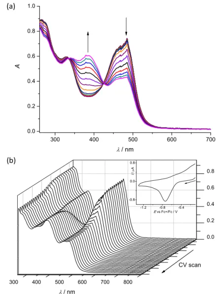
Cyclic voltammetry and spectroelectrochemistry. Cyclic voltammetric experiments with 0.5 mM solutions of 1–4 in 0.1 M nBu4NPF6 (puriss quality from Fluka; dried under reduced pressure at 70 °C for 24 h before use) supporting electrolyte in DMSO (SeccoSolv max. 0.025% H2O, Merck) were performed under argon atmosphere using a three-electrode arrangement with platinum wire as counter electrodes, and silver wire as pseudoreference electrode. Glassy carbon (GC) or platinum wire served as working electrodes. Ferrocene purchased from Sigma Aldrich was used as the internal potential standard without further purification. All potentials in voltammetric studies were quoted vs ferricenium/ferrocene (Fc+/Fc) redox couple. A Heka PG310USB (Lambrecht, Germany) potentiostat with a PotMaster 2.73 software package served for the potential control in voltammetric studies. In situ ultraviolet-visible-near-infrared (UV‒vis‒NIR) spectroelectrochemical measurements were performed on a spectrometer (Avantes, Model AvaSpec-
Dalton Transactions Accepted Manuscript
Open Access Article. Published on 10 May 2019. Downloaded on 5/14/2019 5:52:00 AM. This article is licensed under a Creative Commons Attribution-NonCommercial 3.0 Unported Licence.
View Article Online DOI: 10.1039/C9DT01238A
2048x14-USB2 in the spectroelectrochemical cell kit (AKSTCKIT3) with the Pt- microstructured honeycomb working electrode, purchased from Pine Research Instrumentation. The cell was positioned in the CUV-UV Cuvette Holder (Ocean Optics) connected to the diode-array UV-vis-NIR spectrometer by optical fibers. UV- vis-NIR spectra were processed using the AvaSoft 7.7 software package. Halogen and deuterium lamps were used as light sources (Avantes, Model AvaLight-DH-S- BAL).
Cell lines. Human colonic adenocarcinoma cell lines Colo 205 doxorubicin-sensitive (ATCC-CCL-222) and Colo 320/MDR-LRP multidrug resistant over-expressing ABCB1 (MDR1)-LRP (ATCC-CCL-220.1) were purchased from LGC Promochem, Teddington, UK. The cells were cultured in RPMI 1640 medium supplemented with 10% heat-inactivated fetal bovine serum, 2 mM L-glutamine, 1 mM Na-pyruvate and 10 mM HEPES. The cell lines were incubated at 37 °C, in a 5% CO2, 95% air atmosphere. The semi-adherent human colon cancer cells were detached with Trypsin-Versene (EDTA) solution for 5 min at 37 °C. MRC-5 human embryonal lung fibroblast cell line (ATCC CCL-171) was purchased from LGC Promochem, Teddington, UK. The cells were cultured in Eagle’s Minimal Essential Medium (EMEM, containing 4.5 g/L glucose) supplemented with a non-essential amino acid mixture, a selection of vitamins and 10% heat-inactivated fetal bovine serum. The cell lines were incubated at 37 °C, in a 5% CO2, 95% air atmosphere.
Assay for cytotoxic effect. MRC-5 non-cancerous human embryonic lung fibroblast and human colonic adenocarcinoma cell lines (doxorubicin-sensitive Colo 205 and multidrug resistant Colo 320 colonic adenocarcinoma cells) were used to determine the effect of compounds on cell growth. The effects of increasing concentrations of compounds on cell growth were tested in 96-well flat-bottomed microtiter plates. The compounds were dissolved in DMSO and stock solutions of 10 mM were prepared.
These were further diluted in the appropriate cell culture medium by 2-fold serial dilution starting from 100 µM to 0.195 µM. The adherent human embryonal lung fibroblast cells were cultured in 96-well flat-bottomed microtiter plates, using EMEM supplemented with 10% heat-inactivated fetal bovine serum. The density of the cells was adjusted to 1104 cells in 100 μL per well, the cells were seeded for 24 h at 37
C, 5% CO2, then the medium was removed from the plates containing the cells, and
Dalton Transactions Accepted Manuscript
Open Access Article. Published on 10 May 2019. Downloaded on 5/14/2019 5:52:00 AM. This article is licensed under a Creative Commons Attribution-NonCommercial 3.0 Unported Licence.
View Article Online DOI: 10.1039/C9DT01238A
the dilutions of compounds previously made in a separate plate were added to the cells in 200 μL. In case of the colonic adenocarcinoma cells, the two-fold serial dilutions of compounds were prepared in 100 μL of RPMI 1640, horizontally. The semi-adherent colonic adenocarcinoma cells were treated with Trypsin-Versene (EDTA) solution. They were adjusted to a density of 1104 cells in 100 μL of RPMI 1640 medium, and were added to each well, with the exception of the medium control wells. The final volume of the wells containing compounds and cells was 200 μL. The culture plates were incubated at 37 °C for 72 h; at the end of the incubation period, 20 μL of MTT (thiazolyl blue tetrazolium bromide, Sigma) solution (from a stock solution of 5 mg/mL) were added to each well. After incubation at 37 °C for 4 h, 100 μL of sodium dodecyl sulfate (SDS) (Sigma) solution (10% in 0.01 M HCI) were added to each well and the plates were further incubated at 37˚C overnight. Cell growth was determined by measuring the optical density (OD) at 540/630 nm with Multiscan EX ELISA reader (Thermo Labsystems, Cheshire, WA, USA). Inhibition of the cell growth was determined according to the formula below:
IC50 =100 100
control medium
OD control cell
OD
control medium
OD sample OD
Results are expressed in terms of IC50, defined as the inhibitory dose that reduces the growth of the cells exposed to the tested compounds by 50%. The IC50 values were calculated using GraphPad Prism7 software.
Results and Discussion
Synthesis. The synthesis of the main core structure A in Chart 2 was performed by following the published literature protocols. B was synthesised similarly.18–20 A detailed description for the synthesis of B is given in the ESI†. The lactam derivatives A and B were converted into thiolactams C and D by reaction with P2S5 on Al2O3 in dry boiling acetonitrile or tetrahydrofuran in 82 and 60% yield, respectively.27 Thionation with phosphorus pentasulfide bound to aluminium oxide offers the advantage that the reagent can be removed by filtration after the reaction. In addition, this method was chosen because the standard method for the thionation of paullones10 gave only small yields. Further reaction of C and D with hydrazine hydrate as reagent and solvent afforded hydrazines E and F in 86 and 92% yield, respectively. It should be noted that that F exists in two tautomeric forms in DMSO solution, as evidenced by the appearance of two signal sets in the 1H NMR spectrum.
Dalton Transactions Accepted Manuscript
Open Access Article. Published on 10 May 2019. Downloaded on 5/14/2019 5:52:00 AM. This article is licensed under a Creative Commons Attribution-NonCommercial 3.0 Unported Licence.
View Article Online DOI: 10.1039/C9DT01238A
The predominant species is the one shown in Chart 2 where N13 is protonated and the double bond is endocyclic. The NMR signal of N13H appears as a singlet at δ = 6.36 ppm. In the minor species, (ca. 15% in DMSO), N6 is protonated and the double bond is exocyclic. N6H appears as a triplet with the typical coupling constant of 5.3 Hz at δ = 8.49 ppm. NMR data is given only for the major species in the experimental part, because of the low signal intensity of the minor species and signal overlapping.
For atom numbering scheme see Scheme S1, ESI†. Similar tautomerism has been observed in the case of paullones previously.14 The Schiff bases HL1–HL4 were prepared in 99, 31, 94 and 58% yield, respectively, from hydrazine species E and F and 2-formyl- and 2-acetylpyridine taken in 10% excess in boiling ethanol. 1H and 13C NMR spectra of HL1–HL4 were in agreement with the formulae proposed. Notably, HL4 showed a second set of NMR-signals with an intensity of about 10% of the main species in DMSO-d6. Tautomerism as in the case for F was excluded, because of the presence of the typical triplet signal for N6H in both species. It is likely that a mixture of E and Z isomers is present, similarly to previously reported paullone derived ruthenium(II) and osmium(II) complexes.17 However, low signal intensity and signal overlapping did not allow a more accurate analysis to be performed. ESI mass spectra measured in positive ion mode showed peaks with m/z 352.26, 432.06, 366.16 and 446.08 attributed to [M+H]+. The purity of HL1–HL4 (>95%) was confirmed by elemental analysis. The structure of HL4 was also established by single crystal X-ray diffraction (vide infra). Complexes 1‒4 were prepared by reaction of HL1–HL4 in isopropanol by addition of a methanolic solution of CuCl2·2H2O in 1:1 mol ratio, respectively, in 77 to 99% yields. The positive ion ESI mass spectrum of 1 showed the presence of a peak with m/z 764.18 due to [CuII(L1)(HL1)]+. The mass spectra of 2–4 contain peaks at m/z 528.97 and 492.99, 463.04 and 427.07, 542.96 and 507.00 attributed to [CuIICl(HL2–4)]+ and [CuII(L2–4)]+, respectively. The elemental analysis was in agreement with the structures shown in Chart 2, providing the required purity of bulk samples for biological investigations. The complexes 1‒4 as well as 3´ have been studied by X-ray diffraction.
X-ray Diffraction. The results of X-ray diffraction studies for 1trim, 2, 3, 3´, 4 and HL4 are shown in Figures 1–3, respectively, with selected bond lengths and bond angles quoted in the legends. Complex 1 crystallised as a trimer (1trim), while 2–4 and 3´as monomeric entities. The corresponding ligand is protonated in 1trim, 2–4 and acts as
Dalton Transactions Accepted Manuscript
Open Access Article. Published on 10 May 2019. Downloaded on 5/14/2019 5:52:00 AM. This article is licensed under a Creative Commons Attribution-NonCommercial 3.0 Unported Licence.
View Article Online DOI: 10.1039/C9DT01238A
a tridentate one being coordinated to copper(II) via azepine nitrogen, hydrazine nitrogen and pyridine nitrogen atoms. The ligands can be deprotonated in the presence of a base. In particular, deprotonation of HL3 in 3 in the presence of triethylamine was confirmed by X-ray diffraction study of 3´ (Figure 2b). The coordination geometry of copper(II) ion in the trimer (1trim) and in mononuclear complexes 2–4 is square-pyramidal or distorted square-pyramidal (see legends to Figures 1-3 quoting 5-descriptor values36 for each copper(II) ion), while in 3´ it is slightly distorted square-planar. The coordination polyhedron in each case is completed by two or one chlorido co-ligands. Note that the same coordination geometry was previously established for complexes 5 and 6 with paullone derivatives (5 = 0.10 (5) and 0.05 (6)).15 The bond lengths Cu–N5 = 2.038(3) and 2.0220(15), Cu–N14 = 1.982(3) and 1.9787(15), Cu–N17 = 2.062(3) and 2.0466(15) in 5 and 6, respectively, are significantly longer than those in 2 and 3 (see legends to Figures 1 and 2) by 0.01–0.05 and 0.005–0.04 Å, respectively, even though the metal binding moieties in the corresponding ligands are closely related. Overall a tighter binding of the ligand moiety in 2 and 3 is to be mentioned when compared to that in 5 and 6.
The proligand HL4 and the ligand HL4 in 4 adopt different configurations and distinct proton tautomeric forms as shown in Figure 3 and confirmed by the distribution of bond lengths (electron density) over the fragment C5–N6–C7–N13–N14–C15 quoted in the legend to Figure 3.
Dalton Transactions Accepted Manuscript
Open Access Article. Published on 10 May 2019. Downloaded on 5/14/2019 5:52:00 AM. This article is licensed under a Creative Commons Attribution-NonCommercial 3.0 Unported Licence.
View Article Online DOI: 10.1039/C9DT01238A
Figure 1. ORTEP views of a) the complex cation [Cu3Cl4(HL1)3]2+ in 1trim and b) of [CuCl2(HL2)] in 2. Selected bond distances (Å) and bond angles (deg) in 1trim: Cu1–
N6 1.994(2), Cu1–N14 1.991(2), Cu1–N17 2.044(2), Cu1–Cl1 2.2685(8), Cu1–Cl2 2.4382(8), N6–Cu1–N14 78.75(10), N14–Cu1–N17 77.92(9), Cu2–N27 1.990(2), Cu2–N35 1.975(2), Cu2–N38 2.034(2), Cu2–Cl2 2.4492(8), Cu2–Cl3 2.2781(8), N27–Cu2–N35 79.39(10), N35–Cu2–N38 78.63(10), Cu3–N48 1.995(3), Cu3–N56 1.959(2), Cu3–N59 2.044(2), Cu3–Cl3 2.6404(8), Cu3–Cl4 2.2116(8), N48–Cu3–N56 79.51(10), N56–Cu3–N59 79.20(10). Details of coordination geometry in 1trim: 5
(Cu1) = 0.35, 5 (Cu2) = 0.25, 5 (Cu3) = 0.17; in 2: Cu–N6 1.982(5), Cu–N14 1.971(5), Cu–N17 2.051(5), Cu–Cl1 2.2252(16), Cu–Cl2 2.5762(17), N6–Cu–N14 79.7(2), N14–Cu–N17 78.8(2). Details of coordination geometry in 2: 5 (Cu) = 0.06.
Of note is also that the two Cu–Cl bonds in 1–4 are markedly different. The bond length from copper(II) to basal chlorido co-ligand is by 0.17 to 0.43 Å shorter compared to that between copper(II) and the apical chlorido co-ligand. A similar
Dalton Transactions Accepted Manuscript
Open Access Article. Published on 10 May 2019. Downloaded on 5/14/2019 5:52:00 AM. This article is licensed under a Creative Commons Attribution-NonCommercial 3.0 Unported Licence.
View Article Online DOI: 10.1039/C9DT01238A
situation is also typical for 5 and 6 reported previously.15,25 The corresponding bonds differ by 0.19 and 0.13 Å in 5 and 6, respectively.
Figure 2. ORTEP views of a) one crystallographically independent complex [CuCl2(HL3)] in 3 (co-crystallised solvent is omitted) and b) of [CuCl(L3)] in 3´.
Selected bond distances (Å) and bond angles (deg) in 3: Cu1–N6 1.979(4), Cu1–N14 1.973(4), Cu1–N17 2.034(4), Cu1–Cl1 2.2460(12), Cu1–Cl2 2.4567(14), N6–Cu1–
N14 78.72(17), N14–Cu1–N17 78.33(16), Cu2–N28 1.986(5), Cu2–N36 1.969(4), Cu2–N39 2.031(4), Cu2–Cl3 2.2309(14), Cu2–Cl4 2.5054(13), N28–Cu2–N36 79.09(18), N36–Cu2–N39 78.36(18). Details of coordination geometry in 2: 5 (Cu1)
= 0, 5 (Cu2) = 0.06 (second crystallographically independent molecule); in 3´: Cu–N6 1.937(3), Cu–N14 1.953(3), Cu–N17 2.019(3), Cu–Cl 2.2058(9), N6–Cu–N14 79.52(12), N14–Cu–N17 80.27(12).
Dalton Transactions Accepted Manuscript
Open Access Article. Published on 10 May 2019. Downloaded on 5/14/2019 5:52:00 AM. This article is licensed under a Creative Commons Attribution-NonCommercial 3.0 Unported Licence.
View Article Online DOI: 10.1039/C9DT01238A
![Figure 1. ORTEP views of a) the complex cation [Cu 3 Cl 4 (HL 1 ) 3 ] 2+ in 1 trim and b) of [CuCl 2 (HL 2 )] in 2](https://thumb-eu.123doks.com/thumbv2/9dokorg/1064212.70365/21.892.220.677.98.732/figure-ortep-views-complex-cation-cu-trim-cucl.webp)
![Figure 2. ORTEP views of a) one crystallographically independent complex [CuCl 2 (HL 3 )] in 3 (co-crystallised solvent is omitted) and b) of [CuCl(L 3 )] in 3´](https://thumb-eu.123doks.com/thumbv2/9dokorg/1064212.70365/22.892.257.637.207.659/figure-ortep-crystallographically-independent-complex-crystallised-solvent-omitted.webp)
![Figure 3. ORTEP views of a) proligand HL 4 and b) of [CuCl 2 (HL 4 )] in 4 (co- (co-crystallised solvent is omitted)](https://thumb-eu.123doks.com/thumbv2/9dokorg/1064212.70365/23.892.227.669.100.584/figure-ortep-views-proligand-cucl-crystallised-solvent-omitted.webp)


![Highly Antiproliferative Latonduine and Indolo[2,3‑c]quinoline Derivatives: Complex Formation with Copper(II) Markedly Changes the Kinase Inhibitory Profile](data:image/gif;base64,R0lGODlhAQABAIAAAP///wAAACH5BAEAAAAALAAAAAABAAEAAAICRAEAOw==)