Highly Antiproliferative Latonduine and Indolo[2,3 ‑c ]quinoline Derivatives: Complex Formation with Copper(II) Markedly Changes the Kinase Inhibitory Pro fi le
Christopher Wittmann,
#Felix Bacher,
#Eva A. Enyedy, Orsolya Dömötör, Gabriella Spengler, Christian Madejski, Jóhannes Reynisson, and Vladimir B. Arion*
Cite This:https://doi.org/10.1021/acs.jmedchem.1c01740 Read Online
ACCESS
Metrics & More Article Recommendations*
sı Supporting InformationABSTRACT:
A series of latonduine and indoloquinoline derivatives HL
1−HL8and their copper(II) complexes (1−8) were synthesized and comprehensively characterized. The structures of
five compounds (HL
6, [CuCl(L
1)(DMF)]
·DMF, [CuCl(L
2)- (CH
3OH)], [CuCl(L
3)]·0.5H
2O, and [CuCl
2(H
2L
5)]Cl·2DMF) were elucidated by single crystal X-ray di
ffraction. The copper(II) complexes revealed low micro- to sub-micromolar IC
50values with promising selectivity toward human colon adenocarcinoma multidrug-resistant Colo320 cancer cells as compared to the doxorubicin-sensitive Colo205 cell line. The lead compounds HL
4and 4 as well as HL
8and 8 induced apoptosis e
fficiently in Colo320 cells. In addition, the copper(II) complexes had higher a
ffinity to DNA than their metal-free ligands. HL
8showed selective inhibition for the PIM-1 enzyme, while 8 revealed strong inhibition of
five other enzymes, i.e., SGK-1, PKA, CaMK-1, GSK3
β, and MSK1, from a panel of 50 kinases. Furthermore, molecular modeling of the ligands and complexes showed a good
fit to the binding pockets of these targets.
1. INTRODUCTION
Indolobenzazepines and indoloquinolines are fused heterocyclic sca
ffolds, which have gained a considerable interest in the
field of medicinal chemistry.
1−17Indolo[3,2-d]benzazepines or paul- lones (backbone A in
Chart 1),first synthesized in 1992, were discovered as potential inhibitors of cyclin-dependent kinases (Cdks)
7,18,19with antiproliferative activity similar to that of
flavopiridol, the
first Cdk-inhibitor that reached clinical trials as an anticancer drug. Later, other possible targets have been identi
fied, namely, sirtuins,
20,21GSK3
β,
18,19,22,23and mitochon- drial malate dehydrogenase.
23Indolo[3,2-c]quinolines (back- bone B in
Chart 1) are known to induce apoptosis in cancercells. DNA intercalation and poisoning topoisomerase I/II (topo I/II) are considered the mechanisms of action.
24−26Furthermore, some of the indolo[3,2-c]quinolines are e
ffective and selective KRAS-mutated oncogene G-quadruplex stabilizers causing cancer cell apoptosis.
27Indolo[3,2-d]benzazepines are non-planar heterocyclic systems due to the sp
3-hybridized methylene carbon atom in the seven-membered azepine ring,
whereas the indolo[3,2-c]quinolines are planar, making them e
ffective DNA intercalators and/or topo I/II inhibitors.
Latonduines (backbones C and D in
Chart 1) were first extracted from the Indonesian sponge Stylissa carteri and are not cytotoxic to cancer cells.
28,29However, substitution of their pyrrole ring by an indole unit made them cytotoxic.
30,31The resulting indolo[2,3-d]benzazepine (backbone E in
Chart 1) is amicrotubule destabilizing agent (MDA) targeting the colchicine binding site.
30The two isomeric backbones indolo[3,2- d]benzazepine A and indolo[2,3-d]benzazepine E are related structurally as shown in
Chart 1. Nevertheless, byflipping the indole moiety and shifting the lactam unit in paullone A, one
Received: October 7, 2021© XXXX The Authors. Published by American Chemical Society
A
https://doi.org/10.1021/acs.jmedchem.1c01740 J. Med. Chem.XXXX, XXX, XXX−XXX
Downloaded via 188.156.253.87 on February 2, 2022 at 04:51:47 (UTC). See https://pubs.acs.org/sharingguidelines for options on how to legitimately share published articles.
obtains not only increased cytotoxicity but also a di
fferent mode of action.
30Being intrigued by the activity of indolo[3,2-d]benzazepine- and indolo[3,2-c]quinoline-based molecules as potential anti- cancer drugs, we decided to extend our chemistry to other related isomeric systems, namely, indolo[2,3-d]benzazepine- and indolo[2,3-c]quinoline-derived species, with unexplored chemistry and biological e
ffects. We envisioned exciting new results in the
field of metal-based anticancer drugs and, in particular, new structure
−activity relationships.
One of the major drawbacks of these isomeric sca
ffolds is their limited aqueous solubility and bioavailability. This issue was successfully addressed for many indolo[3,2-d]benzazepine and indolo[3,2-c]quinoline derivatives and several latonduines by creating metal binding sites at their backbones and metal complex formation. Werner-type coordination complexes of copper(II), ruthenium(II), osmium(II), gallium(III), and organometallic compounds were synthesized and investigated as potential anticancer drugs.
32−40The reported results revealed that the metal complexes did not only enhance the aqueous solubility and bioavailability but also augmented their antiproliferative activity both in vitro and in vivo. Nevertheless, bioavailability and aqueous solubility need further improve- ment, requiring other approaches to enhance their pharmaco- logical pro
file.
Morpholine, as a known biologically active moiety, has been attached to the main sca
ffolds since it is considered to improve the necessary pharmacological parameters of drug candi- dates.
41,42In our recent paper,
43we reported that the Schi
ffbase resulted from condensation of the 11-bromo-7-hydrazin-yl derivative of E (Chart 1) with 2-acetylpyridine and its copper(II) complex showed the highest cytotoxicity among the compounds tested. This prompted us to further develop this backbone and prepare 2-acetylpyridine with a morpholine unit.
As a starting material, the respective aldehyde was used, which was recently reported by us.
44Protein kinases represent an excellent target for cancer therapy.
45−49It should be also stressed that the multitargeted kinase inhibitors have become a
“hot topic
”, accounting for about 25% of drug discovery research.
48,49Initial attempts to create highly selective mono-kinase inhibitors to avoid unexpected toxic e
ffects have been steadily displaced by two anticancer therapies that target several kinases and block distinct kinase signaling pathways as they showed therapeutic bene
fits in
the treatment of complex cancer diseases.
46The
first therapy is based on using several selective mono-kinase inhibitors simultaneously, while the second is based on using a single drug as a multikinase inhibitor. Advantages and hurdles of both therapies have been discussed in the literature.
46,48,49The second therapy, which implies the use of a single drug as a multikinase inhibitor that revealed higher potency, permits avoiding the consequences of drug
−drug interactions, which can a
ffect absorption, metabolism, excretion, plasma level, and,
finally, activities, as well as reducing side e
ffects and is much easier to apply.
46Herein, we report the synthesis and characterization of new chelating systems derived from indolo[2,3-d]benzazepine E and indolo[2,3-c]quinoline F and of their copper(II) complexes (Chart 2), speciation in aqueous solution, and antiproliferative
activity. The inhibition ability of the lead compounds in a panel of 50 kinases was investigated in vitro and by molecular modeling, providing insights into the mode of action of the most potent copper(II) complex and its metal-free ligand. These modified molecules offer a broad spectrum of various effects on malign cells, while some of them show a marked increase in aqueous solubility, thus increasing the bioavailability and improving the pharmacological pro
file. Lead compound 8 and its proligand HL
8do not only target cancer specific kinases but also o
ffer an excellent pharmacological pro
file.
2. RESULTS AND DISCUSSION
2.1. Synthesis and Characterization of Starting Building Blocks and Ligands.
The aldehyde G prepared as reported elsewhere
44was converted into secondary alcohol I by reaction with the Grignard reagent (CH
3MgBr) and workup in 92% yield. Swern oxidation of I resulted in ketone J (Scheme 1), which was puri
fied chromatographically to give an easily crystallizable product in 69% yield.
The
1H NMR spectrum of J agreed with the expected structure, which, in addition, has been con
firmed by SC-XRD (see
Chart S1for atom numbering scheme and
Figure S1in the Supporting Information).
The derivative IVb (Scheme 2) has not been reported previously. Its synthesis has been performed by following the procedures described in the literature for unsubstituted indolo[2,3-c]quinoline IVa
50as shown in
Scheme 2. In the first step, ethyl 5-bromo-1-ethoxymethyl-1H-indol-2-carboxy- late was allowed to react with 2-iodoaniline in the presence of Chart 1. Indolo[3,2-d]benzazepine (A, Paullone),
Indolo[3,2-c]quinoline (B), Naturally Occurring
Latonduines C and D, Indolo[2,3-d]benzazepine (E), and Indolo[2,3-c]quinoline (F)
Chart 2. Copper(II) Complexes 1
−8 and Their Metal-Free Ligands HL
1−HL
8https://doi.org/10.1021/acs.jmedchem.1c01740 J. Med. Chem.XXXX, XXX, XXX−XXX B
AlMe
3in dichloromethane (DCM) to give Ib in 84% yield.
Protection of carboxamide nitrogen atom and isolation of IIb were realized in 99% yield by treatment of Ib with di-tert-butyl dicarbonate Boc
2O in dry acetonitrile in the presence of catalytic amount of N,N-dimethyl-4-aminopyridine (DMAP). The intramolecular Heck cyclization reaction of IIb in the presence of Pd(OAc)
2, PPh
3, and Ag
2CO
3followed by workup a
fforded IIIb in 19% yield. Full deprotection of IIIb and formation of IVb were accomplished in 79% yield by re
fluxing IIIb in EtOH:12 M HCl 4:1.
Then, compounds IVa and IVb were chlorinated with excess POCl
3at 120
°C to give rise to Va/Vb in >90% yield. Finally, the treatment of Va/Vb with excess hydrazine hydrate at re
flux delivered the desired species M and N in >95% yield. This pathway to create chelating molecules with some modi
fications was also successful with core structures A and B (Chart 1).
51,52The potential ligands HL
1−HL
8were synthesized by Schi
ffbase condensation reactions of hydrazin-yl derivatives K
−N with aldehyde G or ketone J in anoxic ethanol (Scheme 3) in 57
−98% yields by adapting literature protocols.
43,51,531
H NMR spectra of the potential ligands HL
1−HL
8show the typical peak pattern of the morpholine unit at around 2.35 and 3.55 ppm, as well as proton resonances of the linking methylene group between the morpholine unit and the pyridine ring at around 3.55 ppm sometimes overlapping with H
25for indolo[2,3-d]benzazepines or H
24for indolo[2,3-c]quinolines, respectively (for atom numbering scheme, see
Chart S2). Theadditional methyl group as R
2in
Scheme 3is seen as a singlet at around 2.49 ppm. The 2D NMR spectra provided evidence that indolo[2,3-d]benzazepines HL
1−HL
4are solely present as
tautomers with an exocyclic double bond between C
7and N
13as evidenced by weak coupling between H
6and C
5and triplet resonance in the
1H NMR spectra, suggesting the presence of two protons in the closest vicinity of H
6.
43There were no other tautomeric forms identi
fied. In contrast, NMR spectra of HL
5−HL
8indicate that these indolo[2,3-c]quinolines exist in two tautomeric forms in the solution. The major species possesses an exocyclic double bond with a hydrogen atom at N
5, while the minor species contains an endocyclic double bond with a hydrogen atom at N
12. The ratio between these two species is solvent- and concentration-dependent. At a concentration of about 10 mg/mL in DMSO-d
6, the ratios between the major vs minor species are 1:0.02, 1:0.01, 1:0.80 and 1:0.15 for HL
5−HL
8, respectively. In most cases, a complete assignment of all
1H and
13C resonances was impeded by low signal intensity for minor species and signal overlapping. However, the high signal intensity of the minor species in the case of HL
7made a complete assignment of all signals in both species possible. The chemical shift of NH
5for the species with an exocyclic double bond is 11.97 ppm, while that of NH
12for the species with an endocyclic double bond is 14.54 ppm. This was con
firmed by a long-range
1H
−13C HMBC 2D NMR experiment, where the proton N
5H showed
3J couplings to quaternary carbons C
11cand C
6a, while N
12H revealed such couplings to quaternary carbon C
6aand ternary carbon C
14, which is possible, if the hydrogen is bound to a hydrazinic nitrogen. The structural change in the molecule from an exocyclic to an endocyclic double bond leads to a shift of all
1H and
13C signals. While the
1H and
13C resonances of the morpholine moiety are only marginally a
ffected, those near the hydrazinic moiety show major changes.
In particular, the signals for the hydrogen C
14H and imine carbon are up
field shifted from 8.52 to 7.57 ppm and from 152.19 to 131.14 ppm, respectively, when going from major to minor species (see
Chart S2for the NMR atom numbering scheme in the
Supporting Information). At close inspection ofthe
1H NMR spectrum of HL
7, two more sets of NMR signals with low intensities become apparent, which are presumably attributed to E and Z isomers of the previously described tautomers, leading to a total of four signal sets. However, low signal intensity, signal overlapping, and the absence of non-cross Scheme 1. Synthesis of 2-Acetyl-5-(morpholinomethyl)-
pyridine J Starting from 2-Formyl-5-(morpholinomethyl)- pyridine G
44aaReagents and conditions: (i) MeMgBr, THFdry, 0°C; (ii) (COCl)2, DMSOdry, NEt3, DCMdry,−80°C.
Scheme 2. Synthesis of M and N
aaReagents and conditions: (i) AlMe3, 2-iodoaniline, CH2Cl2dry,−20°C to RT, 19 h; (ii) Boc2O, DMAP, CH3CNdry, RT, 72 h; (iii) Pd(OAc)2, PPh3, Ag2CO3, DMFdry, 100°C, 2 h; (iv) EtOH/HCl, 100°C, 16 h; (v) POCl3, 120°C, 16 h; (vi): N2H4·H2O 131°C, 16 h.
https://doi.org/10.1021/acs.jmedchem.1c01740 J. Med. Chem.XXXX, XXX, XXX−XXX C
peaks in a two-dimensional
1H
−1H NMR NOESY experiment made the identi
fication of isomers di
fficult, if at all possible.
2.2. Synthesis and Characterization of Metal Com- plexes.
Copper(II) complexes 1−8 were prepared by reactions of CuCl
2·2H2O with HL
1−HL8in a boiling mixture of methanol/isopropanol, a procedure used previously for the synthesis of copper(II) complexes with related Schi
ffbases without a morpholine moiety.
32,43The formation of copper(II) complexes 1
−8 was con
firmed by positive ion ESI mass spectra with peaks at m/z 512.14 and 548.11 attributed to [Cu
II(L
1)]
+and [Cu
IICl(HL
1)]
+, respectively (for 1), m/z 592.07 assigned to [Cu
II(L
2)]
+(for 2), at m/z 562.23 attributed to [Cu
IICl- (HL
3)]
+(for 3), at m/z 498.27 assigned to [Cu
II(L
5)]
+(for 5), at m/z 578.14 attributed to [Cu
II(L
6)]
+(for 6), at m/z 512.20 assigned to [Cu
II(L
7)]
+(for 7), and m/z 592.16 attributed to [Cu
II(L
8)]
+(for 8). The negative ion ESI mass spectrum of 4 showed a strong peak at m/z 640.03, which could be easily assigned to [Cu
IICl(L
4)
−H
+]
−. The reaction of HL
8with NiCl
2·6H
2O in methanol in a 1:1 molar ratio delivered a 1:2 nickel-to- ligand complex instead of the desired complex of 1:1 stoichiometry. Recrystallization of this product from DMF a
fforded crystals of the composition [Ni(L
8)(HL
8)]Cl
·2DMF, the structure of which was determined by SC-XRD. Starting from HL
7and NiCl
2·6H
2O in methanol in a 2:1 molar ratio, the complex [Ni(HL
7)
2]Cl
2·H2O was synthesized. The ESI mass spectrum revealed a doubly charged peak at m/z 479.18 corresponding to [Ni(HL
7)
2]
2+.
Elemental analyses were in good agreement with the composition proposed for all isolated complexes, attesting the purity (
≥95%) required for biological assays. The coordination geometry was con
firmed by SC-XRD measurements of complexes [CuCl(L
1)(DMF)]·DMF, [CuCl(L
2)(CH
3OH)], [CuCl(L
3)]·0.5H
2O, and [CuCl
2(H
2L
5)]Cl·2DMF.
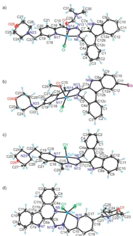
2.3. X-ray Crystallography.
The results of SC-XRD studies of complexes [CuCl(L
1)(DMF)]·DMF, [CuCl(L
2)- (CH
3OH)], [CuCl(L
3)]·0.5H
2O, and [CuCl
2(H
2L
5)]Cl·
2DMF are shown in
Figure 1, while those for the metal-freeindolo[2,3-c]quinoline HL
6and [Ni(L
8)(HL
8)]Cl·2DMF are shown in
Figures S3 and S4, respectively, with pertinent bonddistances (Å), bond angles, and torsion angles (deg) quoted in the legends. Details of data collection and re
finement are given in
Table S1. The complexes crystallized in the monoclinic spacegroups C2/c, P2
1/c, P2
1/c, and non-centrosymmetric triclinic P1, respectively.
The coordination geometry of Cu(II) in [CuCl(L
1)(DMF)]
(Figure 1a) is four-coordinate square-planar, even though very weak coordination of DMF molecule can be considered, taking into account the apical position of oxygen atom O1 with respect to the basal plane determined by the metal ion, the coordinated three nitrogen donor atoms, and the chlorido co-ligand. In this latter case, the coordination geometry can be interpreted as 4 + 1 binding. Comparison with coordination geometry in [CuCl- (L
2)(CH
3OH)] (Figure 1b), which is best described as
five- coordinate square-pyramidal, and bond lengths around copper- (II), which are signi
ficantly expanded when compared to those in [CuCl(L
1)(DMF)], provides further evidence for a more appropriate description of coordination geometry in [CuCl- (L
1)(DMF)] as four-coordinate. The increase in coordination number in [CuCl(L
2)(CH
3OH)] to
five leads to a signi
ficant expanding of interatomic distances between Cu(II) and donor atoms due to increase in interatomic repulsions. The latonduine backbone in both complexes has almost identical folding due to the presence of an sp
3-hybridized carbon atom in the seven- membered azepine ring. The torsion angle
ΘC4a−C5−N6−C7is almost the same in both complexes (see the values quoted in the legends to
Figures 1a,b).Complex [CuCl(L
3)] forms a weak dimeric associate (Figure
S2), in which the coordination environment of Cu1 can bedescribed as slightly distorted square-planar (see also
Figure 1c).The atom Cl1 acts as a bridging
μ-chlorido co-ligand to Cu2 of the second half of the dimeric associate with formation of a long contact of 2.8970(8) Å. Therefore, the coordination geometry of Cu2 can be described as 4 + 1 as was the case for complex [CuCl(L
1)(DMF)]. The latonduine derivatives adopt the same binding mode to both Cu(II) atoms Cu1 and Cu2, and each acts as a monoanionic tridentate ligand. The bond lengths in each chromophore of the two Cu(II) ions are very similar to those in [CuCl(L
1)(DMF)], in accordance with small structural di
ffer- ence between the two coordinated ligands (L
1)
−and (L
3)
−.
In contrast to Cu(II) compounds with strongly folded indolo[2,3-d]benzazepine backbone E ([CuCl(L
1)(DMF)]
·DMF, [CuCl(L
2)(CH
3OH)], and [CuCl(L
3)]
·0.5H
2O), Cu- (II) in [CuCl
2(H
2L
5)]Cl
·2DMF is coordinated by a
flat indolo[2,3-c]quinoline-based tridentate ligand protonated at the nitrogen atom N1 of the morpholine moiety. The positive global charge of the complex cation (Figure 1d) is counter- balanced by a chloride anion. The brominated backbone in the metal-free ligand HL
6is also
flattened and stabilized by two intramolecular H-bonding interactions (Figure S3) N7
−H
···Scheme 3. Synthesis of Proligands HL
1−HL
8https://doi.org/10.1021/acs.jmedchem.1c01740 J. Med. Chem.XXXX, XXX, XXX−XXX D
N13 [N7
···N13 = 2.725(3) Å;
∠N7HN13 = 122(2)
°] and N12
−H
···N16 [N12
···N16 = 2.751(3) Å;
∠N12HN16 = 131(2)
°]. The coordination geometry in [CuCl
2(H
2L
5)]
+is
five-coordinate and can be described as intermediate (
τ5= 0.40) between square-pyramidal (
τ5= 0) and trigonal bipyramidal (
τ5= 1).
54The indolo[2,3-d]benzazepine backbone in [CuCl(L
1)- (DMF)]
·DMF, [CuCl(L
2)(CH
3OH)], and [CuCl(L
3)]
·0.5H
2O is folded due to the presence of one sp
3-hybridized carbon atom in the seven-membered azepine ring. The dihedral angles between the mean plane through C1
−C2
−C3
−C4
−C4a
−C12c and Cu
−N6
−C7
−N13
−N14 are of 121.5, 108.3, and 112.3
°for [CuCl(L
1)(DMF)]
·DMF, [CuCl(L
2)- (CH
3OH)], and [CuCl(L
3)]
·0.5H
2O, respectively. These are
larger than those in copper(II) complexes with closely related Schi
ffbases based on indolo[3,2-d]benzazepine (paullone), reported previously of 99.6
−102.2
°(see
Chart S3in the Supporting Information).
33The presence of a six-membered pyridine-like ring in [CuCl
2(H
2L
5)]
+and HL
6instead of a seven-membered azepine ring makes these indoloquinoline systems
flat, a premise to intercalate into DNA. The SC-XRD studies revealed that HL
1−HL
3and HL
5act as tridentate ligands but adopt di
fferent protonation states depending on conditions.
Therefore, it was of interest to study the solubility of metal-free ligands and Cu(II) complexes as well as their protonation state at physiological pH.
2.4. Solubility Studies.
The thermodynamic solubility of selected metal-free ligands and Cu(II) complexes was characterized in water at pH 7.4 and 5.0 using UV
−vis spectrophotometry for the determination of the concentration of the saturated solutions. The determined solubility (S) values are shown in
Figure 2.The obtained solubility values indicate that the copper(II) complexes are commonly more soluble in water than their corresponding metal-free ligands. The positive e
ffect of the morpholine moiety on the solubility is measurable but minor.
Comparison of the S values for the HL
5and HL
5nm, 5 and 5
nm, and 6 and 6
nmpairs under the conditions used indicates that all compounds have signi
ficantly better solubility at pH 5 than at pH 7.4. For HL
5and HL
5nm, this can be explained by the partial protonation of the morpholine nitrogen and pyridine nitrogen with the decreasing pH. The morpholine-containing complexes (3 and 5
−8) also can get partially protonated at the non- coordinating morpholine nitrogen when the pH is lowered.
However, formation of the aqua complex from the mixed hydroxido species (vide inf ra) can also contribute to the increased solubility even for the non-morpholine complexes at pH 5. The presence of the bromo-substituent (5 vs 6, 5
nmvs 6
nm, and 7 vs 8) results in a considerable decrease of the aqueous solubility at both tested pH values, while the e
ffect of the methyl group at the Schi
ffbase ketimine bond is not signi
ficant.
2.5. Solution Equilibrium Studies.
The protonation processes of HL
1nmand its morpholine counterpart HL
1(Chart 3) and the solution stability of their copper(II) complexes (1
nmand 1) were characterized by UV
−vis spectrophotometry. Since the organic compounds and their copper(II) complexes possess limited aqueous solubility, the solution equilibrium studies were performed in 30% (w/w) DMSO at low concentrations (12.5
μM (HL
1nm) or 50
μM (HL
1)). Based on the characteristic changes in the UV
−vis spectra for HL
1nmin the pH range 2
−6 (Figure 3a), two
Figure 1.ORTEP views of (a)[CuCl(L1)(DMF)], (b)[CuCl(L2)-(CH3OH)], (c)[CuCl(L3)], and (d)[CuCl2(H2L5)]+with thermal ellipsoids at a 50% probability level. Selected bond distances (Å), bond angles (deg), and torsion angles (deg) in (a): Cu−N6 1.949(2), Cu−
N14 1.963(2), Cu−N17 2.017(2), Cu−Cl 2.2355(9), Cu−O1 2.582(2); N6−C7 1.321(4), C7−N13 1.355(4), N13−N14 1.364(3), N14−C15 1.289(3), C15−C16 1.452(4), C16−N17 1.359(4),ΘC4a−C5−N6−C7−71.7(3); in (b): Cu−N6 1.9590(15), Cu−
N14 1.9724(14), Cu−N17 2.0301(14), Cu−Cl 2.2555(4), Cu−O1 2.3493(12); N6−C7 1.322(2), C7−N13 1.366(2), N13−N14 1.354(2), N14−C15 1.290(2), C15−C16 1.455(2), C16−N17 1.366(2), ΘC4a−C5−N6−C7−72.84(19); in (c): Cu−N6 1.9407(19), Cu−N14 1.969(2), Cu−N17 2.0107(18), Cu−Cl 2.2475(7), N6−C7 1.305(3), C7−N13 1.374(3), N13−N14 1.364(3), N14−C15 1.302(3), C15−C16 1.479(3), C16−N17 1.353(3),ΘC4a−C5−N6−C7− 73.6(3); and in (d): Cu−N5 2.013(8), Cu−N13 1.997(7), Cu−N16 2.024(7), Cu−Cl1 2.364(2), Cu−Cl2 2.281(2), N5−C6 1.334(12), C6−N12 1.383(11), N12−N13 1.350(9), N13−C14 1.281(11), C14−
C15 1.458(12), C15−N16 1.366(11),ΘC4a−N5−C6−C6a1.4(13).
Figure 2.Thermodynamic solubility (S) determined for selected metal- free ligands and complexes using UV−vis spectral analysis at pH 5 (20 mM MES buffer) and 7.4 (20 mM HEPES) in water (T= 298 K).
https://doi.org/10.1021/acs.jmedchem.1c01740 J. Med. Chem.XXXX, XXX, XXX−XXX E
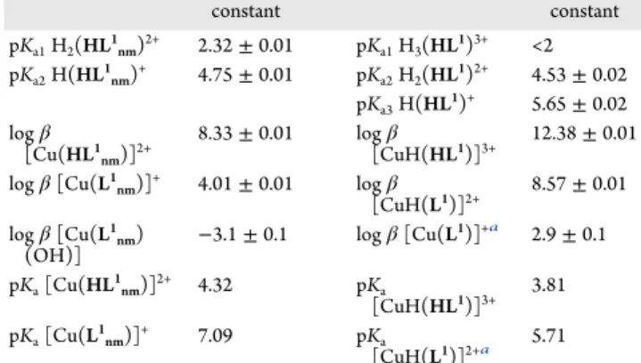
relatively well-separated proton dissociation processes were observed and their pK
avalues were determined (Table 1).
Notably, upon increasing the pH to >∼6.6 precipitation occurred in the solution, leading to the elevation of the base line most probably due to the formation of the neutral metal-free ligand species. The
first proton dissociation step was accompanied by a blueshift (λ
max: 352 nm
→342 nm) in the pH range between 2 and 3.45, while the
λmaxis redshifted (342 nm
→377 nm) upon the second step. These spectral changes and the spectra of the individual ligand species (Figure 3b) calculated by deconvolution of the recorded UV
−vis spectra are fairly similar to those found for the ketimine derivative of HL
1nmin our recent work.
43Thus, a similar deprotonation pattern is feasible for the non-substituted and the methyl-substituted ligands. The neutral species HL
1nmcan be present in two tautomeric forms (due to the rearrangement of the N
C
−NH−N and NH−CN−N bonds) and can be protonated at two sites (Scheme 4). Therefore, the
first proton dissociation step (pK
a1) is attributed to deprotonation of the pyridinium nitrogen, while the second process (pK
a2) is attributed to the deprotonation of the benzazepinium nitrogen. The ligand HL
1nmpossesses somewhat lower pK
avalues than its methyl derivative as a result of the electron-donating property of the methyl group.
Two pK
avalues were determined for HL
1as well (Table 1) by the deconvolution of the recorded spectra (Figure 3b), although the spectral changes were different. This ligand contains also the morpholinium group (Scheme 4). By the careful analysis of the spectral changes, it is suggested that the deprotonation of the pyridinium nitrogen takes place at fairly acidic pH, and its pK
avalue is lower compared to that of HL
1nmas a result of the electron-withdrawing e
ffect of the protonated morpholinium moiety. Even though the deprotonation of the benzazepinium and morpholinium nitrogens is overlapping, the spectra of the individual species (Figure 3d) suggest that pK
a2mostly belongs to the benzazepinium moiety, and the deprotonation of the non- Chart 3. Metal-Free Ligands HL
1and HL
1nmUsed for the
Solution Equilibrium Studies
Figure 3.UV−vis spectra for (a)HL1nmand (b)HL1at various pH values in a 30−70% (w/w) DMSO−water solvent mixture in addition to the computed molar absorbance spectra of the proligand species in the different protonation states: (c)HL1nmand (d)HL1. (cligand= 12.5μM (a), 50μM (b);T= 298 K;I= 0.10 M (KCl); l = 2 cm (a), 0.5 cm (b)).
Table 1. Proton Dissociation Constants (pK
a) of HL
1nmand HL
1, along with Overall Stability Constants (log
β) of TheirCopper(II) Complexes Determined by UV
−vis Titrations in a 30−70% (w/w) DMSO−Water Solvent Mixture (T = 298 K;
I
= 0.10 M (KCl))
constant constant
pKa1H2(HL1nm)2+ 2.32±0.01 pKa1H3(HL1)3+ <2 pKa2H(HL1nm)+ 4.75±0.01 pKa2H2(HL1)2+ 4.53±0.02
pKa3H(HL1)+ 5.65±0.02 logβ
[Cu(HL1nm)]2+
8.33±0.01 logβ
[CuH(HL1)]3+
12.38±0.01 logβ[Cu(L1nm)]+ 4.01±0.01 logβ
[CuH(L1)]2+ 8.57±0.01 logβ[Cu(L1nm)
(OH)] −3.1±0.1 logβ[Cu(L1)]+a 2.9±0.1 pKa[Cu(HL1nm)]2+ 4.32 pKa
[CuH(HL1)]3+
3.81 pKa[Cu(L1nm)]+ 7.09 pKa
[CuH(L1)]2+a 5.71
aFormation of [Cu(L1)]+and [CuH(L1)(OH)]+is overlapping; the two species cannot be well distinguished.
https://doi.org/10.1021/acs.jmedchem.1c01740 J. Med. Chem.XXXX, XXX, XXX−XXX F
chromophoric morpholinium nitrogen is accompanied by a minor spectral change, as expected.
The pK
avalues collected in
Table 1indicate that the neutral form (HL) predominates at physiological pH in solution in the case of both ligands, contributing to their strong lipophilic nature (log D
7.4values +4.75 (HL
1nm) and +4.30 (HL
1) estimated by the MarvinSketch program.
55The UV−vis spectra recorded for 1
nmin the pH range 2−11 (Figure 4a) showed strong similarities to those of the methylated complex reported recently.
43The spectrum measured at pH 2 suggests a considerable extent of complex formation. The tridentate coordination via the N,N,N donor set is assumed in [Cu(HL
1nm)]
2+at such low pH. Increase in the pH
is accompanied by
λmaxshift to a lower wavelength (345 nm
→316 nm) parallel with the development of a new band at 449 nm, suggesting a signi
ficant rearrangement in the coordination sphere. The deprotonation of the non-coordinating hydrazonic nitrogen is likely, and complex [Cu(L
1nm)]
+is formed. Further increase in the pH (pH > 7) resulted in a bathochromic shift (
λmax≈462 nm), indicating a new process, namely the formation of the neutral mixed hydroxido species [Cu(L
1nm)(OH)].
However, the absorbance is decreased in the whole wavelength range due to precipitate formation. By the deconvolution of the recorded UV
−vis spectra, overall stability constants for the complexes [Cu(HL
1nm)]
2+and [Cu(L
1nm)]
+were computed, and log
βfor [Cu(L
1nm)(OH)] was only estimated (Table 1). In the case of the morpholine hybrid, a somewhat di
fferent speciation was obtained as the presence of the pendant morpholine moiety had to be taken into consideration. Log
βvalues for [CuH(HL
1)]
3+, [CuH(L
1)]
2+, and [Cu(L
1)]
+were computed from the spectrophotometric titration data (Table 1).
Since the molar absorbance spectra of [CuH(HL
1)]
3+and [CuH(L
1)]
2+complexes resemble those of [Cu(HL
1nm)]
2+and [Cu(L
1nm)]
+(Figure 4b), the same coordination modes are likely in these species in couples. Therefore, the deprotonation of [CuH(HL
1)]
3+takes place most probably at the hydrazinic nitrogen (characterized by a pK
aof 3.81) and the morpholinium nitrogen remains protonated. Increasing the pH (pH > 5), the absorbance is decreased in the whole wavelength range, no isosbestic points are found (indicating the formation of some precipitate), while the
λmaxis increased. Formation of a mixed hydroxido species [CuH(L
1)(OH)]
+is also possible. However, the deprotonation of the morpholinium group can take place as well (formation of [Cu(L
1)]
+) in this pH range. As these two processes are overlapping, the two species could not be well distinguished, and the obtained stability constant is quite uncertain.
Using the determined stability constants, concentration distribution curves were computed (Figure 5) for both copper(II)
−ligand systems. The results imply that the complexes do not dissociate at the used 12.5
μM concertation at neutral pH (the fraction of the free metal ion is negligible).
However, the concentration of the free metal ion is significantly higher in the acidic pH range than it was found for the methylated complex, suggesting the somewhat lower stability of Scheme 4. Stepwise Proton Dissociation Processes of HL
1and Its Tautomeric Forms
Figure 4.(a) UV−vis spectra recorded for complex1nmat various pH values. (b) Molar absorbance spectra computed for selected complex 1nm(black lines) and1(gray lines) in the various protonation states.
(ccomplex= 12.5μM;T= 298 K;I= 0.10 M (KCl); 30% (w/w) DMSO−
H2O).
https://doi.org/10.1021/acs.jmedchem.1c01740 J. Med. Chem.XXXX, XXX, XXX−XXX G
1
nmand 1. Notably, while the ketimine derivative of 1
nm(or 3
nmin terms of nomenclature used herein) was identi
fied as the predominant species at neutral pH,
43for 1
nmand 1, formation of considerable amounts of mixed hydroxido species is also suggested (Figure 5).
2.6. Stability of Selected Compounds in a Buffered Medium and Blood Serum.
Prior to the biological assays, the aqueous stability of selected compounds (HL
4, 4, HL
8, and 8) was measured as a function of time by UV
−vis spectropho- tometry at pH = 7.40 in 10 mM HEPES in PBS and in blood serum diluted by factor 3 (dilutions were made in HEPES and in PBS as well). In the bu
ffer solutions, slow precipitation of the compounds was observed as shown for 4 in
Figure S5 in the Supporting Information. This process took several hours, and itwas in all cases less pronounced in the PBS bu
ffer than in the HEPES medium.
Measurements with diluted serum show a more elaborate picture. As can be seen in
Figure S6, complex8 appears to react with serum components in a fast process and then slow precipitation and a second type of interaction become dominant.
The
first process (0.1
−13 min) has only moderate e
ffect on the spectral properties of the complex, and the N, N, N coordination sphere is not altered, while slow development of a new band at 400 nm indicates partial decomposition of the complex. The new band cannot be undoubtedly attributed to the liberation of ligand HL
8. For complex 4, a similar behavior was observed, even though changes were smaller. Interestingly, HL
4itself interacts with serum components (Figure S7), and complex formation with metal ions (i.e., Zn(II), Cu(II), and Fe(III)) can be supposed as well.
2.7. Lead Morpholine-Indolo[2,3-c]quinolone and Latonduine Derivatives as well as Their Cu(II) Complexes Exhibit Antiproliferative Activity in a Sub-micromolar Concentration Range and Trigger Apoptosis.
The in vitro antiproliferative activity of the compounds was tested in doxorubicin-sensitive Colo205 and multidrug-resistant Colo320 human colon adenocarcinoma cell lines as well as in normal human embryonal lung
fibroblast cells (MRC-5) by
Figure 5.Concentration distribution curves for complexes (a)1nmand(b)1plotted against the pH. (Dashed lines show the region where precipitate appears.) (ccomplex= 12.5μM;T= 298 K;I= 0.10 M (KCl);
30% (w/w) DMSO−H2O).
Table 2. IC
50Values for Morpholine-Indolo[2,3-c]quinoline and Latonduine Derivatives HL
1−HL
8and Their Copper(II) Complexes 1
−8 as well as for the Ligand and Copper(II) Complex HL
4nmand 4
nm. Selectivity factors (SF) for Colo205 and Colo320 cancer cell lines over non-cancerous MRC-5 cells. SF(Colo205) = IC
50MRC-5/IC
50Colo205, SF(Colo320) = IC
50MRC-5/IC
50Colo320
IC50(μM)
compound Colo320 resistant Colo205 sensitive MRC-5 benign selectivity factor (SF)
mean SD mean SD mean SD Colo320 Colo205
HL1 8.32 0.81 2.545 0.058 3.02 0.23 0.36 1.19
1 2.29 0.19 1.425 0.047 1.90 0.16 0.83 1.33
HL2 4.80 0.48 2.83 0.46 4.86 0.32 1.01 1.72
2 3.19 0.45 2.74 0.12 5.52 0.38 1.73 2.01
HL3 1.41 0.20 0.372 0.099 1.13 0.17 0.80 3.04
3 0.335 0.053 0.23 0.042 0.413 0.03 1.23 1.80
HL4 1.34 0.38 0.267 0.01 0.374 0.025 0.28 1.40
4 0.109 0.008 0.22 0.01 0.397 0.029 3.64 1.80
HL4nm 8.19 0.60 0.091 0.004 0.111 0.009 0.01 1.22
4nm 0.350 0.037 0.083 0.010 0.19 0.04 0.54 2.29
HL5 2.08 0.13 1.40 0.19 0.615 0.054 0.30 0.44
5 2.64 0.17 2.09 0.13 2.30 0.13 0.87 1.10
HL6 1.04 0.12 1.03 0.12 0.043 0.010 0.04 0.04
6 0.851 0.056 1.087 0.034 0.221 0.014 0.26 0.20
HL7 0.392 0.025 0.123 0.012 2.83 0.52 7.22 23.0
7 0.149 0.007 0.098 0.007 0.652 0.063 4.38 6.65
[Ni(HL7)2]Cl2 40.4 2.1 72.6 4.7 52.80 0.78 1.31 0.73
HL8 4.07 0.07 0.016 0.002 1.066 0.051 0.26 68.8
8 0.547 0.003 0.038 0.002 0.165 0.013 0.30 4.38
doxorubicin 5.43 0.94 0.712 0.021 11.0 0.2 2.03 15.44
https://doi.org/10.1021/acs.jmedchem.1c01740 J. Med. Chem.XXXX, XXX, XXX−XXX H
MTT assay. As seen in
Table 2, the morpholine-hybrid ligandsHL
1−HL
8and their Cu(II) complexes 1
−8 exhibit IC
50values in the low micromolar to the sub-micromolar range.
The summarized IC
50values indicate that the indolo[2,3- d]benzazepine derivatives (HL
1−HL4and 1−4) are generally less cytotoxic than the analogous indolo[2,3-c]quinoline compounds (HL
5−HL
8and 5
−8). This observation correlates well with the structural peculiarities of both families of compounds, i.e., better potential ability of
flat indolo[2,3- c]quinolines and their metal complexes (HL
5−HL
8and 5
−8) to intercalate into DNA than that of the folded indolo[2,3- d]benzazepine derivatives (HL
1−HL4and 1−4). Another general trend was observed in our recent work,
43namely that methyl group at positions 15 and 14, respectively (see
Chart S2),has a favorable e
ffect on both the selectivity and cytotoxicity.
Comparison of the IC
50values for the methylated and non- methylated pairs (1 and 3, and 5 and 7) indicates little improvement of selectivity, while the cytotoxicity is enhanced markedly upon the methylation. Methylation of metal-free ligands can slightly improve the electron-donating abilities of the indolobenzazepines and indoloquinolines as ligands and increase their chelating ability with respect to copper(II). It has been established recently
56that higher copper(II) complex stability in the case of thiosemicarbazones correlates well with increased anticancer activity. Comparison of the stability constants of copper(II) complexes of HL
1nm(log
β[Cu- (HL
1nm)]
2+= 8.33; log
β[Cu(L
1nm)]
2+= 4.01) with copper(II) complexes of HL
3nm(log
β[Cu(HL
3nm)]
2+= 10.96; log
β[Cu(L
1nm)]
2+= 6.39)
43indicates that methylation at the Schi
ffbase azomethine bond increases markedly the stability constants and is in agreement with the enhancement of antiproliferative activity.
The bromo-substituent brings much smaller changes in the cytotoxicity but enhances selectivity for cancer cells in the case of methylated (ketimine) Schi
ffbases.
An additional trend can be seen when comparing the IC
50values of metal-free ligands HL
1−HL8with their respective copper(II) complexes 1
−8. Upon complex formation, the IC
50values generally decrease, showing a positive e
ffect on the cytotoxic behavior of the organic molecules in both cancer cell lines except for the HL
5/5 pair. The nickel(II) complex [Ni(HL
7)
2]Cl
2showed inferior cytotoxicity when compared to compound 7 and other copper(II) complexes tested. Even though the stoichiometry of copper(II) complexes and nickel- (II) complexes is di
fferent, we assume that not only the stoichiometry has an e
ffect on cytotoxicity but also the metal ion identity. Most of the proligands as well as their copper(II) complexes (except HL
5, HL
6, and 6) show selectivity toward the doxorubicin-sensitive Colo205 cells over the normal cells (selectivity factor (SF) > 1), while the SF values were smaller with regard to the resistant Colo320 cell line. Notably, the HL
7/ 7 pair in both cell lines and the HL
8/8 pair in the case of Colo205 display very good selectivity. An additional positive e
ffect of complex formation with copper(II), besides the increase in cytotoxicity, is the generally increased selectivity of the complexes toward the cancer cells. Therefore, upon complex formation with copper(II), a marked enhancement of the pharmacological pro
file is noticed. In all, complexes 4 and 7 are not only characterized by lower IC
50values on both malign cell lines than their corresponding metal-free ligands and the reference compound doxorubicin but are also selective.
Additionally, complex 4 is found to be somewhat more cytotoxic
against the resistant Colo320 cells in comparison to the sensitive cells, which is another noteworthy feature.
The in
fluence of the morpholine moiety on both cytotoxicity and selectivity was assessed by comparison of the cytotoxicity for the metal-free ligands HL
4and HL
4nmand complexes 4 and 4
nm(Chart 4).
While the cytotoxicity of HL
4nmand 4
nmin Colo205 cells exceeds that of the respective morpholine-bearing molecules HL
4and 4 by a factor of ca. 3, this trend is inverted for Colo320 cells. Interestingly, the increase in the cytotoxicity in the Colo205 cells upon the complex formation is much smaller for both ligands in comparison to the Colo320 cells. The morpholine-bearing complex 4 is more active than 4
nmin Colo320 cells.
The lead compound 8 and its metal-free ligand HL
8, as well as their indolo[2,3-d]benzazepine analogues (4 and HL
4), were further investigated to elucidate the cytotoxic mechanism. An apoptosis assay was performed by
flow cytometry via the analysis of multidrug-resistant Colo320 cells stained with Annexin-V- FITC and propidium iodide (PI). The compounds were tested at two concentrations in the range of their IC
50values, and 12H- benzophenothiazine (M627) and cisplatin were used as positive controls. Apoptosis is a form of programmed cell death, which is the preferred mode of action for an anticancer drug.
57The
fluorescence of PI (FL3) was plotted versus Annexin-V
fluorescence (FL1) as shown in
Figure 6for the positive controls, for DMSO, and for the tested compounds. The percentage of the gated events regarding the early apoptosis, the late apoptosis and necrosis, and cell death is quoted in
Table S2.These data revealed that all four tested compounds (HL
4, 4, HL
8, and 8) could trigger apoptosis in Colo320 cells more e
fficiently than cisplatin. Of note is the high percentage of early apoptotic (10.7%) and late apoptotic and necrotic cells (20.1%) for HL
4at 2
μM, which further increases for HL
8to 15.8 and 44.5%, respectively. A high population of apoptotic and necrotic cells is also observed for complex 4 (12.7%) at 0.25
μM and for complex 8 (12.1%) at 0.5
μM.
The interaction of lead drug candidates with DNA was further studied to reveal peculiarities in their behavior.
2.8. Lead Cu(II) Complex 8 Interacts with Calf Thymus (ct)-DNA More Effectively than Complex 4.
The interaction of 4 and 8 and HL
4and HL
8with ct-DNA was investigated by spectro
fluorometry in ethidium bromide (EtBr) displacement studies. EtBr is a
fluorescent probe, and its
fluorescence intensity increases upon intercalation into the DNA helix. The ligands did not a
ffect the
fluorescence of the EtBr
−ct-DNA system (Figure
S8). Addition of complexes, however, decreased the emissionintensity. Interestingly, the solubility of 8 (which was low) increased signi
ficantly in the presence of ct-DNA, and this complex reduced most signi
ficantly the
fluorescence of EtBr.
Decrease in the
fluorescence may indicate (i) the displacement Chart 4. Structures of HL
4nmand 4
nm43and HL
4and 4
https://doi.org/10.1021/acs.jmedchem.1c01740 J. Med. Chem.XXXX, XXX, XXX−XXX I
of EtBr or (ii) (partial) quenching of the
fluorescence of the bound probe. In order to separate these processes,
fluorescence lifetime measurements were carried out. These experiments indicated that the decrease in intensity is due to both EtBr displacement and alterations in the close environment of the intercalated EtBr (see
Figure S9in the
Supporting Information).In addition, they provided evidence that the two copper(II)
complexes bind to ct-DNA. However, complex 8 replaced EtBr more e
ffectively than 4. To get further insight into the mechanism of action of lead compounds, their antiproliferative activity in wild-type cells HCT116 and HCT116 cell subline with knocked out p53 gene was investigated.
2.9. Is DNA a Crucial Target for Lead Drug Candidates HL4, HL8, 4, and 8?
The oncosuppressor protein p53 controls
Figure 6.Quantification of apoptosis in Colo320 cells treated withHL4,4,HL8,8, M627, cisplatin (as positive controls), and DMSO (0.8% (v/v)) using the Annexin-V/PI double staining assay at 2 and 4μM (HL8andHL4), 0.5 and 2μM (8), and 0.25 and 0.5μM (4) concentrations, respectively.Colo320 cells were treated at the indicated concentration of the compounds. The dual parametric dot plots combining Annexin-V (FL1) and PI (FL3) fluorescence show the viable cell population in the lower left quadrant Annexin-V−/PI−(Q4), the early apoptotic cells in the lower right quadrant Annexin-V+/PI−(Q3), and the late apoptotic and necrotic cells in the upper right quadrant Annexin-V+/PI+ (Q2). (Number of cells counted: 23,193 (M627), 20,262 (cisplatin), 13,391 (HL4), 22,309 (4), 27,966 (HL8), and 23,468 (8)).
https://doi.org/10.1021/acs.jmedchem.1c01740 J. Med. Chem.XXXX, XXX, XXX−XXX J
the cellular response to DNA strand breaks induced by cytotoxic drugs or by radiation.
58The p53 protein may enhance cell chemosensitivity by promoting apoptosis via di
fferent mecha- nisms including activation of proapoptotic genes such as bax and repression of antiapoptotic genes such as bcl-2 or in contrast increase chemoresistance by promoting p21-mediated and p21- independent growth arrest and DNA repair and by activation of antiapoptotic genes such as bcl-x. There is strong evidence that the modulation of drug sensitivity by p53 may be both drug- and cell type-speci
fic.
59Targeted p53 inactivation in human cancer cells was shown to enhance their chemosensitivity to the drugs able to induce DNA strand breaks such as doxorubicin but at the same time make them quite resistant to drugs with other nucleic- acid-related mechanisms of action, e.g., 5-
fluorouracil (5-FU).
Accordingly, essential di
fferences in chemosensitivity were observed between cells with wild-type p53 gene and cells with knocked out p53 gene by homologous recombination.
60Based on this knowledge, we used two isogenic cell lines, namely the wild-type HCT116 and HCT116 cell line with knocked out p53 gene, and treated them with our lead compounds as well as by cisplatin used as a DNA-damaging (positive control) drug. The results of these MTT assays summarized in
Table 3clearly show
that the sensitivity of a p53-de
ficient HCT116 subline toward compounds HL
4, HL
8, 4, and 8 remains intact when compared to wild-type cells with pro
ficient p53 gene. The sensitivity data are in strong contrast with the response of the cells to DNA cross-linking drug cisplatin, which showed lowered cytotoxicity in the subline with knocked out p53 gene. The data obtained strongly suggest that DNA is not a crucial target for the lead drug candidates evaluated in this study.
Among other possible mechanisms underlying the anti- proliferative activity of the lead drug candidates, kinase inhibition was further considered.
2.10. Cu(II) Complex Formation Changes Dramatically the Kinase Inhibition Profile in a Panel of 50 Kinases.
The two lead compounds, HL
8and 8, showing the highest cytotoxicity with IC
50values in the sub-micromolar concen- tration range and selectivity for the Colo205 cell line (see
Table 2), were submitted to the International Centre for KinasePro
filing at Dundee University and screened against 50 enzymes using an inhibitor concentration of 10
μM.
Figure 7(see also
Table S4) summarizes the results of this assay as a histogramplotting the percentage of the remaining enzyme activity (x-axis) as a function of added lead compound (HL
8: blue trace, 8: red trace) for each of the 50 enzymes assayed (y-axis). These data revealed fully distinct enzyme inhibitory patterns and selectivity for HL
8and its complex 8. HL
8showed good selectivity and notable potency for one of the 50 kinases assayed, namely for the
serine and threonine protein kinase PIM-1, while 8 signi
ficantly inhibited (below 68% of original activity) the activity of
five enzymes, namely of serum and glucocorticoid-regulated kinase SGK-1, cAMP-dependent protein kinase PKA, calcium/calm- odulin-dependent protein kinase CaMK-1, mitogen stress- activated kinase MSK1, and glycogen synthase kinase GSK3
β. Thus, the coordination to copper(II) completely changes the kinase inhibition pro
file of HL
8.
A closer look at these proteins reveals common features. All of these are serine/threonine-protein kinases, which have at least one ATP binding site, indicating a competitive behavior with this molecule. They use ATP to phosphorylate protein residues, e.g.,
L-serine one.
61−66SGK-1 is closely related with cancer growth, survival, and metastasis in a variety of tumors; in these malign tissues, SGK-1 is upregulated.
67−71By downregulating this kinase, tumor growth and metastasis can be slowed down or even stopped. PKA is an important kinase often found in mitochondria that is able to modulate the energy household.
72,73Since cancer cells have a very high demand for energy, suppression of that enzyme can assist in starving them. MSK1 is related to cancer growth, metastasis, and increased aggressiveness with an overall poor survival for certain types of cancers.
74,75By downregulating this protein, the favorable e
ffect of cancer therapy might be enhanced. CaMK-1 is expressed in all tissues but overexpressed in cancers.
76,77Furthermore, there is evidence that CaMK-1 impacts chemoresistance in ovarian cancer.
78This overexpressed kinase that a
ffects cancer survival and growth by controlling the cell cycle
77o
ffers a potent target for anticancer therapy. Upregulation of PIM-1 is directly connected with tumor progression, survival, and even trans- formation
79−81and, therefore, is a good target for chemo- therapy. Due to its ability to initiate the transformation of healthy cells to malign cells, it is considered a proto-oncogene.
Resveratrol inhibits PIM-1 activity via binding to the ATP pocket, reducing cancer cell proliferation and survival.
82Both compounds were screened against the respective enzymes, and IC
50values were determined. The IC
50values for 8 are in the range of 0.75
μM (CaMK-1), 1.64
μM (GSK3
β), 2.97
μM (MSK1), 6.69
μM (PKA), and 8.45
μM (SGK-1), and for HL
8the IC
50value against PIM-1 is 1.18
μM (see
Table S5 and Figures S10−S15). GSK3βwas chosen for determining IC
50values since the previously reported paullones showed both in silico and in vitro inhibition of this enzyme.
22,83,84Hence, we are prone to assume that mono-kinase and multi- kinase inhibition is a more plausible underlying mechanism of cytotoxicity for organic lead drug candidate HL
8, and the copper(II) complex 8. Even though the multikinase inhibitory activity of HL
8is likely, it still has to be con
firmed by increasing the panel of available kinases. At the same time inhibition of enzymes might not be the only and also not the main mode of action contributing to cell death for the selected molecules, and other underlying mechanisms might be responsible for the apoptosis. Among other possible targets, tubulin is worthy to be mentioned as some of the indolo[2,3-d]benzazepine derivatives were reported to e
ffectively inhibit tubulin polymerization.
30To further provide evidence for the potential binding of copper(II) complexes and the respective ligands to PIM-1, CaMK-1, SGK-1, PKA, and GSK3
βkinases and, in particular, for the lead drug candidates HL
8and 8, molecular docking calculations were conducted.
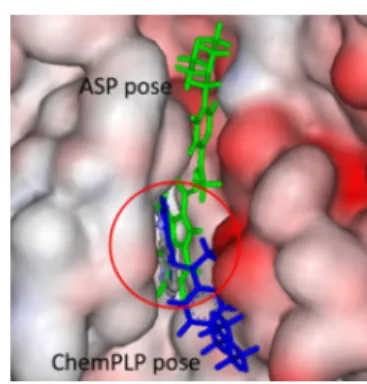
2.11. Lead Cu(II) Complexes are Located in the Binding Sites of Specific Kinases.
Cu(II) complexes 1
−8 and their ligands HL
1−HL
8were docked to the binding sites of Table 3. IC
50Values of Lead Drug Candidates in HCT-116
Colon Carcinoma Cells and an Isogenic p53-Knock-Out Subline
IC50[μM]
compound HCT-116 HCT-116p53ko
mean SD mean SD
HL4 0.14 0.02 0.12 0.02
4 0.13 0.05 0.13 0.03
HL8 0.042 0.003 0.038 0.008
8 0.050 0.005 0.041 0.006
cisplatin 0.78 0.27 1.7 0.4
https://doi.org/10.1021/acs.jmedchem.1c01740 J. Med. Chem.XXXX, XXX, XXX−XXX K
![Table 2. IC 50 Values for Morpholine-Indolo[2,3-c]quinoline and Latonduine Derivatives HL 1 − HL 8 and Their Copper(II) Complexes 1 − 8 as well as for the Ligand and Copper(II) Complex HL 4 nm and 4 nm](https://thumb-eu.123doks.com/thumbv2/9dokorg/731672.29175/8.911.117.397.96.458/values-morpholine-indolo-quinoline-latonduine-derivatives-complexes-complex.webp)