Transactions
PAPER
Cite this:Dalton Trans., 2018,47, 17032
Received 28th July 2018, Accepted 6th November 2018 DOI: 10.1039/c8dt03088j rsc.li/dalton
Impact of copper and iron binding properties on the anticancer activity of 8-hydroxyquinoline derived Mannich bases †
Veronika F. S. Pape, ‡aNóra V. May, bG. Tamás Gál,bIstván Szatmári,c Flóra Szeri, §aFerenc Fülöp,cGergely Szakács *a,dand Éva A. Enyedy *e
The anticancer activity of 8-hydroxyquinolines relies on complex formation with redox active copper and iron ions. Here we employ UV-visible spectrophotometry and EPR spectroscopy to compare proton dis- sociation and complex formation processes of the reference compound 8-hydroxyquinoline (Q-1) and three related Mannich bases to reveal possible correlations with biological activity. The studied derivatives harbor a CH2–N moiety at position 7 linked to morpholine (Q-2), piperidine (Q-3), and chlorine and fluorobenzylamino (Q-4) substituents. Solid phase structures of Q-3, Q-4·HCl·H2O, [(Cu(HQ-2)2)2]·
(CH3OH)2·Cl4·(H2O)2, [Cu(Q-3)2]·Cl2 and [Cu(HQ-4)2(CH3OH)]·ZnCl4·CH3OH were characterized by single-crystal X-ray diffraction analysis. In addition, the redox properties of the copper and iron complexes were studied by cyclic voltammetry, and the direct reaction with physiologically relevant reductants (glutathione and ascorbic acid) was monitored.In vitrocytotoxicity studies conducted with the human uterine sarcoma MES-SA/Dx5 cell line reveal the significant cytotoxicity ofQ-2,Q-3, andQ-4in the sub- to low micromolar range (IC50values 0.2–3.3μM). Correlation analysis of the anticancer activity and the metal binding properties of the compound series indicates that, at physiological pH, weaker copper(II) and iron(III) binding results in elevated toxicity (e.g.Q4: pCu = 13.0, pFe = 6.8, IC50= 0.2μMvs.Q1: pCu = 15.1, pFe = 13.0 IC50= 2.5μM). Although the studied 8-hydroxyquinolines preferentially bind copper(II) over iron(III), the cyclic voltammetry data revealed that the more cytotoxic ligands preferentially stabilize the lower oxidation state of the metal ions. A linear relationship between the pKa(OH) and IC50values of the studied 8-hydroxyquinolines was found. In summary, we identifyQ-4as a potent and selective anti- cancer candidate with significant toxicity in drug resistant cells.
Introduction
The broad pharmacological activity of 8-hydroxyquinoline (Q-1, Chart 1) and its derivatives has been exploited in several medicinal applications ranging from antineurodegenerative,
Chart 1 Structures of the reference compound 8-hydroxyquinoline (Q-1) and its derivatives (Q-2,Q-3, andQ-4) reported in this work in their neutral form.
†Electronic supplementary information (ESI) available: Crystal and UV-Vis spec- tral data. EPR parameters for the Cu(II) complexes, UV-Vis and EPR spectra, dis- tribution diagrams and cyclic voltammograms for the studied complexes. NMR spectra forQ-4. CCDC 1575107–1575111. For ESI and crystallographic data in CIF or other electronic format see DOI: 10.1039/c8dt03088j
‡Present address: Department of Physiology, Semmelweis University, Faculty of Medicine, Tűzoltó utca 37-47, H-1094 Budapest, Hungary.
§Present address: Department of Dermatology and Cutaneous Biology, Sidney Kimmel Medical College, Thomas Jefferson University, 233 S. 10thStreet, 19107 Philadelphia (PA), USA.
aInstitute of Enzymology, Research Centre for Natural Sciences, Hungarian Academy of Sciences, Magyar Tudósok körútja 2, H-1117 Budapest, Hungary
bResearch Centre for Natural Sciences, Hungarian Academy of Sciences, Magyar Tudósok körútja 2, H-1117 Budapest, Hungary
cInstitute of Pharmaceutical Chemistry and Stereochemistry Research Group of Hungarian Academy of Sciences, University of Szeged, Eötvös u. 6, H-6720 Szeged, Hungary
dInstitute of Cancer Research, Medical University of Vienna, Borschkegasse 8a, A-1090 Vienna, Austria. E-mail: gergely.szakacs@meduniwien.ac.at
eDepartment of Inorganic and Analytical Chemistry, Interdisciplinary Excellence Centre, University of Szeged, Dóm tér 7, H-6720 Szeged, Hungary.
E-mail: enyedy@chem.u-szeged.hu Open Access Article. Published on 07 November 2018. Downloaded on 7/30/2019 10:31:43 AM. This article is licensed under a Creative Commons Attribution-NonCommercial 3.0 Unported Licence.
View Article Online
View Journal | View Issue
anti-inflammatory and antimicrobial to anticancer thera- pies.1,2 In all of these applications, the therapeutic effect of 8-hydroxyquinolines is believed to rely on metal chelation.
Indeed, the (N,O) donor set of the planar, heterocyclic scaffold possesses a high affinity for various transition metal ions. For example, clioquinol (5-chloro-7-iodo-8-hydroxyquinoline, CQ), a lipophilic derivative ofQ-1, has been explored as a treatment for Alzheimer’s disease based on its ability to disrupt the inter- action between zinc(II) and copper(II) ions and the Aβpeptide in the brain.3,4 CQ and numerous other 8-hydroxyquinoline derivatives were reported to exhibit a remarkable anticancer effect. The anticancer activity of 8-hydroxyquinolines is also related to complexation with endogenous metals, above all the redox active copper and iron ions.5–7Despite continuous inter- est in novel 8-hydroxyquinolines as candidate anticancer agents, relatively little is known about their exact mechanism of toxicity. Studies have suggested several alternative models, including lysosomal disruption, inhibition of proteasome or histone deacetylase, inhibition of angiogenesis, or the inhi- bition of ribonucleotide reductase.8–10Labile iron and copper pools in the cytoplasm represent only rather low concen- trations under healthy conditions, as most of these metal ions are bound to proteins.11,12 However, the concentration of labile metal ions may be significantly increased in cancer cells.13CQ can also act as an ionophore, inducing the transfer of metal ions across biological membranes, either in or out of cells.10The antitumor activity of 8-hydroxyquinolines may also be linked to the toxicity of their metal complexes, as shown for copper(II),6,14 ternary ruthenium(II),15 half-sandwich organoruthenium,16–19 organorhodium,16 platinum(II)20 and gallium(III).21,22
Mannich bases of 8-hydroxyquinolines were reported to possess high potency against human cancer cells,23,24and our meta-analysis of the cytotoxicity of various 8-hydroxyquino- lines revealed the importance of the CH2–N subunit at position 7 for cytotoxicity.25–27 In this study we have chosen three 8-hydroxyquinoline derivatives in which the crucial CH2–N building block is retained. Morpholine (7-(morpholinomethyl) quinolin-8-ol,Q-2) and piperidine (7-( piperidin-1-ylmethyl)qui- nolin-8-ol,Q-3) moieties are introduced at position 7 (Chart 1) to decrease the hydrophobic character of the reference com- pound Q-1. To include a compound with a more lipophilic character to the study,Q-4was developed, bearing a chlorine at position 5 and a fluorobenzylamino substituent at position 7 (5-chloro-7-((2-fluorobenzylamino)methyl)quinolin-8-ol, Q-4, Chart 1).
In this work we characterize the effect of these derivatiza- tions on the cytotoxicity, proton dissociation processes, lipophilicity, copper(II) and iron(III) binding abilities and also study the redox properties of the relevant metal complexes to establish the relationship between the determined thermo- dynamic parameters and the biological activity of the com- pounds. Correlation analysis exploring the relationship between physico-chemical and biological properties provides an opportunity to better understand the mechanism of action of these compounds. Based on quantitative relationship ana-
lyses between chemical structures and bioactivity, between a set of thermodynamic parameters and toxicity, more reliable structure activity predictions can be made. Elegant examples of these types of correlation studies were shown fore.g.poly- oxometalates regarding their antibacterial activity,28 or for the antitumor and antiangiogenic effect of half-sandwich Ru(η6-p-cymene) and Ir(η5-C5Me5) benzimidazole complexes.29
Results and discussion
Synthesis and characterization of the 8-hydroxyquinoline derived Mannich bases
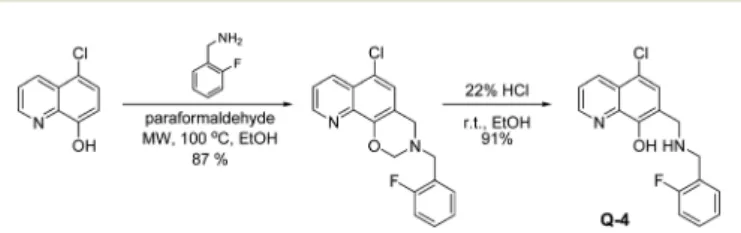
Q-1is a commercially available compound, whileQ-2andQ-3 were obtained from the National Cancer Institute’s Developmental Therapeutics Program (Chart 1).27As shown in Scheme 1,Q-4was synthesized by coupling 5-chloro-8-hydroxy- quinoline with 2-fluorobenzylamine in the presence of para- formaldehydeviaa modified Mannich reaction under microwave conditions. The formed [1,3]oxazino[5,6-h]quinolone derivative was isolated by crystallization from ann-hexane : ethyl acetate mixture and converted to the desired product as Q-4·HCl in ethanolic hydrogen chloride in excellent yield. NMR spectra were in agreement with the expected structures, enabling the assignment of all 1H and 13C resonances. The purity of Q-4·HCl was further confirmed by elemental analyses.
The structures ofQ-3andQ-4·HCl·H2O were established by single crystal X-ray diffraction (Fig. 1). Crystal data and struc- ture refinement parameters of the crystals are given in Table S1;† selected bond distances and angles are listed in
Scheme 1 Two-step synthesis of 5-chloro-7-((2-fluorobenzyl-amino) methyl)quinolin-8-ol Q-4, starting from 5-chloro-8-hydroxyquinoline via a modified Mannich reaction in ethanol (EtOH) at 100 °C, under microwave (MW) conditions, followed by an acidic cleavage of the dihydro-[1,3]-oxazine ring with HCl in ethanolic solution.
Fig. 1 ORTEP views ofQ-3(a) andQ-4·HCl·H2O (b) at the 30% prob- ability level of displacement ellipsoids. Blue lines indicate the suggested intramolecular hydrogen bonds. Selected bond distances and bond angles are listed in Tables S2 and S4.†
Open Access Article. Published on 07 November 2018. Downloaded on 7/30/2019 10:31:43 AM. This article is licensed under a Creative Commons Attribution-NonCommercial 3.0 Unported Licence.
Tables S2 and S4.†Q-3crystallized in its neutral form in the orthorhombic crystal system, in the space group P212121, without solvent inclusion.
The molecular structure is stabilized by the O1–H1O⋯N10 intramolecular hydrogen bond. The crystal structure is stabil- ized by weak C–H⋯O and C–H⋯πinteractions with the partici- pation of the piperidine protons (Fig. S1 and Tables S2, S3†).
Q-4crystallized in its protonated form with Cl−counter ions and a water molecule in the triclinic crystal system, in the space groupP1ˉ. The conformation is stabilized by a weak inter- molecular interaction between O1 and one of the amino protons. The neighboring molecules are connected by O–H⋯N and C–H⋯O hydrogen bonds, and π⋯π(off-centered parallel stacking) interactions (Fig. S1, S2 and Tables S3, S5†).
In vitrotoxicity of Q-1 and its Mannich base derivatives against a cancer cell line and primary hepatocytes
The in vitro cytotoxic activity of compounds Q-1 to Q-4 was determined in the human uterine sarcoma MES-SA/Dx5 cell line and in primary hepatocytes (Table 1). The compounds demonstrated marked cytotoxic activity in the micromolar and submicromolar concentration range in the drug resistant human uterine sarcoma MES-SA/Dx5 cell line, increasing in the following orderQ-2 <Q-1< Q-3< Q-4. Notably,Q-3and Q-4 possess even stronger cytotoxic activity than the FDA approved anticancer agent doxorubicin. To exclude non- specific toxic effects related to the multiple targets of metal chelators,30 primary hepatocytes were also included in the study. Compared to doxorubicin, the newly investigated com- pounds displayed a manifold higher selectivity towards cancer cells (Table 1). Due to the high proliferation rate, malignant cancer cells show increased reliance on metal ions, which are required e.g. as co-factors in enzymes catalyzing essential steps in DNA-synthesis, as well as for cell cycle progression and cellular growth.31–34For example, the rate limiting step in DNA synthesis is the reduction of ribonucleotides, which is catalyzed by the iron dependent enzyme ribonucleotide reductase (RR).35,36Thus, selective toxicity of the studied com- pounds to the human uterine sarcoma cells over hepatocytes might be the result of the altered metal homeostasis of the cancerous cells.
Proton dissociation processes of the studied 8-hydroxyquinolines
The proton dissociation constant (Ka) is a key physico-chemi- cal parameter influencing the pharmacokinetic properties of
the compounds. Determination of the pKa value allows the estimation of the distribution of the chemical forms of the molecule in the various protonation states at a given pH. As the pKa values of the reference compoundsQ-1 andQ-3 are available, although forQ-3under different conditions,16,37we determined the pKavalues of Q-2,Q-3andQ-4by UV-visible (UV-Vis) spectrophotometric titrations in an aqueous solution.
This method requires lower concentrations as compared to pH-potentiometry, which are compatible with the limited water-solubility of the compounds. The UV-Vis spectra of all ligands recorded in the pH range 2 to 11.5 show characteristic spectral changes upon deprotonation processes.
Representative spectra forQ-2, as well as the molar absor- bance spectra of individual ligand species in the different pro- tonated forms are shown in Fig. 2. The recorded absorbance
Fig. 2 UV-Vis absorption spectra ofQ-2(a) recorded at different pH values in aqueous solution and the calculated molar absorption spectra of the individual ligand species (b). (cQ-2= 50 µM;T= 25.0 °C;I= 0.20 M KCl;l= 2 cm).
Table 1 In vitrocytotoxicity (expressed as IC50) of the studied compounds in the human uterine sarcoma MES-SA/Dx5 cell line and in primary hepatocytesa
IC50/μM Q-1 Q-2 Q-3 Q-4 Doxorubicin
MES-SA/Dx5 2.46 ± 0.69 3.27 ± 0.39 0.86 ± 0.26 0.20 ± 0.06 2.13 ± 0.65
Primary hepatocytes 115 ± 18 243 ± 69 163 ± 35 40.4 ± 8.9 2.14 ± 1.11
aDetermined by means of the MTT assay after exposure for 72 h. Values are means ± standard deviations obtained from at least three indepen- dent experiments.
Open Access Article. Published on 07 November 2018. Downloaded on 7/30/2019 10:31:43 AM. This article is licensed under a Creative Commons Attribution-NonCommercial 3.0 Unported Licence.
spectra were deconvoluted resulting in the pKavalues of the compounds and the molar absorbance spectra of the various species (Table S6†). The pKa values were assigned to the particular moieties by careful analysis of the spectral changes (Scheme 2). The deprotonation processes of Q-1 are well- described in the literature,16,37and pK1and pK2values corres- pond to the deprotonation of the quinolinium-NH+ and the phenolic-OH moieties, respectively, as indicated in Scheme 2.
Values obtained forQ-3are in good agreement with those reported in the presence of KNO3instead of KCl.16
The most pronounced spectral changes are linked to the deprotonation of the hydroxyl functional group. The emerging strong bands with higherλmaxvalues originate from the more extended conjugated π-electron system in the deprotonated form.16,37In comparison with the 8-hydroxyquinoline scaffold (Scheme 2), the morpholino-substituent in the 7-position in Q-2increases the pKavalue of the phenolic-OH by almost one order of magnitude, possibly due to the formation of a hydro- gen bond between the OH and the morpholine-N in the HL form of the ligand, which hinders deprotonation. In contrast, the protonated (thus positively charged) amine moieties of the piperidine of Q-3and of the benzylamine in the case ofQ-4 decrease the proton dissociation constants of the OH group by 2.5 and 4.4 orders of magnitude, respectively, as compared to Q-1. These results are consistent with the lower pKa values reported for numerous hydroxamic and carboxylic acids due to the electron withdrawing effect of the protonated piperidinium moiety.38 As confirmed by single crystal X-ray diffraction analysis of the solid compounds Q-3 and Q-4·HCl·H2O (vide supra), possible hydrogen bond formations between the phenolate and the piperidinium moiety ofQ-3(in the zwitter- ionic L form) can additionally contribute to the diminished
pKa values due to the stabilization of the conjugate base.
Intramolecular hydrogen bonding is also probable in the HL form of Q-4 between the phenolate and the protonated benzylamine moieties. The pKaof the OH group inQ-4( pK1) is also affected by the electron withdrawing effects of the chloro- substituent.
pKa values assigned to the deprotonation of the quinoli- nium-NH+moiety of the R7-substituted ligands (pK1forQ-2and Q-3) were found to be considerably lower compared to that of Q-1as a consequence of the electron withdrawing effect of the protonated methylene-amine groups in the Mannich bases. In Q-4 the deprotonation of this moiety could not be observed under the applied experimental conditions, since it most prob- ably takes place at lower pH (<2) due to the additional negative inductive effect of the halogen atom (the predicted pKavalue for the quinolinium-NH+moiety inQ-4is 0.98).39
Based on smaller spectral alterations further pKa values could be assigned corresponding to the deprotonation pro- cesses of the non-coordinating nitrogens, such as the morpho- linium-NH+ of Q-2 and the benzylamino-NH+ of Q-4.
Deprotonation of the piperidinium-NH+ofQ-3most probably takes place only at strongly basic pH ( pKa> 11.5).
On the basis of the obtained pKa values, species distri- bution was calculated at pH 7.4 (Scheme 2). Thus, the neutral forms of these compounds predominate at physiological pH;
however Q-3andQ-4are mostly present in their zwitterionic L and HL forms, respectively. The lipophilicity of the com- pounds was characterized by the n-octanol/water distribution coefficients at pH 7.4 (logD7.4values in Scheme 2). These data revealed that while the introduction of the aliphatic methylene amines in position R7 ofQ-2andQ-3increases the hydrophilic character of the ligands in comparison with the unsubstituted Scheme 2 Proton dissociation steps of the studied 8-hydroxyquinolines and their pKavalues. Predominant species andn-octanol/water distri- bution coefficients (logD7.4) determined at pH 7.4. (T= 25 °C,I= 0.20 M (KCl))aData taken from ref. 43.
Open Access Article. Published on 07 November 2018. Downloaded on 7/30/2019 10:31:43 AM. This article is licensed under a Creative Commons Attribution-NonCommercial 3.0 Unported Licence.
scaffold Q-1, the additional benzyl moiety on the methylene amine in R7 and the halogen substituent in R5 significantly increase the lipophilicity ofQ-4.
Complex formation of the studied 8-hydroxyquinolines with iron(III) and copper(II) in solution
The anticancer activity of 8-hydroxyquinolines may be related to the formation of (redox active) metal complexes with essen- tial ions such as iron and copper7,40Therefore, we investigated complex formation ofQ-2toQ-4with two physiologically rele- vant metal ions (iron(III) and copper(II)), in order to reveal a possible relationship between the metal binding ability of these ligands and their cytotoxicity. Results were compared to data reported forQ-1.41,42
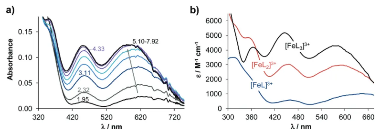
Complexation of the 8-hydroxyquinoline derived Mannich bases with iron(III) and copper(II) was studied by UV-Vis spectrophotometry. During titrations with both metal ions the absorbance values tend to decrease at pH ≳ 8. In the case of iron(III), hydrolysis of the metal ion may suppress complex formation at alkaline pH values, whereas neutral copper(II) complexes precipitate at pH≳9 (Fig. S3†forQ-2and Q-4complexes). Given these possible artefacts, evaluation was restricted to data collected at pH < 8. Spectral changes upon complexation with iron(III) detected in the 350–750 nm wave- length range were the most useful for the calculations of the overall stability constants, since bands located in this range are relatively well-separated from the ligand bands (see the representative spectra forQ-3in Fig. 3).
The stoichiometries of the metal complexes and the overall stability constants (β) furnishing the best fits to the experi- mental data are shown in Table 2.
The molar absorbance spectra of the individual metal com- plexes were also computed (see λmax and molar absorptivity values in Table S7†). As expected, mono and bis complexes are formed with both metal ions, while tris complexes could also be detected with iron(III). In the case of Q-2and Q-4, further protonated complexes were formed, in which the protons were attributed to the non-coordinating protonated morpholinium and secondary amine moieties, respectively (Table 2).
Based on the overall stability constants (Table 2), concen- tration distribution diagrams were computed at various metal- to-ligand ratios (exemplarily diagrams are shown for 5 µM ligand concentration at pH 7.4 in Fig. S4†). These data reveal the different speciation of the four ligands at physiological pH.
Complexation of iron(III) with Q-1 and Q-2 predominantly results in tris-ligand complexes, while withQ-3, the formation of the bis-complex is more pronounced. In the iron(III)−Q-4 system, mono and protonated bis complexes co-exist at pH 7.4.
Notably, the formation of tris complexes is not observed even at a 1 : 3 metal-to-ligand ratio, most probably due to steric reasons. In the case of copper(II) complexation, at a 1 : 2 metal- to-ligand ratio, the coordination sphere of the metal ion is likely to be saturated in the physiologically relevant pH range, as bis complexes are formed almost exclusively under these conditions.
Fig. 3 UV-Vis absorption spectra recorded for the iron(III)–Q-3(1 : 3) system at different pH values (a) and the calculated UV-Vis molar absorption spectra of the various complexes (b). (cQ-3= 50μM;T= 25.0 °C;I= 0.20 M KCl;l= 2 cm).
Table 2 Logarithm of the overall stability constants (logβ) of the copper(II) and iron(III) complexes of ligandsQ-2toQ-4determined by UV-vis spectrophotometric titrations and pM values calculated at pH 7.4. Data forQ-1are also shown for comparison. (Charges of the com- plexes are omitted for clarity. L is neutral forQ-3and singly negatively charged forQ-1,Q-2andQ-4.) (T= 25 °C,I= 0.20 M (KCl))
Q-1 Q-2 Q-3 Q-4
Logβ[FeLH] — 19.80 ± 0.04 — 18.46 ± 0.04
Logβ[FeL] 13.69a 16.90 ± 0.04 11.11 ± 0.02 14.31 ± 0.05
Logβ[FeL2H2] — — — 34.44 ± 0.04
Logβ[FeL2H] — 35.18 ± 0.04 — 28.50 ± 0.06 Logβ[FeL2] 26.3a — 20.32 ± 0.03 —
Logβ[FeL3H] — 49.03 ± 0.03 — —
Logβ[FeL3] 36.9a 42.49 ± 0.03 25.42 ± 0.07 —
pMc 21.1 21.1 16.0 14.8
pM*d 13.0 13.0 8.0 6.8
Logβ[CuLH] — — — 16.87 ± 0.03
Logβ[CuL] 12.1b 16.54 ± 0.04 10.6 ± 0.1 10.96 ± 0.07
Logβ[CuL2H2] — — — 32.54 ± 0.06
Logβ[CuL2H] — 34.67 ± 0.04 — 26.94 ± 0.06 Logβ[CuL2] 23.0b 27.5 ± 0.1 19.4 ± 0.1 —
pMc 15.1 17.4 14.9 13.0
aData taken from ref. 41.bData taken from ref. 42.cpM =−log[M] cal- culated at pH = 7.4,cM= 1μM;cL= 10μM.dpM* =−log([Fe] +i ×
∑[Fei(OH)j]) calculated at pH = 7.4,cM= 1μM;cL= 10μM.
Open Access Article. Published on 07 November 2018. Downloaded on 7/30/2019 10:31:43 AM. This article is licensed under a Creative Commons Attribution-NonCommercial 3.0 Unported Licence.
The overall stability constants of the complexes cannot be compared directly owing to the different basicities of the ligands and the number of dissociable protons. However, the stability constants for the iron(III) species are generally higher than those of the corresponding copper(II) complexes, the sig- nificantly different extent of hydrolysis of these metal ions should also be taken into consideration when the solution stabilities are compared. Therefore, in order to compare the metal binding ability of the four investigated 8-hydroxyquino- lines adequately, pM values (negative decadic logarithm of the equilibrium concentration of the unbound metal ion) were computed at pH 7.4 (Table 2). In the case of iron(III), the for- mation of hydroxido species was also taken into account ( pM*
values in Table 2). At pH 7.4, the iron binding capacity of the investigated ligands follows the orderQ-1∼ Q-2> Q-3> Q-4, and a similar trend (Q-2>Q-1>Q-3>Q-4) was obtained for the copper(II) complexes. A comparison of the pM (of copper(II)) and pM* (of iron(III)) values indicates that at pH 7.4, the studied ligands preferentially bind copper(II) over iron(III).
To further investigate the coordination modes of the copper(II) complexes in solution, electron paramagnetic resonance (EPR) spectroscopic measurements were performed.
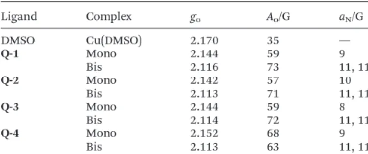
EPR spectra were recorded at room temperature for samples containing copper(II) and the investigated ligands (deproto- nated with one equivalent of NaOH) in equimolar concen- trations and at two-fold ligand excess (Fig. 4). Dimethyl sulfox- ide (DMSO) was used as a solvent in which the complexes are soluble in the mM concentration range. The measured spectra could be deconvoluted into the individual spectra of mono- and bis complexes (and free copper(II)). Notably, the [CuL2] and [CuL2H] complexes could not be distinguished, as side chain deprotonations have a negligible impact on the EPR spectra. The EPR data indicate that the formation of the mono complexes seems to be more pronounced in DMSO than in the
aqueous solution in case ofQ-1andQ-4. Additionally, the frac- tion of the bis complex is much lower forQ-4as compared to the other ligands, most probably due to steric reasons. The iso- tropic EPR parameters obtained by the simulation of room temperature spectra are shown in Table 3.
Thegovalues of the copper(II) complexes support a similar bidentate (N,O−) 8-quinolinolato coordination mode in all complexes. It is worth noting that thegoconstant of the mono complex ofQ-4is higher compared to those of the complexes of the other 3 ligands, indicating a somewhat weaker ligand field.
EPR spectra were also recorded at 77 K in methanol and in toluene. Interestingly, these EPR spectra revealed that while for Q-1and Q-3 the bis complexes form monomers, for Q-2 andQ-4, additional dimeric species were detectable (Fig. S5, Table S8†) in the frozen samples. A doublet peak detected at around 3000 and 3500 G and the half-field signal at 1500 G unambiguously show the appearance of a coupled-spin system (S= 1), which can be established by two neighboring copper(II) centers. From the simulation of the spectra the orientation of the copper(II) centers can be calculated ( polar angles: χ = 28.5°,ψ=−5.0°), showing that one center is above the other. A copper–copper distance of 3.88 Å was calculated from the dipolar coupling by using the point-dipole approach. These results are in good agreement with the obtained X-ray crystallo- graphic results (vide infra) whereχ= 35.1° andψ= 0° could be measured for Q-2andχ = 30.6° andψ= 0° for Q-4, and the copper–copper distances are 3.3935(7) Å and 3.8354(8) Å respectively. From this result we conclude that the bis complexes of Q-2 and Q-4 preferably form dimers in frozen solution as well as in the solid state.
Solid phase characterization of the copper(II) complexes of the studied 8-hydroxyquinolines
Single crystals suitable for X-ray crystallography were obtained from methanolic solution in the case of the copper(II) com- plexes of Q-2, Q-3 and Q-4 as [(Cu(HQ-2)2)2]·(CH3OH)2· Cl4·(H2O)2, [Cu(Q-3)2]·Cl2 and [Cu(HQ-4)2(CH3OH)]·ZnCl4· CH3OH (Fig. 5). Crystal data and structure refinement para- meters are given in Table S1.† Selected bond distances and angles are specified in the legend of Fig. 5, while packing Fig. 4 Experimental (black) and simulated (grey) room temperature EPR
spectra recorded for the copper(II)–ligand systems (Q-1–Q-4) in DMSO atcCu(II)= 2 mM in the presence of one (a) or two (b) equivalents of the ligand. Concentration ratios of the mono and bis complexes obtained from the simulation are indicated in the spectra.
Table 3 Isotropic EPR parameters obtained by the simulation of room temperature EPR spectra recorded in DMSOa
Ligand Complex go Ao/G aN/G
DMSO Cu(DMSO) 2.170 35 —
Q-1 Mono 2.144 59 9
Bis 2.116 73 11, 11
Q-2 Mono 2.142 57 10
Bis 2.113 71 11, 11
Q-3 Mono 2.144 59 8
Bis 2.114 72 11, 11
Q-4 Mono 2.152 68 9
Bis 2.113 63 11, 11
aThe experimental error is ±0.001 forgo, ±1 G forAoandaN.
Open Access Article. Published on 07 November 2018. Downloaded on 7/30/2019 10:31:43 AM. This article is licensed under a Creative Commons Attribution-NonCommercial 3.0 Unported Licence.
arrangements are shown in Fig. S6–8.† As expected based on their structural similarity, the X-ray diffraction studies reveal a similar bidentate (N,O−) 8-quinolinolato coordination mode for copper(II) complexes ofQ-3andQ-4and the reference com- pound Q-1.44 Despite a coordination with the same donor atom set, the copper(II) complex ofQ-2adopts a cyclic dimer [(Cu(HQ-2)2)2]4+form, in which the phenolato oxygen acts as a bridging atom between two metal centers of the neighboring complexes (Fig. S6†). However, the presence of long inter- molecular contacts Cu1⋯O11A or Cu1A⋯O11 (Å) strongly suggests the weak association of complexes into dimers, which most probably dissociate in aqueous solution with the for- mation of monomeric species.
[(Cu(HQ-2)2)2]·(CH3OH)2·Cl4·(H2O)2 crystallized in the monoclinic C2/c space group (Fig. 5 and S6†). The ligand in this complex is in the neutral zwitterionic HL form (with phenolate-O− and morpholinium-NH+). The metal ion is co- ordinated in a square-pyramidal mode, with the basal plane defined by atoms O1 and N1 from oneQ-2ligand and O11 and N21 from another Q-2 ligand in the trans position to each other, while atom O11 from another bis complex occupies the axial position of the metal ion (and vice versa) with a long Cu1–O11 distance (2.681(3) Å). The complex has an inversion centre symmetry, half of the molecule can be found in the asymmetric unit. The charge of the complex is neutralized by chloride counter ions. It is worth mentioning that the O33 atom of a neighbouring morpholine ring comes closer at a distance of 2.810(3) Å to the second axial position of the copper atom (Fig. S6†). The copper–copper distance is 3.3935(7) Å in the dimeric complex. The complex structure is further stabilized by two intramolecular hydrogen bonds C22–H22⋯O1 and C29–H29B⋯O1, and the main inter- molecular interactions are listed in Table S9.†
[Cu(Q-3)2]·Cl2crystallized in the monoclinic crystal system, in the space groupP21/n(Fig. 5 and S7†). The ligand shows a bidentate (N,O−) coordination mode forming a square planar type geometry around the copper center. The planar arrange- ment of the two quinolinolato rings is also secured by the intramolecular hydrogen bond of C2–H2⋯O1. In addition to a N–H⋯Cl bond and three weak C–H⋯Cl hydrogen bonds,π⋯π intermolecular interactions stabilize the crystal lattice between the neighboring quinoline moieties (Fig. S7 and Table S10†).
The closest copper–copper distance is 6.534(5) Å in this structure.
[Cu(HQ-4)2(CH3OH)]·ZnCl4·CH3OH crystallized in the tri- clinic crystal system, space groupP1ˉ(Fig. 5 and S8†). The two HL ligands are coordinatedviathe (N,O−) donor set resulting in a square planar geometry. The fifth (axial) coordination position is occupied by a methanol-OH group with a Cu1–O3 distance of 2.314(4) Å. Although the copper–copper distance (3.8354(8) Å) is significantly larger than in the crystal [Cu(Q-3)2]·Cl2the arrangement of the pair of complexes is very similar to it, and the complexes are placed face to face with their parallel basal planes such as O2 is arranged in the axial position of a neighboring copper ion (and vice versa, see Fig. S8†). Intra- and intermolecular interactions can be found in Table S11.†
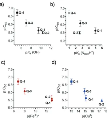
Correlations between solution equilibrium constants and in vitrocytotoxicity
While the low number of the compounds did not allow statisti- cally valid conclusions, we attempted to correlate the solution equilibrium properties of the studied 8-hydroxyquinolines with their biological activity. Correlations between thein vitro cytotoxicity (pIC50) obtained in the MES-SA/Dx5 cancer cell line, lipophilic character (logD7.4), proton dissociation constants Fig. 5 The cyclic dimer structure of the complex cations [(Cu(HQ-2)2)2]4+in [(Cu(HQ-2)2)2]·(CH3OH)2·Cl4·(H2O)2(a), [(Cu(Q-3)2]2+in [Cu(Q-3)2]·Cl2
(b) and [(Cu(HQ-4)2(CH3OH)]2+in [Cu(HQ-4)2(CH3OH)]·ZnCl4·CH3OH (c) at the 30% probability level of displacement ellipsoids. Hydrogens, as well as the chloride and [ZnCl4]2−anions, water and methanol molecules present in the crystal lattice are omitted for clarity. Selected bond lengths (Å) and angles (°) for [(Cu(HQ-2)2)2]4+: Cu1–O1 1.931(3), Cu1–O11 1.954(3), Cu1–N1 1.989(3), Cu1–N21 1.967(3), Cu1–O11A 2.681(3), O1–Cu1–O11 179.5(1), N1–Cu1–N21 171.0(1), O1–Cu1–N1 84.2(1), O1–Cu1–N21 95.7(1), O11A–Cu1–N1 92.8(1), O11A–Cu1–N21 96.1(1), O1–Cu1–O11A 92.4(1), O11–Cu1–O11A 87.2(1). Selected bond lengths (Å) and angles (°) for [(Cu(Q-3)2]2+: Cu2–O1 1.945(6), Cu2–N1 1.951(8), O1–Cu2–N1 85.1(3), O1–Cu2–N1 94.9(3), O1–Cu2–O1 180.0, N1–Cu2–N1 180.0. Selected bond lengths (Å) and angles (°) for [(Cu(HQ-4)2(CH3OH)]2+: Cu1–O1 1.969(2), Cu1–O2 1.952(3), Cu1–N1 1.989(4), Cu1–N21 1.978(4), Cu1–O3 2.314(4), O1–Cu1–O2 174.53(13), N1–Cu1–N21 174.63(15), O1–Cu1–N1 84.26(11), O1–Cu1–N21 97.11(11), O2–Cu1–N1 93.95(13), O2–Cu1–N21 84.22(13), O1–Cu1–O3 87.43(11), O3–Cu1–N1 97.45(14), O3–Cu1–N21 87.81(14), O1–Cu1–O3 87.43(11).
Open Access Article. Published on 07 November 2018. Downloaded on 7/30/2019 10:31:43 AM. This article is licensed under a Creative Commons Attribution-NonCommercial 3.0 Unported Licence.
( pKa) of the compounds and their metal binding abilities ( pM values) were analyzed (Fig. 6). No relationship was found between the pIC50and logD7.4values (Table 1 and Scheme 2) suggesting that lipophilicity as a single parameter is not a pre- dictor of cytotoxicity of the studied ligands, despite the often well-established correlations between the biological activity and lipophilicity of cytotoxic drug molecules, as the latter strongly affects membrane permeability. Whereas there was no strong correlation between pIC50 and pKa (NquinolinidiumH+) values, cytotoxicity seemed to increase with lower pKavalues of the hydroxyl moiety (Fig. 6a and b). Furthermore, compounds with weaker copper(II) and iron(III) binding abilities (at physio- logical pH) seemed to be more toxic, as suggested by the relationship of pM and pIC50values (Fig. 6c and d). The more cytotoxicQ-4andQ-3depending on the metal-to-ligand ratio form positively charged complexes at pH 7.4 with both metal ions (Q-4: [CuL2H]+, [CuL]+ and [FeL2H]2+; Q-3: [CuL2]2+, [CuL]2+ and [FeL2]3+), while the less active compounds (Q-1 andQ-1) mostly form neutral complexes (Q-1: [CuL2], [CuL]+ and [FeL3];Q-2: [CuL2], [CuL2H]+, [CuL]+and [FeL3]).
Redox properties of the studied 8-hydroxyquinolines with iron(III) and copper(II)
In addition to solution stability, the toxicity of metal complexes is also influenced by their redox properties. We therefore characterized the redox features of the copper(II) and iron(III) complexes of the studied 8-hydroxyquinoline ligands. First, the
redox activity of iron(III) and copper(II) complexes was investi- gated by cyclic voltammetry (CV). The CV measurements were carried out in pure DMSO due to the low water solubility of these metal complexes (c≪0.5 mM). However, aprotic organic solvents can modify complex formation processes and redox properties. Reliable data could be collected only for the com- plexes formed withQ-1andQ-3, when quasi-reversible redox processes were observed.
Representative voltammograms recorded for the complexes formed withQ-3at different metal-to-ligand ratios are shown in Fig. 7 and for the complexes formed with the other ligands in Fig. S9.† The corresponding electrochemical data are collected in Table S12.†Based on the voltammograms it could be concluded that upon complex formation withQ-1andQ-3, the half-wave potentials (E1/2) are shifted to more negative potentials compared to those of the corresponding metal salts.
(LigandsQ-3andQ-2were found to be redox active as well, but their identified cathodic peaks appear at much lower poten- tials than the peaks belonging to the metal complexes (Fig. S9 and Table S12†).) On the other hand, redox processes of the copper(II/I) systems occur in lower potential ranges compared to the iron containing systems. Interestingly, the observed shifts from the half-wave potentials of the metal salts were much larger in the case of ligandQ-1as compared toQ-3for both metal ions. Thus, Q-3, the more cytotoxic ligand, stabilizes the lower oxidation states of iron and copper more thanQ-1.
In comparison with the formal potential of −0.26 V reported for the redox couple of oxidized glutathione/gluta- thione (GSSG/GSH) at physiological pH,45the determinedE1/2
value of the iron-Q-3complex is more positive (Table S12†), suggesting that GSH might be able to reduce this iron(III) complex, while the reduction of the iron(III) complex ofQ-1by GSH seems unlikely based on these data. However, since the CV data were obtained in DMSO solutions, care is warranted in
Fig. 7 Cyclic voltammograms of the iron(III)–Q-3(a) and copper(II)–Q-3 (b) systems. The ligand alone (black/deprotonated: grey), FeCl3(brown), iron complexes at the M : L ratios 1 : 1 (bordeaux), 1 : 2 (red) and 1 : 3 (orange), as well as for CuCl2(light blue), and copper complexes at the M : L ratios 1 : 1 (blue) and 1 : 2 (green). (Potentials measured against Ag/AgCl/KCl (3 M);cQ-3= 1.0 mM; solvent: DMSO;I= 0.01 M (TBAClO4);
T= 25 °C; scan rate = 5 mV s−1).
Fig. 6 Correlations between pIC50values measured in MES-SA/Dx5 cell lines (IC50: mol L−1) and pKa(OH) (a), pKa(NquinolinidiumH+) (b), pFe(III)* (c) and pCu(II) (d) of the studied 8-hydroxyquinolines. Data are taken from Tables 1 and 2 and Scheme 2.
Open Access Article. Published on 07 November 2018. Downloaded on 7/30/2019 10:31:43 AM. This article is licensed under a Creative Commons Attribution-NonCommercial 3.0 Unported Licence.
extrapolating the obtained results to the intracellular milieu.
Therefore, the direct reactions of the iron(III) and copper(II) complexes of Q-1 to Q-4 with two physiologically relevant reducing agents GSH and ascorbate (ASC) were followed by UV-Vis spectrophotometry under strictly anaerobic conditions.
Absorbance spectra were analyzed at λ > 310 nm, where neither the reduced nor the oxidized forms of GSH and ASC absorb light.46,47
No significant spectral changes were observed upon reac- tion of the iron(III) complexes with 2.5 equivalents of GSH (in HEPES buffer, pH 7.4) within a time frame of 90 min. It was reported that the reduction of some iron(III)47 and vanadium(V)46,48 complexes by GSH might be kinetically hindered, while the dehydro-L-ascorbic acid/ASC redox pair (E′° at physiological pH = +0.05 V),49reacts considerably faster.
Redox reactions of the iron(III) complexes with ASC were observed at pH 6.87 as shown in Fig. S10,†although the spec- tral changes were still minor under the conditions, especially in the case of Q-3and Q-4. Addition of H2O2 to the samples reverses these changes.
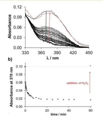
Notably, the copper complexes have lower redox potentials than the iron compounds (Table S12†), thus the copper(II) complexes are less powerful oxidizing agents. Nevertheless, copper(II) complexes ofQ-1toQ-4show characteristic spectral changes upon reaction with GSH, as displayed exemplarily for the complex of Q-2in Fig. 8. The significant decrease of the absorbance values at the λmax of the copper(II) complex
occurred relatively fast, indicating a possible ligand release from the reduced copper(I) complex. In order to assess whether the observed reduction process of the copper(II) complex is reversible, H2O2 was added to the samples at the end of the reaction. Upon addition of the oxidizing agent the spectrum of the original copper(II) complex reappeared, indi- cating the reversibility of the redox process and the possibility of an intracellular redox-cycling reaction. The time courses of the spectral changes induced by the reaction of the various copper(II) complexes with GSH are compared in Fig. S11.†The observed rate constants (kobs) of the redox reactions, calculated based on the initial slope of the ln(A/A0) vs. t plots (not shown), were as follows: 0.15 min−1for Q-1, 0.21 min−1 forQ-2, 0.20 min−1forQ-3, and 0.02 min−1forQ-4. This trend indicates that a stronger copper(II) binding ability of the ligands is accompanied by the faster redox reaction of their complex with GSH. Despite the trend in the kinetics of the reduction of the copper complexes, showing the slowest reac- tion for the most toxic ligand, a clear quantitative relationship could not be established between the redox chemical charac- teristics and the cytotoxic activity of the compounds.
Conclusions
8-Hydroxyquinoline derived Mannich bases were shown to exhibit high potency against human cancer cell lines, and the importance of a CH2–N building block at position 7 has also been described.27As the biological activity of a chelator can be affected by the lipophilicity, pKavalues, metal binding abilities and redox properties of the forming complexes, the possible relationship between these parameters was monitored in detail in the case of 8-hydroxyquinoline (Q-1) and its three derivatives containing the crucial CH2–N moiety. Due to the protonated tertiary amine nitrogens in 7-(morpholinomethyl) quinolin-8-ol (Q-2) and 7-( piperidin-1-ylmethyl)quinolin-8-ol (Q-3), the hydrophilicity is increased as compared toQ-1, while the newly synthesized and characterized 5-chloro-7-((2-fluoro- benzylamino)methyl)quinolin-8-ol (Q-4) possesses a more lipo- philic character. In addition, the various substitutions of the Q-1scaffold altered the size, the acid–base character, charges and the metal ion chelation of the compounds. To evaluate the effect of these alterations on thein vitroanticancer activity of the compounds, cytotoxicity ofQ-1toQ-4was tested against the drug resistant human uterine sarcoma MES-SA/Dx5 cell line as well as against primary hepatocytes. All compounds showed marked cytotoxic activity (IC50values 0.20–3.27μM) in the cancer cells increasing in the following orderQ-2<Q-1<
Q-3 < Q-4. IC50 values obtained in primary hepatocytes revealed a much better selectivity of the studied compounds towards cancer cells than the chemotherapy agent doxo- rubicin. Even though the most lipophilic derivative Q-4 was found to be the most toxic, no clear relationship was apparent between the lipophilicity of the set of derivatives and their tox- icity, suggesting that the cytotoxicity of the compound series is not strictly dependent on membrane permeability.
Fig. 8 Time-dependent UV-Vis spectra of the copper(II) complexes of Q-2(a) upon addition of 2.5 equivalents of GSH at pH 7.40. The black dashed lines denote the initial spectra before the addition of GSH and the red dashed lines denote the spectra upon addition of H2O2 (a).
Absorbance values measured at 370 nm plotted against the time (b). (ccopper(III)= 100μM;cligand= 100μM;cGSH= 250μM;T= 25.0 °C;
I= 0.20 M KCl).
Open Access Article. Published on 07 November 2018. Downloaded on 7/30/2019 10:31:43 AM. This article is licensed under a Creative Commons Attribution-NonCommercial 3.0 Unported Licence.
Relationship between proton dissociation constants and cytotoxicity
Based on the determined pKavalues, the compounds predomi- nate in their neutral forms at physiological pH, althoughQ-3 and Q-4 are mostly present in their zwitterionic L and HL forms, respectively. As compared toQ-1, the pKavalues of the quinolinium-NH+ decrease due to the electron withdrawing effect of the various substituents inQ-2toQ-4, and the pKaof the phenolic-OH moiety is reduced in Q-3 and Q-4. On the other hand, the morpholino substituent in the 7-position increases the pKa(OH) as a consequence of an intramolecular hydrogen bond between the OH and the morpholine-Nin the HL form of the ligand. Interestingly, formation of intra- molecular hydrogen bonding is also feasible in the solidQ-3 andQ-4·HCl·H2O according to their single crystal X-ray diffrac- tion analysis. It was concluded that compounds with lower pKa(OH) values possess lower IC50values in the MES-SA/Dx5 cancer cell lines showing a linear relationship.
Solution speciation and solid structures of the complexes Formation of mono and bis complexes with copper(II) and iron (III) was found based on the UV-visible spectrophotometric titrations, whereas tris iron(III) complexes could also be detected for two of the ligands. At physiological pH, the copper(II) binding strength of the studied compounds follows the orderQ-2>Q-1>Q-3>Q-4and a similar trend was deter- mined for the iron(III) complexes (Q-1∼Q-2>Q-3>Q-4). The studied 8-hydroxyquinolines have a binding preference to copper(II) over iron(III) at pH 7.4. Results of the EPR spectro- scopic measurements and the solid phase structures of [(Cu(HQ-2)2)2]·(CH3OH)2·Cl4·(H2O)2, [Cu(Q-3)2]·Cl2 and [Cu(HQ-4)2(CH3OH)]·ZnCl4·CH3OH characterized by single- crystal X-ray diffraction analysis confirmed the coordination viathe (N,O) donor set in the copper(II) complexes.
Relationship between solution stability, redox properties and cytotoxicity
In the investigated set of compounds, those possessing weaker copper(II) and iron(III) binding abilities at physiological pH were found to be more toxic against the cancer cell line. Based on the cyclic voltammetric data of the iron and copper complexes, the more cytotoxicQ-3stabilizes the lower oxidation states of these metal ions stronger thanQ-1. Glutathione was not able to reduce the iron(III) complexes of the tested 8-hydroxyquinolines at five- fold excess within a time frame of 90 min, while copper(II) com- plexes could be reduced much faster, and in a reversible manner.
The compounds with stronger copper(II) binding abilities showed faster redox reactions and were found to be less cytotoxic.
In all, the established property–activity relationship for the studied compounds can help in understanding the mechanism of action of 8-hydroxyquinolines and can contribute to the develop- ment of anticancer compounds. We found that the introduction of the morpholine moiety at position 7 slightly reduced the cyto- toxicity of 8-hydroxyquinoline in the MES-SA/Dx5 cell line, at the same time the piperidine and fluorobenzylamino substituents
(with chlorine at position 5) increased the activity. The morpholine derivative has slightly higher, while the latter two compounds have much lower pKa(OH) than the reference compoundQ-1. The more cytotoxic ligands possess weaker iron(III) and copper(II) binding abilities thanQ-1. Based on these conclusions,Q-4was selected as a candidate for undergoing preclinicalin vivoexperiments.
Experimental section
Chemicals
Solid KOH, 8-hydroxyquinoline (Q-1), 4-(2-hydroxyethyl)-1- piperazineethanesulfonic acid (HEPES), tetrabutylammonium chloride (TBACl), ferrocene, 5-chloroquinolin-8-ol, 2-fluoro- benzylamine, paraformaldehyde, ascorbic acid (ASC) and glutathione (GSH) were purchased from Sigma-Aldrich and HCl, KCl, CuCl2, FeCl3, and n-octanol were Reanal products (Budapest, Hungary).Q-2(NSC662298) andQ-3(NSC57969) were acquired from the drug repository of the Developmental Therapeutics Program of the National Cancer Institute. Iron(III) and copper(II) stock solutions were prepared by dissolving appro- priate amounts of metal chlorides in known amounts of HCl.
Their concentrations were determined by complexometry via EDTA complexes. Accurate strong acid content of the metal stock solutions was determined by pH-potentiometric titrations. The studied 8-hydroxyquinolines were poorly soluble in water; there- fore stock solutions (0.01 M; 1.4–3.7 g L−1) were in pure DMSO.
Solubility in water was not determined experimentally, but based on the determined log D7.4 values we can conclude that Q-2 andQ-3have somewhat better, whileQ-4has definitely a worse (∼50μM) water solubility thanQ-1(S∼0.56 g L−1,∼4 mM). (The solubility of these compounds strongly depends on the pH.)
Chemicals used for the synthesis were of reagent grade quality or better, obtained from commercial suppliers and used without further purification. Solvents were used as received or dried.
Synthesis and characterization of 5-chloro-7-((2-fluorobenzyl- amino)methyl)quinolin-8-ol hydrochloride (Q-4·HCl)
Q-4·HCl was obtained by the acidic cleavage of 5-chloro-3- (2-fluorobenzyl)-3,4-dihydro-2H-[1,3]oxazino[5,6-h]quinoline, which was prepared by the reaction of 5-chloroquinolin-8-ol (0.50 g, 2.78 mmol), 2-fluorobenzylamine (0.52 g, 4.15 mmol) and paraformaldehyde (0.208 g, 6.95 mmol) using ethanol (17 mL) as the solvent in a 35 mL pressurized reaction vial.
The mixture was heated by microwave irradiation at 100 °C for 45 min. The solvent was then evaporated offand the residue was crystallized withn-hexane : ethyl acetate (5 : 1; 30 mL) and recrystallized from 20 mL of diisopropyl ether. Yield: 0.795 g (87%); mp.: 150–152 °C. 1H NMR (DMSO-d6, Fig. S12†): δ = 3.96 (2H, s); 4.08 (2H, s); 5.11 (2H, s); 7.12–7.26 (2H, m); 7.36 (1H, t,J= 7.6 Hz); 7.41–7.52 (2H, m); 7.65 (1H, dd,J= 8.12 Hz, 3.84 Hz); 8.46 (1H, d, J = 8.5 Hz); 8.92 (1H, s); 13C NMR (DMSO-d6, Fig. S13†):δ= 48.3; 48.7; 61.7; 115.2; 115.4; 118.6;
120.1; 122.5; 124.4; 124.9; 125.0; 126.4; 129.4; 131.1; 132.2;
139.4; 148.8; 149.9. Anal. calcd for C18H14ClFN2O (328.77): C, 65.76; H, 4.29; N, 8.52. Found: C, 66.02; H, 4.31; N, 8.49.
Open Access Article. Published on 07 November 2018. Downloaded on 7/30/2019 10:31:43 AM. This article is licensed under a Creative Commons Attribution-NonCommercial 3.0 Unported Licence.
To obtain Q-4·HCl 0.15 mL (1.0 mmol) of HCl/ethanol (22%) was added to a stirred solution of 5-chloro-3-(2-fluoro- benzyl)-3,4-dihydro-2H-[1,3]oxazino[5,6-h]quinoline (0.20 g;
0.6 mmol) in ethanol (15 mL). The mixture was stirred for 30 min at room temperature. After the evaporation of the solvent and crystallization with diethyl ether (10 mL), the product was obtained in 91% yield (0.19 g); mp.: 211–213 °C.
1H NMR (DMSO-d6, Fig. S14†):δ = 4.25 (2H, s); 4.37 (2H, s);
7.22–7.32 (2H, m); 7.43–7.53 (1H, m); 7.75 (1H, t,J = 7.2 Hz);
7.81 (1H, dd,J= 8.24 Hz, 3.97 Hz ); 8.00 (1H, s); 8.56 (1H, d,J= 8.2 Hz); 9.02 (1H, s); 9.85 (2H, brs); 13C NMR (DMSO-d6, Fig. S15†): δ = 43.0; 44.1; 114.9; 115.6; 118.4; 119.0; 119.1;
123.8; 124.6; 126.2; 129.4; 131.5; 132.5; 133.0; 138.4; 149.3;
151.9. Anal. calcd for C17H15Cl2FN2O (353.22): C, 57.81; H, 4.28; N, 7.93. Found: C, 57.92; H, 4.27; N, 7.95.
1H and13C NMR spectra were recorded in deuterated sol- vents on a 400 (1H: 400 MHz, 13C: 100.6 MHz) MHz spectro- meter at room temperature. Chemical shiftsδare expressed in ppm values using the residual solvent peaks as internal stan- dards (DMSO-d62.50; 39.52 ppm).50Melting points were deter- mined on a Hinotek X-4 type melting point apparatus and are uncorrected. Elemental analyses were performed with a PerkinElmer 2400 CHNS elemental analyzer. Merck Kieselgel 60F254plates were used for TLC. Microwave reactions were per- formed by using a CEM LabMate microwave reactor.
Crystallization, X-ray data collection, structure solution and refinement
Single crystals suitable for X-ray crystallography (SXRD) (Q-3, Q-4·HCl·H2O, [(Cu(HQ-2)2)2]·(CH3OH)2·Cl4·(H2O)2, [Cu(Q-3)2]·Cl2
and [Cu(HQ-4)2(CH3OH)]·ZnCl4·CH3OH) were grown from slow evaporation of a methanolic solution at room temperature. In the case of the crystallisation of the copper complexes the solu- tion contained the metal ion and the ligand at a 1 : 2 ratio.
[Cu2(Q-2)4]·(CH3OH)2·(Cl)4·(H2O)2and [Cu(Q-3)]·(Cl)2 were crys- tallized from methanolic solutions applying vapour diffusion of diethyl ether to reduce the solubility. The complex ofQ-4could not be crystallized under similar conditions, however when ZnCl2 was also present in the solution the crystals of [Cu(Q-4)]·(ZnCl4)·(CH3OH) could be obtained. As Cu and Zn could not be unambiguously distinguished by SXRD, elemental analysis (ICP-OES) has been performed. This crystal contains 2.6(1) mg kg−1of Cu and 4.0(2) mg kg−1of Zn that is a 60/40%
Zn/Cu ratio (with an error of ±5%). If we suppose that in this crystal, Zn is possibly in a tetrahedral ZnCl4form (40%) and Cu is complexed by theQ-4ligand (40%), there is still 20% of Zn shared, half in ZnCl4and the other half possibly occupies the position of copper in the Q-4 complex. It means an 80%
copper(II) and 20% zinc(II) occupation of the central metal posi- tion of the organic ligand complex cation, which may be the reason for the poor crystal quality, which prevented the refine- ment of the metal occupancy on the weak quality SXRD data.
The single crystals were mounted on loops. X-ray diffraction data were collected on a Rigaku RAXIS-RAPID II diffractometer at room temperature (293 K) in the case ofQ-3,Q-4·HCl·H2O, [(Cu(HQ-2)2)2]·(CH3OH)2·Cl4·(H2O)2, and [Cu(Q-3)2]·Cl2, while
the temperature was 103 K during the measurement of [Cu(HQ-4)2(CH3OH)]·ZnCl4·CH3OH. Numerical absorption correction was carried out, except the last crystal where multi- scan absorption correction has been applied using the soft- ware CrystalClear.51
Sir201452and SHELXL53under WinGX54software were used for crystal structure solution and refinement, respectively. The structures were solved by direct methods. The models were refined by full-matrix least squares onF2. Hydrogen atoms were included in structure factor calculations but they were not refined, and their isotropic displacement parameters were approximated from theU(eq)value of the atom they were bonded to. Refinement of non-hydrogen atoms was carried out with anisotropic tempera- ture factors. Selected bond lengths and angles of compounds were calculated using PLATON software.55The graphical represen- tations were done by Mercury,56and the editing of CIF files was performed using PublCif57software, respectively.
Crystallographic data for the crystal structures were de- posited at the Cambridge Crystallographic Data Centre as sup- plementary publication numbers CCDC 1575107–1575111†for the five crystals listed above.
UV-Vis spectrophotometry and determination of the distribution coefficients
The pH-metric measurements for determination of the exact concentrations of HCl and KOH stock solutions used for the spectrophotometric titrations were carried out at 25.0 ± 0.1 °C in aqueous solutions and at an ionic strength of 0.20 M KCl in order to keep the activity coefficients constant. All the titra- tions were performed with carbonate-free KOH solutions of a known concentration (0.10 M) in the presence of 0.1 M KCl.
An Orion 710A pH-meter equipped with a Metrohm combined electrode (type 6.0234.100) and a Metrohm 665 Dosimat burette were used for titrations. The electrode system was cali- brated to the pH =−log[H+] scale by the method suggested by Irvinget al.58The average water ionization constant, pKwater, is 13.76 ± 0.01 at 25 °C. The samples were deoxygenated by bub- bling purified argon forca.10 min prior to the measurements and argon was also passed over the solutions during further titrations. A Hewlett Packard 8452A diode array spectrophoto- meter was used to record the UV-Vis spectra in the 200–800 nm interval. The path length was 1 or 2 cm. The spec- trophotometric titrations were performed on the samples of the ligands Q-3, Q-4 and Q-2 alone or with iron(III) or copper(II). The concentration of the ligand was usually 50 µM (10 µM forQ-4due to its worse water solubility) and the metal ion-to-ligand ratios were 1 : 1, 1 : 2 and 1 : 3 over the pH range between 2 and 11.5 at an ionic strength of 0.20 M (KCl) at 25.0 ± 0.1 °C. The initial volume of the samples was 10.00 mL.
The samples contained 0.5% (v/v) DMSO. The overall protona- tion constants of the ligands (from which the pKavalues were calculated), the overall stability constants of their metal complexes and the individual spectra of the species were com- puted using the program PSEQUAD59and literature data were used for iron(III) hydroxido species.60
Open Access Article. Published on 07 November 2018. Downloaded on 7/30/2019 10:31:43 AM. This article is licensed under a Creative Commons Attribution-NonCommercial 3.0 Unported Licence.