F
ORUMR
EVIEWA
RTICLEAnticancer Thiosemicarbazones:
Chemical Properties, Interaction with Iron Metabolism, and Resistance Development
Petra Heffeter,1,2,* Veronika F.S. Pape,3,4,* E´va A. Enyedy,5Bernhard K. Keppler,2,6 Gergely Szakacs,1,3 and Christian R. Kowol2,6
Abstract
Significance:
During the past decades, thiosemicarbazones were clinically developed for a variety of diseases, including tuberculosis, viral infections, malaria, and cancer. With regard to malignant diseases, the class of
a-N-heterocyclic thiosemicarbazones, and here especially 3-aminopyridine-2-carboxaldehyde thiosemicarbazone (Triapine), was intensively developed in multiple clinical phase I/II trials.
Recent Advances:
Very recently, two new derivatives, namely COTI-2 and di-2-pyridylketone 4-cyclohexyl-4- methyl-3-thiosemicarbazone (DpC) have entered phase I evaluation. Based on the strong metal-chelating/metal- interacting properties of thiosemicarbazones, interference with the cellular iron (and copper) homeostasis is assumed to play an important role in their biological activity.
Critical Issues:
In this review, we summarize and analyze the data on the interaction of (a-N-heterocyclic) thiosemicarbazones with iron, with the special aim of bridging the current knowledge on their mode of action from chemistry to (cell) biology. In addition, we highlight the difference to classical iron(III) chelators such as desferrioxamine (DFO), which are used for the treatment of iron overload.
Future Directions:
We want to emphasize that thiosemicarbazones are not solely removing iron from the cells/
organism. In contrast, they should be considered as iron-interacting drugs influencing diverse biological pathways in a complex and multi-faceted mode of action. Consequently, in addition to the discussion of physicochemical properties (e.g., complex stability, redox activity), this review contains an overview on the diversity of cellular thiosemicarbazone targets and drug resistance mechanisms.
Antioxid. Redox Signal.30, 1062–1082.
Keywords:
thiosemicarbazones, iron, metal complex, ribonucleotide reductase, drug resistance, collateral sensitivity
Introduction
D
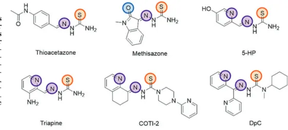
uring the past decades, thiosemicarbazones were clini- cally developed for a variety of diseases, including tuber- culosis, viral infections, malaria, and cancer. The first clinicallyapproved drug of this compound class (introduced in the late 1940s) was p-acetamidobenzaldehyde thiosemicarbazone (Thioacetazone, Fig. 1), which is still used for the treatment of multidrug-resistant tuberculosis (40, 41, 140). The second clinically investigated thiosemicarbazone (commercialized
1Department of Medicine I, Institute of Cancer Research, Comprehensive Cancer Center of the Medical University, Medical University of Vienna, Vienna, Austria.
2Research Cluster ‘‘Translational Cancer Therapy Research,’’ Vienna, Austria.
3Institute of Enzymology, Research Centre for Natural Sciences, Hungarian Academy of Sciences, Budapest, Hungary.
4Department of Physiology, Faculty of Medicine, Semmelweis University, Budapest, Hungary.
5Department of Inorganic and Analytical Chemistry, University of Szeged, Szeged, Hungary.
6Institute of Inorganic Chemistry, Faculty of Chemistry, University of Vienna, Vienna, Austria.
*These authors contributed equally to this work.
ªMary Ann Liebert, Inc.
DOI: 10.1089/ars.2017.7487
1062
Downloaded by University of Szeged Library Collection (Hungary) from www.liebertpub.com at 07/05/19. For personal use only.
in the 1960s) wasN-methylisatin thiosemicarbazone [(Me- thisazone), Fig. 1], an agent developed against smallpox, which, however, due to the development of the smallpox vaccination, is not administered anymore (129). The clinical anticancer research mainly focused on a-N-heterocyclic thiosemicarbazones. As these compounds are very potent chelators for metal ions, including iron, they were originally developed with the aim of targeting the enhanced demand of cancer cells for iron (181). Already in 1956, the first deriv- ative, 2-formylpyridine thiosemicarbazone [(FTSC), Fig. 2], was reported to show activity against leukemia in mice (154).
After extensive structure-activity studies, the most promising compound, 5-hydroxyl-2-formylpyridine thiosemicarbazone [(5-HP), Fig. 1], was tested in clinical phase I trials and was shown to possess anticancer activity in leukemia patients (37, 103). Unfortunately, these tests also revealed severe side effects (mainly gastrointestinal toxicity) and fast inactivation
by glucoronidation, leading to the withdrawal of the com- pound (37, 103). Further optimization led to the development of 3-aminopyridine-2-carboxaldehyde thiosemicarbazone [(Triapine), 3-AP, Fig. 1], a compound that has meanwhile been tested in more than 30 clinical phase I and II trials (4, 47, 56, 91, 132). Similar to the results obtained with 5-HP, Triapine showed promising activity against hematologic diseases, whereas solid cancers proved unresponsive. The reasons for this inefficacy of Triapine monotherapy against solid tumors are still not fully understood. Probable expla- nations (discussed in more detail in the Resistance Against Thiosemicarbazones section) include inappropriate drug delivery due to the very short plasma half-life (47, 132, 143), fast metabolism/excretion (142), and/or rapid development of drug resistance (127). Despite these drawbacks, thiose- micarbazones have remained the focus of interest. Thus, several recent phase I studies have been performed for testing FIG. 1. Chemical structures
of clinically investigated or approved thiosemicarbazones with highlighted donor atoms. 5-HP, 5-hydroxyl-2- formylpyridine thiosemicar- bazone; DpC, di-2-pyridylk- etone 4-cyclohexyl-4-methyl- 3-thiosemicarbazone; Triapine, 3-aminopyridine-2-carboxal- dehyde thiosemicarbazone.
Color images are available online.
FIG. 2. Chemical structu- res of the a-N-heterocyclic thiosemicarbazones FTSC and Dp44mT with N,N,S donor sets, STSC with O,N,S donor atoms, the iron chela- tors phen, 8-HQ and PIH, as well as the clinically approved iron(III) chelators DFO, de- feriprone, and deferasirox.
8-HQ, 8-hydroxyquinoline; DFO, desferrioxamine; Dp44mT, di- 2-pyridylketone 4,4-dimethyl- 3-thiosemicarbazone; FTSC, 2-formylpyridine thiosemi- carbazone; phen, 1,10- phenanthroline; PIH, pyridoxal isonicotinoyl hydrazine; STSC, salicylaldehyde thiosemicarba- zone. Color images are avail- able online.
Downloaded by University of Szeged Library Collection (Hungary) from www.liebertpub.com at 07/05/19. For personal use only.
the safety of Triapine in combination therapy [e.g., with cisplatin (106, 109), gemcitabine (91), cytarabine (137), doxorubicin (163), irinotecan (26), or radiation (109)]. In addition, there are currently two newa-N-heterocyclic thio- semicarbazones undergoing clinical evaluation. In December 2015, COTI-2 (developed by Critical Outcome Technolo- gies, Inc., Fig. 1) (159) entered a clinical phase I trial for the treatment of advanced or recurrent gynecologic malignancies (NCT02433626). According to the information provided by the company, COTI-2 displays strong anticancer activity in the nanomolar range, especially in p53-mutant cell lines, as this thiosemicarbazone [comparable to some other thiosemi- carbazones discovered by the NCI (78)] restores the wild-type function of the p53 protein (see the Impact of thiosemicarba- zones on the cell cycle and other (iron-dependent) cellular pathways section). The second currently tested thiosemi- carbazone is di-2-pyridylketone 4-cyclohexyl-4-methyl-3- thiosemicarbazone [(DpC), developed by Oncochel Ther- apeutics, Fig. 1] (61), which has been under clinical phase I evaluation since January 2016 (NCT02688101) to assess its pharmacokinetic characteristics and the maximum tolerated dose in patients with a range of advanced and resistant tumors.
Based on these promising recent developments, the elu- cidation of the mechanisms underlying the (biological) ac- tivity of the different anticancer thiosemicarbazones remains of high interest. In this review, we summarize and analyze the data on the interaction of (a-N-heterocyclic) thiosemi- carbazones with iron, with the special aim of connecting the chemical properties with the current knowledge on (cell) biological modes of action. In contrast to several drugs such as desferrioxamine (DFO), which are used for the treatment of iron overload and selectively chelate iron(III), a-N- heterocyclic thiosemicarbazones usually form stable com- plexes with both iron(II) and iron(III) and, therefore, are able to redox cycle between these two oxidation states under physiological conditions. Throughout this review, we em- phasize that thiosemicarbazones are not ‘‘simple’’ iron che- lators (solely removing iron from the circulation or the cells), but in contrast have to be considered rather as iron-interacting drugs that affect diverse biological pathways with a complex and multi-faceted mode of action. Based on the broad metal affinity, at least for some thiosemicarbazone derivatives, further biologically relevant metal ions (especially copper and zinc) have been associated with their mechanism(s) of action. However, due to the restriction in the length, this review does not comprehensively cover these additional metal ions. Further, it is beyond the scope of this review to give an overview on human iron homeostasis, especially as there are several excellent reviews in literature covering this field (24, 54, 87).
Important Physico-Chemical Parameters for the Metal-Binding Ability ofa-N-Heterocyclic Thiosemicarbazones
Given the importance of metal binding in the biological activity ofa-N-heterocyclic thiosemicarbazones, the chemi- cal characteristics influencing their metal binding have been intensively investigated over the past few decades. These studies revealed that a profound knowledge of the solution stability, stoichiometry, and redox properties of the metal complexes [especially iron(II)/(III)] under physiological
conditions as well as lipophilicity or pKaof the ligands is mandatory for the understanding of structure-activity rela- tionships of this compound class. In case of thiosemicarba- zone iron complexes, the possibility to readily switch in a one-electron oxidation-reduction reaction between the fer- rous form, Fe(II), and the ferric form, Fe(III), is of high im- portance to understand the biological effects. Consequently, the type of donor atoms that coordinate to the iron ions not only strongly influences the overall stability of the complexes but also determines whether or in which electrochemical window redox cycling between the two iron oxidation states is possible.
In general, thiosemicarbazones coordinate to metal ions by a bidentate binding modeviaa neutral nitrogen/sulfur (N,S) or an anionic (N,S-) donor set. However, metal complexes with much higher stability can be formed when an additional coordinating functionality is present in the molecule as in the case ofa-N- heterocyclic thiosemicarbazones (usually containing a pyridyl moiety), resulting in a (N,N,S) donor set (Fig. 2, coordinating atoms are indicated).
Unfortunately, solution stability constants have been published only for a fewa-N-heterocyclic thiosemicarbazone iron complexes, making the comparison of the different an- alogues difficult. So far, comparable stability data are avail- able only for various derivatives of FTSC, including Triapine (8, 44) and di-2-pyridylketone 4,4-dimethyl-3-thiosemicarba- zone (Dp44mT) (52) with (N,N,S) donor atoms as well as salicylaldehyde thiosemicarbazone (STSC) containing an (O,N,S) donor set (43, 128) (Fig. 2). In the following sections, we will summarize the available data and explain correlations between complex stability in solution, proton dissociation constants, and redox properties.
pKaValues of Metal-Freea-N-Heterocyclic Thiosemicarbazones
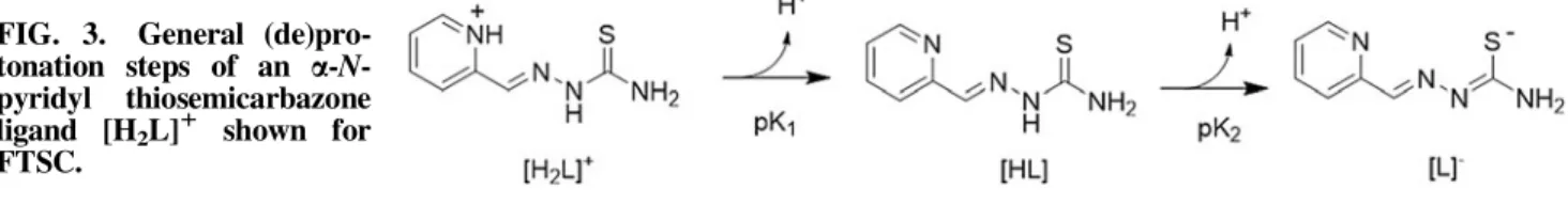
As a first parameter to compare different thiosemicarba- zone complexes, the proton dissociation behavior of the li- gands is of interest to understand the complex stability and the composition of the formed species. In general, simple a-N-pyridyl thiosemicarbazones (such as FTSC) possess two dissociable protons. Here, the pK1(approximately 3-4) can be attributed to the deprotonation of the pyridinium unit and pK2(approximately 10.5-11.5) can be attributed to the de- protonation of the hydrazinic N-H group of the thiosemi- carbazide moiety (Fig. 3). In case of pK2, the resulting negative charge is mainly localized on the S atomvia the thione–thiol tautomeric equilibrium (Fig. 3) and is influenced by the presence of different substituents. Thus, for example, N-terminal dimethylation significantly decreases the pK2
values (43). Based on the pKavalues, at physiological pH, all these a-N-pyridyl thiosemicarbazones are charged neutral (denoted as ‘‘HL form,’’ see Fig. 3), enabling an easier pas- sage across the cell membrane.
Characteristics of Iron(II/III) Thiosemicarbazone Complexes
Due to their tridentate nature,a-N-pyridyl thiosemicarba- zones form octahedral mono- and bis-ligand complexes with iron(II/III) ions, resulting in species such as Fe(HL), FeL, FeL(HL), or FeL2(where L is the completely deprotonated form of the ligand; Fig. 3). In the protonated complexes
Downloaded by University of Szeged Library Collection (Hungary) from www.liebertpub.com at 07/05/19. For personal use only.
Fe(HL) and FeL(HL), which are formed under acidic con- ditions, the HL ligand coordinatesviathe (Npyridyl,N,S) donor set with the proton attributed to the non-coordinating hydrazinic-N atom (44). In contrast, at neutral and basic pH values, FeL and FeL2complexes are formed, with anionic ligand(s) coordinating via(Npyridyl,N,S-) together with de- protonation of the hydrazinic nitrogen. This was also con- firmed by X-ray diffraction measurements for numerous iron(III) species (99, 102, 173).
On the basis of the reported solution equilibrium data for the iron complexes ofa-N-pyridyl thiosemicarbazones (in- cluding Triapine and Dp44mT, Table 1), it can be concluded that at slightly acidic and physiological pH the FeL2-type complexes predominate (44, 52).
However, due to differences in the number of dissociable protons and the basicity of the ligands, the logb values (a cumulative formation constant for the complexes) of the iron complexes shown in Table 1 cannot be directly compared.
Still, logbvalues clearly show that the differences between the constants of the iron(III) and iron(II) complexes strongly depend on the type of ligand. For example, stability constants of the iron(II) and iron(III) complexes of the clinically ap- proved iron chelator DFO (Fig. 2) differ by*20 orders of magnitude. In contrast, among the group of a-N-pyridyl thiosemicarbazones, the difference is, in general, much smaller, encompassing only two to three orders of magnitude (Table 1). The impact of the different donor atom sets in the thiosemicarbazone structure (N,N,S- vs. O-,N,S-) can be demonstrated by comparing the stability of iron(II/III) com- plexes formed by FTSC and STSC (Fig. 4). Thus, in a hy- pothetical metal/FTSC/STSC ternary system, the STSC ligand with the (O,N,S,) donor set has a much higher affinity
for Fe(III) ions, whereas the (N,N,S) donor set of FTSC prefers iron(II) ions. This preference corresponds to the dif- ferent hard-soft character of metal ions and the donor atoms (87, 89). The underlying theory behind the so-called HSAB (hard and soft acid and bases) concept (141) is that transition metals (‘‘Lewis acids’’) as well as donor atoms of the po- tential ligands (‘‘Lewis bases’’) can be classified into soft (low charge/large ionic radius), borderline, and hard (high charge/small ionic radius). According to this concept, soft acids react more efficiently and form stronger bonds with soft bases, whereas hard acids form stronger bonds with hard bases. Thus, the hard Lewis acid iron(III) will be preferen- tially coordinated by the STSC ligand (O,N,S) possessing the hard oxygen donor, whereas due to the softer (N,N,S) donor set, a-N-pyridyl thiosemicarbazones are unambiguously more efficient chelators of iron(II).
Besides the type of the coordinating donor atoms, also substituents on the thiosemicarbazone backbone can distinctly modify the stability of the respective metal complexes. For example, the iron(II)-binding ability of FTSC is weaker compared with that of Triapine, which, in turn, is weaker than that of theN-terminally dimethylated 2-formylpyridine thio- semicarbazone (PTSC) (44). Notably, the iron(II)-binding ability is in strong correlation with the anticancer activity of the compounds, with PTSC being the compound with the highest cytotoxicity and FTSC possessing the lowest cyto- toxicity (in the nMand lMrange, respectively) (102). This correlation also fits to the much lower stability of the STSC iron(II) complex and its low cytotoxic activity (45, 117, 148).
Overall, these data highlight the importance of the iron(II)- binding affinity of thiosemicarbazones in the optimization of their anticancer effects. However, it has to be considered that the analytical data for determination of the complex stability are usually measured in a concentration range of*10lMto 1 mM. Consequently, binding affinities in the lowlMto nM range are based on extrapolations, which make a profound estimation of the interaction of thiosemicarbazones with iron ions especially in the low nM range very difficult. This is especially of relevance in case of several highly active deriv- atives such as PTSC (42), DpC (116), and Dp44mT (14), in whichN-terminal dimethylation (or, more general, dialkyla- tion) results in a dramatically increased cytotoxic activity as compared with derivatives with an unsubstituted terminal NH2 moiety (e.g., FTSC and Triapine). In case of these thiosemi- carbazones, we can only speculate as to whether they are, indeed, still able to bind iron(II)/(III) ions at their nanomolar IC50 concentrations. Noteworthy, detailed structure-activity relationship studies revealed that this increased activity only occurs if no NH2or even NH group (with the exception of the hydrazinic NH) is present in the molecule (101, 102). Conse- quently, the terminally dimethylated derivative of Triapine has a cytotoxic activity comparable to Triapine itself (102) and only dimethylation of both amino groups results in nanomolar ac- tivity (101). However, the underlying mechanisms and the role FIG. 3. General (de)pro-
tonation steps of an a-N- pyridyl thiosemicarbazone ligand [H2L]1 shown for FTSC.
Table1. Cumulative Stability Constants (Logb) of Iron(III) and Iron(II) Complexes
of Desferrioxamine and Selected Thiosemicarbazones
Logb DFO Triapine FTSC Dp44mT STSC
Fe(III)L 30.4a NR NR NR NR
Fe(II)L 10b NR NR NR NR
Fe(III)L2 NR 26.25c 25.36c 23.6d 34.02e Fe(II)L2 NR 22.55c 22.31c 21.2d 24.73e
aHarris and Aisen (69).
bSpasojevic´et al.(172).
cData obtained in 30% (w/w) DMSO/H2O (44).
dValues estimated from the conditional constants obtained at pH 5:
Fe(III)L2—12.67 and Fe(II)L2—10.25 (52).
eData obtained in 30% (w/w) DMSO/H2O (45).
DFO, desferrioxamine; Dp44mT, di-2-pyridylketone 4,4- dimethyl-3-thiosemicarbazone; FTSC, 2-formylpyridine thiosemi- carbazone; NR, not relevant for the given ligand system; STSC, salicylaldehyde thiosemicarbazone; Triapine, 3-aminopyridine-2- carboxaldehyde thiosemicarbazone.
Downloaded by University of Szeged Library Collection (Hungary) from www.liebertpub.com at 07/05/19. For personal use only.
of iron complexation in this highly increased cytotoxic activity are widely unknown. There are some indications that thiosemi- carbazones being active in the nanomolar concentration range are characterized by an additional mode of action (and also induce different resistance patterns), which might involve inter- action with copper, as discussed in more detail in the Copper(II) Complexes of Thiosemicarbazones section (52, 83, 101).
Solution Stability and Redox Potential
The ratio between the cumulative stability constants of the iron(II) and iron(III) complexes is strongly connected to the redox potential. The reason is that the formal redox potential (e¢) of the Fe(III)/Fe(II) redox couple is determined by the ratio of the cumulative stability constants of the iron(III) and the iron(II) complexes according to the general Equation [1] (172):
e¢Fe IIIð Þcomplex=Fe IIð Þcomplexe¢Fe IIIð Þaqua=Fe IIð Þaqua¼ 59:15 · log[bFe IIIð Þcomplex=bFe IIð Þcomplex] [1]
Therefore, an increasing formal potential obtained at pH 7.4 represents an increasing affinity of the ligand toward iron(II) compared with iron(III) (Table 2). As a consequence, for example, the iron complexes formed with the aromatic nitrogen donor atom-containing bidentate ligand 1,10-
phenanthroline (phen) (Fig. 2) are characterized by a very high formal potential of the iron complex [+1140 mV vs.
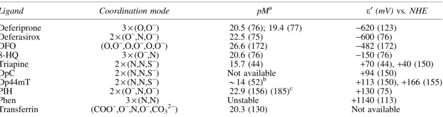
normal hydrogen electrode (NHE) (113)] and, therefore, the iron(III) complex shows low stability in aqueous solution (122). In contrast, the iron complexes of chelators applied for the treatment of iron overload such as DFO, deferiprone, and deferasirox (Fig. 2) are characterized by very low redox po- tentials (Table 2) and, consequently, they have very high iron(III)-binding abilities (reflected by high pM values, which is the negative logarithm of the equilibrium concen- trations of the unbound metal ion). For example, in case of DFO, which has a e¢ of –482 mV versus NHE (172), the iron(II) complex has much lower stability compared with the respective iron(III) complex (46, 69). Under anaerobic con- ditions (at physiological pH), DFO is even able to oxidize iron(II) to iron(III) (46). The reason for this behavior is that these agents mainly contain hard oxygen donor atoms, which are ideal for iron(III) binding (87, 89). This feature results in a very high binding efficacy and provides the enormous se- lectivity of these ligands for iron(III), in comparison not only to iron(II) but also to other essential bivalent metal ions such as zinc(II) and copper(II). In addition, due to the lowe¢val- ues, iron complexes of these chelators do not redox cycle under physiological conditions (76), preventing generation of reactive oxygen species (ROS)viaFenton-type reactions.
FIG. 4. Distribution of iron(II) and iron(III) between two different types of thiosemicarbazone com- plexes in a hypothetic metal/FTSC/
STSC (1:2:2) ternary system at pH 7.4 (cFe51 mM) calculated based on the stability constants (43, 44). The STSC ligands with the harder (O,N,S,) donor set have a much higher affinity for Fe(III) ions, whereas the (N,N,S) donor set of FTSC prefers iron(II) ions.
N,S, nitrogen/sulfur.
Table2. Physico-Chemical Properties of Selected Iron(III) Complexes Formed with Ligands Containing Different Donor Atoms
Ligand Coordination mode pMa e¢(mV)vs. NHE
Deferiprone 3·(O,O-) 20.5 (76); 19.4 (77) -620 (123)
Deferasirox 2·(O-,N,O-) 22.5 (75) -600 (76)
DFO (O,O-,O,O-,O,O-) 26.6 (172) -482 (172)
8-HQ 3·(O-,N) 20.6 (76) -150 (76)
Triapine 2·(N,N,S-) 15.7 (44) +70 (44),+40 (150)
DpC 2·(N,N,S-) Not available +94 (150)
Dp44mT 2·(N,N,S-) *14 (52)b +113 (150),+166 (155)
PIH 2·(O-,N,O-) 22.9 (156) (185)c +130 (75)
Phen 3·(N,N) Unstable +1140 (113)
Transferrin (COO-,O-,N,O-,CO32-) 20.3 (130) Not available
apM values (pM=–log[Fe(III)] at cFe(III)=1lM; cL=10lM; pH=7.4) and formal (e¢) potentialsversusNHE of the iron(III)/iron(II) complexes (pH=7.4).
bpM is estimated on the basis of the conditional constants reported at pH 5 in Gaalet al.(52) and transferred to pH 7.4 with the use of the protonation constants of the ligand and the hydrolysis constants of iron(III) (8a).
cpM is calculated from equilibrium constants reported in Richardsonet al.(156) and Vitoloet al.(185).
8-HQ, 8-hydroxyquinoline; DpC, di-2-pyridylketone 4-cyclohexyl-4-methyl-3-thiosemicarbazone; NHE, normal hydrogen electrode;
phen, 1,10-phenanthroline; PIH, pyridoxal isonicotinoyl hydrazine; pM, negative logarithm of the equilibrium concentrations of the unbound metal ion (pM–log[M]).
Downloaded by University of Szeged Library Collection (Hungary) from www.liebertpub.com at 07/05/19. For personal use only.
In contrast to the (mainly oxygen-containing) chelators de- veloped for the treatment of iron overload, anticancer a-N- pyridyl thiosemicarbazones such as Triapine, DpC, or Dp44mT coordinate metal ionsviatheir (Npyridyl,N,S-) donor set. As a result of this binding mode, the redox potentials of their iron complexes are significantly higher (in the range of-170 to +170 mVvs. NHE) (44, 155) compared with DFO, deferiprone, or deferasirox (-482 to –620 mV vs. NHE; see Table 2).
Consequently, a-N-pyridyl thiosemicarbazones show a con- siderably high affinity toward both iron(II) and iron(III) ions.
This property enables the iron thiosemicarbazone complexes to redox cycle between the two oxidations states, as these redox potentials are accessible to intracellular oxidants and reductants (66, 87, 155). Redox cycling is considered an important factor in the antiproliferative activity of these compounds (although the correlation between the redox potential and cytotoxicity is not as evident as the relation between stability and cytotoxici- ty). In line with the relevance of redox cycling for anticancer thiosemicarbazones, iron(III)-specific chelators such as DFO are generally not efficient antitumor agents and can be safely used to treat iron overload (23, 116, 197). This already indicates that ‘‘iron depletion’’ in itself is not sufficient to explain the anticancer activity of thiosemicarbazones.
Interaction of Thiosemicarbazones with Body Iron Based on the features of thiosemicarbazones described earlier in this review, there is no doubt that interaction with iron plays an important role in the biological activity of Triapine as well as many othera-N-heterocyclic derivatives.
In addition to cell culture studies (discussed in more detail in the Cancer-Associated Cellular Targets of Thiosemicarba- zones section), which investigated the role of iron and other metal ions in the activity of thiosemicarbazones on a cellular level, there are reports from clinical trials on the interaction of thiosemicarbazones with iron metabolism. For example, in case of 5-HP, excretion of the green-colored iron complex was observed in the urine of treated patients within a few hours after drug administration (103). In contrast, in Triapine-treated patients (or animals) (47, 132, 143), no coloring of urine but methemoglobin formation [oxidation of iron(II) to iron(III) in hemoglobin] was observed, which is a prominent indica- tion for its interaction with body iron. This type of adverse effect, observed in *23% of Triapine-treated patients (94, 107), was also reported for several preclinically developeda- N-heterocyclic thiosemicarbazones such as Dp44mT (150) (in this study, methemoglobin formation was analyzed in mice as well as in isolated human red blood cells) (Table 3). With regard to the underlying mechanism, the iron(III) complex of Triapine is believed to oxidize the iron(II) in the hemoglobin molecule by direct electron transfer (183). In line with the supposed role of a redox reaction in the thiosemicarbazone- induced methemoglobin formation, clinical trials with Triapine additionally indicated that patients with a glucose-6-phosphate deficiency suffered from enhanced methemoglobin levels and even hemolysis after treatment (199). Since glucose-6- phosphate is important in the protection of red blood cells from oxidative stress (184), this supports the assumption that Triapine treatment interferes with redox homeostasis. Met- hemoglobin formation seems to be strongly dependent on the interaction of the terminally unsubstituted NH2groups of the
thiosemicarbazone iron complexes with the carboxylic acid groups of the porphyrins of hemoglobin (Fig. 5) (11, 12, 143).
Consequently, novel a-N-heterocyclic thiosemicarbazones such as DpC (and probably also COTI-2) carrying a bulky substituent at this position are far less potent methemoglobin inducers than their predecessors (12).
Besides methemoglobin formation, also other iron- associated biological parameters were affected by Triapine treatment. Wadler et al. (186) described that after 96 h of continuous Triapine infusion, serum iron and ferritin levels transiently increased by 104% and 77%, respectively. Re- markably, the total iron-binding capacity of the blood re- mained unchanged in these patients, indicating that there was no net loss of iron from the body. Consequently, the authors concluded that [in contrast to 5-HP or the traditional iron(III) chelators (103)] Triapine-bound iron might be recovered during its hepatic metabolism and the increased delivery of iron to the liver could induce production of ferritin, explaining the elevated serum levels of this protein. Interestingly, de- spite these distinct impacts on several iron-associated clinical parameters, all attempts to detect the formation of an iron- Triapine complexin vivoor even in cell culture have failed so far (95, 96, 143).
Cancer-Associated Cellular Targets of Thiosemicarbazones
Ribonucleotide reductase
The iron-containing enzyme ribonucleotide reductase (RR) is most probably the best described target of anticancer a-N-heterocyclic thiosemicarbazones (Table 3). This me- talloenzyme catalyzes the conversion of ribonucleoside di- phosphates (NDPs) to deoxyribonucleoside diphosphates (dNDPs). Therefore, it is crucial for DNA synthesis as well as DNA damage repair and is frequently overexpressed in cancer cells, making it an attractive target for treatment. The RR is a tetrameric protein composed of two large subunits (hRRM1) that are important for substrate binding, and two small iron-containing R2 subunits being responsible for its enzymatic function (5, 25). Two different R2 subtypes have been described: (i) the hRRM2 subunit, which is a cell cycle- dependent protein, strongly upregulated at the beginning of the S phase and actively degradedviaubiquitination during the G2 phase of the cell cycle (60); (ii) p53R2 (also called hRRM2B), a subunit that is expressed at lower levels than hRRM2 but is not degraded during mitosis (138) and, therefore, its levels remain constant during the cell cycle. In addition, reports suggest that expression of this subunit can be induced in a p53-dependent manner. Consequently, it is assumed that the so-formed protein is involved in several cellular functions, including DNA repair, cell cycle arrest, and mitochondrial homeostasis (5, 25).
Inhibition of the tyrosyl radical in the active center of the hRRM2/p53R2 subunit was demonstrated for several thio- semicarbazones (including Triapine and Dp44mT) as well as diverse iron chelators (such as phen and DFO), mainly in cell- free systems (6, 59, 74, 99, 167, 178, 198, 202). The best- investigated thiosemicarbazones with regard to RR inhibition are Triapine and its closest analogs, for which detailed in vitro studies were performed during the past decades. In addition, a phase I trial with Triapine demonstrated the
Downloaded by University of Szeged Library Collection (Hungary) from www.liebertpub.com at 07/05/19. For personal use only.
depletion of dATP and dGTP pools together with a decrease of DNA synthesis in circulating leukemia cells (56).
With regard to the mechanism, it was initially assumed that Triapine inhibits the hRRM2 subunit through iron chelation, either from the active site of the protein or from the cellular labile iron pool (5). However, this theory was questioned by several observations, indicating that, in fact, Triapine seems to be a surprisingly inefficient iron chelator in cell culture.
For example, it was shown that, in contrast to DFO, Triapine had a relatively weak effect on cellular transferrin receptor levels of SK-N-MC cells (23), perhaps due to the weaker iron(III)-binding ability compared with DFO. Also, electron paramagnetic resonance spectroscopic analyses of Triapine- treated K562 cells (5 or 500lM) showed no change in the size of the g=4.3 signals (typical for intracellular iron- chelate complexes) after 0.5, 3, and 12 h of treatment, which would be indicative for depletion of the intracellular iron(III) pools (6). The same authors showed that in the presence of Triapine the loss of iron bound inside the RR molecule was five times lower than the destruction of the tyrosyl radical (5, 6) and subsequent investigations of Triapine-treated COS-1 cells indicated iron-loaded hRRM2 (with a quenched tyrosyl radical) as the main remaining protein species, arguing
against iron chelation from the enzyme. Moreover, in these COS-1 cells, Triapine did not change the aconitase activity (6). This is of interest, as this protein contains an essential [4Fe4S] cluster, which is ROS sensitive and could be influ- enced by depletion of the labile iron pool (6). Consequently, the authors of this study concluded that Triapine is not only ineffective in sequestering iron inside the cell, but also that it is unlikely to be involved in the disruption of biosynthesis or the repair of metallo-cofactor-requiring proteins (6).
Based on the repeated observations, that in cell-free systems metal-bound Triapine [particularly the iron(II) complex] pos- sesses a dramatically increased ability to inhibit hRRM2 compared with the metal-free ligand (6, 146, 167, 202), several authors speculated that the active species responsible for cell killing is, in fact, the iron complex of Triapine. In more detail, it was assumed that on intracellular formation of the redox-active iron complex, ROS are generated, which are ultimately re- sponsible for RR inhibition (167). Interestingly, the pro- nounced activity of Triapine and close derivatives against hRRM2 in the presence of iron was not observed in cell-free systems of p53R2, and co-incubation with iron had even pro- tective effects against the inhibition of this enzyme (202).
Further, there are several reports suggesting that at biologically Table3. Overview on Biological Thiosemicarbazone Targets Described
Target Drug Level of evidence (used method)
Hemoglobin Triapine In humans (methemoglobinemia) (94, 107)
Dp44mT In mice and isolated human red blood cells (methemoglobin formation) (150)
RR Triapine In humans (depletion of dNDP pools in leukemia cells) (56, 109)
Cell culture (whole-cell EPR, inhibition of dCTP production) (6, 99) Cell-free enzyme inhibition (EPR) (167, 202)
Dp44mT, Dp4eT Cell culture (whole-cell EPR) (198)
IQ-1 Cell-free enzyme inhibition (EPR) (178)
DNA Triapine Cell culture (H2AX phosphorylation) (83, 108)
In mice (immunofluorescence stain, H2AX phosphorylation in subcutaneous tumors, unpublished results)
Dp44mT Cell culture (H2AX phosphorylation) (83, 151)
STAT3 DpC, Dp44mT In mice (immunofluorescence stain of subcutaneous tumors) (119) Cell culture (Western blot, DNA-binding activity) (119, 187) NDRG1/2/3 Dp44mT In mice (immunofluorescence stain of subcutaneous tumors) (110)
Cell culture (Western blot) (110, 120, 187)
DpC Cell culture (Western blot) (97)
Cyclin A Triapine Cell culture (Western blot) (23)
TSC24 Cell culture (Western blot) (7)
Cyclin D Triapine Cell culture (Western blot) (23)
Dp44mT Cell culture (Western blot) (110, 111)
DpC Cell culture (Western blot) (97)
CDK2/4 Triapine Cell culture (Western blot) (23)
Bp4eT, Dp44mT Cell culture (CDK2 activity by immunoprecipitation) (35) PDE4D Triapine Cell culture (loss in resistant cell lines) (126)
P53 NSC319725,
NSC319726, NSC328784
Cell culture (restoring p53-wt confirmation) (196)
P21 DpC Cell culture (Western blot) (97, 131)
TSC24 Cell culture (Western blot) (7)
Trx2 Triapine Cell culture (Western blot) (134)
Prx3 Triapine, Dp44mT Cell culture (Western blot) (133)
AMPK Dp44mT Cell culture (Western blot) (104)
AMPK, adenosine monophosphate-activated protein kinase; CDK, cyclin-dependent kinase; dNDP, deoxyribonucleoside diphosphate;
EPR, electron paramagnetic resonance; NDRG1/2/3,N-myc downstream-regulated gene 1 or 2 or 3; PDE4D, cAMP-specific 3¢,5¢-cyclic phosphodiesterase 4D; Prx3, peroxiredoxin 3; RR, ribonucleotide reductase; STAT3, signal transducer and activator of transcription 3; Trx, thioredoxin; TSC24, N,N-dimethyl-2-(quinolin-2-ylmethylene)hydrazinecarbothioamide.
Downloaded by University of Szeged Library Collection (Hungary) from www.liebertpub.com at 07/05/19. For personal use only.
active concentrations no substantial ROS generation was de- tected after treatment with metal-free Triapine or other close derivatives (100, 115, 134). In line with these observations, recent studies have revealed no oxidation of hRRM2 residues or the accumulation of oxidized cellular proteins (neither in cell-free nor in cell culture experiments) after Triapine treat- ment, suggesting that global ROS generation might not be mandatory for RR inhibition by Triapine (5, 6). Notably, as already mentioned earlier, despite several attempts (96, 143), intracellular formation of a Triapine-iron complex has not been directly demonstrated eitherin vitroorin vivo. Taken together, although the importance and necessity of iron binding in the anticancer activity of thiosemicarbazones is proven without doubt [derivatives and metabolites lacking a metal-binding domain are completely inactive (142, 147, 200)], the exact mode of action remains elusive.
Consequently, several alternative theories have been published to offer an explanation as to how Triapine could inhibit the RR without the direct chelation of the enzyme- bound iron or the production of ROS. On the one hand, it was suggested (comparably to the interaction with hemoglobin in the methemoglobin formation) that the iron(II) complex of Triapine could act as reductant, directly reducing the tyrosyl radical of the hRRM2 subunit (6). This is of interest as direct binding of Triapine to hRRM2 and p53R2 was shown by radioactive labeling (202), which was only slightly affected by the addition of iron. In line with these experimental data, docking studies of mouse RR revealed a possible Triapine- binding site with the pyridine ring of the thiosemicarbazone located in the pocket of the four amino acids Phe237, Phe241, Ser238, and Tyr324 (146). Based on these results, it can be hypothesized that, after the binding of (metal-free) Triapine to the enzyme, an iron complex is formed locally, using the iron liberated during each catalytic cycle of the RR. Another possible mechanism is based on the effect of Triapine on intracellular thiol homeostasis. Here, especially the effects on the thioredoxin (Trx) system seem to be of importance, as Trx or glutaredoxin is necessary to reactivate the RR enzyme after the end of each catalytic cycle. In detail, during the conversion cycle of NDPs to dNDPs, a disulfide bond is formed in the hRRM1 subunit, which has to be reduced be- fore the RR can enter a new enzymatic cycle (60, 166). Some reports indicate that Triapine and Dp44mT inhibit thior- edoxin reductase (TrxR1) (134, 198). However, as these effects were observed only at very high concentrations [>50-
fold above the IC50values (133)], it remains unclear whether this is of biological relevance. Interestingly, murine L1210 leukemia cells selected for resistance against 4-methyl-5- amino-1-formylisoquinoline thiosemicarbazone (MAIQ) were [besides the overexpression of several efflux pumps (29)] also characterized by overexpression of protein disulfide isomer- ases (30), which are thiol oxidoreductase chaperones from the Trx superfamily (171).
Impact of thiosemicarbazones on the cell cycle and other (iron-dependent) cellular pathways
During the past decades, it was repeatedly reported that treatment with diverse iron chelators leads to cell cycle arrest in G0/G1or early S phase (27, 71, 79, 147, 151, 170). One explanation certainly is the earlier described inhibition of the RR. However, several other proteins involved in DNA rep- lication and repair also require iron as a cofactor and, thus, could be influenced by thiosemicarbazones and other iron- chelating drugs. These proteins include, for example, three DNA polymerases (Pol a, Pol d, and Pol e), several DNA helicases (including Rad3/XPD, Dna2, RTEL1, FANCJ, and ChIR1), as well as the DNA primase regulator subunit PRIM2 (201). In contrast to RR, these polymerases, heli- cases, and primases contain an Fe-S cluster, which is nec- essary for the formation of active holoproteins (201).
Interestingly, inhibition of RNA polymerases seems to be very important for the antiviral activity of several thiosemi- carbazones, indicated by point mutations in these enzymes, leading to drug-resistant virus variants (20, 21, 31, 149).
However, there are only a few studies (based on cell-free systems) linking polymerase inhibition to cancer (63) and to our knowledge, there is so far no indication that inhibition of polymerases also occurs in cancer cells after thiosemicarba- zone treatment. Likewise, the activity of the [4Fe-4S] cluster- containing aconitase (201) was not affected by Triapine (6), indicating that, in general, human Fe-S cluster proteins are not (heavily) affected by thiosemicarbazone treatment.
In addition to its direct role in proteins executing DNA synthesis, iron also influences cell cycle regulationvia cy- clins and cyclin-dependent kinases (CDKs) (201). Thus, it has been repeatedly reported that treatment with diverse iron chelators such as DFO or L-mimosine (an amino acid de- rivative and predecessor of deferiprone) and also thiosemi- carbazones results in the downregulation of cyclin A and D (7, 23, 27, 105, 110, 136) as well as CDK2/4 (23, 35, 36, 105) (Table 3). Overall, the mechanisms underlying these effects are not fully understood and there might be several expla- nations. For example, in case of CDK2, it was reported that the binding of iron ions to the protein occurs only to its phosphorylated form, indicating a direct regulation of this protein by the cellular metal homeostasis (9). With regard to cyclin A, little is known about the mechanisms explaining how thiosemicarbazones could influence the expression levels of this protein. Of interest in this context might be a recent report on the downregulation of cyclin A (in addition to cyclin D) by rolipram, a PDE4 inhibitor (121), which also strongly protects cells from Triapine treatment. In addition, loss of cAMP-specific 3¢,5¢-cyclic phosphodiesterase 4D (PDE4D) was the only genomic alteration found in Triapine- resistant SW480 cells (126). With regard to its biological function, PDE4 is a negative regulator of cyclic adenosine FIG. 5. Possible interaction of the iron complex of a
terminally unsubstituted thiosemicarbazone and the porphyrin system of hemoglobin [drawn according to reference Bashaet al.(11)].
Downloaded by University of Szeged Library Collection (Hungary) from www.liebertpub.com at 07/05/19. For personal use only.
monophosphate (cAMP), which, in turn, is one of the most abundant second messengers regulating various physiological processes such as cell survival, differentiation, proliferation, and apoptosis (10). One major downstream target of cAMP is cAMP-dependent protein kinase (protein kinase A, PKA), which then activates the transcription factor cAMP-responsive element-binding protein (CREB) (157). Consequently, the interaction of thiosemicarbazones with this regulatory path- way could also serve as an explanation for the observed effects on cyclin A.
More information is available on the mechanisms linking disturbed cellular iron homeostasis to the reduced expression of cyclin D1. For example, DFO and 311 (2-hydroxy-1- naphthylaldehyde isonicotinoyl hydrazine, a congener of pyri- doxal isonicotinoyl hydrazine [PIH], Fig. 2) resulted in active degradation of cyclin D1 (136), whereas DpC (and in some cell lines also Dp44mT) downregulated the expression of cyclin D1 by the inhibition of signal transducer and activator of tran- scription 3 (STAT3) dimerization and activation (possibly viainhibition of upstream kinases such as Src and Abl) (119, 187). Interestingly, the described effects were similar for the metal-free ligands and preformed iron complexes. In addition to STAT3, treatment with DFO (110), deferasirox (120), L- mimosine (27), as well as Dp44mT (110, 114, 120, 187) and DpC (97) downregulated cyclin D1 also via N-myc downstream-regulated gene 1 or 2 (NDRG1/2), repeatedly shown in cell culture and for some drugs also in mouse models (Table 3). However, NDRG1 also has multiple other cellular targets, including the TGFb, the Wnt/b-catenin (114), the FAK/
paxillin (188), the NF-jB (192), and the ErbB signaling path- ways (98). Consequently, it is not surprising that besides inhi- bition of cell cycle progression, the drug-mediated stimulation of NDRG1/2 can be linked to several other effects, including reduced aggressiveness or metastatic potential, which has been described, for example, for Dp44mT- or DpC-treated hepato- cellular and breast carcinoma cells (114, 187). Interestingly, NDRG1 has recently also been identified as a downstream target of the PKA/CREB signaling axis mentioned earlier (81).
Finally, cyclin D1 is also a well-known target of p21waf, which was reported to be stimulated by not only diverse iron(III) che- lators, including DFO, deferasirox, 311, but also Dp44mT, DpC, and TSC24 [N,N-dimethyl-2-(quinolin-2-ylmethylene)hydra- zinecarbothioamide, a terminally dimethylated analogue of FTSC] (7, 23, 32, 97, 120). P21wafis a downstream target of the DNA damage sensor p53 (78). Considering the enhanced DNA damage (indicated e.g., by phosphorylation of the damage marker H2XA) that was observed after treatment with different RR inhibitors (including Triapine, Dp44mT) not only in cell culture (83, 108, 138, 151) but also in mouse models (unpub- lished results from our group), it is not unexpected that stimu- lation of p53 expression has been described for some RR inhibitors (112, 131) (Table 3). However, there is a surprisingly low number of reports on p53 activation by thiosemicarbazones, and no correlation between p53 status and anticancer activity was found for several Dp44mT derivatives or Triapine (189, 196). This led (at least in case of Dp44mT) to the suggestion that mouse double-minute 2 homolog (MDM2)-mediated protein degradation could be responsible for p53-independent p21waf regulation (131). Nevertheless, Yuet al.identify three thiose- micarbazones (NSC319725 and NSC319726, both terminally substituted FTSC derivatives, as well as NSC328784, a seleno- semicarbazone derivative of COTI-2) in the NCI60 screen
database, which showed preferential anticancer activity in p53-mutant cell models harboring the R175 mutation (196).
Subsequent analysis revealed that these effects are based on the restoration of a p53 ‘‘wild type-like confirmation’’ in an iron-sensitive manner (in case of NSC319726, addition of FeSO4completely abrogated the activity) (196).
Interestingly, there are also reports on the interaction of cyclin D1 with the mitochondrial regulation (145). Thus, knock-out of cyclin D1 leads to increased mitochondrial size and activity. Accordingly, inactivation of the CDK/cyclin substrate Rb and overexpression of p21 resulted in inhibition of the mitochondrial metabolism (145). Further, there are several heme proteins such as cytochromes or nitric oxyge- nase, which are crucial for mitochondrial function (201). In line with this, there are some reports on how thiosemicarba- zones affect the mitochondria. On the one hand, induction of apoptosisviathe intrinsic (mitochondria-mediated) pathway has been described for diverse representatives of this com- pound class (1, 7, 97, 144, 147, 170, 200). However, as thio- semicarbazones induce apoptosis usually only at quite high drug concentrations (far above the IC50values), they can be considered rather as cytostatic agents that mainly execute their activityvia cell cycle arrest. On the other hand, at least for Triapine and Dp44mT, also a perturbation of the mitochon- drial redox homeostasis was reported (133, 134). Thus, treat- ment resulted in oxidation of mitochondrial Trx2 and, subsequently, the inability of the treated cells to maintain the redox state of Trx2-dependent proteins such as peroxiredoxin 3 (Prx3) (134) (Table 3). In line with these findings, Triapine treatment was distinctly more efficient in cells with Prx3 and Trx2 knockdown (134). However, in this study, no signs of global redox stress in the cytosol of Triapine-treated cells were detected (134). Thus, for example, several cytosolic redox proteins such as Trx1, and Prx1, or endoplasmic Prx4 re- mained unchanged at clinically relevant concentrations (low lMrange) (133). This preferential oxidation of mitochondrial Trx2 and Prx3 after Triapine treatment could be explained by an organelle-specific accumulation of the iron(III) complex of Triapine, as lipophilic cations can accumulate at several hundred-fold higher levels inside the mitochondria because of the large mitochondrial membrane potential (134). On the other hand, it is also possible that Triapine increases the mi- tochondrial peroxide (or other ROS) levels that may directly promote Prx3 oxidation. Also, in case of Dp44mT (and to some extent DFO), interaction with the mitochondria was in- dicated by reports of treatment-induced upregulation of adenosine monophosphate-activated protein kinase (AMPK), a protein that is important for the restoration of the cellular energy levels (104) at low ATP levels, for example, in re- sponse to mitochondrial dysfunction or stress (82).
Besides inhibition of cell cycle, progression and (to some extent the induction of apoptotic cell death), more recently, also induction of ER stress and autophagy have been described for several thiosemicarbazones [e.g., Triapine and its terminally dimethylated derivative PTSC (180), Dp44mT (111, 125, 158), as well as a combination of DpC with N,N,N¢,N¢-tetrakis- [2-pyridylmethyl]-ethylenediamine (195)] in cancer cells.
However, autophagy induction was also reported for two thiosemicarbazones, which are unlikely to form stable metal complexes [2-(acridin-9-ylmethylene)-N-phenylhydrazinecar- bothioamide in breast cancer cells (33) and 4-nitrobenz- aldehyde thiosemicarbazone in leishmania (162)], indicating
Downloaded by University of Szeged Library Collection (Hungary) from www.liebertpub.com at 07/05/19. For personal use only.
that this mode of action is not primarily attributed to their in- teraction with cellular metal metabolism.
Copper(II) Complexes of Thiosemicarbazones
Due to the generally strong metal-binding abilities of thiosemicarbazones, besides their interaction with iron(II/III) ions, also complex formation with other biologically relevant metals has been repeatedly suggested (87, 90). In this respect, especially copper complexation has been discussed in the mode of action for several thiosemicarbazones, as botha-N- pyridyl- and salicylaldehyde-type thiosemicarbazones form mono-ligand copper(II) complexes with very high stabilities (43–45). In fact, thiosemicarbazones have typically a much stronger affinity toward copper(II) ions compared with iron ions and the stability of the formed copper(II) complexes, for example, for Triapine is so high that their decomposition at pH 7.4 even at 1lMconcentration is negligible (£1%) (43).
In line, for Dp44mT, a strong preference for copper com- plexation compared with iron(II) or zinc(II) was reported (52). In contrast to the trend observed for the iron complexes, in case of copper(II),N-terminal dimethylation of the thio- semicarbazone ligands had only minor effects on the com- plex stability (39). However, it has to be considered that intracellular iron is rather easily accessibleviathe labile iron pool, whereas under healthy conditions ‘‘available’’ copper cannot be found as most of the copper is protein bound.
During the past decades, multiple cell culture studies were performed investigating the anticancer activity of pre- formed copper thiosemicarbazone complexes (2, 17, 85, 135, 161). Here, the generation of ROS based on the reduction of copper(II) to copper(I) by intracellular reducing agents is so far assumed to be the major mode of action underlying the anticancer activity of such copper(II) thiosemicarbazone complexes (2, 17, 85, 135, 161). However, our own studies indicated that this might not be a general rule, as, for ex- ample, in case of Triapine, the intracellular milieu seems not to be sufficient to allow significant redox cycling and ROS generation of the respective copper(II) complex (100).
With regard to metal-free ligands and their cellular inter- action with copper, the available data are quite controversial.
As already mentioned earlier, there are several reports stating that at IC50concentrations no signs of global ROS formation (e.g., by using 2¢,7¢-dichlorodihydrofluorescein diacetate [DCF-DA] stains) were detectable when metal-free thiosemi- carbazone ligands were used (100, 115, 134). Moreover, similar to iron thiosemicarbazone complexes, so far no ex- perimental proof of intracellular copper complex formation exists. Both aspects could be explained by a low-level and organelle-specific formation of such complexes. Conse- quently, in case of Dp44mT (and its derivatives), for which an important role of copper has been proposed, Lovejoyet al.
suggested that due to its ‘‘polyprotic nature’’ Dp44mT and its copper complex are specifically trapped in the acidic ly- sosomes (115). However, detailed solution equilibrium studies proving this hypothesis are still missing. This lysosomal ac- cumulation subsequently leads to oxidative damage of the ly- sosomal membrane and induction of lysosomal cell death (62).
In general, it can be summarized that intracellular complex formation of thiosemicarbazones with copper(II) ions is mainly discussed for terminally di-alkylated thiosemicarba- zones such as Dp44mT and DpC, which are characterized by
cytotoxic activity in cell culture in the nanomolar range.
These compounds also show a very strong synergistic activity with copper(II) salts, in contrast to thiosemicarbazones such as Triapine with even antagonistic effects with copper(II) (83, 101). As the stability of the copper(II) complexes, however, does not seem to be the crucial parameter, other factors such as the thermodynamics and kinetics of the reduction process in the cellular environment are probably more important in this aspect.
Drug Resistance: Background
Drug resistance is still one of the main reasons for the failure of anticancer therapies, especially at the late stage of the dis- ease, when tumor cells have spread throughout the patient’s body (67). Even in case of novel targeted therapeutics such as tyrosine kinase inhibitors that were designed based on the in- creased understanding of the so-called ‘‘cancer hallmarks’’
(67, 68), clinical trials of the past 15 years proved that cancer cells can rapidly become resistant through multiple mecha- nisms (15, 55, 57, 58, 88, 92, 190). The challenges experienced with targeted therapeutics highlight the importance of research on traditional chemotherapeutic approaches. Given the com- plexity and heterogeneity of cancer, ‘‘dirty drugs’’ targeting multiple enzymes may avoid resistance mechanisms, limiting the success of existing treatment schemes (48). Several che- motherapeutics have been shown to modulate more than a single molecular target or pathway, suggesting that such promiscuous behavior may be key to long-term therapeutic efficiency (3, 16).
Also, metal-interacting drugs, including thiosemicarbazones, hit multiple targets related to tumor growth, drug resistance, and metastasis [‘‘triad of death,’’ see Janssonet al.(84)], which is why they are considered prime examples of the success and promise of ‘‘polypharmacology.’’ As described earlier, the mechanisms of action of anticancer thiosemicarbazones are not entirely understood, and unfortunately even less is known about the mechanisms of resistance against these drugs.
In general, drug resistance can be classified into two types (72). On the one hand, tumor cells might prove unresponsive to therapy already at the first treatment (‘‘intrinsic drug re- sistance’’). On the other hand, drug-resistant tumors can arise after initial response to therapy (‘‘acquired resistance’’) (72).
Several resistance mechanisms inhibit the delivery of drugs to their cellular target. Reduced delivery can be based on the tumor-specific microenvironment (e.g., poor vascularization of the malignant tissue) or cellular mechanisms spanning from enhanced drug efflux, reduced drug uptake, and altered drug distribution to (especially in case of targeted therapeu- tics) target mutation/modification, resulting in loss of binding and steric hindrance (Fig. 6) (58, 72, 88, 92). Even if the target is reached, tumor cells may still survive by the inac- tivation of programmed cell death execution pathways, en- hanced damage repair (in case of DNA-damaging agents), or upregulation of oncogenic by-pass mechanisms (in case of targeted drugs) (Fig. 6) (58, 72, 88, 92). In addition, activa- tion of cell cycle checkpoints can contribute to survival during therapy. A frequently observed problem is the occur- rence of multidrug resistance (MDR), when cells become cross-resistant to multiple structurally and mechanistically unrelated drugs (57). The most prominent and best-understood MDR mechanism is the active drug export due to the over- expression of efflux pumps [ATP-binding cassette (ABC)
Downloaded by University of Szeged Library Collection (Hungary) from www.liebertpub.com at 07/05/19. For personal use only.
transporters]. This family of ATP-driven, membrane-spanning proteins is involved in the transmembrane transport of a vast array of potentially toxic molecules that include toxins, peptides, sugars, lipids, but also chemotherapeutics and diverse pharmaceutical agents (34, 49, 57, 177). Cancer cells frequently hijack these physiological protection mechanisms to shield themselves from the toxic insult of therapy (72, 160). In particular, anticancer drug resistance has been linked to the overexpression of P-glycoprotein (ABCB1), multidrug resistance-associated proteins 1 and 2 (ABCC1/2, MRP1/2), and the breast cancer resistance protein (ABCG2, BCRP) (176).
Resistance Against Thiosemicarbazones
As already pointed out earlier, results of clinical trials assessing the activity of Triapine, especially in solid tumors, were overall rather disappointing. One important aspect is the rapid excretion and, thus, very short plasma half-life of this thiosemicarbazone (<1 h in humans) (56, 91, 143, 186). Gi- ven the cytostatic (rather than cytotoxic) activity of Triapine, one could hypothesize that the transient Triapine plasma levels are insufficient for efficient drug delivery and long- lasting RR inhibition in solid tumor nodules. Moreover, as mentioned earlier, cancer cells can adapt to treatment by transiently arresting proliferation through the activation of cell cycle checkpoints, until the drug levels are below phar- macologically active concentrations.
To better understand the mechanisms leading to resistance against thiosemicarbazones, several representatives have
been studied by usingin vitromodels, typically based on cell lines cultured in the presence of increasing concentrations of the thiosemicarbazone. Early studies on a murine leukemia cell line selected for resistance against MAIQ suggested that Triapine is recognized and transported by ABCB1 and ABCC1 (29, 152) [interestingly, this cell line was later described to additionally overexpress disulfide isomerase, a protein of the Trx superfamily (30)]. Similarly, screening of diverse com- pounds resulted in a number of other thiosemicarbazones, which were found to be substrates for ABCB1 (65, 127), ABCG2 (38, 73), or other (non-human) ABC transporters (13, 18). Also, the Triapine-resistant SW480 cell line developed in our lab showed massive overexpression of ABCB1viaacti- vation of phosphokinase C (PKC) (127). However, in contrast to the earlier mentioned previous report (152), Triapine proved to be only a weak ABCB1 substrate in two human MDR models and ABCB1 inhibition did not re-sensitize the Triapine- resistant SW480 cells to thiosemicarbazone treatment (127). In contrast, as already described earlier, the resistance in Triapine- resistant SW480 cells was based on the hyper-activation of the cAMP-signaling pathway (126).
Interestingly, some reports suggest that common ABCB1 (and ABCC1) haplotypes may influence the sensitivity of patients to Triapine treatment (26, 179). Thus, patients with wild-type ABCB1 exhibited higher plasma levels along with an increased occurrence of dose-limiting toxicities (26), whereas the C3435T variant was associated with prolonged overall survival (179). Unfortunately, since tumoral ABCB1 levels were not assessed, it remains unclear as to whether FIG. 6. Cellular mechanisms of resistance.The development of MDR is a multifactorial process, and the MDR phenotype implies a number of cellular changes. Resistance can emerge as a result of genetic changes. Here, targets can be overexpressed or structurally modified in a manner that drug binding is reduced, or sterically hindered. Adaption can also rely on increased repair of the damage (e.g., DNA adducts). Especially in case of targeted drugs, alternative signaling pathways can be modified, to overcome the dependence on the drug target. Altered cellular metabolism can lead to the inactivation or decreased activation of the cytotoxic agent, accompanied by changes in metabolic enzymes, as well as by changes in cellular energy metabolism, metal- and redox homeostasis. Further, the cellular response to drugs can also be altered by lowering intracellular drug concentrations as a result of the reduced uptake or the enhanced efflux of compounds. Usually, efflux is based on energy- dependent transport mediated by ABC proteins, such as ABCB1. In addition, altered-intracellular drug distribution has been frequently described for drug-resistant cells. ABC, ATP-binding cassette; ABCB1, P-glycoprotein; MDR, multidrug resis- tance. Color images are available online.
Downloaded by University of Szeged Library Collection (Hungary) from www.liebertpub.com at 07/05/19. For personal use only.
tumor-associated ABCB1 had an impact on the response in these clinical trials.
Especially with regard to drug resistance and the impact of ABC transporters, strong structure-activity relationships were observed for several thiosemicarbazones, indicating that already small modifications of the structure may result in distinct changes of the drug resistance profile. For ex- ample, methylation of the pyridine- and terminal amino- groups of Triapine resulted in increased activity against Triapine-resistant SW480 cells (101). In addition, N- terminal dimethylation of Triapine derivatives led to rec- ognition by ABCC1 as well as ABCG2 and (probably glutathione-associated) efflux from resistant cancer cells (73), resulting in distinct differences in the resistance pattern of the derivatives being active at nanomolar concentra- tions compared with other thiosemicarbazones. Interest- ingly, structural modification of certain isatin derivatives led to an increased, rather than decreased activity in ABCB1-overexpressing cell lines (80, 118). This phenom- enon is called ‘‘collateral sensitivity’’ and will be discussed in more detail in the next section.
As already indicated in the section above (Drug Resistance:
Background), drug resistance is frequently associated with changed intracellular drug levels. Besides enhanced efflux (most prominentlyviaABC transporters), also reduced drug uptake is frequently observed (72). For example, in case of the clinically approved drugs cisplatin and carboplatin, the copper transporter 1 (Ctr1) seems to be responsible for cellular ac- cumulation and, consequently, cells with acquired drug resis- tance were characterized by Ctr1 downregulation. Notably, also the isatin-b-thiosemicarbazone NSC73306 has been identified as a Ctr1 substrate (and its uptake was blocked by cisplatin coincubation) (50). Moreover, in case of Dp44mT and other derivatives of the DpT class (but not Triapine or most derivatives of the BpT or ApT series), uptake via a carrier-/receptor-mediated mechanism was suggested (125). It is still a matter of debate (124, 193) as to whether the (rather weak) albumin binding of Dp44mT is involved in this cellular uptake mechanism. Nevertheless, it is interesting that in sev- eral cell lines co-incubation of Dp44mT with human serum albumin resulted in enhanced cellular uptake of the thiose- micarbazone (124). A possible explanation is the (direct) in- teraction of albumin with Ctr1 (93, 168), which could point toward a role of Ctr1 also in the uptake of Dp44mT. However, as the knockdown of Ctr1 did only marginally affect the sen- sitivity of KB cells to NSC73306 (50), the role of Ctr1 in resistance to thiosemicarbazones is unclear and needs further investigation.
Besides the changed intracellular drug levels (and the earlier mentioned impact of RNA polymerase mutations in case of antiviral activity of some thiosemicarbazones), the cellular mechanisms of thiosemicarbazone resistance have not been investigated in depth. Considering the central role of RR in the anticancer activity of thiosemicarbazones, it is worth men- tioning that although overexpression of hRRM2 is a common resistance mechanism for clinically approved RR inhibitors such as hydroxyurea or gemcitabine, multiple RR-overexpressing cell lines (e.g., HU-7-S7, L1210/ED1, L1210/ED2, KB-HU, or KB- GEM) displayed only a trend toward drug resistance against MAIQ or diverse Triapine derivatives (19, 28, 202). In some cases, even enhanced sensitivity of these cell lines to thiosemi- carbazone treatment was observed (202). Overall, this indicates
that the RR expression levels do not have a central role in the resistance against thiosemicarbazones.
Thiosemicarbazones with MDR-Selective Activity As already mentioned earlier, certain thiosemicarbazones (mainly isatin derivatives, including NSC73306 but also Dp44mT) show increased, rather than decreased toxicity against ABCB1-expressing MDR cells (51, 65, 86, 118, 139, 164, 165, 175, 176, 182, 194). This ‘‘MDR-selective’’ toxicity is shared by several other compounds that are able to chelate metal ions, including some 8-hydroxyquinoline (8-HQ) or phen derivatives (51, 70, 74, 80, 182). This paradoxical hypersensitivity (collat- eral sensitivity) of multidrug-resistant cells suggests that, in addition to limiting the efficacy of chemotherapy, ABCB1 should be considered a ‘‘double-edged sword,’’ which may be turned against cancer cells. However, as for many aspects of the thiosemicarbazone compound class, the exact mode of MDR-selective toxicity is not understood.
After the discovery of the lysosomal accumulation of the redox-active copper complex of Dp44mT (115), it was pos- tulated that the increased toxicity against MDR cells is due to the function of lysosomal ABCB1 (86). ABCB1 usually re- sides in the plasma membrane, whereas glucose modulation was found to specifically increase the lysosomal accumulation of ABCB1 together with an increase of the sensitivity of MDR cells toward Dp44mT (164). A similar mechanism was pos- tulated for related ligands, including 2-acetylpyridine 4,4- dimethyl-3-thiosemicarbazone (Ap44mT; the acetylpyridine analogue of PTSC) and DpC as well as their zinc complexes, after trans-metalation to the respective copper complexes (174, 194). The functionality of intracellular (lysosomal) ABCB1 could be demonstrated by increased lysosomotropic drug se- questration (194), which led to a model suggesting that in MDR cells Dp44mT is targeted to the lysosomes by ABCB1 (86). Congruently with this hypothesis, Dp44mT did not prove to be selectively toxic to MDR cells expressing ABCB1 in the plasma membrane, suggesting that sensitivity against Dp44mT is restricted to MDR cells with lysosomal ABCB1 (51). At present, the clinical relevance of lysosomal ABCB1 remains to be verified. Given the complexity of the mode of action of Dp44mT (Table 3), additional molecular alterations restricted to particular cell models may be necessary for the paradoxical increase of its toxicity in MDR cells.
Several MDR-selective compounds with chelating moieties (including isatin-b-thiosemicarbazones such as NSC73306) were identified as possessing increased toxicity against MDR cells expressing ABCB1 solely in the plasma membrane (51). These were identified by a pharmacoge- nomic approach, correlating gene expression and drug ac- tivity patterns recorded against 60 cancer cell lines of different tissue origin (NCI 60 Screen) (118, 153, 169, 176).
In contrast to serendipitously found MDR-targeting com- pounds whose preferential toxicity is often limited to specific cells, the MDR-selective toxicity of compounds identified based on the pharmacogenomic approach is mediated by surface-expressed ABCB1 (51, 175, 182). These reports support the notion that ABCB1, which is considered a uni- versal marker of drug resistance, should be interpreted as a trait that can be targeted by new drugs (64, 65, 175).
Structure-activity relationships predicting chemical fea- tures responsible for susceptibility to transport (Triapine and
Downloaded by University of Szeged Library Collection (Hungary) from www.liebertpub.com at 07/05/19. For personal use only.