1
Antioxidants and Redox Signaling Anticancer thiosemicarbazones: chemical properties, interaction with iron metabolism, and resistance development (DOI: 10.1089/ars.2017.7487) This paper has been peer-reviewed and accepted for publication, but has yet to undergo copyediting and proof correction. The final published version may differ from this proof.
FORUM REVIEW ARTICLE
Anticancer thiosemicarbazones: chemical properties, interaction with iron metabolism, and resistance development
Petra Heffetera,e,§*, Veronika F.S. Papeb,f§, Eva A. Enyedyc, Bernhard K. Kepplerd,e, Gergely Szakacsa,b, Christian R. Kowold,e*
a Institute of Cancer Research, Department of Medicine I and Comprehensive Cancer Center of the Medical University, Medical University of Vienna, Borschkegasse 8a, A-1090 Vienna, Austria
b Institute of Enzymology, Research Centre for Natural Sciences, Hungarian Academy of Sciences, Magyar Tudósok körútja 2, H-1117 Budapest, Hungary
c Department of Inorganic and Analytical Chemistry, University of Szeged, Dóm tér 7, H- 6720 Szeged, Hungary
d Institute of Inorganic Chemistry, Faculty of Chemistry, University of Vienna, Waehringer Str. 42, A-1090 Vienna, Austria
e Research Cluster ‘‘Translational Cancer Therapy Research’’, Waehringer Str. 42, A-1090 Vienna, Austria
f Department of Physiology, Semmelweis University, Faculty of Medicine, Tűzoltó utca 37- 47, H-1094 Budapest, Hungary
§ These authors contributed equally
Authors for correspondence: Petra Heffeter, Institute of Cancer Research, Medical University of Vienna, Borschkeg. 8a, A-1090 Vienna, Austria. Phone: +43-1-40160-57594.
Fax: +43-1-40160-957555. E-mail: petra.heffeter@meduniwien.ac.at
Christian R. Kowol, Institute of Inorganic Chemistry, University of Vienna, Waehringer Str.
42, A-1090 Vienna, Austria. Phone: +43-1-4277-52609. Fax: +43-1-4277-52680. E-mail:
christian.kowol@univie.ac.at.
Downloaded by Kings College London-journal Section from online.liebertpub.com at 01/16/18. For personal use only.
2
Antioxidants and Redox Signaling Anticancer thiosemicarbazones: chemical properties, interaction with iron metabolism, and resistance development (DOI: 10.1089/ars.2017.7487) This paper has been peer-reviewed and accepted for publication, but has yet to undergo copyediting and proof correction. The final published version may differ from this proof.
Abbreviated title: Thiosemicarbazones and iron metabolism
Word count (excluding references and figure legends): 8654 Reference number: 202
Number greyscale illustrations: 3 Number color illustrations: 4 (online 4)
Manuscript keywords: thiosemicarbazones, iron, metal complex, ribonucleotide reductase, drug resistance, collateral sensitivity
Downloaded by Kings College London-journal Section from online.liebertpub.com at 01/16/18. For personal use only.
3
Antioxidants and Redox Signaling Anticancer thiosemicarbazones: chemical properties, interaction with iron metabolism, and resistance development (DOI: 10.1089/ars.2017.7487) This paper has been peer-reviewed and accepted for publication, but has yet to undergo copyediting and proof correction. The final published version may differ from this proof.
Abstract
Significance: During the last decades, thiosemicarbazones have been clinically developed for a variety of diseases including tuberculosis, viral infections, malaria and cancer. With regard to malignant diseases, the class of α-N-heterocyclic thiosemicarbazones, and here especially Triapine, was intensively developed in multiple clinical phase I/II trials.
Recent Advances: Very recently two new derivatives, namely COTI-2 and DpC have entered phase I evaluation. Based on the strong metal-chelating/metal-interacting properties of thiosemicarbazones, interference with the cellular iron (and copper) homeostasis is assumed to play an important role in their biological activity.
Critical Issues: In this review, we summarize and analyze the data on the interaction of (α- N-heterocyclic) thiosemicarbazones with iron, with the special aim to bridge the current knowledge on their mode of action from chemistry to (cell) biology. In addition, we highlight the difference to classical iron(III) chelators such as desferrioxamine (DFO) which are used for the treatment of iron overload.
Future Directions: We want to emphasize that thiosemicarbazones are not solely removing iron from the cells/organism. In contrast, they should be considered as iron- interacting drugs influencing diverse biological pathways with a complex and multi- facetted mode of action. Consequently, in addition to the discussion of physicochemical properties (e.g. complex stability, redox activity), this review contains an overview on the diversity of cellular thiosemicarbazone targets and drug resistance mechanisms.
Downloaded by Kings College London-journal Section from online.liebertpub.com at 01/16/18. For personal use only.
4
Antioxidants and Redox Signaling Anticancer thiosemicarbazones: chemical properties, interaction with iron metabolism, and resistance development (DOI: 10.1089/ars.2017.7487) This paper has been peer-reviewed and accepted for publication, but has yet to undergo copyediting and proof correction. The final published version may differ from this proof.
Introduction
During the last decades, thiosemicarbazones have been clinically developed for a variety of diseases including tuberculosis, viral infections, malaria and cancer. The first clinically approved drug of this compound class (introduced in the late 1940s) was p- acetamidobenzaldehyde thiosemicarbazone (Thioacetazone, Figure 1), which is still used for the treatment of multidrug-resistant tuberculosis (40,41,140). The second clinically investigated thiosemicarbazone (commercialized in the 1960s) was N-methylisatin thiosemicarbazone (Methisazone, Figure 1), an agent developed against smallpox, which, however, due to the development of the smallpox vaccination, is not administered anymore (129). The clinical anticancer research mainly focused on α-N-heterocyclic thiosemicarbazones. As these compounds are very potent chelators for metal ions including iron, they were originally developed with the aim to target the enhanced demand of cancer cells for iron (181). Already in 1956, the first derivative, 2-formylpyridine thiosemicarbazone (FTSC, Figure 2) was reported to show activity against leukemia in mice (154). After extensive structure-activity studies the most promising compound, 5-hydroxyl- 2-formylpyridine thiosemicarbazone (5-HP, Figure 1), was tested in clinical phase I trials and was shown to possess anticancer activity in leukemia patients (37,103). Unfortunately, these tests also revealed severe side effects (mainly gastrointestinal toxicity) and fast inactivation by glucoronidation leading to the withdrawal of the compound (37,103).
Further optimization led to the development of 3-aminopyridine-2-carboxaldehyde thiosemicarbazone (Triapine, 3-AP, Figure 1), a compound which has meanwhile been tested in more than 30 clinical phase I and II trials (4,47,56,91,132). Similarly to the results obtained with 5-HP, Triapine showed promising activity against hematologic diseases, while solid cancers proved unresponsive. The reasons for this inefficacy of Triapine monotherapy against solid tumors are still not fully understood. Probable explanations (discussed in more detail below) include inappropriate drug delivery due to the very short plasma half-life (47,132,143), fast metabolism/excretion (142) and/or rapid development of drug resistance (127). Despite these drawbacks, thiosemicarbazones have remained the focus of interest. Thus, several recent phase I studies have been performed testing the safety of Triapine in combination therapy (e.g. with cisplatin (106,109), gemcitabine (91), cytarabine (137), doxorubicin (163), irinotecan (26) or radiation (109)). In addition, there
Downloaded by Kings College London-journal Section from online.liebertpub.com at 01/16/18. For personal use only.
5
Antioxidants and Redox Signaling Anticancer thiosemicarbazones: chemical properties, interaction with iron metabolism, and resistance development (DOI: 10.1089/ars.2017.7487) This paper has been peer-reviewed and accepted for publication, but has yet to undergo copyediting and proof correction. The final published version may differ from this proof.
are currently two new α-N-heterocyclic thiosemicarbazones undergoing clinical evaluation.
In December 2015, COTI-2 (developed by Critical Outcome Technologies Inc., Figure 1) (159) entered a clinical phase I trial for treatment of advanced or recurrent gynecologic malignancies (NCT02433626). According to the information provided by the company, COTI-2 displays strong anticancer activity in the nanomolar range, especially in p53-mutant cell lines, as this thiosemicarbazone (comparable to some other thiosemicarbazones discovered by the NCI (78)) restores the wild-type function of the p53 protein (see below).
The second currently tested thiosemicarbazone is di-2-pyridylketone 4-cyclohexyl-4- methyl-3-thiosemicarbazone (DpC, developed by Oncochel Therapeutics, Figure 1) (61), which has been under clinical phase I evaluation since January 2016 (NCT02688101) in order to assess its pharmacokinetic characteristics and the maximum tolerated dose in patients with a range of advanced and resistant tumors.
Based on these promising recent developments, the elucidation of the mechanisms underlying the (biological) activity of the different anticancer thiosemicarbazones remains of high interest. In this review we summarize and analyze the data on the interaction of (α- N-heterocyclic) thiosemicarbazones with iron, with the special aim to connect the chemical properties with the current knowledge on (cell) biological modes of action. In contrast to several drugs such as desferrioxamine (DFO), which are used for the treatment of iron overload and selectively chelate iron(III), α-N-heterocyclic thiosemicarbazones usually form stable complexes with both iron(II) and iron(III) and therefore are able to redox cycle between these two oxidation states under physiological conditions. Throughout this review we emphasize that thiosemicarbazones are not “simple” iron chelators (solely removing iron from the circulation or the cells), but in contrast have to be considered rather as iron-interacting drugs, which affect diverse biological pathways with a complex and multi-facetted mode of action. Based on the broad metal affinity, at least for some thiosemicarbazone derivatives, further biologically relevant metal ions (especially copper and zinc) have been associated with their mechanism(s) of action. However, due to the restriction in the length, this review does not comprehensively cover these additional metal ions. Furthermore, it is beyond the scope of this review to give an overview on human iron homeostasis, especially as there are several excellent reviews in literature covering this field (24,54,87).
Downloaded by Kings College London-journal Section from online.liebertpub.com at 01/16/18. For personal use only.
6
Antioxidants and Redox Signaling Anticancer thiosemicarbazones: chemical properties, interaction with iron metabolism, and resistance development (DOI: 10.1089/ars.2017.7487) This paper has been peer-reviewed and accepted for publication, but has yet to undergo copyediting and proof correction. The final published version may differ from this proof.
Important physico-chemical parameters for the metal-binding ability of α-N-heterocyclic thiosemicarbazones
Given the importance of metal binding in the biological activity of α-N-heterocyclic thiosemicarbazones, the chemical characteristics influencing their metal binding have been intensively investigated over the last few decades. These studies revealed that a profound knowledge of the solution stability, stoichiometry and redox properties of the metal complexes (especially iron(II)/(III)) under physiological conditions as well as lipophilicity or pKa of the ligands is mandatory for the understanding of structure-activity relationships of this compound class. In case of thiosemicarbazone iron complexes, the possibility to readily switch in an one-electron oxidation-reduction reaction between the ferrous form, Fe(II), and the ferric form, Fe(III) is of high importance to understand the biological effects. Consequently, the type of donor atoms which coordinate to the iron ions strongly influences not only the overall stability of the complexes but also determines if or in which electrochemical window redox cycling between the two iron oxidation states is possible.
In general, thiosemicarbazones coordinate to metal ions by a bidentate binding mode via a neutral nitrogen/sulfur (N,S) or anionic (N,S‒) donor set. However, metal complexes with much higher stability can be formed when an additional coordinating functionality is present in the molecule as in the case of α-N-heterocyclic thiosemicarbazones (usually containing a pyridyl moiety) resulting in a (N,N,S) donor set (Figure 2, coordinating atoms are indicated).
Unfortunately, solution stability constants have been published only for a few α-N- heterocyclic thiosemicarbazone iron complexes, making the comparison of the different analogues difficult. So far, comparable stability data are available only for various derivatives of 2-formylpyridine thiosemicarbazone (FTSC) including Triapine (8,44) and di- 2-pyridylketone 4,4-dimethyl-3-thiosemicarbazone (Dp44mT) (52) with (N,N,S) donor atoms as well as salicylaldehyde thiosemicarbazone (STSC) containing an (O,N,S) donor set (43,128) (Figure 2). In the following sections we will summarize the available data and
Downloaded by Kings College London-journal Section from online.liebertpub.com at 01/16/18. For personal use only.
7
Antioxidants and Redox Signaling Anticancer thiosemicarbazones: chemical properties, interaction with iron metabolism, and resistance development (DOI: 10.1089/ars.2017.7487) This paper has been peer-reviewed and accepted for publication, but has yet to undergo copyediting and proof correction. The final published version may differ from this proof.
explain correlations between complex stability in solution, proton dissociation constants and redox properties.
pKa values of metal-free α-N-heterocyclic thiosemicarbazones
As a first parameter to compare different thiosemicarbazone complexes, the proton dissociation behavior of the ligands is of interest to understand the complex stability and the composition of the formed species. In general, simple α-N-pyridyl thiosemicarbazones (such as FTSC) possess two dissociable protons. Here, the pK1 (~3‒4) can be attributed to deprotonation of the pyridinium unit and pK2 (~10.5‒11.5) to the deprotonation of the hydrazinic N-H group of the thiosemicarbazide moiety (Figure 3). In case of pK2, the resulting negative charge is mainly localized on the S atom via the thione–thiol tautomeric equilibrium (Figure 3) and is influenced by the presence of different substituents. Thus, for example, N-terminal dimethylation significantly decreases the pK2 values (43). Based on the pKa values, at physiological pH all these α-N-pyridyl thiosemicarbazones are charged neutral (denoted as “HL form”) enabling an easier passage across the cell membrane.
Characteristics of iron(II/III) thiosemicarbazone complexes
Due to their tridentate nature, α-N-pyridyl thiosemicarbazones form octahedral mono- and bis-ligand complexes with iron(II/III) ions resulting in species such as Fe(HL), FeL, FeL(HL), or FeL2 (where L is the completely deprotonated form of the ligand; Figure 3).
In the protonated complexes Fe(HL) and FeL(HL), which are formed under acidic conditions, the HL ligand coordinates via the (Npyridyl,N,S) donor set with the proton attributed to the non-coordinating hydrazinic-N atom (44). In contrast, at neutral and basic pH values FeL and FeL2 complexes are formed, with anionic ligand(s) coordinating via (Npyridyl,N,S‒) together with deprotonation of the hydrazinic nitrogen. This was also confirmed by X-ray diffraction measurements for numerous iron(III) species (99,102,173).
On the basis of the reported solution equilibrium data for the iron complexes of α- N-pyridyl thiosemicarbazones (including Triapine and Dp44mT, Table 1), it can be
Downloaded by Kings College London-journal Section from online.liebertpub.com at 01/16/18. For personal use only.
8
Antioxidants and Redox Signaling Anticancer thiosemicarbazones: chemical properties, interaction with iron metabolism, and resistance development (DOI: 10.1089/ars.2017.7487) This paper has been peer-reviewed and accepted for publication, but has yet to undergo copyediting and proof correction. The final published version may differ from this proof.
concluded that at slightly acidic and physiological pH the FeL2-type complexes predominate (44,52).
However, due to differences in the number of dissociable protons and the basicity of the ligands, the log β values (a cumulative formation constant for the complexes) of the iron complexes shown in Table 1 cannot be directly compared. Still, log β values clearly show that the differences between the constants of the iron(III) and iron(II) complexes strongly depend on the type of ligand. For example, stability constants of the iron(II) and iron(III) complexes of the clinically approved iron chelator DFO (Figure 2) differ by ~20 orders of magnitude. In contrast, among the group of α-N-pyridyl thiosemicarbazones the difference is in general much smaller encompassing only 2–3 orders of magnitude (Table 1). The impact of the different donor atom sets in the thiosemicarbazone structure (N,N,S‒ vs. O‒ ,N,S‒) can be demonstrated by comparing the stability of iron(II/III) complexes formed by FTSC and STSC (Figure 4). Thus, in a hypothetical metal/FTSC/STSC ternary system, the STSC ligands with the (O,N,S,) donor set have a much higher affinity for Fe(III) ions, whereas the (N,N,S) donor set of FTSC prefers iron(II) ions. This preference corresponds to the different hard-soft character of metal ions and the donor atoms (87,89). The underlying theory behind the so-called HSAB (hard and soft acid and bases) concept (141) is that transition metals (“Lewis acids”) as well as donor atoms of the potential ligands (“Lewis bases”) can be classified into soft (low charge/large ionic radius), borderline, and hard (high charge/small ionic radius). According to this concept, soft acids react more efficiently and form stronger bonds with soft bases, whereas hard acids form stronger bonds with hard bases. Thus, the hard Lewis acid Fe(III) will be preferentially coordinated by the STSC ligand (O,N,S) possessing the hard oxygen donor, while due to the softer (N,N,S) donor set, α-N-pyridyl thiosemicarbazones are unambiguously more efficient chelators of iron(II).
Beside the type of the coordinating donor atoms, also substituents on the thiosemicarbazone backbone can distinctly modify the stability of the respective metal complexes. For example, the iron(II)-binding ability of FTSC is weaker compared to that of
Downloaded by Kings College London-journal Section from online.liebertpub.com at 01/16/18. For personal use only.
9
Antioxidants and Redox Signaling Anticancer thiosemicarbazones: chemical properties, interaction with iron metabolism, and resistance development (DOI: 10.1089/ars.2017.7487) This paper has been peer-reviewed and accepted for publication, but has yet to undergo copyediting and proof correction. The final published version may differ from this proof.
Triapine, which in turn is weaker than that of the N-terminally dimethylated 2- formylpyridine thiosemicarbazone (PTSC) (44). Notably, the iron(II)-binding ability is in strong correlation with the anticancer activity of the compounds, with PTSC being the compound with the highest and FTSC the lowest cytotoxicity (in the nM and µM range, respectively) (102). This correlation also fits to the much lower stability of the STSC iron(II) complex and its low cytotoxic activity (45,117,148). Overall, these data highlight the importance of the iron(II)-binding affinity of thiosemicarbazones in the optimization of their anticancer effects. However, it has to be considered that the analytical data for determination of the complex stability are usually measured in a concentration range of
~10 µM to 1 mM. Consequently, binding affinities in the low µM to nM range are based on extrapolations, which make a profound estimation of the interaction of thiosemicarbazones with iron ions especially in the low nM range very difficult. This is especially of relevance in case of several highly active derivatives such as PTSC (42), DpC (116), and Dp44mT (14), in which N-terminal dimethylation (or more general dialkylation) results in a dramatically increased cytotoxic activity as compared to derivatives with an unsubstituted terminal NH2 moiety (e.g. FTSC and Triapine). In case of these thiosemicarbazones, we can only speculate, whether they are indeed still able to bind iron(II)/(III) ions at their nanomolar IC50 concentrations. Noteworthy, detailed structure- activity relationship studies revealed that this increased activity only occurs if no NH2 or even NH group (with the exception of the hydrazinic NH) is present in the molecule (101,102). Consequently, the terminally dimethylated derivative of Triapine has a cytotoxic activity comparable to Triapine itself (102) and only dimethylation of both amino groups results in a nanomolar activity (101). However, the underlying mechanisms and the role of iron complexation in this highly increased cytotoxic activity are widely unknown. There are some indications that thiosemicarbazones being active in the nanomolar concentration range are characterized by an additional mode of action (and also induce different resistance patterns), which might involve interaction with copper, which is discussed in more detail below (53,83,101).
Downloaded by Kings College London-journal Section from online.liebertpub.com at 01/16/18. For personal use only.
10
Antioxidants and Redox Signaling Anticancer thiosemicarbazones: chemical properties, interaction with iron metabolism, and resistance development (DOI: 10.1089/ars.2017.7487) This paper has been peer-reviewed and accepted for publication, but has yet to undergo copyediting and proof correction. The final published version may differ from this proof.
Solution stability and redox potential
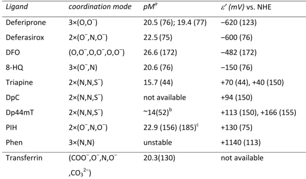
The ratio between the cumulative stability constants of the iron(II) and iron(III) complexes is strongly connected to the redox potential. The reason is that the formal redox potential (ε’) of the Fe(III)/Fe(II) redox couple is determined by the ratio of the cumulative stability constants of the iron(III) and the iron(II) complexes according to the general Eq.1.(172)
’Fe(III)complex/Fe(II)complex ‒ ’Fe(III)aqua/Fe(II)aqua = ‒59.15×log(Fe(III)complex / Fe(II)complex) (Eq.1).
Therefore, an increasing formal potential obtained at pH 7.4 represents an increasing affinity of the ligand towards iron(II) compared to iron(III) (Table 2). As a consequence, e.g.
the iron complexes formed with the aromatic nitrogen donor atom-containing bidentate ligand 1,10-phenantholine (Figure 2) are characterized by a very high formal potential of the iron complex (+1140 mV vs. normal hydrogen electrode (NHE) (113)) and, therefore, the iron(III) complex shows low stability in aqueous solution (122). In contrast, the iron complexes of chelators applied for the treatment of iron overload such as DFO, deferiprone and deferasirox (Figure 2) are characterized by very low redox potentials (Table 2) and, consequently, very high iron(III)-binding abilities (reflected by high pM values, which is the negative logarithm of the equilibrium concentrations of the unbound metal ion). For example, in case of DFO, which has a formal redox potential of –482 mV vs.
NHE (172), the iron(II) complex has much lower stability compared to the respective iron(III) complex (46,69). Under anaerobic conditions (at physiological pH) DFO is even able to oxidize iron(II) to iron(III) (46). The reason for this behavior is that these agents mainly contain hard oxygen donor atoms, which are ideal for iron(III) binding (87,89). This feature results in a very high binding efficacy and provides the enormous selectivity of these ligands for iron(III), not only in comparison to iron(II) but also to other essential bivalent metal ions like zinc(II) and copper(II). In addition, due to the low ε’ values, iron complexes of these chelators do not redox cycle under physiological conditions (76) preventing generation of reactive oxygen species (ROS) via Fenton-type reactions.
Downloaded by Kings College London-journal Section from online.liebertpub.com at 01/16/18. For personal use only.
11
Antioxidants and Redox Signaling Anticancer thiosemicarbazones: chemical properties, interaction with iron metabolism, and resistance development (DOI: 10.1089/ars.2017.7487) This paper has been peer-reviewed and accepted for publication, but has yet to undergo copyediting and proof correction. The final published version may differ from this proof.
In contrast to the (mainly oxygen-containing) chelators developed for the treatment of iron overload, anticancer α-N-pyridyl thiosemicarbazones such as Triapine, DpC or Dp44mT coordinate metals via their (Npyridyl,N,S‒) donor set. As a result of this binding mode, the redox potentials of their iron complexes are significantly higher (in the range of ‒170 mV to +170 mV vs. NHE) (44,155) compared to DFO, deferiprone or deferasirox (–482 mV to – 620 mV vs. NHE; see Table 2). Consequently, α-N-pyridyl thiosemicarbazones show a considerably high affinity towards both iron(II) as well as iron(III) ions. This property enables the iron thiosemicarbazone complexes to redox cycle between the two oxidations states, as these redox potentials are accessible to intracellular oxidants and reductants (66,87,155). Redox cycling is considered as an important factor in the antiproliferative activity of these compounds (although the correlation between the redox potential and cytotoxicity is not as evident as the relation between stability and cytotoxicity). In line with the relevance of redox cycling for anticancer thiosemicarbazones, iron(III)-specific chelators such as DFO are generally not efficient antitumor agents and can be safely used to treat iron overload (23,116,197). This already indicates that “iron depletion” in itself is not sufficient to explain the anticancer activity of thiosemicarbazones.
Interaction of thiosemicarbazones with the body iron
Based on the above-described features of thiosemicarbazones, there is no doubt that interaction with iron plays an important role in the biological activity of Triapine as well as many other α-N-heterocyclic derivatives. In addition to cell culture studies (discussed in more detail below), which investigated the role of iron and other metal ions in the activity of thiosemicarbazones on a cellular level, there are reports from clinical trials on the interaction of thiosemicarbazones with iron metabolism. For example, in case of 5-HP, excretion of the green-colored iron complex was observed in the urine of treated patients within a few hours after drug administration (103). In contrast, in Triapine-treated patients (or animals) (47,132,143)) no coloring of urine but methemoglobin formation (oxidation of iron(II) to iron(III) in hemoglobin) was observed which is a prominent indication for its interaction with body iron. This type of adverse effect, observed in ~23% of Triapine-
Downloaded by Kings College London-journal Section from online.liebertpub.com at 01/16/18. For personal use only.
12
Antioxidants and Redox Signaling Anticancer thiosemicarbazones: chemical properties, interaction with iron metabolism, and resistance development (DOI: 10.1089/ars.2017.7487) This paper has been peer-reviewed and accepted for publication, but has yet to undergo copyediting and proof correction. The final published version may differ from this proof.
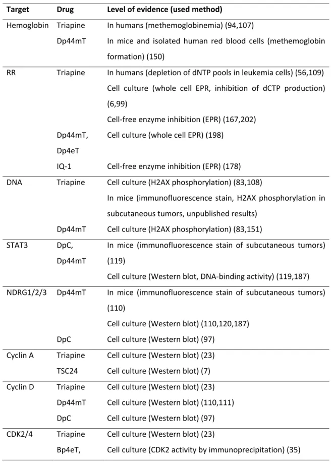
treated patients (94,107), was also reported for several preclinically developed α-N- heterocyclic thiosemicarbazones such as Dp44mT (150) (in this study methemoglobin formation was analyzed in mice as well as in isolated human red blood cells) (Table 3).
With regard to the underlying mechanism, the iron(III) complex of Triapine is believed to oxidize the iron(II) in the hemoglobin molecule by direct electron transfer (183). In line with the supposed role of a redox reaction in the thiosemicarbazone-induced methemoglobin formation, clinical trials with Triapine additionally indicated that patients with a glucose-6-phosphate deficiency suffered from enhanced methemoglobin levels and even hemolysis after treatment (199). Since glucose-6-phosphate is important in the protection of red blood cells from oxidative stress (184), this supports the assumption that Triapine treatment interferes with redox homeostasis. Methemoglobin formation seems to be strongly dependent on the interaction of the terminally unsubstituted NH2 groups of the thiosemicarbazone iron complexes with the carboxylic acid groups of the porphyrins of hemoglobin (Figure 5) (11,12,143). Consequently, novel α-N-heterocyclic thiosemicarbazones such as DpC (and probably also COTI-2) carrying a bulky substituent at this position are far less potent methemoglobin inducers than their predecessors (12).
Besides methemoglobin formation, also other iron-associated biological parameters were affected by Triapine treatment. Wadler et al. (186) described that after 96 h of continuous Triapine infusion serum iron and ferritin levels transiently increased by 104% and 77%, respectively. Remarkably, the total iron-binding capacity of the blood remained unchanged in these patients, indicating that there was no net loss of iron from the body.
Consequently, the authors concluded that (in contrast to 5-HP or the traditional iron(III) chelators (103)) Triapine-bound iron might be recovered during its hepatic metabolism and the increased delivery of iron to the liver could induce production of ferritin explaining the elevated serum levels of this protein. Interestingly, despite these distinct impacts on several iron-associated clinical parameters, all attempts to detect the formation of an iron- Triapine complex in vivo or even in cell culture have failed so far (95,96,143).
Downloaded by Kings College London-journal Section from online.liebertpub.com at 01/16/18. For personal use only.
13
Antioxidants and Redox Signaling Anticancer thiosemicarbazones: chemical properties, interaction with iron metabolism, and resistance development (DOI: 10.1089/ars.2017.7487) This paper has been peer-reviewed and accepted for publication, but has yet to undergo copyediting and proof correction. The final published version may differ from this proof.
Cancer-associated cellular targets of thiosemicarbazones
1) Ribonucleotide reductase
The iron-containing enzyme ribonucleotide reductase (RR) is most probably the best described target of anticancer α-N-heterocyclic thiosemicarbazones (Table 3). This metalloenzyme catalyzes the conversion of ribonucleoside diphosphates (NTPs) to deoxyribonucleoside diphosphates (dNTPs). Therefore, it is crucial for DNA synthesis as well as DNA damage repair and is frequently overexpressed in cancer cells making it an attractive target for treatment. The RR is a tetrameric protein composed of two large subunits (hRRM1) that are important for substrate binding, and two small iron-containing R2 subunits being responsible for its enzymatic function (5,25). Two different R2 subtypes have been described: i) the hRRM2 subunit, which is a cell cycle-dependent protein, strongly upregulated at the beginning of the S-phase and actively degraded via ubiquitination during the G2 phase of the cell cycle (60); ii) p53R2 (also called hRRM2B), a subunit which is expressed at lower levels than hRRM2 but is not degraded during mitosis (138) and therefore its levels remain constant during the cell cycle. In addition, reports suggest that expression of this subunit can be induced in a p53-dependent manner.
Consequently, it is assumed that the so formed protein is involved in several cellular functions including DNA repair, cell cycle arrest and mitochondrial homeostasis (5,25).
Inhibition of the tyrosyl radical in the active center of the hRRM2/p53R2 subunit was demonstrated for several thiosemicarbazones (including Triapine and Dp44mT) as well as diverse iron chelators (like phen and DFO) mainly in cell-free systems (6,59,74,99,167,178,198,202). The best-investigated thiosemicarbazones with regard to RR inhibition are Triapine and its closest analogs, for which detailed in vitro studies were performed during the last decades. In addition, a phase I trial with Triapine demonstrated the depletion of dATP and dGTP pools together with a decrease of DNA synthesis in circulating leukemia cells (56).
With regard to the mechanism, it was initially assumed that Triapine inhibits the hRRM2 subunit through iron chelation, either from the active site of the protein or from the
Downloaded by Kings College London-journal Section from online.liebertpub.com at 01/16/18. For personal use only.
14
Antioxidants and Redox Signaling Anticancer thiosemicarbazones: chemical properties, interaction with iron metabolism, and resistance development (DOI: 10.1089/ars.2017.7487) This paper has been peer-reviewed and accepted for publication, but has yet to undergo copyediting and proof correction. The final published version may differ from this proof.
cellular labile iron pool (5). However, this theory was questioned by several observations indicating that in fact Triapine seems to be a surprisingly inefficient iron chelator in cell culture. For example, it was shown that, in contrast to DFO, Triapine had a relatively weak effect on cellular transferrin receptor levels of SK-N-MC cells (23) perhaps due to the weaker iron(III)-binding ability compared to DFO. Also, electron paramagnetic resonance (EPR) spectroscopic analyses of Triapine-treated K562 cells (5 or 500 µM) showed no change in the size of the g = 4.3 signals (typical for intracellular iron-chelate complexes) after 0.5, 3 and 12 h of treatment, which would be indicative for depletion of the intracellular iron(III) pools (6). The same authors showed that in the presence of Triapine the loss of iron bound inside the RR molecule was five times lower than the destruction of the tyrosyl radical (5,6) and subsequent investigations of Triapine-treated COS-1 cells indicated iron-loaded hRRM2 (with a quenched tyrosyl radical) as the main remaining protein species, arguing against iron chelation from the enzyme. Moreover, in these COS-1 cells Triapine did not change the aconitase activity (6). This is of interest, as this protein contains an essential [4Fe4S] cluster, which is ROS-sensitive and could be influenced by depletion of the labile iron pool (6). Consequently, the authors of this study concluded that Triapine is not only ineffective in sequestering iron inside the cell, but also that it is unlikely to be involved in disruption of biosynthesis or repair of metallo-cofactor-requiring proteins (6).
Based on the repeated observations, that in cell-free systems metal-bound Triapine (particularly the iron(II) complex) possesses a dramatically increased ability to inhibit hRRM2 compared to the metal-free ligand (6,146,167,202), several authors speculated that the active species responsible for cell killing is in fact the iron complex of Triapine. In more detail, it was assumed that upon intracellular formation of the redox-active iron complex, ROS are generated, which are ultimately responsible for RR inhibition (167).
Interestingly, the pronounced activity of Triapine and close derivatives against hRRM2 in the presence of iron was not observed in cell-free systems of p53R2, and co-incubation with iron had even protective effects against the inhibition of this enzyme (202).
Furthermore, there are several reports suggesting that at biologically active concentrations no substantial ROS generation was detected after treatment with metal-free Triapine or
Downloaded by Kings College London-journal Section from online.liebertpub.com at 01/16/18. For personal use only.
15
Antioxidants and Redox Signaling Anticancer thiosemicarbazones: chemical properties, interaction with iron metabolism, and resistance development (DOI: 10.1089/ars.2017.7487) This paper has been peer-reviewed and accepted for publication, but has yet to undergo copyediting and proof correction. The final published version may differ from this proof.
other close derivatives (100,115,134). In line with these observations, recent studies have revealed no oxidation of hRRM2 residues or the accumulation of oxidized cellular proteins (neither in cell-free nor in cell culture experiments) after Triapine treatment, suggesting that global ROS generation might not be mandatory for RR inhibition by Triapine (5,6).
Notably, as already mentioned before, despite several attempts (96,143) intracellular formation of a Triapine-iron complex has not been directly demonstrated either in vitro or in vivo. Taken together, while the importance and necessity of iron-binding in the anticancer activity of thiosemicarbazones is proven without doubt (derivatives and metabolites lacking a metal-binding domain are completely inactive (142,147,200)), the exact mode of action remains elusive.
Consequently, several alternative theories have been published to offer an explanation how Triapine could inhibit the RR without the direct chelation of the enzyme-bound iron or the production of ROS. On the one hand, it was suggested (comparably to the interaction with hemoglobin in the methemoglobin formation) that the iron(II) complex of Triapine could act as reductant, directly reducing the tyrosyl radical of the hRRM2 subunit (6). This is of interest as direct binding of Triapine to hRRM2 and p53R2 was shown by radioactive labelling (202), which was only slightly affected by addition of iron. In line with these experimental data, docking studies of mouse RR revealed a possible Triapine-binding site with the pyridine ring of the thiosemicarbazone located in the pocket of the four amino acids Phe237, Phe241, Ser238 and Tyr324 (146). Based on these results, it can be hypothesized that, following the binding of (metal-free) Triapine to the enzyme, an iron complex is formed locally, using the iron liberated during each catalytic cycle of the RR.
Another possible mechanism is based on the effect of Triapine on intracellular thiol homeostasis. Here, especially the effects on the thioredoxin system seem to be of importance, as thioredoxin (Trx) or glutaredoxin are necessary to reactivate the RR enzyme after the end of each reaction cycle. In detail, during the conversion cycle of NTPs to dNTPs a disulphide bond is formed in the hRRM1 subunit, which has to be reduced before the RR can enter a new enzymatic cycle (60,166). Some reports indicate that Triapine as well as Dp44mT inhibit thioredoxin reductase (TrxR1) (134,198). However, as these effects were observed only at very high concentrations (>50-fold above the IC50 values (133)), it remains
Downloaded by Kings College London-journal Section from online.liebertpub.com at 01/16/18. For personal use only.
16
Antioxidants and Redox Signaling Anticancer thiosemicarbazones: chemical properties, interaction with iron metabolism, and resistance development (DOI: 10.1089/ars.2017.7487) This paper has been peer-reviewed and accepted for publication, but has yet to undergo copyediting and proof correction. The final published version may differ from this proof.
unclear whether this is of biological relevance. Interestingly, murine L1210 leukemia cells selected for resistance against 4-methyl-5-amino-1-formylisoquinoline thiosemicarbazone (MAIQ) were (besides the overexpression of several efflux pumps (29)) also characterized by overexpression of protein disulfide isomerases (PDI) (30), which are thiol oxidoreductase chaperones from the thioredoxin superfamily (171).
2) Impact of thiosemicarbazones on the cell cycle and other (iron-dependent) cellular pathways.
During the last decades, it has been repeatedly reported that treatment with diverse iron chelators leads to cell cycle arrest in G0/G1 or early S-phase (27,71,79,147,151,170). One explanation certainly is the above-described inhibition of the RR. However, several other proteins involved in DNA replication and repair also require iron as a cofactor and thus could be influenced by thiosemicarbazones and other iron-chelating drugs. These proteins include for example three DNA polymerases (Pol α, Pol δ and Pol ε), several DNA helicases (including Rad3/XPD, Dna2, RTEL1, FANCJ and ChIR1) as well as the DNA primase regulator subunit PRIM2 (201). In contrast to RR, these polymerases, helicases and primases contain an Fe-S cluster, which is necessary for the formation of active holoproteins (201).
Interestingly, inhibition of RNA polymerases seems to be very important for the antiviral activity of several thiosemicarbazones, indicated by point mutations in the enzymes leading to drug-resistant virus variants (20,21,31,149). However, there are only a few studies (based on cell-free systems) linking polymerase inhibition to cancer (63) and to our knowledge there is so far no indication that inhibition of polymerases also occurs in cancer cells after thiosemicarbazone treatment. Likewise, the activity of the [4Fe-4S] cluster- containing aconitase (201) is not affected by Triapine (6), indicating that, in general, human Fe-S cluster proteins are not (heavily) affected by thiosemicarbazone treatment.
In addition to its direct role in proteins executing DNA synthesis, iron also influences cell cycle regulation via cyclins and cyclin-dependent kinases (CDKs) (201). Thus, it has been repeatedly reported that treatment with diverse iron chelators such as DFO or L-mimosine (an amino acid derivative and predecessor of deferiprone) and also thiosemicarbazones
Downloaded by Kings College London-journal Section from online.liebertpub.com at 01/16/18. For personal use only.
17
Antioxidants and Redox Signaling Anticancer thiosemicarbazones: chemical properties, interaction with iron metabolism, and resistance development (DOI: 10.1089/ars.2017.7487) This paper has been peer-reviewed and accepted for publication, but has yet to undergo copyediting and proof correction. The final published version may differ from this proof.
results in the downregulation of cyclin A and D (7,23,27,105,110,136) as well as CDK2/4 (23,35,36,105) (Table 3). Overall, the mechanisms underlying these effects are not fully understood and there might be several explanations. For example, in case of CDK2 it was reported that binding of iron ions to the protein occurs only to its phosphorylated form, indicating a direct regulation of this protein by the cellular metal homeostasis (9). With regard to cyclin A, little is known about the mechanisms explaining how thiosemicarbazones could influence the expression levels of this protein. Of interest in this context might be a recent report on the downregulation of cyclin A (in addition to cyclin D) by rolipram, a PDE4 inhibitor (121), which also strongly protects cells from Triapine treatment. In addition, loss of PDE4D was the only genomic alteration found in Triapine- resistant SW480 cells (126). With regard to its biological function PDE4 is a negative regulator of cyclic AMP (cAMP), which in turn is one of the most abundant second messengers regulating various physiological processes like cell survival, differentiation, proliferation and apoptosis (10). One major downstream target of cAMP is cAMP- dependent protein kinase (protein kinase A, PKA), which then activates the transcription factor cAMP responsive element-binding protein (Creb) (157). Consequently, interaction of thiosemicarbazones with this regulatory pathway could also serve as an explanation for the observed effects on cyclin A.
More information is available on the mechanisms linking disturbed cellular iron homeostasis to the reduced expression of cyclin D1. For example, DFO and 311 (2-hydroxy- 1-naphthylaldehyde isonicotinoyl hydrazine, a congener of PIH) resulted in active degradation of cyclin D1 (136), whereas DpC (and in some cell lines also Dp44mT) downregulated the expression of cyclin D1 by inhibition of STAT3 dimerization and activation (possibly via inhibition of upstream kinases such as Src and Abl) (119,187).
Interestingly, the described effects were similar for the metal-free ligands and preformed iron complexes. In addition to STAT3, treatment with DFO (110), deferasirox (120), L- mimosine (27), as well as Dp44mT (110,114,120,187) and DpC (97) downregulated cyclin D1 also via N-myc downstream-regulated gene 1 or 2 (NDRG1/2), repeatedly shown in cell culture and for some drugs also in mouse models (Table 3). However, NDRG1 also has multiple other cellular targets including the TGFβ, the Wnt/β-catenin (114), the
Downloaded by Kings College London-journal Section from online.liebertpub.com at 01/16/18. For personal use only.
18
Antioxidants and Redox Signaling Anticancer thiosemicarbazones: chemical properties, interaction with iron metabolism, and resistance development (DOI: 10.1089/ars.2017.7487) This paper has been peer-reviewed and accepted for publication, but has yet to undergo copyediting and proof correction. The final published version may differ from this proof.
FAK/paxillin (188), the NF-κB (192), and the ErbB signaling pathways (98). Consequently, it is not surprising that besides inhibition of cell cycle progression, the drug-mediated stimulation of NDRG1/2 can be linked to several other effects including reduced aggressiveness or metastatic potential, which has been described e.g. for Dp44mT- or DpC- treated hepatocellular and breast carcinoma cells (114,187). Interestingly, NDRG1 has recently also been identified as downstream-target of the above mentioned PKA/CREB signaling axis (81). Finally, cyclin D1 is also a well-known target of p21waf, which was reported to be stimulated by diverse iron(III) chelators including DFO, deferasirox, 311, but also Dp44mT, DpC, and TSC24 (N,N-dimethyl-2-(quinolin-2- ylmethylene)hydrazinecarbothioamide, a terminally dimethylated analogue of FTSC) (7,23,32,97,120). P21waf is a down-stream target of the DNA damage sensor p53 (78).
Considering, the enhanced DNA damage (indicated for example by phosphorylation of the damage marker H2XA) which was observed after treatment with different RR inhibitors (including Triapine, Dp44mT) in cell culture (83,108,138,151) but also in mouse models (unpublished results from our group), it is not unexpected that stimulation of p53 expression has been described for some RR inhibitors (112,131) (Table 3). However, there is a surprisingly low number of reports on p53 activation by thiosemicarbazones and no correlation between p53 status and anticancer activity was found for several Dp44mT derivatives or Triapine (189,196). This led (at least in case of Dp44mT) to the suggestion that mouse double minute 2 homolog (MDM2)-mediated protein degradation could be responsible for p53-independent p21waf regulation (131). Nevertheless, Yu et al. identify three thiosemicarbazone derivatives (NSC319725 and NSC319726, both terminally substituted FTSC derivatives, as well as NSC328784, a selenosemicarbazone derivative of COTI-2) in the NCI60 screen database, which showed preferential anticancer activity in p53-mutant cell models harboring the R175 mutation (196). Subsequent analysis revealed that these effects are based on the restoration of a p53 “wild type-like confirmation” in an iron-sensitive manner (in case of NSC319726 addition of FeSO4 completely abrogated the activity) (196).
Interestingly, there are also reports on the interaction of cyclin D1 with the mitochondrial regulation (145). Thus, knock-out of cyclin D1 leads to increased mitochondrial size and
Downloaded by Kings College London-journal Section from online.liebertpub.com at 01/16/18. For personal use only.
19
Antioxidants and Redox Signaling Anticancer thiosemicarbazones: chemical properties, interaction with iron metabolism, and resistance development (DOI: 10.1089/ars.2017.7487) This paper has been peer-reviewed and accepted for publication, but has yet to undergo copyediting and proof correction. The final published version may differ from this proof.
activity. Accordingly, inactivation of the CDK/cyclin substrate Rb and overexpression of p21 resulted in inhibition of the mitochondrial metabolism (145). Furthermore, there are several heme proteins such as cytochromes or nitric oxygenase, which are crucial for mitochondrial function (201). In line with this, there are some reports how thiosemicarbazones affect the mitochondria. On the one hand, induction of apoptosis via the intrinsic (mitochondria-mediated) pathway has been described for diverse representatives of this compound class (1,7,97,144,147,170,200). However, as thiosemicarbazones induce apoptosis usually only at quite high drug concentrations (far above the IC50 values), they can be considered rather as cytostatic agents which mainly execute their activity via cell cycle arrest. On the other hand, at least for Triapine and Dp44mT also a perturbation of the mitochondrial redox homeostasis was reported (133,134). Thus, treatment resulted in oxidation of mitochondrial Trx2 and, subsequently, the inability of the treated cells to maintain the redox state of Trx2-dependent proteins such as peroxiredoxin 3 (Prx3) (134) (Table 3). In line with these findings, Triapine treatment was distinctly more efficient in cells with Prx3 and Trx2 knockdown (134).
However, in this study no signs of global redox stress in the cytosol of Triapine-treated cells were detected (134). Thus, for example several cytosolic redox proteins like Trx1, and Prx1, or endoplasmic Prx4 remained unchanged at clinically relevant concentrations (low µM range) (133). This preferential oxidation of mitochondrial Trx2 and Prx3 after Triapine treatment could be explained by an organelle-specific accumulation of the iron(III) complex of Triapine, as lipophilic cations can accumulate at several hundred-fold higher levels inside the mitochondria because of the large mitochondrial membrane potential (134). On the other hand, it is also possible that Triapine increases the mitochondrial peroxide (or other ROS) levels that may directly promote Prx3 oxidation. Also in case of Dp44mT (and to some extent DFO), interaction with the mitochondria was indicated by reports of treatment-induced upregulation of adenosine monophosphate-activated protein kinase (AMPK) a protein which is important for the restoration of the cellular energy levels (104) at low ATP levels, e.g. in response to mitochondrial dysfunction or stress (82).
Downloaded by Kings College London-journal Section from online.liebertpub.com at 01/16/18. For personal use only.


![Figure 3: General (de)protonation steps of an α-N-pyridyl thiosemicarbazone ligand [H 2 L] + shown for FTSC](https://thumb-eu.123doks.com/thumbv2/9dokorg/1316015.105948/59.918.166.765.161.356/figure-general-protonation-steps-pyridyl-thiosemicarbazone-ligand-shown.webp)


