1
Antioxidants and Redox Signaling High copper complex stability and slow reduction kinetics as key parameters for improved activity, paraptosis induction and impact on drug-resistant cells of anticancer thiosemicarbazones (DOI: 10.1089/ars.2019.7854) This paper has been peer-reviewed and accepted for publication, but has yet to undergo copyediting and proof correction. The final published version may differ from this proof.
Original Research Communication
High copper complex stability and slow reduction kinetics as key parameters for improved activity, paraptosis induction and impact
on drug-resistant cells of anticancer thiosemicarbazones
Sonja Hager,1,2$ Veronika F.S. Pape,3,4$ Vivien Pósa,5,6 Bianca Montsch,1,2 Lukas Uhlik,1,2 Gergely Szakács,1,3 Szilárd Tóth,3 Nikolett Jabronka,3 Bernhard K. Keppler,2,7 Christian R.
Kowol,2,7 Éva A. Enyedy,5,6* Petra Heffeter,1,2*
1 Institute of Cancer Research, Medical University of Vienna, Borschkegasse 8a, A-1090 Vienna, Austria
2 Research Cluster ‘Translational Cancer Therapy Research’, A-1090 Vienna, Austria
3 Institute of Enzymology, Research Centre for Natural Sciences, Hungarian Academy of Sciences, Magyar Tudósok körútja 2, H-1117 Budapest, Hungary
4 Department of Physiology, Semmelweis University, Tűzoltó utca 37-47, H-1094 Budapest, Hungary
5 Department of Inorganic and Analytical Chemistry, Interdisciplinary Excellence Centre, University of Szeged, Dóm tér 7, H-6720 Szeged, Hungary
6 MTA-SZTE Lendület Functional Metal Complexes Research Group, University of Szeged, Dóm tér 7, H-6720 Szeged, Hungary
7 Institute of Inorganic Chemistry, Faculty of Chemistry, University of Vienna, Waehringer Str. 42, A-1090 Vienna, Austria
$ These authors contributed equally to the main findings of this manuscript.
Running title: Insights into the MoA of nM TSCs
Keywords: solution stability, thiosemicarbazones, copper complexes, protein disulfide isomerase, paraptosis, superoxide dismutase
Authors for correspondence: University of Szeged, Department of Inorganic and Analytical Chemistry, Interdisciplinary Excellence Centre, Dóm tér 7, H-6720 Szeged, Hungary. Phone:
+36-62-544334. E-mail: enyedy@chem.u-szeged.hu
Downloaded by Northwestern University from www.liebertpub.com at 05/03/20. For personal use only.
2
Antioxidants and Redox Signaling High copper complex stability and slow reduction kinetics as key parameters for improved activity, paraptosis induction and impact on drug-resistant cells of anticancer thiosemicarbazones (DOI: 10.1089/ars.2019.7854) This paper has been peer-reviewed and accepted for publication, but has yet to undergo copyediting and proof correction. The final published version may differ from this proof.
Medical University of Vienna, Institute of Cancer Research, Borschkeg. 8a, A-1090 Vienna, Austria. Phone: +43-1-40160-57594. Fax: +43-1-40160-957555. E-mail:
petra.heffeter@meduniwien.ac.at.
Word count: 8 172 (incl. Abstract, Introduction, Results, Discussion, Materials & Methods) References: 60
Greyscale illustrations: 4 Color illustrations: 1
Downloaded by Northwestern University from www.liebertpub.com at 05/03/20. For personal use only.
3
Antioxidants and Redox Signaling High copper complex stability and slow reduction kinetics as key parameters for improved activity, paraptosis induction and impact on drug-resistant cells of anticancer thiosemicarbazones (DOI: 10.1089/ars.2019.7854) This paper has been peer-reviewed and accepted for publication, but has yet to undergo copyediting and proof correction. The final published version may differ from this proof.
Abstract
Aims: Due to their significant biological activity, thiosemicarbazones (TSCs) are promising candidates for anticancer therapy. In part, the efficacy of TSCs is linked to their ability to chelate essential metal ions such as copper and iron. Triapine, the best-studied anticancer TSC, has been tested clinically with promising results in hematological diseases. During the last years, a novel subclass of TSCs with improved anticancer activity was found to induce paraptosis, a recently characterized form of cell death. The aim of this study was to identify structural and chemical properties associated with anticancer activity and paraptosis induction of TSCs.
Results: When testing a panel of structurally related TSCs, compounds with nanomolar anticancer activity and paraptosis-inducing properties showed higher copper(II) complex solution stability and a slower reduction rate, which resulted in reduced redox activity. In contrast, TSCs with lower anticancer activity induced higher levels of superoxide that rapidly stimulated superoxide dismutase expression in treated cells, effectively protecting the cells from drug-induced redox stress.
Innovation: Consequently, we hypothesize that in case of close Triapine derivatives, intracellular reduction leads to rapid dissociation of intracellularly formed copper complexes. In contrast, TSCs characterized by highly stable, slowly reducible copper(II) complexes are able to reach new intracellular targets such as the ER-resident protein disulfide isomerase.
Conclusions: The additional modes of actions observed with highly active TSC derivatives are based on intracellular formation of stable copper complexes, offering a new approach to combat (drug-resistant) cancer cells.
Downloaded by Northwestern University from www.liebertpub.com at 05/03/20. For personal use only.
4
Antioxidants and Redox Signaling High copper complex stability and slow reduction kinetics as key parameters for improved activity, paraptosis induction and impact on drug-resistant cells of anticancer thiosemicarbazones (DOI: 10.1089/ars.2019.7854) This paper has been peer-reviewed and accepted for publication, but has yet to undergo copyediting and proof correction. The final published version may differ from this proof.
Introduction
Thiosemicarbazones (TSCs) possess significant biological activity, which resulted in their development as pharmaceuticals against several diseases, including cancer (24). In part, the efficacy of TSCs is linked to their ability to chelate essential metal ions such as copper and iron. Cancer cells, in particular, require higher amounts of these metal ions due to their increased rate of replication (3,60). As a result, the ability of α-N-heterocyclic TSCs to form stable metal complexes is an important property for their development as anticancer agents (26).
Initially, the mechanism of action of α-N-heterocyclic TSCs was thought to primarily rely on the depletion of iron and the consequential inhibition of the iron-containing enzyme ribonucleotide reductase (57). However, the role of other metals (especially copper), metalloenzymes and metal-interacting proteins is gaining more attention (16,22). For example, the ability of copper(II)-TSC complexes to undergo redox cycling in the presence of reducing agents, with production of reactive oxygen species (ROS) (a process also called
“activation by reduction” (22)), and the disruption of the cellular thiol redox homeostasis is increasingly discussed as relevant contributions to TSC activity (11,23,27,44). Furthermore, the interaction with copper ions has been recently suggested to be involved in collateral sensitivity of P-glycoprotein (P-gp, ABCB1)-overexpressing multidrug-resistant cancer cells to the nanomolar-active TSC di-2-pyridylketone 4,4-dimethyl-3-thiosemicarbazone (Dp44mT) (21). Interestingly, recently performed studies confirmed the enhanced sensitivity of certain P-gp-overexpressing cells (e.g. MES-SA/Dx5) to several metal chelators (including Dp44mT), but also suggested that the collateral sensitivity of these MDR cells may rely on other (more complex) mechanisms independent of P-gp transport function (9,40).
With regard to the clinical situation, the best studied anticancer TSC, Triapine, has been already tested in several phase I and II trials with promising results especially against hematological diseases (12,25,54,56,59). Currently, this drug is being investigated as chemo- and radiosensitizer in an ongoing clinical phase III study assessing Triapine in combination with cisplatin and radiation therapy (NCT02466971) (30). In addition, two other α-N-heterocyclic TSCs di-2-pyridylketone 4-cyclohexyl-4-methyl-3- thiosemicarbazone (DpC) and 4-(pyridine-2-yl)-N-{[(8E)-5,6,7,8-tetrahydroquinolin-8-
Downloaded by Northwestern University from www.liebertpub.com at 05/03/20. For personal use only.
5
Antioxidants and Redox Signaling High copper complex stability and slow reduction kinetics as key parameters for improved activity, paraptosis induction and impact on drug-resistant cells of anticancer thiosemicarbazones (DOI: 10.1089/ars.2019.7854) This paper has been peer-reviewed and accepted for publication, but has yet to undergo copyediting and proof correction. The final published version may differ from this proof.
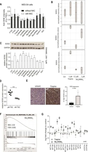
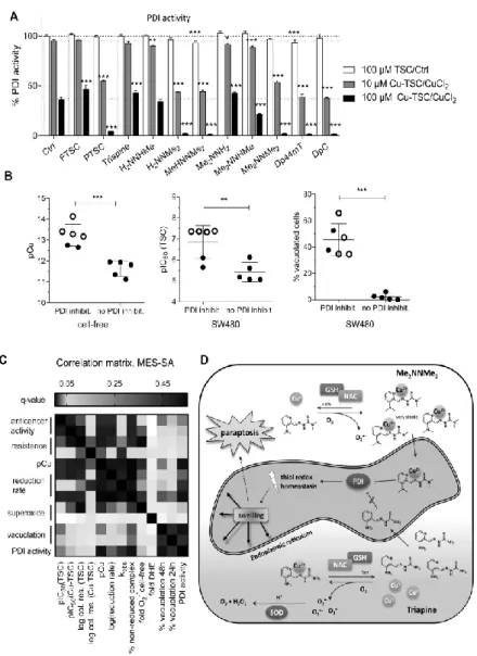
ylidene]amino})piperazine-1-carbothioamide (Coti-2) recently entered clinical phase I trials (www.clinicaltrials.gov). Noteworthy, these two compounds as well as Dp44mT (the predecessor of DpC) and our dimethylated Triapine derivative Me2NNMe2 represent a subclass of TSCs characterized by a ~500-fold higher anticancer activity compared to Triapine in cell culture (20,37). Recent studies suggested that this efficiency could be based on an additional mode-of-action, associated with the formation of intracellular copper complexes (16,19,34). In particular, our group discovered that these compounds are able to induce paraptosis, a novel form of programmed cell death (13). Paraptosis is a caspase- independent cell death discernable by the appearance of cytoplasmic vesicles originating from the endoplasmic reticulum (ER) (33,50). TSC-induced paraptosis appears to be associated with the (copper-dependent) inhibition of the ER-resident protein disulfide isomerase (PDI) (13). In light of these recent data, the aim of this study was to elucidate the role of copper complex formation of various TSCs in anticancer activity, paraptosis induction and collateral sensitivity. For this purpose, we investigated solution stability and redox properties of a selected panel of structurally related α-N-heterocyclic TSCs and their respective copper(II) complexes. The measured physico-chemical properties were then correlated with biological parameters (anticancer activity, resistance ratio of multidrug- resistant (MDR) cancer cells, paraptosis induction and PDI inhibition). Thus, we show that highly active TSCs (with IC50 values in the nanomolar range) form copper(II) complexes characterized by high stability and slow reduction kinetics. Our data suggest that these stability and redox properties protect the copper(II) complexes of this TSC subclass from premature reduction-induced ligand liberation allowing the interaction of the copper(II) complexes with intracellular targets such as the ER-resident PDI. In contrast, the copper complexes of TSCs such as Triapine are sensitive to rapid reduction and thus ligand liberation inside the cancer cells, which are efficiently protected from the generated superoxide by the upregulated enzyme superoxide dismutase (SOD) in cell culture as well as in a tumor model in vivo.
Downloaded by Northwestern University from www.liebertpub.com at 05/03/20. For personal use only.
6
Antioxidants and Redox Signaling High copper complex stability and slow reduction kinetics as key parameters for improved activity, paraptosis induction and impact on drug-resistant cells of anticancer thiosemicarbazones (DOI: 10.1089/ars.2019.7854) This paper has been peer-reviewed and accepted for publication, but has yet to undergo copyediting and proof correction. The final published version may differ from this proof.
Results
Anticancer activity and paraptosis-inducing potential of the TSC panel
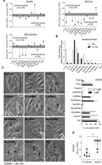
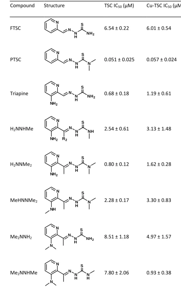
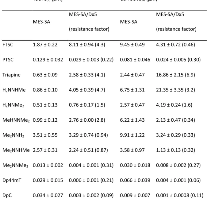
As a first step, the in vitro anticancer activity of the selected α-N-pyridyl TSCs (see Table 1 for their chemical formulae) and their preformed (i.e. in situ generated) copper(II) complexes (in a 1:1 metal-to-ligand ratio) was measured in human colon adenocarcinoma SW480 and uterine sarcoma MES-SA cells as well as the multidrug-resistant subline MES- SA/Dx5. SW480 cells were investigated by conventional MTT assay, while the fluorescently labelled MES-SA and MES-SA/Dx5 cells were tested in co-culture using an automated system (Figure 1A and B, Table 1 and 2 as well as Suppl. Table 1 - 4) (55). In accordance to previous reports (27-29,47,52), following 72 or 144 h incubation, the three terminally dimethylated compounds pyridine-2-carbaldehyde thiosemicarbazone (PTSC), Me2NNMe2, Dp44mT and the disubstituted derivative DpC showed distinctly higher anticancer activities than the other compounds (2-formylpyridine thiosemicarbazone (FTSC), Triapine, H2NNHMe, H2NNMe2, MeHNNMe2, Me2NNH2, Me2NNHMe). This was especially pronounced in the MES-SA/Dx5 cell model, reflecting the collateral sensitivity of the P-gp- overexpressing cells to this subtype of TSCs (Table 1 and 2, Figure 1B).
In order to gain more insight into the time dependency of these effects, the activity of the compounds was also assessed after 3 and 24 h. None of the metal-free ligands exhibited relevant anticancer activity after 3 h resulting in IC50 values above the highest tested concentration of 25 µM. With the exception of PTSC, Dp44mT and DpC, the compounds did not decrease viability following a 24 h-long incubation with the tested cancer cells.
Interestingly, in contrast to the later time points (Figure 1B), collateral sensitivity of MES- SA/Dx5 cells was not observed at 24 h (Suppl. Table 2 and 3), indicating that the collateral sensitivity might be based on enhanced cell cycle arrest rather than enhanced apoptosis.
The anticancer activity of copper(II) complexes was slightly different (Table 1 and 2 as well as Suppl. Table 1 - 4). Several complexes were already active after a relatively short incubation time (24 h) in SW480 cells (Suppl. Table 1 - 3), showing the same activity pattern as observed in the 72 h experiment (Table 1). However, after 72 h IC50 values of all tested copper complexes were in a similar range as the respective metal-free ligands (Figure 1A, Table 1), with the exception of Me2NNHMe, which was significantly more active
Downloaded by Northwestern University from www.liebertpub.com at 05/03/20. For personal use only.
7
Antioxidants and Redox Signaling High copper complex stability and slow reduction kinetics as key parameters for improved activity, paraptosis induction and impact on drug-resistant cells of anticancer thiosemicarbazones (DOI: 10.1089/ars.2019.7854) This paper has been peer-reviewed and accepted for publication, but has yet to undergo copyediting and proof correction. The final published version may differ from this proof.
as a copper(II) complex. Thus, the long-germ anticancer activity of the copper complexes of the nanomolar-active TSCs was similar to that of the respective metal-free ligand.
In order to investigate the paraptosis-inducing potential of the compounds, perinuclear vesicle formation was investigated by microscopy after drug treatment (Figure 1C). In agreement with previous results (13), Me2NNMe2, Dp44mT and DpC induced high levels of vacuolization in SW480 cells already at 0.1 µM drug concentrations (Figure 1C and D). In addition, also incubation with the terminally dimethylated PTSC, H2NNMe2 and MeHNNMe2 likewise induced paraptosis (however, vacuolization at 0.1 µM was observed only in case of PTSC, while for the other two derivatives, the effect was visible only at 1 µM, reflecting their higher IC50 values). Similar results were obtained in MES-SA cells (Suppl. Figure 2).
Proton dissociation processes and pKa values of the investigated TSCs
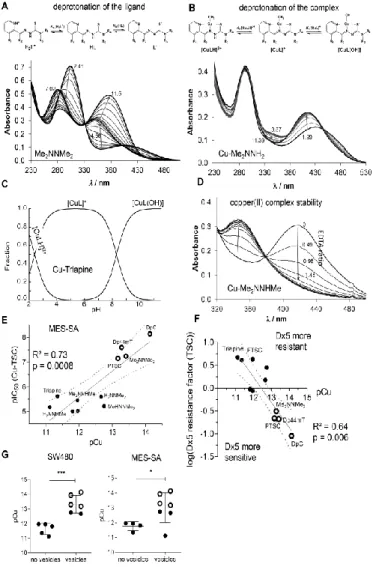
As there is a distinct structure-activity relationship in our TSCs panel, subsequently, we characterized whether these effects are reflected by differences in their chemical properties. While for Triapine and some derivatives several reports on proton dissociation processes are available (5-7), for most of the compounds in this study this parameter has not been characterized so far (especially not in pure aqueous solution). First, pKa values were determined by UV-visible (UV-vis) spectrophotometric titrations in pure water at compound concentrations of 10-50 µM. In case of DpC, addition of some dimethyl sulfoxide (DMSO) was necessary and the pKa values of DpC in pure water were estimated by extrapolation (Table 3; Suppl. Figure 3A). For all compounds, the measured UV-vis spectra revealed characteristic changes upon increasing pH, as it is depicted exemplarily for Me2NNMe2 in Figure 2A. The pH-dependence of the absorbance values always showed two well-separated deprotonation steps: one at pH 2.5‒6.0 and one at 8.0-11.5. Therefore, two pKa values could be determined (Table 3). The λmax and values of the ligand species in the different protonation states are collected in Suppl. Table 5. The calculated individual spectra (e.g. for Me2NNMe2 in Suppl. Figure 3B) represent significant differences between the molar absorptivities of the various ligand species (Suppl. Table 5). Notably, the pKa of the second pyridyl moiety in Dp44mT and DpC could not be determined under the applied conditions due to its fairly acidic character. Analysis of the pKa values revealed that N- terminal dimethylation results in slightly higher pKa (H2L+) and lower pKa (HL) (Triapine vs.
Downloaded by Northwestern University from www.liebertpub.com at 05/03/20. For personal use only.
8
Antioxidants and Redox Signaling High copper complex stability and slow reduction kinetics as key parameters for improved activity, paraptosis induction and impact on drug-resistant cells of anticancer thiosemicarbazones (DOI: 10.1089/ars.2019.7854) This paper has been peer-reviewed and accepted for publication, but has yet to undergo copyediting and proof correction. The final published version may differ from this proof.
H2NNMe2 or FTSC vs. PTSC etc.), whereas dimethylation of the pyridine amine increases slightly both pKa values (Triapine vs. Me2NNH2). At the same time monomethylations have only a minor influence. The dipyridyl derivatives Dp44mT, DpC as well as FTSC and PTSC possess decreased pKa H2L+ by almost one order of magnitude, but no clear trend was evident for pKa HL. In general, not only the electron-donating/withdrawing properties of the substituents should be considered to explain differences in pKa values, but also the ability of the derivatives to be stabilized by mesomeric/resonance effects (e.g. the thione/thiol equilibrium). As apparent from the determined pKa values all TSCs investigated in this study are charge neutral (in HL form) at physiological pH.
High copper(II) complex solution stability is an important parameter for anticancer activity
Having characterized the (de)protonation behavior of our TSCs, we evaluated the stability of the respective copper complexes in aqueous solution. Based on our previous findings (5,6), we assumed that some of the studied α-N-pyridyl TSCs (Triapine, FTSC, PTSC, H2NNMe2) form very stable [CuL]+ complexes in a wide pH range. At lower pH, the balance shifts to [CuLH]2+ complexes, containing the protonated ligand. In contrast, a mixed hydroxido complex [CuL(OH)] is prevalent in the basic pH range, while at ligand excess further species (e.g. [CuL2], [Cu2L3]+) can be found (6).
Equimolar aqueous solutions of the metal ion and the respective ligands (10 or 25 µM concentration) were titrated and the deprotonation processes of the complexes were followed spectrophotometrically (Figure 2B for Cu-Me2NNH2). Deconvolution of the spectra resulted in the pKa values of the copper(II) complexes and their individual absorbance spectra (Suppl. Table 6, Suppl. Figure 3C). All pKa values were in the range of 2.1-2.6 (for [CuLH]2+) and 8.1-8.8 (for [CuL]+) and thus well comparable to reported data of FTSC (with 1% DMSO) (1). In general, the pKa value of [CuLH]2+ was by 7.2-8.6 orders of magnitude lower compared to that of the metal-free HL form, revealing that displacement of the dissociable proton in the complex is mediated by the metal ion coordination. As the formation of the complex [CuL]+ is predominant in a wide pH range (4‒6.5) (Figure 2C for Cu-Triapine) and assumed to be quantitative due to the high solution stability, apparent (conditional) formation constants (β′) for this type of complexes were subsequently determined by competition experiments with EDTA (24) (Figure 2D for 1:1 Cu-Me2NNHMe
Downloaded by Northwestern University from www.liebertpub.com at 05/03/20. For personal use only.
9
Antioxidants and Redox Signaling High copper complex stability and slow reduction kinetics as key parameters for improved activity, paraptosis induction and impact on drug-resistant cells of anticancer thiosemicarbazones (DOI: 10.1089/ars.2019.7854) This paper has been peer-reviewed and accepted for publication, but has yet to undergo copyediting and proof correction. The final published version may differ from this proof.
and Suppl. Figure 3D). Since the displacement was found to be relatively slow, 1–2 h equilibration time was applied for this reaction (the reverse experiment using Cu-EDTA + FTSC resulted in the same endpoint; see Suppl. Figure 4). Increasing amounts of EDTA resulted in decreasing absorbance at the wavelength characteristic for the S→Cu charge transfer band (e.g. 422 nm in case of Me2NNHMe in Figure 2D). Taken into account the conditional stability constants determined (Suppl. Table 6) as well as the proton dissociation constants of the ligands (Table 3), the overall stability constants (β) of the complexes [CuL]+ were calculated (Table 3). Furthermore, also the β values of the other two types of complexes [CuLH]2+ and [CuL(OH)] were computed (Table 3) using the pKa of [CuLH]2+ and [CuL]+ (Suppl. Table 6). Based on these data, it can be concluded that at physiological pH, [CuL]+ is the most predominant species, accompanied by a smaller fraction (3.9-16.3%) of [CuL(OH)] (Table 3, Figure 2C).
Since a direct comparison of the logβ [CuL]+ constants is not adequate due to the different basicity of the ligands, pCu (−log [Cu(II)]) values were calculated in order to compare the copper(II)-binding ability of the studied TSCs at pH 7.4 (a higher pCu value indicates a stronger metal ion-binding ability of the ligand) (Table 3). The calculated pCu values revealed that dimethylation at both the terminal and the pyridine amino group increased complex stability. On the contrary, the effect of monomethylation on the copper(II)- binding ability was minor at both positions. Undoubtedly, the four compounds with activity in the nanomolar range form copper(II) complexes with the highest stability, followed by the two trimethylated compounds MeHNNMe2 and Me2NNHMeand all other derivatives (Table 3).
To evaluate the effect of copper complex stability on the biological activity, the pCu and 72 h pIC50 values of either the ligands or their in situ generated copper complexes were correlated. These analyses revealed that the ligands with high pIC50 values possess a higher copper(II)-binding ability (indicated by higher pCu values) and the correlation is even more pronounced for the pIC50 values of the copper(II) complexes (Figure 2E and Suppl. Figure 4). Accordingly, the higher copper complex stability was also associated with higher activity in the multidrug-resistant cells after long-term incubations (Figure 2F) and paraptosis induction (Figure 2G), indicating an important role of the copper(II) complex in these TSC-induced effects.
Downloaded by Northwestern University from www.liebertpub.com at 05/03/20. For personal use only.
10
Antioxidants and Redox Signaling High copper complex stability and slow reduction kinetics as key parameters for improved activity, paraptosis induction and impact on drug-resistant cells of anticancer thiosemicarbazones (DOI: 10.1089/ars.2019.7854) This paper has been peer-reviewed and accepted for publication, but has yet to undergo copyediting and proof correction. The final published version may differ from this proof.
Reduced reduction rate of copper(II) complex has a strong impact on TSC activity
In addition to the stability of the formed copper(II) complexes, also their redox properties may have an impact on their biological activities. In order to investigate, whether there are differences in the reduction rates of our TSC panel, the redox reactions of the in-situ generated copper(II) complexes with two physiological reducing agents, namely ascorbic acid (AA) and L-glutathione (GSH), were studied. Reduction of the copper(II) complexes was followed spectrophotometrically in aqueous solution at pH 7.4 under anaerobic conditions.
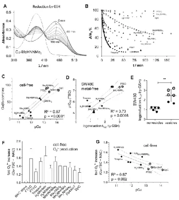
In good agreement with the literature (11), no time-dependent spectral changes were observed in the case of AA which suggests that these metal complexes cannot be reduced by AA under the applied conditions (see Suppl. Figure 5). This could be explained by the relatively weak reducing power of ascorbate (formal potential at pH 7.4: +0.05 V for dehydro-L-ascorbate/AA) (15). In contrast, the stronger reducing agent GSH (formal potential at pH 7.4: −0.26 V for GSSG/GSH (45) reduced all studied copper(II) TSC complexes, although with different rates (Figure 3A and B).
The first recorded spectrum after mixing the reactants showed a small shift of the λmax
value (e.g. 448 → 452 nm, as shown for Cu-MeHNNMe2 in Figure 3A) most probably due to the formation of a mixed ligand complex with GSH, as it is reported for various TSC complexes (22,44). This shift was followed by a significant decrease of the absorbance at this λmax, while the absorbance value at the λmax of the free ligand (~382 nm) increased, probably as a result of the decomposition of the generated unstable copper(I) complex.
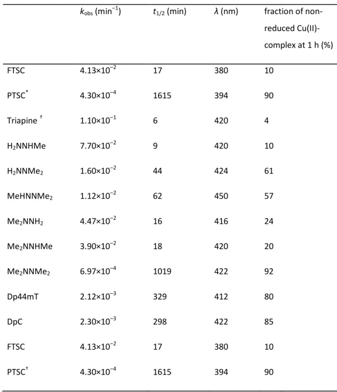
Noteworthy, oxygenizing (bubbling O2 into) the solution regenerated the original copper(II) complexes, confirming the reversibility of the redox process (data not shown). The other studied TSCs behaved similarly. However, significant differences were observed regarding the reduction rates of the respective complexes (Figure 3B). In order to obtain comparable data, the recorded absorbance/time curves were further analyzed at the λmax of the complexes. The calculated kobs, half-lives (t1/2) and percentage of non-reduced complex after 1 h in the presence of 50 equiv. (1.25 mM) of GSH are collected in Table 4. For selected complexes (Triapine, PTSC), kinetic runs were additionally performed at other equivalents of GSH, and kobs were found to be very sensitive to the concentration of the reducing agent, namely slower reaction rates with decreasing excess of GSH were
Downloaded by Northwestern University from www.liebertpub.com at 05/03/20. For personal use only.
11
Antioxidants and Redox Signaling High copper complex stability and slow reduction kinetics as key parameters for improved activity, paraptosis induction and impact on drug-resistant cells of anticancer thiosemicarbazones (DOI: 10.1089/ars.2019.7854) This paper has been peer-reviewed and accepted for publication, but has yet to undergo copyediting and proof correction. The final published version may differ from this proof.
observed (data not shown). In accordance to the reports from Santoro et al. (44) under the applied conditions, the reduction of the copper(II) complexes was incomplete, especially with nanomolar-active compounds (see plateau in Figure 3B and % non-reduced copper(II) complex after 1 h in Table 4). Interestingly, the remaining fraction of non-reduced copper(II) complex after 1 h (Table 4) showed a strong correlation with the pCu value and, therefore, with the solution stability of the copper(II) complexes (Figure 3C). These data demonstrate that the copper(II) complexes of α-N-pyridyl TSC ligands bearing higher solution stability can only be reduced by GSH in a slower and much less effective way.
When these reduction rates were subsequently correlated with the anticancer activity (after 72 h), it became apparent that a slower reduction rate is associated with higher anticancer activity (Figure 3D and Suppl. Figure 6A) and paraptosis-inducing potential (Figure 3E and Suppl. Figure 6B) of the metal-free ligands as well as the copper complexes.
Overall, these results are surprising, as so far it has been assumed that the anticancer activity of copper TSC complexes is mainly based on intracellular redox activity, either by the copper(II) TSC itself or by the copper release from the complex in the cell (22,44).
Copper(II) complex stability influences redox behavior of TSCs under cell-free conditions It is widely accepted in the literature (22,27,38,44), that reduction of copper(II) complexes to copper(I) and their subsequent re-oxidation under aerobic conditions, results in the generation of superoxide radicals and thus redox stress in treated cells. In order to compare the obtained reduction rates of the copper(II) complexes by GSH to their superoxide production potential, formation of cell-free superoxide was measured spectrophotometrically using the nitroblue tetrazolium (NBT) assay. In line with the activation by reduction theory for this compound class (16), without the addition of a reducing agent, none of the tested TSCs (neither as metal-free ligand nor as copper(II) complex) induced any positive signal (data not shown). In contrast, as expected when the GSH precursor and reducing agent N-acetyl cysteine (NAC) was added to the copper(II) complexes, superoxide generation (up to 1.6-fold compared to the control) was detected (Figure 3F). In agreement with the above shown results, distinct differences between the individual ligands were observed. Thus, indeed copper(II) complexes with a higher stability and slower reduction rate produced also less cell-free superoxide (Figure 3G and Suppl.
Figure 7). In contrast, copper ions, which under certain conditions have been reported to
Downloaded by Northwestern University from www.liebertpub.com at 05/03/20. For personal use only.
![Table 3: pK a values of the TSC ligands determined by spectrophotometric titrations and overall stability constants for the copper(II) TSC complexes (logβ) * calculated from the conditional stability constants (logβ’ 5.9 of [CuL] + ) and pK a values](https://thumb-eu.123doks.com/thumbv2/9dokorg/787418.36613/38.893.104.780.311.1136/determined-spectrophotometric-titrations-stability-constants-complexes-calculated-conditional.webp)